Embed Size (px)
Citation preview

RESEARCH ARTICLE
Bacterial persisters in long-term infection:
Emergence and fitness in a complex host
environment
Jennifer A. Bartell1☯, David R. Cameron2,3☯, Biljana Mojsoska4☯¤, Janus Anders
Juul Haagensen1, Tacjana Pressler5, Lea M. Sommer4, Kim Lewis2*, Søren Molin1*, Helle
Krogh JohansenID4,6*
1 The Novo Nordisk Foundation Center for Biosustainability, Technical University of Denmark, Kgs. Lyngby,
Denmark, 2 Antimicrobial Discovery Center, Department of Biology, Northeastern University, Boston,
Massachusetts, United States of America, 3 Department of Intensive Care Medicine, Inselspital, Bern
University Hospital, University of Bern, Bern, Switzerland, 4 Department of Clinical Microbiology,
Rigshospitalet, Copenhagen, Denmark, 5 Cystic Fibrosis Center, Rigshospitalet, Copenhagen, Denmark,
6 Department of Clinical Medicine, University of Copenhagen, Copenhagen, Denmark
☯ These authors contributed equally to this work.
¤ Current address: Department of Science and Environment, Roskilde University, Roskilde, Denmark
* [email protected] (KL); [email protected] (SM); [email protected] (HKJ)
Abstract
Despite intensive antibiotic treatment, Pseudomonas aeruginosa often persists in the air-
ways of cystic fibrosis (CF) patients for decades, and can do so without antibiotic resistance
development. Using high-throughput screening assays of bacterial survival after treatment
with high concentrations of ciprofloxacin, we have determined the prevalence of persisters
in a large patient cohort using 460 longitudinal isolates of P. aeruginosa from 39 CF patients.
Isolates were classed as high persister variants (Hip) if they regrew following antibiotic treat-
ment in at least 75% of the experimental replicates. Strain genomic data, isolate phenotyp-
ing, and patient treatment records were integrated in a lineage-based analysis of persister
formation and clinical impact. In total, 19% of the isolates were classified as Hip and Hip
emergence increased over lineage colonization time within 22 Hip+ patients. Most Hip+ line-
ages produced multiple Hip isolates, but few Hip+ lineages were dominated by Hip. While
we observed no strong signal of adaptive genetic convergence within Hip isolates, they gen-
erally emerged in parallel or following the development of ciprofloxacin resistance and slo-
wed growth. Transient lineages were majority Hip-, while strains that persisted over a
clinically diagnosed ‘eradication’ period were majority Hip+. Patients received indistinguish-
able treatment regimens before Hip emergence, but Hip+ patients overall were treated sig-
nificantly more than Hip- patients, signaling repeated treatment failure. When subjected to in
vivo-similar antibiotic dosing, a Hip isolate survived better than a non-Hip in a structured bio-
film environment. In sum, the Hip phenotype appears to substantially contribute to long-term
establishment of a lineage in the CF lung environment. Our results argue against the exis-
tence of a single dominant molecular mechanism underlying bacterial antibiotic persistence.
We instead show that many routes, both phenotypic and genetic, are available for persister
formation and consequent increases in strain fitness and treatment failure in CF airways.
PLOS PATHOGENS
PLOS Pathogens | https://doi.org/10.1371/journal.ppat.1009112 December 14, 2020 1 / 25
a1111111111
a1111111111
a1111111111
a1111111111
a1111111111
OPEN ACCESS
Citation: Bartell JA, Cameron DR, Mojsoska B,
Haagensen JAJ, Pressler T, Sommer LM, et al.
(2020) Bacterial persisters in long-term infection:
Emergence and fitness in a complex host
environment. PLoS Pathog 16(12): e1009112.
https://doi.org/10.1371/journal.ppat.1009112
Editor: Matthew C. Wolfgang, University of North
Carolina at Chapel Hil, UNITED STATES
Received: January 19, 2020
Accepted: October 31, 2020
Published: December 14, 2020
Copyright: © 2020 Bartell et al. This is an open
access article distributed under the terms of the
Creative Commons Attribution License, which
permits unrestricted use, distribution, and
reproduction in any medium, provided the original
author and source are credited.
Data Availability Statement: All relevant data (trait
measurements and isolate metadata) are provided
within the manuscript and supporting information
files.
Funding: This work was supported by Cystic
Fibrosis Foundation Pilot and Feasibility Award to
KL (www.cff.org). HKJ was supported by The Novo
Nordisk Foundation (NNF12OC1015920 and
NNF15OC0017444, https://novonordiskfonden.dk),
by Rigshospitalet (R88-A3537, Rammebevilling
2015-17, www.rigshospitalet.dk), by the Lundbeck
Author summary
The persister phenotype, the ability of bacterial cells to survive antibiotic treatment with-
out development of antibiotic resistance, is hypothesized to be an important contributor
to treatment failure in recurrent bacterial infections. Using isolates of the bacterial patho-
gen Pseudomonas aeruginosa collected over a decade from the airways of 39 young cystic
fibrosis patients, we investigated the emergence, continuity, and contribution to fitness of
the persister phenotype in a clinical scenario with high levels of antibiotic treatment. We
observe high-persister variants in 56% of the patients, but no signal of adaptive genetic
convergence supporting their appearance, and no difference in patient treatment regi-
mens before variant emergence. However, bacterial lineages (distinct bacterial strains
infecting a patient over time) producing high-persister variants also produce isolates with
antibiotic resistance and/or slowed growth rate. These lineages are also significantly less
likely to be transient and more likely to persist in patient lungs over long periods of time
without detection in the clinic. In sum, we conclude that the persister phenotype can
emerge by many adaptive routes and offers important fitness contributions in the complex
in vivo environment of cystic fibrosis airways.
Introduction
Antibiotic-tolerant persister cells are suspected to be a significant clinical problem. Compared
with antibiotic-resistant bacteria, far less is understood about the contribution of persisters to
treatment failure, even though persisters were in fact described shortly after the clinical intro-
duction of antibiotics [1]. Persisters are distinct from antibiotic-resistant mutants, as they do
not grow in the presence of antibiotics. Instead, they survive during antibiotic exposure but
retain the capacity to resuscitate and restore the population when antibiotic concentrations
drop [2–4]. However, our understanding of the physiology and clinical relevance of persister
cells is limited, given the difficulty in reliably isolating what is theorized to be a stochastic phe-
notype in vitro, much less monitoring this phenotype in routine clinical care. Thus, while a
few characterizations of small environmental isolate collections have shown that formation of
persisters varies across strains [5–8], few studies have assayed persister formation in clinical or
other complex environmental scenarios. One study of oral carriage (0–19 weeks) of Candidaalbicans isolates from 22 cancer patients undergoing chemotherapy found that patients with
carriage of greater than 8 weeks had significantly higher persister levels than those with less
than 8 weeks of carriage, but did not address the underlying mechanisms of persistence for
this pathogen [9]. To examine the underpinnings and long-term impact of the high-persister
phenotype in a clinical scenario, both a large, aligned patient cohort that places the bacteria
under similar environmental stresses as well as isolate sampling at a resolution that captures
the emergence and longevity of the phenotype are needed.
P. aeruginosa is the most frequent cause of chronic airway infections in patients with CF
[10,11]. Mutations in the cystic fibrosis transmembrane conductance regulator (CFTR) gene
often result in inefficient mucociliary clearance of bacteria from the airways, creating opportu-
nities for bacterial colonization [12,13]. Upon entering the host, environmental P. aeruginosaadapts to the CF lung environment, ultimately establishing an incurable airway infection
[14,15]. Despite intensive antibiotic treatment from the first discovery of the bacterium in the
lung, resistance emergence in the first years of infection is surprisingly low [16,17]. In the
absence of clinically defined antibiotic resistance, survival of the bacteria is likely enabled by
PLOS PATHOGENS Bacterial persisters in long-term infection: Emergence and fitness in a complex host environment
PLOS Pathogens | https://doi.org/10.1371/journal.ppat.1009112 December 14, 2020 2 / 25
Foundation (R167-2013-15229, www.
lundbeckfonden.com), by Region Hovedstaden
(R144-A5287, Rammebevilling, www.regionh.dk)
and by Independent Research Fund Denmark (DFF-
4183-00051, https://dff.dk). JAB was supported by
postdoctoral fellowships from the Whitaker
Foundation (www.whitaker.org) and the Cystic
Fibrosis Foundation (BARTEL18F0, www.cff.org).
SM and JAB were supported by the Novo Nordisk
Foundation Center for Biosustainability (CfB, www.
biosustain.dtu.dk), Technical University of
Denmark. The funders had no role in study design,
data collection and analysis, decision to publish, or
preparation of the manuscript.
Competing interests: The authors have declared
that no competing interests exist.

diverse and often co-occurring traits including slowed growth rate, biofilm formation, and the
production of antibiotic tolerant persister populations [18–20]. How persister cells interrelate
with other co-selected changes (e.g. slowed growth rate, biofilm formation), and the additional
contribution of host factors (e.g. the immune system) is rarely accounted for in in vitro per-
sister studies, but is likely clinically important.
Co-evolving traits also complicate the search for genetic mechanisms of the high-persister
phenotype that are clinically impactful. While persister cells are stochastic phenotypic variants
in any bacterial population, genetic changes in bacterial populations have been shown to pro-
duce a high persister state, producing increased numbers of antibiotic tolerant cells following
exposure to antibiotics in in vitro studies of pathogenic species [21,22]. Some of these genetic
changes have also been observed in clinical isolates; within a set of 477 commensal or urinary
tract infection isolates of Escherichia coli, 24 exhibited a mutation in the canonical persister
gene hipA, and the causality between a hipA7mutation and a Hip phenotype confirmed by
deleting this allele from one of the clinical isolates [23]. An investigation in young CF patients
showed an increase in high-persister phenotype in early/late infection isolate pairs from 14
patients. In this study, 35 longitudinal P. aeruginosa isolates taken from one child over a
96-month period showed increased levels of persister cells over time as well as an accumula-
tion of 68 mutations between the first and last isolate [18]. However, the mutations in the sin-
gle patient resembled those known to accumulate in other CF patients over infection rather
than any mutations previously associated with the Hip phenotype in persister-focused in vitrostudies (S1 Table).
To acquire a high-resolution pan-cohort perspective of high-persister emergence, genetic
mechanism, and impact in long-term infections, we have screened 460 longitudinal isolates of
P. aeruginosa collected from 39 young CF patients over a 10-year period from early coloniza-
tion onward for high-persister variants (Hip, defined by survival of at least 75% of replicates)
tolerant to the antibiotic ciprofloxacin. This unique isolate collection allows us to determine
Hip prevalence and dynamics during each colonizing strain’s transition from environmental
isolate to persistent pathogen. We describe relationships between the Hip phenotype and
response to each drug, the age of the isolate, and other adaptive traits in longitudinal infec-
tions. We show that the Hip phenotype, defined in this study as a strong and reliable recovery
from antibiotic challenge that is a serious concern for the clinic, is a widespread trait. We fur-
ther search for genetic and phenotypic changes associated with the Hip phenotype in indepen-
dent clonal lineages within distinct patients, which may suggest adaptive routes to producing
this phenotype. Finally, we show that the Hip phenotype generally accumulates over time in
patients via several archetypal patterns, appears to contribute to long-term persistence of line-
ages despite high levels of antibiotic treatment, and increases the fitness of colonizing popula-
tions of P. aeruginosa in antibiotic-treated CF patient lungs.
Results
The isolate collection
We examined a collection of 460 P. aeruginosa airway isolates obtained from 39 young CF
patients over a 10 year period while they were treated at the Copenhagen CF Centre at Rig-
shospitalet [24]. These patients represent a cohort aligned at the early infection stage and
undergoing similar treatment regimens per CF Centre guidelines, with repeated culture of P.
aeruginosa from their monthly sputum sampling within a time frame of 2–10 years. Patient
inclusion was on a rolling basis over the study period in order to capture all early colonization
cases. This resulted in a median patient age of 8.11 (age range: 1.44–24.14) at their first isolate
included this collection, and a median of 11 isolates per patient (range: 2–28) collected over a
PLOS PATHOGENS Bacterial persisters in long-term infection: Emergence and fitness in a complex host environment
PLOS Pathogens | https://doi.org/10.1371/journal.ppat.1009112 December 14, 2020 3 / 25
median of 4.9 years (range 0.17–10.18). Early isolates therefore represent bacteria that have
not been exposed to decades of antibiotic treatment (a usual concern in CF isolate collections
from adults) before the study start excepting rare cases of strain transmission from another
patient.
The bacterial CF isolates have been grouped into 52 genetically distinct clone types [24], and
while many patients retained a monoclonal infection during the entire course of infection, half
(n = 20, 51.3%) were infected at least transiently with another clone type. To effectively account
for these multi-clonal infections, clinical isolates are described by their patient-specific lineage
combining the clone type and the patient of origin (74 lineages in total). Throughout this paper,
we will also refer to ‘Time since first detection’ for each isolate, which represents the length of
time between first detection and subsequent isolations of the same patient-specific lineage.
Identification of high-persister (Hip) isolates by high-throughput
screening
We screened the collection of P. aeruginosa isolates for the propensity to survive in the presence
of high concentrations of antibiotics. We chose the fluoroquinolone antibiotic ciprofloxacin
because it is frequently used to treat early P. aeruginosa infections in CF patients and is bacteri-
cidal toward stationary phase P. aeruginosa as it targets DNA gyrase [25,26]. Briefly, P. aerugi-nosa subcultures in micro-titer plates were grown for 48 hours until they reached stationary
phase, after which they were challenged with ciprofloxacin (100 μg/ml) for 24 hours before sur-
vival was assessed (Fig 1A). This antibiotic concentration was orders of magnitude above the
0.5 μg/ml resistance breakpoint from the European Committee on Antimicrobial Susceptibility
Testing (EUCAST), minimising the chance that the screen selected for isolates with modestly
elevated minimum inhibitory concentrations (MICs). Isolates were assayed eight times (techni-
cal quadruplicates performed in duplicate biological experiments with a positive growth control
for at least 3 of 4 replicates in each experiment) and scored based on the capacity to re-grow
after antibiotic treatment. An isolate was given a score of 0 if it failed to re-grow in any replicate
of an experiment, a score of 1 if it grew once in both biological duplicates, a score of 2 if it grew
in half of the technical replicates in each experiment, a score of 3 if it grew in at least three repli-
cates in each experiment, and a score of 4 if it grew in all replicates (Fig 1B).
We defined high-persister (Hip) isolates as those scoring either 3 or 4 (i.e. at least six of
eight technical replicates re-grew) and low persister (Lop) isolates as those scoring between 0
and 2. This stringent scoring system was used to minimize the mis-classification of false Hips
and focus our analysis on isolates reliably producing high levels of persister cells, representing
the most concerning phenotype in a clinical environment. Isolates with a score of 4 made up
the largest Hip group, while the largest Lop group consisted of isolates scored as 0, failing to
grow in any replicate (Fig 1B). To validate this classification system, we selected six isolates
from the same patients, three of which were putative Hips, and three of which were classed as
Lops and performed (i) time-dependent killing assays, (ii) concentration-dependent killing
assays, and (iii) persister regrowth killing assays. The isolates displayed typical killing kinetics,
with a ‘persister plateau’ observable following 24 hours of treatment (Fig 1C). Each of the three
putative Hip strains harbored a greater subpopulation of surviving persister cells (>4
log10CFU/ml), and none of the Lop strains reached the detection limit of the high throughput
screen after 24 hours (3.3 log10CFU/ml, dotted black line) when examined using the time-
dependent killing assay. Each of the three Hip strains produced the same number of surviving
persister cells following 24 hours of ciprofloxacin treatment at both 10μg/ml and 100μg/ml (S1
Fig). Finally, for each putative Hip, three independent colonies formed on agar plates following
treatment (100 μg/ml) were regrown and re-exposed to ciprofloxacin. The killing kinetics were
PLOS PATHOGENS Bacterial persisters in long-term infection: Emergence and fitness in a complex host environment
PLOS Pathogens | https://doi.org/10.1371/journal.ppat.1009112 December 14, 2020 4 / 25
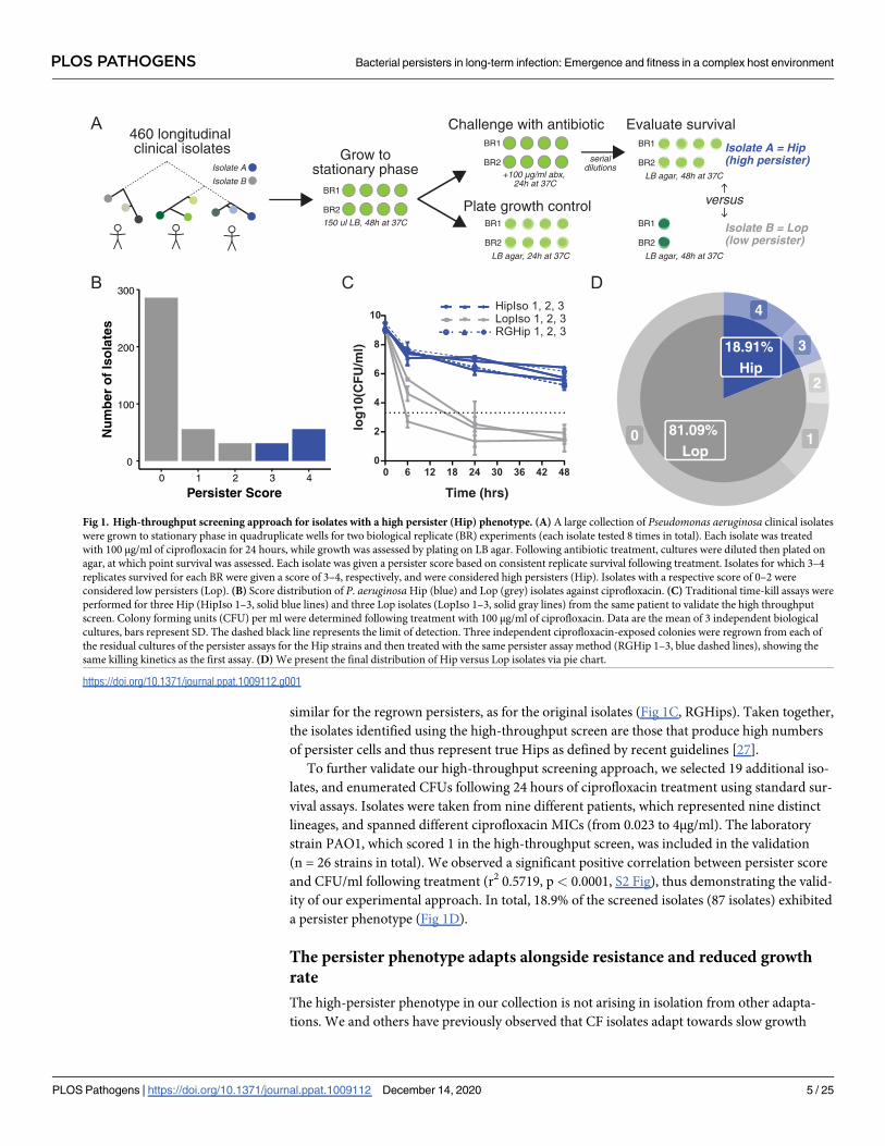
similar for the regrown persisters, as for the original isolates (Fig 1C, RGHips). Taken together,
the isolates identified using the high-throughput screen are those that produce high numbers
of persister cells and thus represent true Hips as defined by recent guidelines [27].
To further validate our high-throughput screening approach, we selected 19 additional iso-
lates, and enumerated CFUs following 24 hours of ciprofloxacin treatment using standard sur-
vival assays. Isolates were taken from nine different patients, which represented nine distinct
lineages, and spanned different ciprofloxacin MICs (from 0.023 to 4μg/ml). The laboratory
strain PAO1, which scored 1 in the high-throughput screen, was included in the validation
(n = 26 strains in total). We observed a significant positive correlation between persister score
and CFU/ml following treatment (r2 0.5719, p< 0.0001, S2 Fig), thus demonstrating the valid-
ity of our experimental approach. In total, 18.9% of the screened isolates (87 isolates) exhibited
a persister phenotype (Fig 1D).
The persister phenotype adapts alongside resistance and reduced growth
rate
The high-persister phenotype in our collection is not arising in isolation from other adapta-
tions. We and others have previously observed that CF isolates adapt towards slow growth
460 longitudinal clinical isolates Grow to
stationary phaseBR1
BR2
150 ul LB, 48h at 37C
BR1
BR2+100 µg/ml abx,
24h at 37C
Challenge with antibiotic
Plate growth controlBR1
BR2
LB agar, 24h at 37C
BR1
BR2
LB agar, 48h at 37C
BR1
BR2
LB agar, 48h at 37C
Isolate A
Isolate B
Evaluate survival
Isolate A = Hip(high persister)
Isolate B = Lop(low persister)
versus
serialdilutions
0
100
200
300
0 1 2 3 4
Persister Score
Nu
mb
er o
f Is
ola
tes
A
B C D
Time (hrs)
log
10(C
FU
/ml)
4
3
2
10
18.91%Hip
81.09%Lop
6 12 18 24 30 36 42 480
2
4
6
8
10HipIso 1, 2, 3LopIso 1, 2, 3RGHip 1, 2, 3
0
Fig 1. High-throughput screening approach for isolates with a high persister (Hip) phenotype. (A) A large collection of Pseudomonas aeruginosa clinical isolates
were grown to stationary phase in quadruplicate wells for two biological replicate (BR) experiments (each isolate tested 8 times in total). Each isolate was treated
with 100 μg/ml of ciprofloxacin for 24 hours, while growth was assessed by plating on LB agar. Following antibiotic treatment, cultures were diluted then plated on
agar, at which point survival was assessed. Each isolate was given a persister score based on consistent replicate survival following treatment. Isolates for which 3–4
replicates survived for each BR were given a score of 3–4, respectively, and were considered high persisters (Hip). Isolates with a respective score of 0–2 were
considered low persisters (Lop). (B) Score distribution of P. aeruginosaHip (blue) and Lop (grey) isolates against ciprofloxacin. (C) Traditional time-kill assays were
performed for three Hip (HipIso 1–3, solid blue lines) and three Lop isolates (LopIso 1–3, solid gray lines) from the same patient to validate the high throughput
screen. Colony forming units (CFU) per ml were determined following treatment with 100 μg/ml of ciprofloxacin. Data are the mean of 3 independent biological
cultures, bars represent SD. The dashed black line represents the limit of detection. Three independent ciprofloxacin-exposed colonies were regrown from each of
the residual cultures of the persister assays for the Hip strains and then treated with the same persister assay method (RGHip 1–3, blue dashed lines), showing the
same killing kinetics as the first assay. (D) We present the final distribution of Hip versus Lop isolates via pie chart.
https://doi.org/10.1371/journal.ppat.1009112.g001
PLOS PATHOGENS Bacterial persisters in long-term infection: Emergence and fitness in a complex host environment
PLOS Pathogens | https://doi.org/10.1371/journal.ppat.1009112 December 14, 2020 5 / 25

rates and increased resistance to antibiotics, and some lineages develop towards a biofilm life-
style [20,28,29]. Supporting a theory that persistence enables resistance, studies have observed
the emergence of persister isolates before resistant isolates in vitro [8,30], with a recent study
showing this relationship in vivo for two bacteremia patients infected by Staphylococcus aureus[31]. A specific association between slowing growth rate and the Hip phenotype has also been
proposed [32]. To broadly survey overlap with other phenotypes, we used a principal compo-
nent analysis to evaluate the distribution of Hip (blue diamonds) versus Lop (grey circles) vari-
ants by multiple traits under selection pressure in the CF lung using previously published
phenotype data for most isolates (described further in Materials and Methods) [20]. We see
that Hip variants group with isolates exhibiting more adapted traits (increased antibiotic MICs
and slowing growth), but they also appear across the full phenotypic space alongside Lop iso-
lates (Fig 2). We further specifically identified the first Hip variant (FirstHip–blue ellipse with
yellow fill) of each lineage (a clone type infecting a given patient) in an attempt to assess these
isolates’ other traits and state of adaptation at first appearance, when the accumulated impact
of co-evolving traits is lowest on the Hip isolates. We see that FirstHips overlap substantially
with both Lops and Hips. This variation of initial adaptive state could be due to different adap-
tive trajectories with patients as well as lapses of time between Hip emergence and isolation.
Given recent literature on resistance and slowed growth, we specifically compared isolate
persister class versus ciprofloxacin resistance as well as slowed growth rate. Thirty-nine per-
cent of the isolates were characterized as resistant to ciprofloxacin based on the EUCAST
breakpoint (176 of 451 isolates with available data). Sixty-three of 87 Hip isolates were resistant
(72.4%), while 113 of 251 Lops were resistant (31%). Forty percent of isolates showed growth
rate reduction (183 of 460 isolates), defined as less than 70% of the growth rate of robustly
growing lab strain P. aeruginosa PAO1. Fifty-nine of 87 Hip isolates were slow growers (87%),
whereas 124 of 373 Lops were slow growers (33.2%). These numbers suggest associations
between growth rate reduction, resistance, and Hip isolates. However, we intentionally avoid
statistical tests for significance as these isolates are affected by lineage bias in both parallel trait
adaptation and varying number of isolates cultured for each patient lineage.
Instead, we contrasted the time of emergence of these trait adaptations within each affected
lineage both individually and in combination with the date of first Hip isolation (Table 1). We
note that our lineage classifications do not guarantee trait continuance within a lineage or co-
occurrence within the same isolate, i.e. lineages are capable of producing isolates with trait(s),
but individual isolates may not share the set of phenotypes. First, 24 of 74 lineages showed Hip
isolate emergence (Hip+ lineages), 35 lineages showed resistant isolate emergence, and 34 line-
ages showed growth-reduced isolate emergence. Resistant isolates preceded Hip isolates in 8
lineages, emerged simultaneously in 11 lineages, and follow Hip in 2 lineages. With respect to
growth rate reduction, we observed that reduction precedes persistence in 9 lineages, emerges
simultaneously in 7 lineages, and follows persistence in 5 lineages.
Importantly, we can confirm when comparing data in the ‘Either trait’ versus ‘Both traits’
columns that persister, resistance, and growth reduction traits are only partially overlapping as
they evolve. For example, both traits adapt before Hip emergence in 4 lineages, but at least one
adapts before Hip emergence in 13 lineages. In contrast, persisters emerge in 5 lineages before
at least one of the traits has adapted, but only emerge earlier in 2 lineages if either trait is
assessed. Only 7 lineages show emergence of both slowed growth isolates and resistance iso-
lates without Hips (‘Both Traits’), while Hip isolates emerge alone in only 1 lineage (‘Either
Trait’). The multi-way mosaic plot of Fig 2A shows presence-absence of isolates with each trait
across all lineages (not accounting for emergence time), which emphasizes the significance of
this lone persister-only lineage (red). In total, these results emphasize the complexity of
PLOS PATHOGENS Bacterial persisters in long-term infection: Emergence and fitness in a complex host environment
PLOS Pathogens | https://doi.org/10.1371/journal.ppat.1009112 December 14, 2020 6 / 25

A
B
−2.2
0.0
2.0
4.0
6.0
Pearsonresiduals:
p−value =1.2639e−14
Cip Resistant
Gro
wth
Rat
e R
edu
ced
Per
sist
er
No Yes
Yes
Yes
No
No
Yes
No
30
6
7
7
1
2 19
2
# Lineages with isolates exhibiting trait(s)
Lop HipFirstHip
Adhesion
cipGR_LB
−0.10
−0.05
0.00
0.05
0.10
0.15
−0.05 0.00 0.05 0.10 0.15PC1 (45.97%)
PC
2 (3
3.2%
)
Fig 2. High-persisters in the multi-trait landscape. (A) Lop (grey circle) and Hip (blue diamonds were analyzed via
principle component analysis with respect to their similarity with other infection-linked traits: growth rate (GR_LB),
adhesion, and ciprofloxacin MIC (cip). 446 isolates with complete trait sets were included. Hip isolates do not
consistently cluster with any one additional trait. Each symbol represents a P. aeruginosa isolate. The first Hip isolates
from a lineage (FirstHip, yellow triangles) were highlighted as Hip variants with mitigated effects of other
accumulating mutations within the lineage to improve cross-lineage comparison. In each case, FirstHip and the
remaining Hip isolates shift to various degrees from ‘naïve’ towards ‘adapted’ levels given the particular Hip dataset.
We illustrate this using data ellipse enclosing samples approximately within the first standard deviation (t distribution,
68% of the set) for isolate sets characterized as FirstHip (yellow ellipse), and the remaining Hips (blue ellipse). (B) We
visualized the association between lineages that produced Hips versus resistant isolates and/or slow growing isolates
(identified by the minimum growth rate of lineage isolates falling below 70% of the P. aeruginosa PAO1 growth rate
based on a 45 minute generation time in LB in microtiter wells). Association between variables is illustrated by a
PLOS PATHOGENS Bacterial persisters in long-term infection: Emergence and fitness in a complex host environment
PLOS Pathogens | https://doi.org/10.1371/journal.ppat.1009112 December 14, 2020 7 / 25
selection pressures at play over time on these lineages, resulting in concurrent adaptation of
distinct traits that likely influence each other and have related genetic underpinnings.
Evolution of the persister phenotype is not genetically convergent across
patient-specific lineages
Sequencing and identification of genetic variations accumulating within each clone type was
performed in a previous study [24] for most of the isolates included in the current screen (403
isolates, 46 lineages). In that previous study, genes targeted in convergent evolution were iden-
tified by the significant enrichment of observed lineages with mutations in those genes com-
pared to the number of lineages expected to have mutations in the same genes according to
genetic drift (derived from a simulated evolution where lineages accumulate an equivalent
number of mutations randomly for 1000 independent evolution simulations) [24]. In our cur-
rent analysis, we split our dataset into Hip and Lop variants, and then performed this same
observed versus expected lineage enrichment analysis for each population (see Materials and
Methods for further details). The ratio of lineage enrichment of mutated genes for Hip versus
Lop variants allowed us to identify candidate ‘hip’ genes for each set (Table 2). For complete-
ness, we also performed an additional genetic analysis focusing on mutations in non-coding
sequences (S2 Dataset).
In general, searching for hip genes accumulating non-synonymous mutations in Hip+ line-
ages revealed only a weak signal for convergent evolution. Only one lineage assessed in the
genetic screen had only Hips present (the only isolate of the lineage assessed in our screen), so
practically all lineages with Hip isolates (Hip+ lineages, 29 included in the genetic study) also
contained Lops. Thus, mutated genes that were enriched 2–3 fold in independently evolved
Hip+ lineages were also frequently present in Lop isolates of the same lineage. Our lineage
enrichment ratio ultimately identified 12 mutated genes enriched in ciprofloxacin Hip+ line-
ages (Table 2).
Of note, there was a surprising lack of the most prominent ‘hip’ genes previously identified
in in vitro studies and screens of P. aeruginosa (S1 Table). None of the lineage enrichment data
pointed toward RNA endonuclease-type toxin-antitoxin systems under adaptive selection,
which supports recent research that has questioned the contribution of these systems to persis-
tence in numerous bacterial pathogens [33–35]. Instead, they belonged to diverse functional
mosaic plot (multi-way contingency table visualization) where color indicates significant deviation from the expected
frequency of lineages in each cell under trait independence using Pearson’s chi-squared test. Presence of isolates with a
given trait in a lineage is indicated by ‘Yes’, absence by ‘No’.
https://doi.org/10.1371/journal.ppat.1009112.g002
Table 1. Emergence of adapted trait isolates versus persister isolates across 74 lineages. Traits assessed included resistance to ciprofloxacin according to the EUCAST
breakpoint of 0.5 μg/ml and reduction in growth (isolates which grew slower than 70% of the growth rate of robust lab strain P. aeruginosa PAO1 grown in similar condi-
tions, i.e. a generation time of greater than 65 minutes). Date of first trait emergence was calculated for the adaptation of each trait, the minimum first date of either trait
(in lineages where both adapted) as ‘Either Trait’, and the date by which both traits had adapted (Both Traits). These dates were then compared with the date of first Hip
emergence.
Category # Lineages affected
Both Traits Ciprofloxacin Resistance Growth Rate Reduction Either TraitTrait before persister adaptation 4 8 9 13
Simultaneous emergence 10 11 7 8
Persister before trait adaptation 5 2 5 2
Trait adaptation alone 7 14 13 20
Persister adaptation alone 5 3 3 1
https://doi.org/10.1371/journal.ppat.1009112.t001
PLOS PATHOGENS Bacterial persisters in long-term infection: Emergence and fitness in a complex host environment
PLOS Pathogens | https://doi.org/10.1371/journal.ppat.1009112 December 14, 2020 8 / 25

categories including transcriptional regulation/two component regulatory systems (3 genes),
energy metabolism (2 genes) DNA replication and repair (1 gene), and virulence (2 genes, iron
uptake and a type VI protein secretion system). Table 2 includes major regulators rpoN,
known to induce a growth defect when functionally mutated [36], and retS, which when func-
tionally mutated induces an array of phenotypic changes linked to chronic infection such as a
non-motile biofilm lifestyle [37,38]. retS and hypothetical protein PA0977 also overlap with
the ‘pathoadaptive’ mutationally enriched gene list identified in our prior study of convergent
evolution across all lineages [24].
Hip variants emerge via diverse incidence patterns
The lack of strong genetic signatures differentiating Hip from Lop isolates motivated us to
examine the temporal dynamics of high persister incidence from a lineage perspective,
where each strain (clone-type) infecting each patient is classed as an independent lineage.
In half of the patients, the earliest P. aeruginosa isolate in our collection is also the first-ever
identified P. aeruginosa in the clinic and the other patients’ isolates also cover most of the
initial colonization phase. We can thus estimate the emergence of the Hip phenotype as P.
aeruginosa adapts from a wild type-similar naïve state into an adapted persistent pathogen.
Previous findings have indicated that the number of Hip variants from a lineage may
increase over time as the bacteria adapt to the antibiotic pressure in the host, and that once
a Hip isolate is observed, it is assumed to persist in the infecting population of the patient
[9,18].
To illustrate the range of persister dynamics we observe, we grouped each lineage by an
array of descriptors. The lineage descriptors include Hip presence versus absence (Hip+ vs
Hip-), presence of multiple Hips in the same lineage (MultiHip), transience of the lineage
(whether it appears for less than 2 years, less than half the length of a patient’s infection and is
afterwards replaced by another lineage), continuity of Hip variants (whether Hips are present
for at least 3 sampling dates in a row), and whether a Hip variant initiates the lineage. Fig 3A
shows the ordered distribution of the lineages in 10 different groups based on descriptor sets,
illustrating the diversity of lineage Hip dynamics. We see that: 1) 24 of 74 lineages are Hip+,
Table 2. Lineage-based mutation enrichment analysis. Mutated genes enriched in Hip versus Lop dataset as assessed from a convergent evolution perspective account-
ing for lineage adaptation. Lineage enrichment ratio was calculated by dividing lineage-based gene mutation enrichment within Hip variants by that within Lop variants
for each gene. Top Hip-linked genes were selected via the following criteria: greater than 2 lineages presenting mutations in that gene in the Hip population and a lineage
enrichment ratio greater than 2.
Locus Gene
Name
Gene Function Hip+ Lineage
Count
Hip- Lineage
Count
Lineage Enrichment
Ratio
Total Lineages
Hit
PA0339 hypothetical protein 3 2 3.098 3
PA2894 hypothetical protein 4 3 3.062 4
PA4462 rpoN RNA polymerase sigma-54 factor 3 2 2.985 3
PA4856 retS RetS (Regulator of Exopolysaccharide and Type III
Secretion)
5 5 2.482 5
PA0977 hypothetical protein 3 3 2.395 3
PA3183 zwf glucose-6-phosphate 1-dehydrogenase 4 4 2.391 4
PA1224 probable NAD(P)H dehydrogenase 3 3 2.186 3
PA1600 probable cytochrome c 3 3 2.179 3
PA2685 vgrG4 VgrG4 3 3 2.099 3
PA3640 dnaE DNA polymerase III, alpha chain 3 3 2.093 3
PA1380 probable transcriptional regulator 3 3 2.057 3
PA2403 fpvG FpvG 3 3 2.006 3
https://doi.org/10.1371/journal.ppat.1009112.t002
PLOS PATHOGENS Bacterial persisters in long-term infection: Emergence and fitness in a complex host environment
PLOS Pathogens | https://doi.org/10.1371/journal.ppat.1009112 December 14, 2020 9 / 25
2), 17 of 24 Hip+ lineages produce multiple Hip isolates, 3) 30 of 50 Hip- lineages are transient,
while 2 Hip+ lineages are transient, 4) 6 lineages exhibit continuous periods of Hip variants,
and 5) 4 lineages have initiating Hip+ variants. Thus, the fraction (18.9%, Fig 1D) of total iso-
lates with a Hip phenotype appears to be distributed over a subset of lineages (32.4%) in both
stable (continuous) and stochastic patterns of incidence (Fig 3A), rather than present in every
evolving lineage.
A
B D
Lineage 9
Lineage 8
Lineage 7
Lineage 6
Lineage 5
Lineage 4
Lineage 3
Lineage 2
Lineage 1
Rep
rese
nta
tive
Lin
eag
e
−2.6
0.0
2.0
3.1
Pearsonresiduals:
p−value =4.8124e−06
Transient Lineage
Per
sist
er P
rese
nt
Hip
−H
ip+
Yes No New
2 16 6
30 7 13
30
20
10
5
4
2
1
1
1
102030
Number of Lineages
LopO
nly
Tran
sient
CT
HipPre
sent
Mul
tiHip
Contin
uous
Initia
ting
0 2 4 6Time since First Detection (years)
lop hip
Hip+ Hip+ (Accumulated)Hip-Time since First Detection (y)
Lin
eag
es
0
20
40
60
0 2 4 6 8
+++++++++++++
++++
+++
+ ++
+
0.00
0.25
0.50
0.75
1.00
0 2 4 6
Pro
bab
ility
of
no
Hip
em
erg
ence
Time since First Detection (y)
C
Fig 3. High-persister incidence patterns from a lineage-based perspective. (A) Lineages were classed according to several nested characteristics: transient versus non-
transient lineages, Hip presence, presence of multiple Hips, continuous periods of isolated Hips, and lineage-initiating Hips. Lineages representing each combination of
traits are shown on the left (Hip blue diamonds, Lop grey circles), while characteristic sets are identified and enumerated for the entire collection on the right. (B) The
continuous lineage count of Hip- lineages (grey circles) versus Hip+ lineages (blue diamonds) for the prior year of colonization is plotted, while the accumulating count
of Hip+ lineages from time 0 is shown by black diamonds. (C) A survival curve shows the probability of no Hip emergence over time since first detection of a lineage,
based on time from first isolate to first Hip isolate for each lineage. The step function is shown in dark gray with confidence band in light gray. Lineages which never
produce a Hip isolate are censored at time of last isolate (black crosses). (D) Transient lineages (lineages of shorter than 2 years duration, less than 50% of total patient
infection length, and which are followed by the appearance of a new lineage) are significantly associated with lineages lacking Hips (Hip-), while non-transient lineages
are associated with the presence of Hips based on Pearson’s chi-squared test (via a mosaic plot visualizing a multi-way contingency table). Transience-unclassifiable
lineages of shorter than 2 years’ duration at the end of a patient’s collection period are shown for context (‘New’).
https://doi.org/10.1371/journal.ppat.1009112.g003
PLOS PATHOGENS Bacterial persisters in long-term infection: Emergence and fitness in a complex host environment
PLOS Pathogens | https://doi.org/10.1371/journal.ppat.1009112 December 14, 2020 10 / 25
Hip variants accumulate over colonization time and occur rarely in
transient lineages
We summarized the incidence of Hip variants over each lineage’s time of colonization in Fig
3B. Here we plot the continuous counts of patients exhibiting the Hip phenotype (Hip+) ver-
sus no Hip presence (Hip-) within the previous year (dashed lines) as well as the accumulation
of lineages that have exhibited a Hip variant at least once by a certain age of colonization (solid
line). This illustrates the number of lineages assessed at a given colonization age and the
increasing fraction of Hip+ lineages over time, respectively. Overall, Hip variants affect 24 line-
ages (Fig 3B) and 56% of the patients in our study cohort (S3 Fig) by the end of the study
period. Fig 3C shows the probability of no Hip emergence (i.e. lineage survival without Hip)
over time since the lineage was first detected, which shows the impact of lineages without Hips
disappearing (the black crosses indicate where they have been censored at their last collected
isolate date) in addition to the step decreases associated with the date of first Hip in Hip+ line-
ages. We investigated this phenomenon further by evaluating the relationship between lineage
transience and Hip presence. We first mark the lineages present for less than 2 years in a
patient at the end of their monitoring period as ‘New’ lineages since we cannot determine tran-
sience without additional samples. Of the 55 remaining lineages, non-transient lineages are
significantly associated with the Hip+ lineage status (Fig 3D). Thus, a given patient often has
multiple infecting lineages, but the Hip- lineages are much more likely to disappear over the
course of infection.
Hip variants associate with frequent antibiotic treatment and eradication
failure in patients
The few persister studies that incorporate clinical data have shown the emergence of Hips after
antibiotic treatment, with a recent work showing how multi-drug treatment induces tolerance
before resistance in two patients infected with methicillin-resistant Staphylococcus aureus [31].
Here, we have the opportunity to analyze the relationship between treatment and the Hip phe-
notype within a substantial patient population under similar treatment regimens. At the
Copenhagen CF Center, patients are treated aggressively with a broad range of antibiotics,
including 9 classes of P. aeruginosa targeting treatments (aminoglycosides, carbapenems,
cephalosporins, fluoroquinolones, macrolides, monobactams, penicillins, and penicillins
paired with beta-lactamases). Patients are regularly prescribed combination therapy using flu-
oroquinolones, polymyxins, and aminoglycosides (ciprofloxacin-colistin or ciprofloxacin-
tobramycin), and treatment is adjusted and recorded at ~monthly clinic visits.
Here, we compare Hip incidence with a patient’s length of treatment time as well as counts
of the months a given drug is prescribed (collapsing IV versus per-oral versus inhaled treat-
ment together for expediency). We assess the data in a patient-specific manner from one year
prior to their first isolate assessed in this study to their last isolate (Hip+ and Hip- patient cate-
gories) as well as from one year prior to their first isolate to their first Hip isolation date (Hip
+ before FirstHip, truncating the Hip+ patient records). Fig 4A and 4B shows different metrics
of fluoroquinolone treatment for these groups: Total time treated with a fluoroquinolone nor-
malized by patient monitoring timespan and the longest continuous treatment period (by
months in a row that a drug is prescribed). These data show that while Hip+ patients receive
on average much more fluoroquinolone treatment than Hip- patients, assessing Hip- patients
versus Hip+ patients before their first Hip shows similar antibiotic usage in these more directly
comparable timeframes (Fig 4A and 4B, means compared by unpaired Wilcoxon test). Even
non-normalized total time of fluoroquinolone treatment is not significantly different between
Hip- patients versus Hip+ patients before their first Hip. Furthermore, an assessment of
PLOS PATHOGENS Bacterial persisters in long-term infection: Emergence and fitness in a complex host environment
PLOS Pathogens | https://doi.org/10.1371/journal.ppat.1009112 December 14, 2020 11 / 25
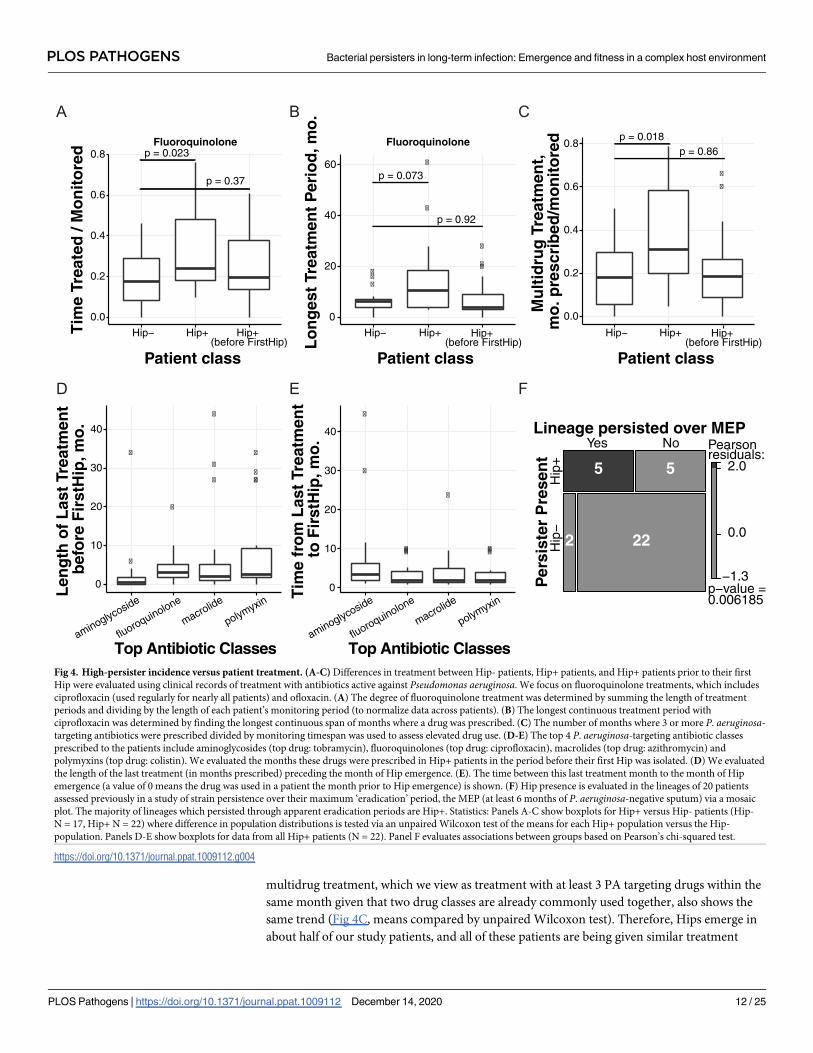
multidrug treatment, which we view as treatment with at least 3 PA targeting drugs within the
same month given that two drug classes are already commonly used together, also shows the
same trend (Fig 4C, means compared by unpaired Wilcoxon test). Therefore, Hips emerge in
about half of our study patients, and all of these patients are being given similar treatment
p = 0.023
p = 0.37
0.0
0.2
0.4
0.6
0.8
Hip− Hip+ Hip+ (before FirstHip)
Patient class
Tim
e Tr
eate
d /
Mo
nit
ore
d
Fluoroquinolone
���
�
�
�
��
p = 0.073
p = 0.92
0
20
40
60
Hip− Hip+
Patient class
Lo
ng
est
Trea
tmen
t P
erio
d, m
o.
Fluoroquinolone
�
�
p = 0.86
0.0
0.2
0.4
0.6
0.8
Hip− Hip+
Patient class
Mu
ltid
rug
Tre
atm
ent,
m
o. p
resc
rib
ed/m
on
ito
red
�
�
�
�
�
�
�
���
0
10
20
30
40
aminoglycoside
fluoroquinolone
macrolide
polymyxin
Len
gth
of
Las
t Tr
eatm
ent
bef
ore
Fir
stH
ip, m
o.
�
�
����
�
���
0
10
20
30
40
Tim
e fr
om
Las
t Tr
eatm
ent
to F
irst
Hip
, mo
.Hip+
(before FirstHip)Hip+
(before FirstHip)
aminoglycoside
fluoroquinolone
macrolide
polymyxin
−1.3
0.0
2.0
Pearsonresiduals:
p−value =0.006185
Lineage persisted over MEP
Per
sist
er P
rese
nt
Hip
−H
ip+
Yes No
5 5
222
A
D E
Top Antibiotic Classes Top Antibiotic Classes
F
B Cp = 0.018
Fig 4. High-persister incidence versus patient treatment. (A-C) Differences in treatment between Hip- patients, Hip+ patients, and Hip+ patients prior to their first
Hip were evaluated using clinical records of treatment with antibiotics active against Pseudomonas aeruginosa. We focus on fluoroquinolone treatments, which includes
ciprofloxacin (used regularly for nearly all patients) and ofloxacin. (A) The degree of fluoroquinolone treatment was determined by summing the length of treatment
periods and dividing by the length of each patient’s monitoring period (to normalize data across patients). (B) The longest continuous treatment period with
ciprofloxacin was determined by finding the longest continuous span of months where a drug was prescribed. (C) The number of months where 3 or more P. aeruginosa-targeting antibiotics were prescribed divided by monitoring timespan was used to assess elevated drug use. (D-E) The top 4 P. aeruginosa-targeting antibiotic classes
prescribed to the patients include aminoglycosides (top drug: tobramycin), fluoroquinolones (top drug: ciprofloxacin), macrolides (top drug: azithromycin) and
polymyxins (top drug: colistin). We evaluated the months these drugs were prescribed in Hip+ patients in the period before their first Hip was isolated. (D) We evaluated
the length of the last treatment (in months prescribed) preceding the month of Hip emergence. (E). The time between this last treatment month to the month of Hip
emergence (a value of 0 means the drug was used in a patient the month prior to Hip emergence) is shown. (F) Hip presence is evaluated in the lineages of 20 patients
assessed previously in a study of strain persistence over their maximum ‘eradication’ period, the MEP (at least 6 months of P. aeruginosa-negative sputum) via a mosaic
plot. The majority of lineages which persisted through apparent eradication periods are Hip+. Statistics: Panels A-C show boxplots for Hip+ versus Hip- patients (Hip-
N = 17, Hip+ N = 22) where difference in population distributions is tested via an unpaired Wilcoxon test of the means for each Hip+ population versus the Hip-
population. Panels D-E show boxplots for data from all Hip+ patients (N = 22). Panel F evaluates associations between groups based on Pearson’s chi-squared test.
https://doi.org/10.1371/journal.ppat.1009112.g004
PLOS PATHOGENS Bacterial persisters in long-term infection: Emergence and fitness in a complex host environment
PLOS Pathogens | https://doi.org/10.1371/journal.ppat.1009112 December 14, 2020 12 / 25

rather than any extra treatment applied to the Hip+ population before Hip emergence. How-
ever, Hip+ patients do on average undergo more aggressive treatment than Hip- patients
when their entire monitoring period is assessed (Fig 4A, 4B, and 4C).
For the most commonly prescribed drug classes in our cohort, we look further into the Hip
+ dataset before the first Hip is isolated (Fig 4D and 4E). First, we see that a few patients have
not been treated with fluoroquinolone at all prior to their first Hip (Fig 4D). The median
length of the fluoroquinolone treatment directly before the first Hip is 3 months where the
antibiotic was prescribed, but this value is not effectively different than the median for macro-
lides or polymyxins (2 and 2.5 months, respectively, Fig 4D). We show in Fig 4E that the
median of time between the last fluoroquinolone treatment and first Hip isolation (N = 20 of
22 Hip+ patients) is 2 months, and this trend again also holds true for macrolides (N = 21 of
22 Hip+ patients) and polymyxins (N = 20 of 22 Hip+ patients). Our first Hips therefore often
arise after a recent treatment for multiple months with a panel of PA targeting drugs. However,
15 of 17 Hip- patients also receive similar multi-drug treatments. Ultimately, we cannot con-
clude that fluoroquinolone treatment, rather than a panel of drugs, is specifically driving the
development of and selection for Hips identified via a ciprofloxacin screen, and we again do
not observe evidence for multi-drug treatment inducing the first Hip isolate.
While the relationship between antibiotic treatment and the Hip phenotype is complex, we
see more definitive patterns in an assessment of treatment failure which connects our analysis
of Hip presence in non-transient lineages to a clinically relevant diagnostic (Fig 3C). A recent
study of strain eradication, defined in the Copenhagen clinic as a failure to culture P. aerugi-nosa from a six month period of routine sputum sampling, evaluated whether strains in fact
persisted over the longest supposed eradication period in a patient’s clinical history (the maxi-
mum eradication period, or MEP). Historically, eradication periods are viewed as markers of
treatment success, and strains that recur after eradication periods are assumed to represent
new infections, but our study showed that over 40% of patients had a strain persist over their
maximum eradication period[39]. As these adapted strains are likely less susceptible to treat-
ment than a new infection, this misdiagnosis of eradication is of great concern for the clinic
and should influence treatment decisions. We assessed whether strains that persisted versus
were eradicated over the MEP produced Hip isolates in the 20 patients which were also
included in this study. As Fig 4F shows via a mosaic plot, 5 of 7 strains that persisted in 7
respective patients also produced Hip isolates. Four of these strains produced a Hip before the
MEP within the representative isolates which we assayed here, and one produced a Hip before
a second ‘eradication’ period nearly equal to the length of the MEP (.86 versus .89 years ‘eradi-
cated’). In contrast, only 5 of 22 strains that disappeared from patient lungs prior to a true
MEP actually produced Hips. This association supports the theory that the Hip phenotype
contributes to treatment failure, and provides an explanation for why the clinic’s culture-based
eradication metric and associated treatment guidelines are insufficient for many infections.
Hip variants show increased fitness in patient-similar biofilms
We directly tested whether Hip isolates are able to survive antibiotic treatment better than Lop
isolates with similar antibiotic susceptibilities and growth properties in more complex condi-
tions. We simulated antibiotic treatment of CF patients in a recently developed biofilm Phar-
macokinetic/Pharmacodynamic (PK/PD) system, in which the bacteria are challenged with
antibiotics in much the same way as in patients [40]. We chose this model because P. aerugi-nosa often appears as structured populations in lungs of CF patients [41], because biofilms
have been shown to harbor increased levels of persister cells [42], and because our model mim-
ics the bacterial exposure to ciprofloxacin treatment as described for CF patients [40,43]. The
PLOS PATHOGENS Bacterial persisters in long-term infection: Emergence and fitness in a complex host environment
PLOS Pathogens | https://doi.org/10.1371/journal.ppat.1009112 December 14, 2020 13 / 25
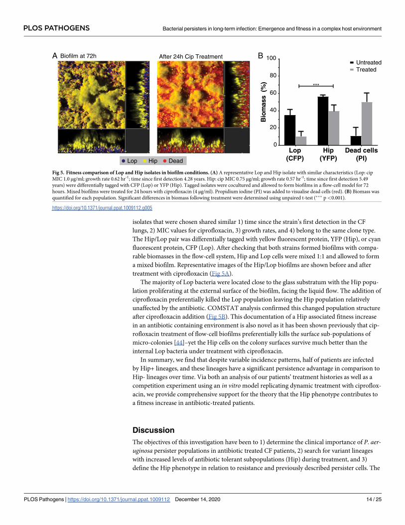
isolates that were chosen shared similar 1) time since the strain’s first detection in the CF
lungs, 2) MIC values for ciprofloxacin, 3) growth rates, and 4) belong to the same clone type.
The Hip/Lop pair was differentially tagged with yellow fluorescent protein, YFP (Hip), or cyan
fluorescent protein, CFP (Lop). After checking that both strains formed biofilms with compa-
rable biomasses in the flow-cell system, Hip and Lop cells were mixed 1:1 and allowed to form
a mixed biofilm. Representative images of the Hip/Lop biofilms are shown before and after
treatment with ciprofloxacin (Fig 5A).
The majority of Lop bacteria were located close to the glass substratum with the Hip popu-
lation proliferating at the external surface of the biofilm, facing the liquid flow. The addition of
ciprofloxacin preferentially killed the Lop population leaving the Hip population relatively
unaffected by the antibiotic. COMSTAT analysis confirmed this changed population structure
after ciprofloxacin addition (Fig 5B). This documentation of a Hip associated fitness increase
in an antibiotic containing environment is also novel as it has been shown previously that cip-
rofloxacin treatment of flow-cell biofilms preferentially kills the surface sub-populations of
micro-colonies [44]–yet the Hip cells on the colony surfaces survive much better than the
internal Lop bacteria under treatment with ciprofloxacin.
In summary, we find that despite variable incidence patterns, half of patients are infected
by Hip+ lineages, and these lineages have a significant persistence advantage in comparison to
Hip- lineages over time. Via both an analysis of our patients’ treatment histories as well as a
competition experiment using an in vitromodel replicating dynamic treatment with ciproflox-
acin, we provide comprehensive support for the theory that the Hip phenotype contributes to
a fitness increase in antibiotic-treated patients.
Discussion
The objectives of this investigation have been to 1) determine the clinical importance of P. aer-uginosa persister populations in antibiotic treated CF patients, 2) search for variant lineages
with increased levels of antibiotic tolerant subpopulations (Hip) during treatment, and 3)
define the Hip phenotype in relation to resistance and previously described persister cells. The
Lop(CFP)
Hip(YFP)
Dead cells(PI)
0
20
40
60
80
100
Bio
mas
s(%
)
UntreatedTreated
***
Lop Hip Dead
Biofilm at 72h After 24h Cip TreatmentA B
Fig 5. Fitness comparison of Lop and Hip isolates in biofilm conditions. (A) A representative Lop and Hip isolate with similar characteristics (Lop: cip
MIC 1.0 μg/ml; growth rate 0.62 hr-1; time since first detection 4.28 years. Hip: cip MIC 0.75 μg/ml; growth rate 0.57 hr-1; time since first detection 5.49
years) were differentially tagged with CFP (Lop) or YFP (Hip). Tagged isolates were cocultured and allowed to form biofilms in a flow-cell model for 72
hours. Mixed biofilms were treated for 24 hours with ciprofloxacin (4 μg/ml). Propidium iodine (PI) was added to visualise dead cells (red). (B) Biomass was
quantified for each population. Significant differences in biomass following treatment were determined using unpaired t-test (��� p<0.001).
https://doi.org/10.1371/journal.ppat.1009112.g005
PLOS PATHOGENS Bacterial persisters in long-term infection: Emergence and fitness in a complex host environment
PLOS Pathogens | https://doi.org/10.1371/journal.ppat.1009112 December 14, 2020 14 / 25

background is an increasing awareness of antibiotic treatment failure in the CF clinics, which
cannot be ascribed to antibiotic resistance development in the early years of infection.
We have mapped the prevalence of high-persisters in a large, aligned cohort of patients
under intensive antibiotic treatment for a 10 year period [24,43]. Of 460 P. aeruginosa isolates
from the airways of 39 young CF patients (74 lineages in total), 18.9% of the isolates were
scored as robustly persisting Hip using a high-throughput screening approach to assay persis-
tence against ciprofloxacin (Fig 1). We show that the isolates display different levels of persist-
ers, in accordance with the variance previously found between species and within strains
[5,45,46]. Most adaptive changes occur during the first few years of colonization [20,47],
which matches our objective of searching for signs of increased fitness of Hip variants in
patients treated continuously with antibiotics. We show that in a young CF patient cohort
impacted by early longitudinal colonization by P. aeruginosa strains, Hip variants were sam-
pled from 56% of the patients (N = 22) during a 10-year observation window. Our analysis is a
new and useful comparative baseline for developing effective surveillance, impact assessment,
and eventual control of the persister phenotype in the clinic.
Multiple relationships between the Hip phenotype and other phenotypic traits such as
growth rate and antibiotic resistance have been suggested in the literature. While some studies
point out that there is no correlation between the mean growth rates of isolates and the
observed Hip phenotype [48–50], reduced growth rates have been associated with high per-
sister phenotypes in E. coli [32]. A recent study in Salmonella enterica further supports that
slow growth (regardless of mechanism) promotes the high-persister phenotype [35]. Drug-tol-
erant cells have also been proposed to facilitate evolution of true antibiotic resistance in E. coli,where intermittent antibiotic exposure of a batch culture selected for mutant clones harboring
tolerance mutations that increased the growth lag-time, during which tolerance to killing by
ampicillin selected for MIC-increasing mutations [30]. Early isolates of a S. aureus lineage
from a patient treated with antibiotics also acquired tolerance, and subsequently resistance
[31]. Though P. aeruginosa in the CF lung is also exposed to fluctuating concentrations of anti-
biotic, we see that our lineages which produce Hips are also often producing isolates with
reduced growth rate and/or ciprofloxacin resistance over time (Fig 2). The stringently defined
persister phenotype described in the current study is often observed after one of these trait
adaptations if not both. In summary, our results suggest that the Hip phenotype is an early and
common advantageous adaptation [20] arising stochastically in infected patients alongside
other beneficial adaptations.
Many studies have shown the increased survival of persister cells under antibiotic treatment
[32,51] and then screened for genetic determinants of persistence, but few have evaluated the
fitness of Hip versus Lop variants in direct competition experiments [45]. In our study, we
tested a single Lop/Hip pair of isolates matched by genotype, phenotype, and colonization age
in order to characterize the selective advantage of the Hip phenotype in a biofilm under treat-
ment-replicating antibiotic exposure [40]. We show that Hip cells survived ciprofloxacin treat-
ment far better than Lop isolates. It is also striking that the in vitro biofilm fitness assessment
shows efficient elimination of the Lop strain in the presence of ciprofloxacin, whereas Hip var-
iants often coexist with Lop variants in vivo (Fig 4A). This suggests that in the patient, direct
competition is likely impacted by uneven distribution of antibiotics, many separate regional
niches and selection forces, and influence of the host [52]. Our analysis is an illustration of a
Hip associated fitness increase in the presence of antibiotics, limited to one Lop/Hip pairing,
and additional experimentation is warranted to determine the broader applicability of the cur-
rent findings.
In many ways, investigations of the genetic underpinnings of high-persisters have been per-
formed analogously to studies of antibiotic resistance, i.e. it was expected that a relatively
PLOS PATHOGENS Bacterial persisters in long-term infection: Emergence and fitness in a complex host environment
PLOS Pathogens | https://doi.org/10.1371/journal.ppat.1009112 December 14, 2020 15 / 25
limited set of genes defines the phenotype. In a study of urinary tract infection E. coli isolates, a
gain-of-function mutation in the HipA toxin was commonly observed [23]. In contrast, a lack
of common targeted genes in a small collection of clinical Hip strains ofMycobacterium tuber-culosis suggested utilization of multiple genetic pathways [53]. Working at a much larger col-
lection scale in a faster adapting organism, we do not see enrichment of mutations which
previously have been associated with Hip phenotypes. Furthermore, the enrichment level of
identified mutated genes is low, with few targets associated with a high number of Hip+ line-
ages. We therefore conclude that a Hip phenotype may derive from a diverse array of accumu-
lating genetic changes, and it is likely that more than one mutation often determines the
persister level in the respective bacterial populations[54,55]. We also cannot know whether
our enriched mutations are gain or loss of function mutations, nor can we be sure of their
effects without reversion in their own strain background rather than a more friendly lab strain;
as other traits clearly may be mutational targets versus our persister phenotype (Fig 2), we did
not pursue this. Our results likely reflect the multiple and dynamic selection pressures in vivo,which challenge Hip variants in antibiotic-treated populations very differently than those
assessed in steady state in vitro conditions with only one selective force.
Our comprehensive integration of clinical data, both treatment records for 39 patients and
clinical metrics of eradication failure for 20 patients, provides novel context for Hip formation
and fitness contributions in the patient environment at a new scale. Antibiotic treatment is
aggressive and diverse in our patient population, but when evaluating comparable Hip-free
time periods for Hip+ and Hip- patients, we do not observe elevated treatment with fluoro-
quinolone or other PA targeting drugs that suggests a specific regimen consistently selects for
Hips. However, heavier treatment follows the emergence of a Hip isolate in Hip+ patients
compared to Hip- patients, and Hip+ lineages are more likely to persist over long ‘eradication’
periods than Hip- lineages. We interpret these phenomena as due to a likely intensification of
treatment failure after Hip emergence which necessitates ever more aggressive (yet failing)
measures against the persisting, Hip producing strain in comparison to Hip- patients. We
demonstrate this association between Hip presence and treatment failure in a worst-case sce-
nario, where patients are treated intensively. Ultimately, identifying altered treatment regi-
mens that might prevent Hip emergence is still difficult, given that Hip- and Hip+ patients
undergo such similar treatment prior to the first Hip isolate. However, monitoring the emer-
gence of Hip isolates appears promising as a prognostic for strain persistence in the clinic, and
could also support development of treatment regimens which actively target persisters or
reduce the fitness benefit of this phenotype in patients once it emerges.
In summary, we have shown that Hip variants of P. aeruginosa emerge frequently in young
CF patients, and our results provide a first window into the evolving landscape of persistence
across a whole patient cohort. As pathogens increase their fitness in patients over time, they
clearly evolve a high-persister phenotype as an important component in their survival reper-
toire and can do so from the earliest stages of infection. It is still premature to conclude that
the high-persister phenotype described here differs from what has been identified as Hip in invitro experimental conditions, but we consistently find a much broader bacterial repertoire for
survival in patient lungs. Notably, there seems to be no direct link between presence of the rel-
evant antibiotic (ciprofloxacin) and Hip appearance, and Hip variants do not seem to be
mutated in genes previously found from in vitro experiments to associate with Hip or in any
strongly conserved genetic route. We suggest that the difference in complexity of selection
pressures when comparing in vitro and in vivo environmental conditions results in highly dif-
ferent evolutionary trajectories, and that a Hip phenotype may be selected for by other forces
than presence of antibiotics. With our investigation, we provide an important platform for
PLOS PATHOGENS Bacterial persisters in long-term infection: Emergence and fitness in a complex host environment
PLOS Pathogens | https://doi.org/10.1371/journal.ppat.1009112 December 14, 2020 16 / 25

broader clinically based studies and contribute important new context for monitoring and one
day hopefully preventing the high persister phenotype in the clinic.
Materials and methods
Ethics statement
All patients (or their legal guardians) consented to the collection of sputum samples as a part
of routine treatment at the Cystic Fibrosis Center at Rigshospitalet, Copenhagen which
includes regular culture, biobanking and clone typing of bacteriological isolates. All samples
and data used in this study (patient origin of bacterial isolate and associated clinical metadata)
were pseudonymized according to local ethics guidelines, so further patient consent was not
required. The local ethics committee at the Capital Region of Denmark (Region Hovedstaden)
approved the use of the stored P. aeruginosa isolates (registration number H-4-2015-FSP). The
Danish Agency for Patient Safety (registration number 31-1521-428) approved the analysis of
pseudonymized microbiological records from the clinical microbiology database (MADS) and
treatment data (journal number 2008-41-2682). These data were provided by the management
of the Department of Clinical Microbiology at Rigshospitalet who also approved of the study.
We confirm that all methods were performed in accordance with the relevant guidelines and
regulations.
Strain collection
In total, we analyzed 460 P. aeruginosa airway isolates from young CF patients followed at the
Copenhagen CF-clinic at Rigshospitalet (S1 Dataset). The local ethics committee at the Capital
Region of Denmark (Region Hovedstaden) approved the use of the stored P. aeruginosa iso-
lates: registration number H-4-2015-FSP. Phenotyping data for 434 isolates of this strain col-
lection (growth rate in LB, adhesion in LB, and ciprofloxacin MIC) have been previously
published [20]. We include additional MIC measurements and expand the complete trait data-
set to 446 isolates. All available trait data is provided in S1 Dataset along with the persister clas-
sification of each isolate and descriptive data.
Of the 460 isolates examined in the study, 403 isolates from 32 patients were described pre-
viously in Marvig et. al. [24] and the remaining isolates were taken from seven previously
undescribed patients. The isolates were collected and stored at the Department of Clinical
Microbiology at Rigshospitalet, Copenhagen, Denmark, between 2002 and 2014. Of the
patients included in this study, 35.9% were diagnosed as chronically infected with P. aerugi-nosa by the end of the study period. We defined chronicity based on the Copenhagen CF Cen-
tre definition, whereby either P. aeruginosa has been detected in six consecutive monthly
sputum samples or fewer consecutive sputum samples combined with observation of two or
more P. aeruginosa-specific precipitating antibodies [43,56]. Intermittently colonized patients
were defined as patients where at least one isolate of P. aeruginosa is detected, and normal lev-
els of precipitating antibiotics against P. aeruginosa were observed.
Pseudonymized patient treatment data was assembled using records kept by the Copenha-
gen CF Centre administration for the following PA-targeting antibiotics: ciprofloxacin, colis-
tin, tobramycin, azithromycin, meropenem, piperacillin/tazobactam, clarithromycin,
ceftazidime, aztreonam, ofloxacin, amikacin, imipenem, and piperacillin. Per-oral, inhaled
and intravenous (IV) treatments of these antibiotics were collapsed into broader antibiotic
classes (with focus on the length of time drugs from a given class were applied rather than
attempting a more complex estimate of combined antibiotic dosing). Treatment length (origi-
nally recorded as weeks of treatment in the CF Centre database) and count of months a given
drug was prescribed (whether a 2 week IV course or a month of inhalation) were then used as
PLOS PATHOGENS Bacterial persisters in long-term infection: Emergence and fitness in a complex host environment
PLOS Pathogens | https://doi.org/10.1371/journal.ppat.1009112 December 14, 2020 17 / 25

treatment metrics. For example, 2 weeks of tobramycin IV therapy one month might be fol-
lowed by a full month of inhaled treatment, which we allow as essentially continuous tobramy-
cin therapy when looking for a stressful period of treatment that might select for the Hip
phenotype. Records for the Hip+ and Hip- patient populations ran from 1 year prior to each
patient’s first isolate included in our study to their last isolate included in our study (termed
patient-specific monitoring periods). A subset of the Hip+ treatment data, from 1 year prior to
each patient’s first isolate to the date of their first Hip isolate, was also evaluated. The Danish
Agency for Patient Safety and the administrators of the Copenhagen Cystic Fibrosis Centre
approved the analysis of treatment data: registration number 31-1521-428.
High-throughput screening for Hip mutants
To determine the frequency at which P. aeruginosaHip mutants emerge in CF patients, we
screened 460 isolates for the ‘persister’ phenotype against ciprofloxacin. Stock 96-well microti-
ter plates containing 4 technical replicates of each isolate stored in glycerol (25% v/v) were pre-
pared and stored at -80˚C. Using a 96-well spot replicator, bacteria were transferred from the
stock plates into sterile 96-well microtiter plates containing 150 μl of Lysogeny Broth (LB)
media. Plates were incubated statically for 48 hours at 37˚C until the bacteria reached the sta-
tionary phase of growth. To determine the initial viability of bacteria in each well, the replica-
tor was used to spot bacteria onto LB agar plates. Subsequently, 100 μg/ml of ciprofloxacin was
added to each well and the microtiter plates were incubated statically for a further 20–24 hours
at 37˚C. Serial dilutions were performed in 96 well microtiter plates containing 0.9% NaCl
using an automated fluid handling robot (Viaflo3844/ Integra Biosciences AG). Each dilution
was spotted onto LB agar plates using the replicator. Approximately 0.5μL was transferred
using the replicator, resulting in a limit for detection of 2x10³CFU/ml. Plates were incubated
at 37˚C for at least 48 hours. The growth of the bacteria was then compared by counting colo-
nies whenever possible and visually inspecting growth on the plates before and after antibiotic
treatment. Experiments were performed in duplicate.
Persister assay validation
Time-kill experiments were performed for six isolates from the same lineage (3 Hip and 3
Lop). P. aeruginosa were inoculated in 3 ml of LB media in 14 ml culture tubes and incubated
for 48 hours at 37˚C with shaking at 250 rpm. Following incubation, each culture was serially
diluted using sterile 0.9% NaCl, plated onto LB agar and incubated at 37˚C to determine the
initial colony forming units (CFU). The remaining culture was treated with either 10 μg/ml or
100 μg/ml of ciprofloxacin and incubated at 37˚C with shaking. Cultures were washed and
diluted in sterile 0.9% NaCl, then spot plated onto LB agar 6 and 24 hours after the addition of
antibiotic. Plates were incubated for 24 hours at 37˚C. Bacteria survival was measured by
counting CFU per ml. To determine if surviving bacteria (i.e. persisters) produce the same kill-
ing kinetics as the initial isolate, three independent colonies were picked following 24 hours of
ciprofloxacin treatment (100 μg/ml) and then persister assays were repeated as described
above. For an additional 19 isolates, the same validation experiment was performed, however
cultures were only plated out after 24 hours, which was within the ‘persister plateau’. Persister
validation assays were performed using at least three biological replicates.
Phenotype screening
The same frozen library of isolates used in the persister screening was also used for assay of
minimum inhibitory concentrations (MICs), bacterial growth, and adhesion as described
below and in Bartell et al [20]. MICs for ciprofloxacin were determined using E-test
PLOS PATHOGENS Bacterial persisters in long-term infection: Emergence and fitness in a complex host environment
PLOS Pathogens | https://doi.org/10.1371/journal.ppat.1009112 December 14, 2020 18 / 25

methodology according to the manufacturer’s recommendations (Liofilchem, Italy). To assay
growth rate, bacteria were replicated from frozen plates into 96 well plates containing 150μL of
LB medium, and incubated for 20 hours at 37˚C with constant shaking. OD 630 nm measure-
ments were taken every 20 minutes using a microplate reader (Holm & Halby, Copenhagen,
Denmark/Synergy H1). Generation times (Td) were determined on the best-fit line of a mini-
mum of 3 timepoints during exponential growth of the bacterial isolate. Growth rates (hr-1)
were calculated using the formula log (2)/ Td using semi-automated code described in Bartell
et al [20]. Adhesion was measured via attachment assays in 96-well plates using NUNC peg
lids and 96 well plates with 150μl Luria broth medium. OD600nm was measured after incuba-
tion for 20 hours at 37˚C and subsequently, a “washing microtiter plate” with 180μl PBS was
used to wash the peg lids and remove non-adhering cells. After transfer of the peg lids to a
microtiter plate containing 160μl 0.01% crystal violet (CV), they were left to stain for 15 min.
To remove unbound crystal violet, the lids were then washed again three times in three indi-
vidual “washing microtiter plates” with 180μl PBS. Adhesion was measured by detaching
adhering CV stained cells through washing the peg lids in a microtiter plate containing 180μl
99% ethanol. An ELISA reader was then used to measure the CV density at OD590nm. (Microti-
ter plates were bought at Fisher Scientific, NUNC Cat no. 167008, peg lids cat no. 445497).
Pharmacokinetic/Pharmacodynamic (PK/PD) flow chamber biofilm model
For fitness experiments, we used a PK/PD biofilm model system combined with confocal laser-
scanning microscopy. This system simulates the changing antibiotic concentrations in CF
patients during intravenous dosing in addition to retaining a similar profile of antibiotic decay
as the one taking place in CF patients [40]. First, Hip and Lop isolates were differentially tagged
with a yellow fluorescent protein (YFP) or cyan fluorescent protein (CFP) respectively [16]. Flow
chambers were inoculated with a 1:1 mixture of Hip and Lop bacteria (each isolate had an initial
OD600 of 0.5). Bacteria were incubated for one hour at 30˚C, then nutrient flow was applied to
each chamber (40x diluted LB at a rate of 20 ml/h using a Watson Marlow 205S peristaltic
pump). Biofilms were allowed to form for 72 hours, at which point flow was stopped and
medium containing ciprofloxacin was added. Peak ciprofloxacin concentrations were calculated
to be 4 mg/L based on PK parameters generated from healthy patients and CF patients [57]. The
medium was pumped from the dilution flask through the antibiotic flask to the flow chambers at
a constant rate calculated to mimic the elimination rate constant of the antibiotic for 24 hrs. A
confocal laser-scanning microscope (Zeiss LSM 510) equipped with an argon/krypton laser and
detectors was used to monitor YFP (excitation 514 nm, emission 530 nm), CFP (excitation 458
nm, emission 490 nm), and dead cells (propidium iodine, excitation 543 nm, emission 565 nm).
Multichannel simulated fluorescent projections (SFPs) and sections through the biofilms were
generated using Imaris software (Bitplane AG, Switzerland). The images were later analyzed
using COMSTAT [58]. The PK/PD biofilm experiments were performed using two independent
Hip/Lop isolate pairs. Pairs were taken from the same patient at a similar time since first detec-
tion and had similar growth rates and ciprofloxacin MICs (Fig 5 legend). The data presented are
from 2 biological experiments with 4 independent images taken from each experiment.
Lineage-based genetic analysis
To generate a list of mutated genes associated with the Hip phenotype, we used previously gen-
erated whole-genome sequencing data and variant calling filtered to obtain nonsynonymous
mutations that had accumulated within a lineage after the first isolate [24] to evaluate differen-
tial mutation patterns for Lop and Hip variants for 403 sequenced isolates. In this filtering pro-
cess, we also removed mutations associated with any known ‘hypermutator’ isolates based on a
PLOS PATHOGENS Bacterial persisters in long-term infection: Emergence and fitness in a complex host environment
PLOS Pathogens | https://doi.org/10.1371/journal.ppat.1009112 December 14, 2020 19 / 25
mutation inmutS ormutL to avoid the influence of high random mutation in these isolates on
the analysis. To identify genes that were mutated more than would have been expected by
drift/random mutation while accounting for lineage-based mutation accumulation over time,
we adapted a statistical analysis of the relative mutation enrichment by lineage. After separat-
ing Lop and Hip variants, we compared the mutated-gene lineage enrichment ratios for each
group—the number of lineages with observed mutation(s) in a given gene divided by the num-
ber of lineages expected to have mutations in that gene according to random mutation. This
enrichment metric was obtained as follows for each group: we determined the observed num-
ber of lineages mutated (sum-obs) in each gene. Then we estimated the average number of lin-
eages (avg-exp) that would have been mutated in each gene if mutations were spread out
randomly over the PAO1 genome. Using a random-roulette algorithm, the number of genes
that were observed to be mutated in a given lineage was spread out over the PAO1 genome for
1000 iterations, providing amgene by niteration matrix of randomly mutated gene profiles for
each lineage. For the same iteration n across all lineages, it was noted whether a given gene was
mutated. This allowed us to determine an average number of lineages expected to be mutated
over 1000 iterations. If a gene was hit by chance more than once in a single iteration, this
would still only be denoted as one hit; this is in alignment with our observed mutation assess-
ment, where multiple isolates could be hit in the same gene but we only noted whether or not
the lineage was hit by unique mutations in the specific gene. After obtaining the relative
enrichment by lineage, a Poisson distribution was used to calculate the probability of the
observed given random drift (expected). We also divided the lineage enrichment metric for
genes mutated in Hip variants by that for Lop variants to obtain a lineage enrichment ratio to
identify targeted genes particularly impactful in the evolution of the Hip population.
Data analysis and statistics
Analyses were conducted in RStudio v. 1.0.143 and R v. 3.4.0 with visualization package
ggplot2 v. 3.0.0. Lineage set analysis was performed using UpSetR v. 1.3.3 in R [59]. Principal
component analysis was performed in R using ‘prcomp’ with centered and scaled phenotype
data (S1 Dataset). Mosaic plots (visualizing multi-way contingency tables) showing the associ-
ation between two variables via the conditional relative frequency and significant associations
based on a Pearson X2 test were created using vcd v. 1.4–4 in R [60].
Supporting information
S1 Fig. Surviving colony forming units of Hip strains in persister assays using different
ciprofloxacin concentrations. P. aeruginosaHip isolates from the patient examined in Fig 1C
were treated with 10 versus 100 μg/ml of ciprofloxacin for 24 hours, then plated on agar for
surviving CFU determination. Each isolate was tested independently 3 times. The data are rep-
resented by the mean and SD.
(EPS)
S2 Fig. Correlation between persister score and colony forming units following treatment
with ciprofloxacin. P. aeruginosa isolates representing each of the scores possible from the
high-throughput screen (0–4) were treated with 100 μg/ml of ciprofloxacin for 24 hours, then
plated on agar for surviving CFU determination. Laboratory strain PAO1 was included as a
control. Each isolate was tested independently at least 4 times. The data are represented by the
mean and SEM.
(TIF)
PLOS PATHOGENS Bacterial persisters in long-term infection: Emergence and fitness in a complex host environment
PLOS Pathogens | https://doi.org/10.1371/journal.ppat.1009112 December 14, 2020 20 / 25
S3 Fig. Hip accumulation in patients. Hip- (gray squares) and Hip+ (blue diamonds) show
the continuous count of patients with Hip- lineage(s) versus Hip+ lineage(s) for the prior year
of colonization, while the accumulating count of patients with Hip+ lineages from time 0 is
shown by black circles.
(EPS)
S1 Table. Comparison of persister genes identified in previous P. aeruginosa studies to
mutated genes highlighted by our lineage analysis.
(XLSX)
S1 Dataset. Isolate collection data, metadata, and screen results. Description of all isolates
including age, genotypic, and phenotypic information used in the analysis. Specific legend
included below and in dataset file.
(XLSX)
S2 Dataset. Lineage-based mutation enrichment analysis for coding genes and noncoding
regions. Mutated genes enriched in Hip versus Lop isolates as assessed from a convergent evo-
lution perspective accounting for lineage adaptation. Lineage enrichment ratio was calculated
by dividing lineage-based gene mutation enrichment within Hip variants by that within Lop
variants for each gene. Top Hip-linked genes were selected via the following criteria: greater
than 2 lineages presenting mutations in that gene in the Hip population and a lineage enrich-
ment ratio greater than 2.
(XLSX)
Acknowledgments
We thank Katja Bloksted, Ulla Rydahl Johansen, Helle Nordbjerg Andersen, Sarah Buhr Bend-
ixen, Camilla Thranow, Pia Poss, Bonnie Horsted Erichsen and Rakel Schiøtt for excellent
technical assistance at the Department of Clinical Microbiology, Rigshospitalet. We thank
Prof. Vasili Hauryliuk for helpful comments.
Author Contributions
Conceptualization: Kim Lewis, Søren Molin.
Data curation: Jennifer A. Bartell, David R. Cameron.
Formal analysis: Jennifer A. Bartell, David R. Cameron, Biljana Mojsoska, Lea M. Sommer.
Funding acquisition: Helle Krogh Johansen.
Investigation: Jennifer A. Bartell, David R. Cameron, Biljana Mojsoska, Janus Anders Juul
Haagensen, Lea M. Sommer.
Methodology: Jennifer A. Bartell, David R. Cameron, Biljana Mojsoska.
Resources: Tacjana Pressler, Helle Krogh Johansen.
Supervision: Kim Lewis, Søren Molin, Helle Krogh Johansen.
Validation: David R. Cameron, Biljana Mojsoska.
Visualization: Jennifer A. Bartell.
Writing – original draft: Jennifer A. Bartell, David R. Cameron, Biljana Mojsoska, Søren
Molin.
PLOS PATHOGENS Bacterial persisters in long-term infection: Emergence and fitness in a complex host environment
PLOS Pathogens | https://doi.org/10.1371/journal.ppat.1009112 December 14, 2020 21 / 25

Writing – review & editing: Jennifer A. Bartell, David R. Cameron, Biljana Mojsoska, Janus
Anders Juul Haagensen, Lea M. Sommer, Kim Lewis, Søren Molin, Helle Krogh Johansen.
References1. Bigger JW. Treatment of staphylococcal infections with penicillin. Lancet. 1944; 2: 497–500. Available:
http://www.embase.com/search/results?subaction=viewrecord&from=export&id=L17139584
2. Wood TK, Knabel SJ, Kwan BW. Bacterial persister cell formation and dormancy. Appl Environ Micro-
biol. 2013; 79: 7116–7121. https://doi.org/10.1128/AEM.02636-13 PMID: 24038684
3. Rowe SE, Conlon BP, Keren I, Lewis K. Persisters: Methods for Isolation and Identifying Contributing
Factors—A Review. Michiels J, Fauvart M, editors. Methods in Molecular Biology. New York, NY:
Springer New York; 2016. https://doi.org/10.1007/978-1-4939-2854-5
4. Lewis K. Persister Cells. Annu Rev Microbiol. 2010 [cited 3 Aug 2010]. https://doi.org/10.1146/annurev.
micro.112408.134306 PMID: 20528688
5. Stewart B, Rozen DE. Genetic variation for antibiotic persistence in Escherichia coli. Evolution (N Y).
2011; 66: 933–939. https://doi.org/10.1111/j.1558-5646.2011.01467.x PMID: 22380453
6. Hofsteenge N, van Nimwegen E, Silander OK. Quantitative analysis of persister fractions suggests dif-
ferent mechanisms of formation among environmental isolates of E. coli. BMC Microbiol. 2013; 13: 25.
https://doi.org/10.1186/1471-2180-13-25 PMID: 23379956
7. Vogwill T, Comfort AC, Furio V, MacLean RC. Persistence and resistance as complementary bacterial
adaptations to antibiotics. J Evol Biol. 2016; 29: 1223–1233. https://doi.org/10.1111/jeb.12864 PMID:
26999656
8. Windels EM, Michiels JE, Fauvart M, Wenseleers T, Van den Bergh B, Michiels J. Bacterial persistence
promotes the evolution of antibiotic resistance by increasing survival and mutation rates. ISME J. 2019;
13: 1239–1251. https://doi.org/10.1038/s41396-019-0344-9 PMID: 30647458
9. LaFleur MD, Qi Q, Lewis K. Patients with Long-Term Oral Carriage Harbor High-Persister Mutants of
Candida albicans. Antimicrob Agents Chemother. 2010; 54: 39–44. https://doi.org/10.1128/AAC.
00860-09 PMID: 19841146
10. Salsgiver EL, Fink AK, Knapp EA, LiPuma JJ, Olivier KN, Marshall BC, et al. Changing Epidemiology of
the Respiratory Bacteriology of Patients With Cystic Fibrosis. Chest. 2016; 149: 390–400. https://doi.
org/10.1378/chest.15-0676 PMID: 26203598
11. Parkins MD, Somayaji R, Waters VJ. Epidemiology, Biology, and Impact of Clonal Pseudomonas aeru-
ginosa Infections in Cystic Fibrosis. Clin Microbiol Rev. 2018; 31: 1–38. https://doi.org/10.1128/CMR.
00019-18 PMID: 30158299
12. Dalemans W, Barbry P, Champigny G, Jallat S, Dott K, Dreyer D, et al. Altered chloride ion channel
kinetics associated with the ΔF508 cystic fibrosis mutation. Nature. 1991; 354: 526–528. https://doi.org/
10.1038/354526a0 PMID: 1722027
13. Boucher RC. Cystic fibrosis: a disease of vulnerability to airway surface dehydration. Trends Mol Med.
2007; 13: 231–240. https://doi.org/10.1016/j.molmed.2007.05.001 PMID: 17524805
14. Folkesson A, Jelsbak L, Yang L, Johansen HK, Ciofu O, Høiby N, et al. Adaptation of Pseudomonas
aeruginosa to the cystic fibrosis airway: an evolutionary perspective. Nat Rev Microbiol. 2012; 10: 841–
851. https://doi.org/10.1038/nrmicro2907 PMID: 23147702
15. Gibson RL, Burns JL, Ramsey BW. Pathophysiology and management of pulmonary infections in cystic
fibrosis. Am J Respir Crit Care Med. 2003; 168: 918–951. https://doi.org/10.1164/rccm.200304-505SO
PMID: 14555458
16. Frimodt-Møller J, Rossi E, Haagensen JAJ, Falcone M, Molin S, Johansen HK. Mutations causing low
level antibiotic resistance ensure bacterial survival in antibiotic-treated hosts. Sci Rep. 2018; 8: 1–13.
https://doi.org/10.1038/s41598-017-17765-5 PMID: 29311619
17. Burns JL, Van Dalfsen JM, Shawar RM, Otto KL, Garber RL, Quan JM, et al. Effect of Chronic Intermit-
tent Administration of Inhaled Tobramycin on Respiratory Microbial Flora in Patients with Cystic Fibro-
sis. J Infect Dis. 1999; 179: 1190–1196. https://doi.org/10.1086/314727 PMID: 10191222
18. Mulcahy LR, Burns JL, Lory S, Lewis K. Emergence of Pseudomonas aeruginosa strains producing
high levels of persister cells in patients with cystic fibrosis. J Bacteriol. 2010; 192: 6191–9. https://doi.
org/10.1128/JB.01651-09 PMID: 20935098
19. van Gestel J, Vlamakis H, Kolter R. Division of labor in biofilms: the ecology of cell differentiation. Micro-
biol Spectr. 2015; 3: 1–24. https://doi.org/10.1128/microbiolspec.MB-0002-2014 PMID: 26104716
PLOS PATHOGENS Bacterial persisters in long-term infection: Emergence and fitness in a complex host environment
PLOS Pathogens | https://doi.org/10.1371/journal.ppat.1009112 December 14, 2020 22 / 25

20. Bartell JA, Sommer LM, Haagensen JAJ, Loch A, Espinosa R, Molin S, et al. Evolutionary highways to
persistent bacterial infection. Nat Commun. 2019; 10: 629. https://doi.org/10.1038/s41467-019-08504-
7 PMID: 30733448
21. Moyed HS, Bertrand KP. hipA, a newly recognized gene of Escherichia coli K-12 that affects frequency
of persistence after inhibition of murein synthesis. J Bacteriol. 1983; 155: 768–75. Available: http://
www.ncbi.nlm.nih.gov/pubmed/6348026%0Ahttp://www.pubmedcentral.nih.gov/articlerender.fcgi?
artid=PMC217749 https://doi.org/10.1128/JB.155.2.768-775.1983 PMID: 6348026
22. Cameron DR, Shan Y, Zalis EA, Isabella V, Lewis K. A Genetic Determinant of Persister Cell Formation
in Bacterial Pathogens. O’Toole G, editor. J Bacteriol. 2018; 200: 1–11. https://doi.org/10.1128/JB.
00303-18 PMID: 29941425
23. Schumacher MA, Balani P, Min J, Chinnam NB, Hansen S, VulićM, et al. HipBA-promoter structures
reveal the basis of heritable multidrug tolerance. Nature. 2015; 524: 59–66. https://doi.org/10.1038/
nature14662 PMID: 26222023
24. Marvig RL, Sommer LM, Molin S, Johansen HK. Convergent evolution and adaptation of Pseudomonas
aeruginosa within patients with cystic fibrosis. Nat Genet. 2015; 47: 57–64. https://doi.org/10.1038/ng.
3148 PMID: 25401299
25. Eliopoulos GM, Gardella A, Moellering RC. In vitro activity of ciprofloxacin, a new carboxyquinoline anti-
microbial agent. Antimicrob Agents Chemother. 1984; 25: 331–335. https://doi.org/10.1128/aac.25.3.
331 PMID: 6721464
26. Su HC, Ramkissoon K, Doolittle J, Clark M, Khatun J, Secrest A, et al. The development of ciprofloxacin
resistance in Pseudomonas aeruginosa involves multiple response stages and multiple proteins. Anti-
microb Agents Chemother. 2010; 54: 4626–4635. https://doi.org/10.1128/AAC.00762-10 PMID:
20696867
27. Balaban NQ, Helaine S, Lewis K, Ackermann M, Aldridge B, Andersson DI, et al. Definitions and guide-
lines for research on antibiotic persistence. Nat Rev Microbiol. 2019; 17: 441–448. https://doi.org/10.
1038/s41579-019-0196-3 PMID: 30980069
28. La Rosa R, Johansen HK, Molin S. Convergent Metabolic Specialization through Distinct Evolutionary
Paths in Pseudomonas aeruginosa. MBio. 2018; 9: e00269–18. https://doi.org/10.1128/mBio.00269-18
PMID: 29636437
29. Winstanley C, O’Brien S, Brockhurst MA. Pseudomonas aeruginosa Evolutionary Adaptation and Diver-
sification in Cystic Fibrosis Chronic Lung Infections. Trends Microbiol. 2016; 24: 327–337. https://doi.
org/10.1016/j.tim.2016.01.008 PMID: 26946977
30. Levin-Reisman I, Ronin I, Gefen O, Braniss I, Shoresh N, Balaban NQ. Antibiotic tolerance facilitates
the evolution of resistance. Science (80-). 2017; 355: 826–830. https://doi.org/10.1126/science.aaj2191
PMID: 28183996
31. Liu J, Gefen O, Ronin I, Bar-Meir M, Balaban NQ. Effect of tolerance on the evolution of antibiotic resis-
tance under drug combinations. Science (80-). 2020; 367: 200–204. https://doi.org/10.1126/science.
aay3041 PMID: 31919223
32. Balaban NQ, Merrin JM, Chait R, Kowalik L, Leibler S. Bacterial Persistence as a Phenotypic Switch.
Science (80-). 2004; 305: 1622–1625. https://doi.org/10.1126/science.1099390 PMID: 15308767
33. Goormaghtigh F, Fraikin N, Putrins M, Hallaert T, Hauryliuk V, Garcia-Pino A, et al. Reassessing the
Role of Type II Toxin-Antitoxin Systems in Formation of Escherichia coli Type II Persister Cells. Storz
G, editor. MBio. 2018; 9: 1–14. https://doi.org/10.1128/mBio.00640-18 PMID: 29895634
34. Conlon BP, Rowe SE, Gandt AB, Nuxoll AS, Donegan NP, Zalis EA, et al. Persister formation in Staphy-
lococcus aureus is associated with ATP depletion. Nat Microbiol. 2016; 1: 16051. https://doi.org/10.
1038/nmicrobiol.2016.51 PMID: 27398229
35. Pontes MH, Groisman EA. Slow growth determines nonheritable antibiotic resistance in Salmonella
enterica. Sci Signal. 2019; 12: eaax3938. https://doi.org/10.1126/scisignal.aax3938 PMID: 31363068
36. Viducic D, Murakami K, Amoh T, Ono T, Miyake Y. RpoN Promotes Pseudomonas aeruginosa Survival
in the Presence of Tobramycin. Front Microbiol. 2017; 8: 839. https://doi.org/10.3389/fmicb.2017.00839
PMID: 28553272
37. Goodman AL, Kulasekara B, Rietsch A, Boyd D, Smith RS, Lory S. A signaling network reciprocally reg-
ulates genes associated with acute infection and chronic persistence in Pseudomonas aeruginosa. Dev
Cell. 2004; 7: 745–54. https://doi.org/10.1016/j.devcel.2004.08.020 PMID: 15525535
38. Ventre I, Goodman AL, Vallet-Gely I, Vasseur P, Soscia C, Molin S, et al. Multiple sensors control recip-
rocal expression of Pseudomonas aeruginosa regulatory RNA and virulence genes. Proc Natl Acad Sci.
2006; 103: 171–176. https://doi.org/10.1073/pnas.0507407103 PMID: 16373506
PLOS PATHOGENS Bacterial persisters in long-term infection: Emergence and fitness in a complex host environment
PLOS Pathogens | https://doi.org/10.1371/journal.ppat.1009112 December 14, 2020 23 / 25
39. Bartell JA, Sommer LM, Marvig RL, Skov M, Pressler T, Molin S, et al. Omics-based tracking of Pseudo-
monas aeruginosa persistence in “eradicated” CF patients. Eur Respir J. 2020; 2000512. https://doi.
org/10.1183/13993003.00512-2020 PMID: 33093121
40. Haagensen JAJ, Verotta D, Huang L, Spormann A, Yang K. New in vitro model to study the effect of
human simulated antibiotic concentrations on bacterial biofilms. Antimicrob Agents Chemother. 2015;
59: 4074–4081. https://doi.org/10.1128/AAC.05037-14 PMID: 25918138
41. Singh PK, Schaefer AL, Parsek MR, Moninger TO, Welsh MJ, Greenberg EP. Quorum-sensing signals
indicate that cystic fibrosis lungs are infected with bacterial biofilms. Nature. 2000; 407: 762–764.
https://doi.org/10.1038/35037627 PMID: 11048725
42. Spoering AL, Lewis K. Biofilms and planktonic cells of Pseudomonas aeruginosa have similar resis-
tance to killing by antimicrobials. J Bacteriol. 2001; 183: 6746–6751. https://doi.org/10.1128/JB.183.23.
6746-6751.2001 PMID: 11698361
43. Johansen HK, Nørregaard L, Gøtzsche PC, Pressler T, Koch C, Hølby N. Antibody response to Pseu-
domonas aeruginosa in cystic fibrosis patients: a marker of therapeutic success?—A 30-year cohort
study of survival in Danish CF patients after onset of chronic P. aeruginosa lung infection. Pediatr Pul-
monol. 2004; 37: 427–432. https://doi.org/10.1002/ppul.10457 PMID: 15095326
44. Pamp SJ, Gjermansen M, Johansen HK, Tolker-Nielsen T. Tolerance to the antimicrobial peptide colis-
tin in Pseudomonas aeruginosa biofilms is linked to metabolically active cells, and depends on the pmr
and mexAB-oprM genes. Mol Microbiol. 2008; 68: 223–240. https://doi.org/10.1111/j.1365-2958.2008.
06152.x PMID: 18312276
45. Stepanyan K, Wenseleers T, Dueñez-Guzman EA, Muratori F, Van Den Bergh B, Verstraeten N, et al.
Fitness trade-offs explain low levels of persister cells in the opportunistic pathogen Pseudomonas aeru-
ginosa. Mol Ecol. 2015; 24: 1572–1583. https://doi.org/10.1111/mec.13127 PMID: 25721227
46. Bink A, Vandenbosch D, Coenye T, Nelis H, Cammue BPA, Thevissen K. Superoxide dismutases are
involved in Candida albicans biofilm persistence against miconazole. Antimicrob Agents Chemother.
2011; 55: 4033–4037. https://doi.org/10.1128/AAC.00280-11 PMID: 21746956
47. Yang L, Jelsbak L, Marvig RL, Damkiaer S, Workman CT, Rau MH, et al. Evolutionary dynamics of bac-
teria in a human host environment. Proc Natl Acad Sci. 2011; 108: 7481–7486. https://doi.org/10.1073/
pnas.1018249108 PMID: 21518885
48. Fung DKC, Chan EWC, Chin ML, Chan RCY. Delineation of a bacterial starvation stress response net-
work which can mediate antibiotic tolerance development. Antimicrob Agents Chemother. 2010; 54:
1082–1093. https://doi.org/10.1128/AAC.01218-09 PMID: 20086164
49. Keren I, Minami S, Rubin E, Lewis K. Characterization and transcriptome analysis of Mycobacterium
tuberculosis persisters. MBio. 2011; 2: 3–12. https://doi.org/10.1128/mBio.00100-11 PMID: 21673191
50. Wakamoto Y, Dhar N, Chait R, Schneider K, Signorino-Gelo F, Leibler S, et al. Dynamic persistence of
antibiotic-stressed Mycobacteria. Science (80-). 2013; 339: 91–95. https://doi.org/10.1126/science.
1229858 PMID: 23288538
51. Spoering AL, VulićM, Lewis K. GlpD and PlsB participate in persister cell formation in Escherichia coli.
J Bacteriol. 2006; 188: 5136–5144. https://doi.org/10.1128/JB.00369-06 PMID: 16816185
52. Jorth P, Staudinger BJ, Wu X, Bruce JE, Timothy L, Singh PK, et al. Regional Isolation Drives Bacterial
Diversification within Cystic Fibrosis Lungs. Cell Host Microbe. 2015; 18: 307–319. https://doi.org/10.
1016/j.chom.2015.07.006 PMID: 26299432
53. Torrey HL, Keren I, Via LE, Lee JS, Lewis K. High Persister Mutants in Mycobacterium tuberculosis.
Kaufmann GF, editor. PLoS One. 2016; 11: e0155127. https://doi.org/10.1371/journal.pone.0155127
PMID: 27176494
54. Girgis HS, Harris K, Tavazoie S. Large mutational target size for rapid emergence of bacterial persis-
tence. Proc Natl Acad Sci. 2012; 109: 12740–12745. https://doi.org/10.1073/pnas.1205124109 PMID:
22802628
55. Pontes MH, Groisman EA. A Physiological Basis for Nonheritable Antibiotic Resistance. Garsin DA, edi-
tor. MBio. 2020; 11: 1–13. https://doi.org/10.1128/mBio.00817-20 PMID: 32546621
56. Høiby N, Flensborg E, Beck B, Friis B, Jacobsen S, Jacobsen L. Pseudomonas aeruginosa infection in
cystic fibrosis—Diagnostic and prognostic significance of Pseudomonas aeruginosa precipitins deter-
mined by means of crossed immunoelectrophoresis. Scand J Respir Dis. 1977; 58: 65–79. PMID:
404701
57. Touw DJ, Knox AJ, Smyth A. Population pharmacokinetics of tobramycin administered thrice daily and
once daily in children and adults with cystic fibrosis. J Cyst Fibros. 2007; 6: 327–333. https://doi.org/10.
1016/j.jcf.2006.12.007 PMID: 17276147
58. Heydorn A, Ersboll B, Kato J, Hentzer M, Parsek MR, Tolker-Nielsen T, et al. Statistical Analysis of
Pseudomonas aeruginosa Biofilm Development: Impact of Mutations in Genes Involved in Twitching
PLOS PATHOGENS Bacterial persisters in long-term infection: Emergence and fitness in a complex host environment
PLOS Pathogens | https://doi.org/10.1371/journal.ppat.1009112 December 14, 2020 24 / 25
Motility, Cell-to-Cell Signaling, and Stationary-Phase Sigma Factor Expression. Appl Environ Microbiol.
2002; 68: 2008–2017. https://doi.org/10.1128/aem.68.4.2008-2017.2002 PMID: 11916724
59. Conway JR, Lex A, Gehlenborg N. UpSetR: An R package for the visualization of intersecting sets and
their properties. Bioinformatics. 2017; 33: 2938–2940. https://doi.org/10.1093/bioinformatics/btx364
PMID: 28645171
60. Meyer D, Zeileis A, Hornik K. The Strucplot Framework: Visualizing Multi-way Contingency Tables with
vcd. J Stat Softw. 2006; 17: 1–48. https://doi.org/10.18637/jss.v017.i03
PLOS PATHOGENS Bacterial persisters in long-term infection: Emergence and fitness in a complex host environment
PLOS Pathogens | https://doi.org/10.1371/journal.ppat.1009112 December 14, 2020 25 / 25


![The Bacterial Cytoskeleton Modulates Motility, Type 3 ... · spectrum of disease ranging from gastroenteritis to typhoid fever [1,2]. The emergence of multidrug resistant salmonellae](https://img.pdfslide.net/doc/110x75/60951996c06d21275937fd5c/the-bacterial-cytoskeleton-modulates-motility-type-3-spectrum-of-disease-ranging.jpg)

![Monodisperse Emulsion Drop Microenvironments for Bacterial ... · bial world: of persistence in bacterial subpopulations, [ 25 ] of the acceleration and emergence of bacterial antibiotic](https://img.pdfslide.net/doc/110x75/5f10c9d97e708231d44ad240/monodisperse-emulsion-drop-microenvironments-for-bacterial-bial-world-of-persistence.jpg)