Embed Size (px)
Citation preview
HAL Id: hal-00560852https://hal.archives-ouvertes.fr/hal-00560852
Submitted on 31 Jan 2011
HAL is a multi-disciplinary open accessarchive for the deposit and dissemination of sci-entific research documents, whether they are pub-lished or not. The documents may come fromteaching and research institutions in France orabroad, or from public or private research centers.
L’archive ouverte pluridisciplinaire HAL, estdestinée au dépôt et à la diffusion de documentsscientifiques de niveau recherche, publiés ou non,émanant des établissements d’enseignement et derecherche français ou étrangers, des laboratoirespublics ou privés.
Bacteriological and molecular investigations of in dairygoats
T. Mørk, B. Kvitle, T. Mathisen, H.J. Jørgensen
To cite this version:T. Mørk, B. Kvitle, T. Mathisen, H.J. Jørgensen. Bacteriological and molecular inves-tigations of in dairy goats. Veterinary Microbiology, Elsevier, 2010, 141 (1-2), pp.134.�10.1016/j.vetmic.2009.08.019�. �hal-00560852�
Accepted Manuscript
Title: Bacteriological and molecular investigations ofStaphylococcus aureus in dairy goats
Authors: T. Mørk, B. Kvitle, T. Mathisen, H.J. Jørgensen
PII: S0378-1135(09)00378-2DOI: doi:10.1016/j.vetmic.2009.08.019Reference: VETMIC 4548
To appear in: VETMIC
Received date: 19-5-2009Revised date: 9-8-2009Accepted date: 11-8-2009
Please cite this article as: Mørk, T., Kvitle, B., Mathisen, T., Jørgensen, H.J.,Bacteriological and molecular investigations of Staphylococcus aureus in dairy goats,Veterinary Microbiology (2008), doi:10.1016/j.vetmic.2009.08.019
This is a PDF file of an unedited manuscript that has been accepted for publication.As a service to our customers we are providing this early version of the manuscript.The manuscript will undergo copyediting, typesetting, and review of the resulting proofbefore it is published in its final form. Please note that during the production processerrors may be discovered which could affect the content, and all legal disclaimers thatapply to the journal pertain.
Page 1 of 30
Accep
ted
Man
uscr
ipt
1
Bacteriological and molecular investigations of Staphylococcus aureus in dairy goats 1
2
T. Mørk,* B. Kvitle, T. Mathisen, H. J. Jørgensen 3
National Veterinary Institute, Oslo, Norway 4
5
6
7
*Corresponding author: 8
Tormod Mørk 9
National Veterinary Institute 10
P.O.Box 750 Sentrum NO-0106 Oslo, Norway. 11
Phone: +47 23 21 6380; fax: + 47 23 21 6301. 12
E-mail: [email protected] 13
14
Running title: Staphylococcus aureus in dairy goats 15
16
17
18
Manuscript
Page 2 of 30
Accep
ted
Man
uscr
ipt
2
Abstract 1
In order to investigate reservoirs of Staphylococcus aureus in dairy goats, samples for 2
bacteriological analyses were collected from 7 herds. S. aureus was detected in 353 (6.2%) of 3
5671 milk samples, 53 (9.9%) of 535 teat skin swabs, 392 (68.9%) of 569 nasal swabs and in 4
180 (31.6%) of 569 vaginal swabs. Vaginal swabs were more often S. aureus-positive after 5
kidding (44.9%) than before drying off (19.1%), while nasal swabs were more often positive 6
before drying off (75.6%) than after kidding (62.0%). Retrieved S. aureus isolates were 7
compared by pulsed-field gel electrophoresis (PFGE), and selected isolates were tested for 8
enterotoxin genes (se) by PCR. By PFGE, 505 S. aureus isolates were divided into 33 9
pulsotypes (PTs). The 5 most prevalent PTs included 73.3% of the isolates and were found in 10
3─5 herds. Pairs of S. aureus isolates from persistent intramammary infections (IMI), 11
repeated vaginal swabs, and from milk and teat skin from the same animal were usually 12
identical. Paired isolates from other body sites of the same animal, including from bilateral 13
IMI, were identical in less than 50% of the situations. The majority (71.9%) of analysed S. 14
aureus isolates were se-positive. The genes sec, sell and tst were detected almost exclusively, 15
but no correlation was observed between persistence of IMI and the enterotoxin gene profile 16
of the causal S. aureus strains. The frequent presence of S. aureus on the mucous membranes 17
may contribute to dispersal of the bacteria among dairy goats, hampering effective 18
transmission control in dairy goat herds. 19
Keywords: Staphylococcus aureus; dairy goats; epidemiology; typing; enterotoxin genes 20
21
22
Page 3 of 30
Accep
ted
Man
uscr
ipt
3
1. Introduction 1
Staphylococcus aureus is an important udder pathogen in small ruminants (Bergonier et al., 2
2003; White, 2007), and infected udders represent a source of bacterial spread and 3
contamination of raw milk and raw milk products (Headrick et al., 1998; Jørgensen et al., 4
2005b; Zottola and Smith, 1993). 5
The infected mammary gland is often considered to be the primary reservoir of S. aureus in 6
ruminants, but S. aureus may also be isolated from skin, especially from udder and teat skin, 7
and from the mucous membranes of the nose and vagina of both cattle (Davidson, 1961; 8
Haveri et al., 2008; Jørgensen et al., 2005b; Roberson et al., 1994) and small ruminants 9
(Bergonier et al., 2003; Valle et al., 1991; Vautor et al., 2005; White, 2007). However, the 10
importance of extramammary reservoirs of S. aureus remains to be elucidated, especially for 11
small ruminants. 12
The virulence of S. aureus is believed, in part, to be conferred by exotoxins, including the 13
staphylococcal enterotoxins (SE) and SE-like proteins (SEl) (Foster, 2005). Both the SEs and 14
SEls are superantigens (SAg) and the SEs cause emesis in primates when ingested (Dinges et 15
al., 2000). The exact role of SAgs in staphylococcal infections remains unclear, but they may 16
contribute to the pathogenesis of S. aureus intramammary infections (IMI) in ruminants by 17
inducing aberrant activation of T cell populations which suppresses the immune response 18
(Ferens et al., 1998; Park et al., 2006). 19
The main goal of this study was to identify reservoirs of S. aureus in dairy goats, and to 20
compare isolates from milk, teat skin, and from the mucous membranes of the nose and 21
vagina by pulsed-field gel electrophoresis (PFGE). The second goal was to investigate the 22
prevalence of enterotoxin determinants among retrieved S. aureus isolates, and to determine 23
whether or not there are differences in the SE-gene profiles of S. aureus isolates that cause 24
persistent vs. those that cause temporary IMI in dairy goats. 25

Page 4 of 30
Accep
ted
Man
uscr
ipt
4
1
2. Material and Methods 2
2.1. Herds 3
Seven commercial dairy goat herds (designated A─G) from 5 different counties in Norway 4
were included in the study. They were selected following an advertisement asking for 5
voluntary participants. The flock sizes varied from 35 to 280 milking goats. All the herds used 6
machine milking, and the animals were housed during the winter, and grazed on mountain 7
pastures during the summer. Small ruminant lentiviruses had been eradicated in herd A, C, D, 8
E and F (http://leine.no/htg/sanering/). The newborn kids were, in all 7 herds, separated from 9
their mothers at birth or within a few hours of birth. 10
11
2.2. Samples 12
Samples were collected between August 2005 and September 2006. Each farm was visited 4 13
times in order to follow one lactation; before drying off, within 4 weeks of kidding, at the 14
onset of the outdoor season, and again at drying off. 15
Milk samples from both udder halves from all lactating goats were collected at each farm 16
visit, with the exception of herd A where all animals were sampled only at the first visit. From 17
this herd, 100 lactating goats were randomly selected for sampling at the next 3 visits. 18
Body site swab samples from teat skin and the mucous membranes of the nose and vagina 19
were collected from a random subset of the lactating goats in farms A–D within 4 weeks of 20
kidding and at the time of drying off in 2006. 21
In addition, swab samples from teat skin and the mucous membranes of the nose and vagina 22
were collected from a random selection of kids within 4 weeks after birth (n=16) and 6-8 23
month after birth (n=23) in farms A–D. 24
Samples from animals with clinical mastitis were not included in the study. 25
Page 5 of 30
Accep
ted
Man
uscr
ipt
5
1
2.3. Sample collection 2
All samples were collected by 7 veterinarians (one for each farm) following detailed 3
instructions. They wore disposable latex gloves that were changed between each animal. 4
Udder secretions were collected aseptically in 10-ml sterile plastic vials as recommended by 5
the International Dairy Federation (1981), frozen and kept at –20oC until submission by 6
express mail to the National Veterinary Institute (NVI) in Oslo. 7
Swab samples were collected using coal swabs (Eurotubo®, Rubi, Spain) moistened in sterile 8
saline. Separate swabs were used for each sampling site. Teat skin samples were collected by 9
moving a swab back and forth over the external surface of one teat. Nasal swabs were 10
collected by inserting the same swab into both nostrils and gently rolling it against the nasal 11
wall. The vaginal mucosa was sampled by inserting a swab 1─5 cm into the vagina and 12
rotating it. All swabs were sent chilled in polystyrene boxes by express mail to the NVI, 13
where microbiological analyses were started upon arrival. 14
15
2.4. Bacteriological procedures and identification of S. aureus 16
Microbiological analyses of udder secretions were performed essentially as recommended by 17
the International Dairy Federation (1981). Briefly, milk samples were brought to room 18
temperature, shaken, and 10 l were plated on blood agar (BA) (Oxoid, Basingstoke, United 19
Kingdom) containing 5% washed bovine erythrocytes. 20
The swabs were placed in 10 ml Voegel-Johnson broth (Oxoid) with 0.5% agar in sterile glass 21
test tubes, vortexed and incubated at 37°C for 4 h. One hundred microliters of the broth was 22
then plated on Baird Parker agar with a Rabbit Plasma Fibrinogen supplement (BP+RPF; 23
bioMérieux, Marcy-l’Etoile, France). 24
All agar plates were incubated at 37 C for 48 h and read after 24 and 48 h. 25
Page 6 of 30
Accep
ted
Man
uscr
ipt
6
From BA, suspected staphylococcal colonies were Gram-stained, tested for catalase 1
production and for coagulase by the tube coagulase test (Becton Dickinson, Sparks, Md). 2
Gram-positive cocci, that were catalase- and coagulase-positive were streaked on peptone 3
agar (p-agar) (Difco, Sparks, Md.) supplemented with 7 mg/l of acriflavin (Sigma-Aldrich 4
Chemie, Steinheim, Germany). Typical colonies from BP+RPF (glistening black and 5
surrounded by an opaque halo) were considered to be coagulase-positive staphylococci and 6
were streaked directly onto p-agar with acriflavine. Some colonies without halos on BP+RPF 7
were tested for coagulase production as described above prior to plating of p-agar. P-agar 8
plates were incubated at 37 C for 24 h, and bacterial growth in the full length of the streak 9
was considered to be a positive reaction and confirmation of S. aureus (Capurro et al., 1999). 10
One or two selected colonies were purified and frozen in heart infusion broth (Difco) with 11
15% glycerol at –70oC. 12
13
2.5. Isolates for PFGE 14
Before PFGE-results were available, infections and colonizations were predefined as 15
“persistent” when S. aureus was detected in 2 consecutive samplings and as “temporary” 16
when a positive sample was followed by a negative sample. These definitions were used when 17
selecting isolates for PFGE. 18
A total of 505 S. aureus isolates were selected for PFGE analyses. 19
To investigate whether bilateral IMI are caused by the same or different S. aureus genotypes 28 20
pairs of isolates (n=56) from bilateral IMI were selected. Each pair included one isolate collected 21
from each mammary gland of the same animal on the same day. 22
To investigate whether persistent IMI are caused by the same or different S. aureus genotypes 76 23
pairs of isolates (n=152) from persistent IMI were selected. Each pair included one isolate 24
collected from 2 consecutive samplings of the same mammary gland. 25
Page 7 of 30
Accep
ted
Man
uscr
ipt
7
To investigate whether persistent extramammary colonizations are caused by the same or different 1
S. aureus genotypes 40 pairs of isolates (n=80) collected from persistent extramammary 2
colonizations were selected. Each pair included one isolate collected from 2 consecutive 3
samplings of the same body site. 4
To investigate whether goats may carry several S. aureus genotypes at the same time 194 isolates 5
were selected from 76 goats collected at 84 samplings. Isolates obtained from one goat collected 6
on the same day were compared with each other. 7
Finally, 20 isolates from unbred kids and 53 isolates from temporary IMI were selected for PFGE. 8
9
2.6. PFGE 10
Preparation of chromosomal DNA and enzymatic digestion with Sma1 were performed as 11
described by Bannerman et al. (1995) with previously described modifications (Mørk et al., 12
2005). 13
14
2.7. Differentiation of PFGE profiles 15
Differentiation of PFGE banding patterns was performed using BioNumerics (version 4.0; 16
Applied Maths, Kortrijk, Belgium) and by visual inspection. DNA fragments between 17
approximately 45 kb and 650 kb were included in the analysis, and a unique electrophoretic 18
banding pattern was defined as a pulsotype (PT). 19
One representative for each PT was selected to create a dendrogram using BioNumerics. 20
Pairwise similarity coefficients were calculated using the Dice formula, and a dendrogram 21
was created using the unweighted pair group method with arithmetic averages. Optimization 22
and position tolerance settings were 1% and 1.5%, respectively. 23
24
2.8. PCR for se- and sel-genes 25

Page 8 of 30
Accep
ted
Man
uscr
ipt
8
In order to investigate whether or not potentially enterotoxigenic S. aureus isolates are more 1
likely to cause persistent infections of the caprine mammary gland than se-negative isolates, 2
153 PFGE-analyzed isolates from persistent (n=100) and temporary (n=53) IMI were selected 3
for se- and sel-gene screening. All 153 isolates were tested for the genes sea–see, seg–selj by 4
a multiplex PCR method (m-PCR) (Lovseth et al., 2004). In addition, 110 of the isolates were 5
tested by conventional PCR for selk–selo, selq, selr and selu. Primers are listed in Table 1. 6
These 110 isolates were selected as follows; all isolates from temporary IMI were included 7
(n=53). If the 2 isolates in a pair from persistent IMI were identical by PFGE, one of the 8
isolates was randomly selected (n=43), while if the 2 isolates of a pair were different by 9
PFGE, both isolates were tested (n=14). 10
For DNA extraction, bacterial isolates were grown over night at 37ºC with shaking in heart 11
infusion broth (Difco). Cells were pelleted and DNA was extracted using NucliSens® 12
easyMAGTM
(bioMérieux). 13
The PCR program included denaturation at 95°C for 10 min followed by 35 cycles of 20 s 14
denaturation at 95°C, 20 s annealing at 55°C, and 20 s extension at 72°C. A final extension 15
for 10 min at 72°C completed the amplification. A set of in house S. aureus isolates that 16
between them contained all the genes tested for in the PCR were included as positive controls 17
for the PCR. Milli-Q water was used as negative control. 18
19
2.9. Statistical analyses 20
The difference between S. aureus isolates from persistent vs. temporary IMI with respect to 21
se-gene profiles (se-positive or se-negative) were compared using contingency table analyses 22
and the Chi squared statistic using JMP® version 7.0.1 (SAS institute Inc., Cary, NC, USA). 23
Results were considered significant if p < 0.05. 24
25
Page 9 of 30
Accep
ted
Man
uscr
ipt
9
3. Results 1
3.1. Isolation of S. aureus 2
Results from the bacteriological analyses with respect to sampling sites and herds are 3
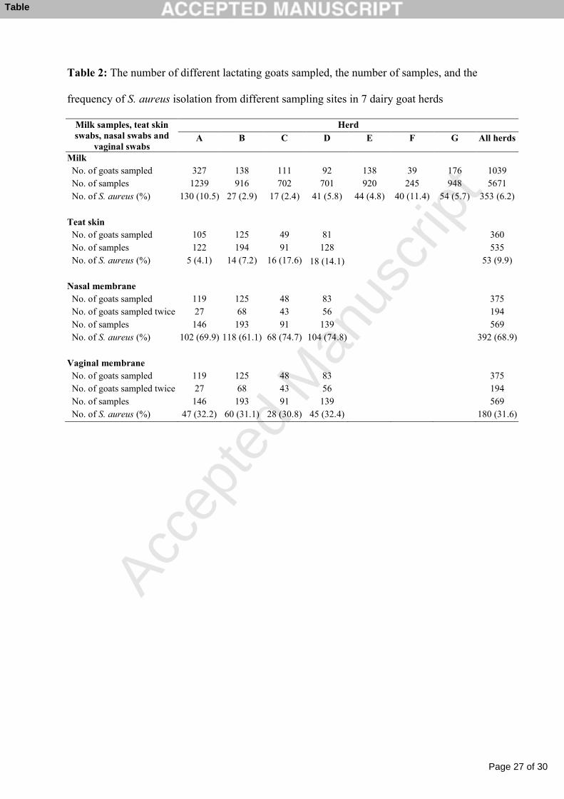
presented in Table 2. S. aureus was detected in 353 (6.2%) of the 5671 milk samples and in 4
625 (37.4%) of the 1673 swab samples from lactating goats. From kids, S. aureus was isolated 5
from one (4%), 28 (61%) and 3 (8%) swab samples from teat skin, nose and vagina, 6
respectively. 7
A noticeably higher percentage of vaginal swab samples were S. aureus-positive after kidding 8
(44.9%) than before drying off (19.1%). Conversely, a higher percentage of nasal swab 9
samples were S. aureus-positive before drying off (75.6%) than after kidding (62.0%). 10
Altogether 122 goats with S. aureus-positive nasal swabs after kidding were resampled before 11
drying off and 90 (73.8%) of these were S. aureus-positive. Eighty two goats that had S. 12
aureus-positive vaginal swabs after kidding were resampled before drying off and 22 (26.8%) 13
were still S. aureus-positive. 14
15
3.2. PFGE 16
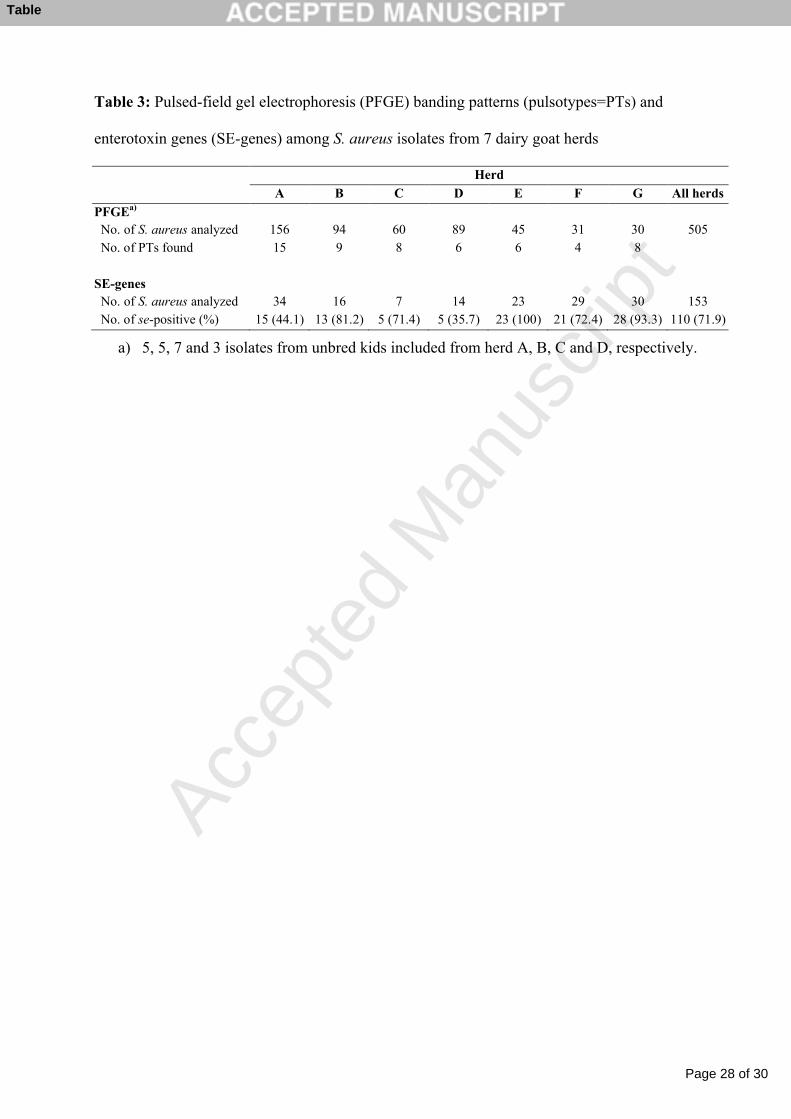
All the 505 analysed S. aureus isolates were typeable by PFGE, and were divided into 33 17
different PTs (Fig. 1). In a dendrogram with one representative for each PT, 6 different 18
clusters (A─F) were defined visually. The minimum similarity between PTs within these 19
clusters was 68.7% (Fig 1). Pairwise comparisons of the 33 PTs showed that with 4 20
exceptions all PTs within a cluster differed by <6 bands and all PTs differed by >5 bands to 21
all PTs outside its cluster. 22
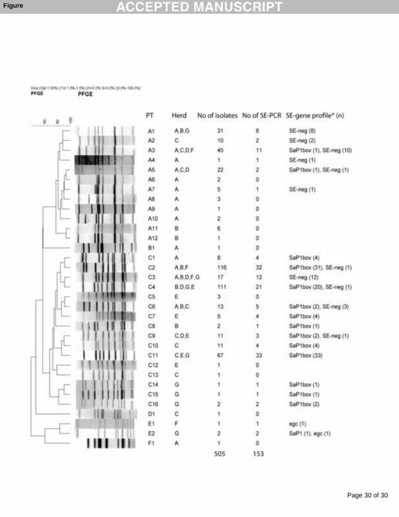
The most prevalent PT (C2) comprised 116 (23.0%) isolates. Two PTs, C4 and C11, each 23
differed by 2 and 4 bands from C2 and comprised 111 (22.0%) and 67 (13.3%) of the isolates, 24
respectively. Three hundred and seventy (73.3%) isolates belonged to the 5 most prevalent 25
Page 10 of 30
Accep
ted
Man
uscr
ipt
10
PTs (C2, C4, C11, A3, A1) and each of these PTs were found in 3−5 herds. With the 1
exception of PT A1, isolates belonging to these 5 PTs were obtained from all the 4 major 2
sampling sites. With 2 exceptions, C2, C4 and C11 were the predominant PT in each of the 3
herds where they were found. 4
Within each herd, 4 to 15 different PTs were identified (Table 3). The greatest diversity was 5
found in herd A with 15 PTs assigned to 4 clusters. PTs from more than one cluster were 6
found in all herds except for herd E. Each of the 7 herds had a predominant PT which 7
included 34 to 74% of the analysed isolates from that herd. 8
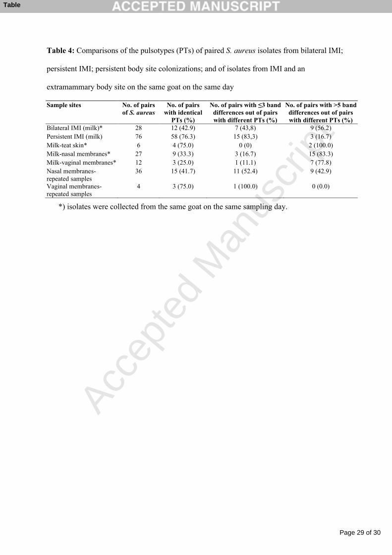
Comparisons of PTs of paired S. aureus isolates from bilateral IMI, persistent IMI, persistent 9
body site colonizations and of isolates collected from IMI and extramammary sites of the 10
same goat on the same day are shown in Table 4. The majority of isolate-pairs from persistent 11
IMI, from repeated vaginal swabs and from milk and teat skin were identical, while remaining 12
paired isolates were identical in less than 50% of the situations. 13
Out of the paired S. aureus isolates that had different PTs, the number of pairs that differed by 14
≤3 and >5 band differences are presented in Table 4. Noticeably, in the majority of situations 15
where paired S. aureus isolates from bilateral IMI, from milk and the nose, and from milk and 16
the vagina were different, the PTs differed by >5 bands. In situations where paired isolates 17
from persistent IMI, from milk and teat skin, from the nose (repeated samples), and from the 18
vagina (repeated samples) had different PTs, most of the pairs differed by 3 bands or less. 19
When S. aureus isolates (n=194) from 76 goats, collected at 84 samplings were compared, 20
different PTs were found among isolates from 55 (65.5%) of the samplings. From 36 (65.5%) 21
of the samplings where isolates had different PTs, >5 band differences were found. 22
23
3.3. Toxin gene detection 24
Page 11 of 30
Accep
ted
Man
uscr
ipt
11
Out of the 153 isolates tested by the m-PCR (for sea–see, seg–selj and tst), at least one SE-1
gene was detected in 110 (71.9%) of the isolates (Table 3). The additional testing of 110 of 2
the isolates by conventional PCR (for selk–selo, selq, selr and selu) did not increase the number 3
of se-positive isolates. 4
There was no significant difference between the se-profiles of isolates from predefined 5
persistent vs. temporary IMI. This result remained the same also when the 7 pairs of isolates 6
that were different by PFGE, but that had been predefined as from persistent IMI, were 7
excluded from the analysis. 8
By m-PCR the genes sec and tst were found in 109 (71.2%) isolates and were always co-9
detected. Seventy eight of these isolates were also tested by conventional PCR and all were 10
sell-positive. Three isolates from two animals were positive for seg, and sei by m-PCR. Two 11
of these isolates, from the same animal, were also positive for sec and tst. Conventional PCR 12
was performed on 2 of the seg- and sei-positive isolates (one from each animal), and both were 13
positive for selm, seln, selo and selu. The genes sea, seb, sed, selk, selq and selr were not 14
detected in any of the isolates. 15
The 153 isolates tested for SE-genes belonged to 22 of the PTs described above. There was a 16
noticeable correspondence between SE-gene profile and PT and cluster (Fig. 1). With only 2 17
exceptions, isolates belonging to cluster A (PTs A1─A5 and A7) were all se-negative. 18
Conversely, most isolates (87.7%) belonging to cluster C were positive for sec and tst (and 19
sell). 20
21
4. Discussion 22
In the present study, 6.2% of the goat milk samples were S. aureus positive, with a variation 23
at herd level between 2.4 and 11.4%. In earlier studies S. aureus has generally been isolated 24
from less than 3% of the goats (Menzies and Ramanoon, 2001). 25
Page 12 of 30
Accep
ted
Man
uscr
ipt
12
The nose was the extramammary body site most often colonized with S. aureus. Almost 70% 1
of the sampled goats and 61% of the kids had S. aureus-positive nasal swabs. This indicates 2
that the nose is an important colonization site for S. aureus in goats, and that kids are 3
colonized during or soon after birth. Previous reports have indicated a S. aureus nasal carriage 4
rate of 30% in dairy sheep (Vautor et al., 2005) and 6% in dairy goats (Valle et al., 1991), and 5
that 9% of dairy heifers were colonized with S. aureus in the muzzle area (Roberson et al., 6
1994). On the other hand, in 2 studies from Norway (Jørgensen et al., 2005b) and the United 7
Kingdom (Smith et al., 2005), respectively, S. aureus was not found in nasal swabs from dairy 8
cows despite a high frequency of S. aureus IMI in both herds. 9
A higher S. aureus colonization rate was found in the vagina after kidding (45%) than before 10
drying off (19%). Uterine involution and decreased secretions may explain the lower 11
colonization rate before drying off. It is likely that presence of S. aureus on the mucous 12
membranes of the vagina and in vaginal secretions contributes to the spread of S. aureus from 13
goats to kids. The kids sampled in this study were removed from their mothers immediately 14
after birth and received one feed of colostrum. They are unlikely, therefore, to have picked up 15
S. aureus during muzzling and suckling. 16
Although the PFGE analyses revealed a great genetic diversity among the S. aureus isolates it 17
was also evident that certain genotypes dominated. This was the case both when comparing 18
isolates from individual herds, and when comparing the complete set of 505 isolates from all 7 19
herds. The 3 most commonly observed PTs were, with 2 exceptions, also the dominant PT in 20
the herds where they were found. It appears that S. aureus strains belonging to certain 21
genotypes are more successful at spreading in the Norwegian dairy goat population than 22
others. This is in agreement with a previous study where a limited number of closely related 23
PTs were found responsible for a great proportion of the cases of S. aureus mastitis in dairy 24
Page 13 of 30
Accep
ted
Man
uscr
ipt
13
cows, dairy goats and meat-producing sheep (Mørk et al., 2005). Similar findings have been 1
reported regarding S. aureus in dairy sheep (Vautor et al., 2003). 2
In the present study almost all the included goats were colonized or infected with S. aureus, 3
and individual goats could be colonised with several S. aureus genotypes. In fact in 65% of 4
the situations, where more than one body site on the same goat was S. aureus-positive after 5
sampling on the same day, isolates belonged to different PTs. 6
The majority of S. aureus IMI that were predefined as persistent were confirmed as persistent 7
infections because two consecutively collected isolates were identical by PFGE. Isolating the 8
same genotype on three consecutive occasions would have strengthened the definition of these 9
infections as truly persistent, but this was not practicable in this study. 10
Although 18 out of the 76 compared S. aureus pairs had different PTs, 14 of these differed by 11
only 2 bands. It could be argued that these belonged to the same infection because an infecting 12
strain may undergo genetic change during the course of an infection (Tenover et al., 1995). 13
In contrast to the situation with persistent IMI, bilateral IMI were equally often caused by 14
distinguishable PTs as by identical PTs. In the majority of situations where S. aureus pairs 15
differed by PFGE, the PTs also belonged to different clusters. This indicates that S. aureus 16
from different sources most often caused IMI in the 2 udder halves of bilateral IMI. 17
The nares were found to be a major S. aureus colonization site in Norwegian dairy goats. One 18
might have expected that the nasal strain would also be the most frequent cause of IMI in 19
individual goats. However, more than 50% of the isolate pairs, collected from the nose and an 20
IMI of the same goat on the same day, differed by 4 bands or more by PFGE. A similar 21
observation was made regarding pairs of isolates from the vagina and an IMI. This indicates 22
that the mucous membranes are not the main reservoir of S. aureus IMI for individual goats. 23
However, the frequent presence of S. aureus on the mucous membranes probably contributes 24
to extensive dispersal of the bacteria in the environment, hampering effective transmission 25
Page 14 of 30
Accep
ted
Man
uscr
ipt
14
control in the herds. Out of the paired isolates from repeated nasal swabs, that were compared 1
by PFGE, 25% of the pairs differed by >5 band. This indicates frequent strain change in the 2
nasal membranes consistent with intermittent rather than persistent carriage (Kluytmans et al., 3
1997). 4
In agreement with previous observations (Jørgensen et al., 2005a) and investigations by others 5
(da Silva et al., 2005; Haveri et al., 2007; Scherrer et al., 2004; Smyth et al., 2005), the 6
majority of analysed S. aureus isolates were SE-gene positive. The genes sec, sell and tst were 7
detected almost exclusively, and indicates a wide distribution of the pathogenicity island 8
Sap1bov (Fitzgerald et al., 2001b) among caprine S. aureus in Norway. It seems plausible that 9
the toxins encoded by Sap1bov play a role in survival of S. aureus on its caprine host. 10
However, no correlation was found between persistence of IMI and the SE-gene profile of the 11
causal S. aureus strains. The observation that certain S. aureus PTs and PFGE clusters mostly 12
had the same SE-gene profile indicates that it is clonal spread of toxin-encoding S. aureus 13
strains rather than horizontal transfer of the toxin encoding genetic elements that is 14
responsible for the wide distribution of Sap1bov. 15
In conclusion, S. aureus is frequently present on the mucous membranes of the nose and 16
vagina of Norwegian dairy goats and the epidemiology is complex. The presence of S. aureus 17
on other body sites than the mammary gland may contribute to the spread of S. aureus in dairy 18
goat herds and thus add to mastitis control problems. The toxin genes sec, sell and tst were 19
found to be widespread among the caprine S. aureus isolates, but the role of these toxins in 20
colonizations and infections of goats remain to be elucidated. 21
22
Acknowledgements 23
The Norwegian Research Council is acknowledged for economic support of this work (grant 24
number 164293). We also thank the veterinarians Olav Hermansen, Marianne Vinje Kilvær, 25
Page 15 of 30
Accep
ted
Man
uscr
ipt
15
Jostein Rise, Kåre Rydningen, Kristin Ryum, Maria Skavnes, and Gunnar Valdal who 1
collected all the samples in the study. 2
3
Page 16 of 30
Accep
ted
Man
uscr
ipt
16
References 1
Bannerman, T.L., Hancock, G.A., Tenover, F.C., Miller, J.M., 1995. Pulsed-field gel 2
electrophoresis as a replacement for bacteriophage typing of Staphylococcus aureus. J. Clin. 3
Microbiol. 33, 551-555. 4
Bergonier, D., de Cremoux, R., Rupp, R., Lagriffoul, G., Berthelot, X., 2003. Mastitis of dairy 5
small ruminants. Vet. Res. 34, 689-716. 6
Capurro, A., Concha, C., Nilsson, L., Östensson, K., 1999. Identification of coagulase-7
positive staphylococci isolated from bovine milk. Acta Vet. Scand. 40, 315-321. 8
Cremonesi, P., Luzzana, M., Brasca, M., Morandi, S., Lodi, R., Vimercati, C., Agnellini, D., 9
Caramenti, G., Moroni, P., Castiglioni, B., 2005. Development of a multiplex PCR assay for 10
the identification of Staphylococcus aureus enterotoxigenic strains isolated from milk and 11
dairy products. Mol. Cell Probes 19, 299-305. 12
da Silva, E.R., do Carmo, L.S., da Silva, N., 2005. Detection of the enterotoxins A, B, and C 13
genes in Staphylococcus aureus from goat and bovine mastitis in Brazilian dairy herds. Vet. 14
Microbiol. 106, 103-107. 15
Davidson, I., 1961. The epidemiology of staphylococcal mastitis. Vet. Rec. 73, 1015-1018. 16
Dinges, M.M., Orwin, P.M., Schlievert, P.M., 2000. Exotoxins of Staphylococcus aureus. 17
Clin. Microbiol. Rev. 13, 16-34. 18
Ferens, W.A., Davis, W.C., Hamilton, M.J., Park, Y.H., Deobald, C.F., Fox, L., Bohach, G., 19
1998. Activation of bovine lymphocyte subpopulations by staphylococcal enterotoxin C. 20
Infect. Immun. 66, 573-580. 21
Page 17 of 30
Accep
ted
Man
uscr
ipt
17
Fitzgerald, J.R., Monday, S.R., Foster, T.J., Bohach, G.A., Hartigan, P.J., Meaney, W.J., 1
Smyth, C.J., 2001a. Characterization of a putative pathogenicity island from bovine 2
Staphylococcus aureus encoding multiple superantigens. J. Bacteriol. 183, 63-70. 3
Fitzgerald, J.R., Sturdevant, D.E., Macki, S.M., Gill, S.R., Musser, J.M., 2001b. Evolutionary 4
genomics of Staphylococcus aureus: insight into the origin of methicillin-resistant strains and 5
the toxic shock syndrome epidemic. Proc. Natl. Acad. Sci. USA 98, 8821-8826. 6
Foster, T.J., 2005. Immune evasion by staphylococci. Nat. Rev. Microbiol. 3, 948-958. 7
Haveri, M., Hovinen, M., Roslöf, A., Pyörälä, S., 2008. Molecular types and genetic profiles 8
of Staphylococcus aureus strains isolated from bovine intramammary infections and 9
extramammary sites. J. Clin. Microbiol. 46, 3728-3735. 10
Haveri, M., Roslöf, A., Rantala, L., Pyörälä, S., 2007. Virulence genes of bovine 11
Staphylococcus aureus from persistent and nonpersistent intramammary infections with 12
different clinical characteristics. J. Appl. Microbiol. 103, 993-1000. 13
Headrick, M.L., Korangy, S., Bean, N.H., Angulo, F.J., Altekruse, S.F., Potter, M.E., Klontz, 14
K.C., 1998. The epidemiology of raw milk-associated foodborne disease outbreaks reported in 15
the United States, 1973 through 1992. Am. J. Public Health 88, 1219-1221. 16
International Dairy Federation, 1981. Laboratory methods for use in mastitis work. IDF 17
Document 132, Brussels, Belgia. 18
Jarraud, S., Peyrat, M.A., Lim, A., Tristan, A., Bes, M., Mougel, C., Etienne, J., Vandenesch, 19
F., Bonneville, M., Lina, G., 2001. egc, a highly prevalent operon of enterotoxin gene, forms a 20
putative nursery of superantigens in Staphylococcus aureus. J. Immunol. 166, 669-677. 21
Page 18 of 30
Accep
ted
Man
uscr
ipt
18
Jørgensen, H.J., Mørk, T., Høgåsen, H.R., Rørvik, L.M., 2005a. Enterotoxigenic 1
Staphylococcus aureus in bulk milk in Norway. J. Appl. Microbiol. 99, 158-166. 2
Jørgensen, H.J., Mørk, T., Rørvik, L.M., 2005b. The occurrence of Staphylococcus aureus on 3
a farm with small-scale production of raw milk cheese. J. Dairy Sci. 88, 3810-3817. 4
Kluytmans, J., van, B.A., Verbrugh, H., 1997. Nasal carriage of Staphylococcus aureus: 5
epidemiology, underlying mechanisms, and associated risks. Clin. Microbiol. Rev. 10, 505-6
520. 7
Kuroda, M., Ohta, T., Uchiyama, I., Baba, T., Yuzawa, H., Kobayashi, I., Cui, L., Oguchi, A., 8
Aoki, K., Nagai, Y., Lian, J., Ito, T., Kanamori, M., Matsumaru, H., Maruyama, A., 9
Murakami, H., Hosoyama, A., Mizutani-Ui, Y., Takahashi, N.K., Sawano, T., Inoue, R., 10
Kaito, C., Sekimizu, K., Hirakawa, H., Kuhara, S., Goto, S., Yabuzaki, J., Kanehisa, M., 11
Yamashita, A., Oshima, K., Furuya, K., Yoshino, C., Shiba, T., Hattori, M., Ogasawara, N., 12
Hayashi, H., Hiramatsu, K., 2001. Whole genome sequencing of meticillin-resistant 13
Staphylococcus aureus. Lancet 357, 1225-1240. 14
Letertre, C., Perelle, S., Dilasser, F., Fach, P., 2003. Identification of a new putative 15
enterotoxin SEU encoded by the egc cluster of Staphylococcus aureus. J. Appl. Microbiol. 95, 16
38-43. 17
Lovseth, A., Loncarevic, S., Berdal, K.G., 2004. Modified multiplex PCR method for 18
detection of pyrogenic exotoxin genes in staphylococcal isolates. J. Clin. Microbiol. 42, 3869-19
3872. 20
Menzies, P.I., Ramanoon, S.Z., 2001. Mastitis of sheep and goats. Vet. Clin. North Am. Food 21
Anim. Pract. 17, 333-358. 22
Page 19 of 30
Accep
ted
Man
uscr
ipt
19
Mørk, T., Tollersrud, T., Kvitle, B., Jørgensen, H.J., Waage, S., 2005. Comparison of 1
Staphylococcus aureus genotypes recovered from cases of bovine, ovine, and caprine mastitis. 2
J. Clin. Microbiol. 43, 3979-3984. 3
Park, Y.H., Lee, S.U., Ferens, W.A., Samuels, S., Davis, W.C., Fox, L.K., Ahn, J.S., Seo, 4
K.S., Chang, B.S., Hwang, S.Y., Bohach, G.A., 2006. Unique features of bovine lymphocytes 5
exposed to a staphylococcal enterotoxin. J. Vet. Sci. 7, 233-239. 6
Roberson, J.R., Fox, L.K., Hancock, D.D., Gay, J.M., Besser, T.E., 1994. Ecology of 7
Staphylococcus aureus isolated from various sites on dairy farms. J. Dairy Sci. 77, 3354-3364. 8
Scherrer, D., Corti, S., Muehlherr, J.E., Zweifel, C., Stephan, R., 2004. Phenotypic and 9
genotypic characteristics of Staphylococcus aureus isolates from raw bulk-tank milk samples 10
of goats and sheep. Vet. Microbiol. 101, 101-107. 11
Smith, E.M., Green, L.E., Medley, G.F., Bird, H.E., Dowson, C.G., 2005. Multilocus 12
sequence typing of Staphylococcus aureus isolated from high-somatic-cell-count cows and the 13
environment of an organic dairy farm in the United Kingdom. J. Clin. Microbiol. 43, 4731-14
4736. 15
Smyth, D.S., Hartigan, P.J., Meaney, W.J., Fitzgerald, J.R., Deobald, C.F., Bohach, G.A., 16
Smyth, C.J., 2005. Superantigen genes encoded by the egc cluster and SaPIbov are 17
predominant among Staphylococcus aureus isolates from cows, goats, sheep, rabbits and 18
poultry. J. Med. Microbiol. 54, 401-411. 19
Tenover, F.C., Arbeit, R.D., Goering, R.V., Mickelsen, P.A., Murray, B.E., Persing, D.H., 20
Swaminathan, B., 1995. Interpreting chromosomal DNA restriction patterns produced by 21
pulsed-field gel electrophoresis: criteria for bacterial strain typing. J. Clin. Microbiol. 33, 22
2233-2239. 23
Page 20 of 30
Accep
ted
Man
uscr
ipt
20
Valle, J., Piriz, S., de la, F.R., Vadillo, S., 1991. Staphylococci isolated from healthy goats. 1
Zentralbl. Veterinarmed. B 38, 81-89. 2
Vautor, E., Abadie, G., Guibert, J.M., Chevalier, N., Pepin, M., 2005. Nasal carriage of 3
Staphylococcus aureus in dairy sheep. Vet. Microbiol. 106, 235-239. 4
Vautor, E., Abadie, G., Guibert, J.M., Huard, C., Pepin, M., 2003. Genotyping of 5
Staphylococcus aureus isolated from various sites on farms with dairy sheep using pulsed-6
field gel electrophoresis. Vet. Microbiol. 96, 69-79. 7
White, E. 2007, The prevalence of mastitis in small ruminants and the effect of mastitis on 8
small ruminant production. In: Proceedings of the NMC 46th Annual Meeting. National 9
Mastitis Council, Madison, Wis, pp. 119-127. 10
Zottola, E.A., Smith, L.B., 1993. Growth and survival of undesirable bacteria in cheese. In: 11
Fox, P.E. (Ed.), Cheese: chemistry, physics and microbiology, Chapman & Hall, London, pp. 12
471-492. 13
14
15
16
Page 21 of 30
Accep
ted
Man
uscr
ipt
21
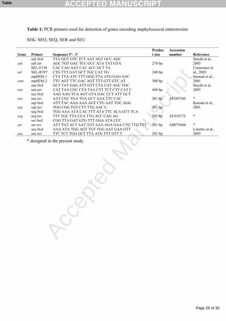
Table 1: PCR primers used for detection of genes encoding staphylococcal enterotoxins 1
SEK–SEO, SEQ, SER and SEU 2
3
Page 22 of 30
Accep
ted
Man
uscr
ipt
22
Table 2: The number of different lactating goats sampled, the number of samples, and the 1
frequency of S. aureus isolation from different sampling sites in 7 dairy goat herds 2
3
Page 23 of 30
Accep
ted
Man
uscr
ipt
23
Table 3: Pulsed-field gel electrophoresis (PFGE) banding patterns (pulsotypes=PTs) and 1
enterotoxin genes (SE-genes) among S. aureus isolates from 7 dairy goat herds 2
3
Page 24 of 30
Accep
ted
Man
uscr
ipt
24
Table 4: Comparisons of the pulsotypes (PTs) of paired S. aureus isolates from bilateral IMI; 1
persistent IMI; persistent body site colonizations; and of isolates from IMI and an 2
extramammary body site on the same goat on the same day 3
4
Page 25 of 30
Accep
ted
Man
uscr
ipt
25
Figure 1: Pulsed-field gel electrophoresis of SmaI digested DNA of S. aureus isolates 1
representing the 33 pulsotypes (PTs) found in the 7 goat dairy herds. The name of the PTs, the 2
presence in different farms, the total number of isolates analyzed by PFGE, the number of 3
isolates analyzed by PCR and the enterotoxin gene profile (SE-gene profile) found are shown. 4
* SE-neg = no SE-genes detected; SaP1bov = detected genes belonging to the 5
pathogenicity island SaP1bov (sec, sell and tst) (Fitzgerald et al., 2001a); egc = detected genes 6
belonging to the enterotoxin gene cluster (egc) (sei, seg, selm, seln, selo, and selu) (Jarraud et 7
al., 2001). 8
9
10
11
12
Page 26 of 30
Accep
ted
Man
uscr
ipt
Table 1: PCR primers used for detection of genes encoding staphylococcal enterotoxins
SEK–SEO, SEQ, SER and SEU
Gene Primer Sequence 5’– 3’Product size
Accession number Reference
seksek fwd sek rev
TTA GGT GTC TCT AAT AGT GCC AGC AGC TGT GAC TCC GCC ATA TAT GTA 278 bp
Smyth et al, 2005
selSEL-F158 SEL-R397
CAC CAG AAT CAC ACC GCT TACTG TTT GAT GCT TGC CAT TG 240 bp
Cremonesi et al., 2005
semmpSEM-1 mpSEM-2
CTA TTA ATC TTT GGG TTA ATG GAG AACTTC AGT TTC GAC AGT TTT GTT GTC AT 300 bp
Jarraud et al., 2001
sensen fwdsen rev
GCT TAT GAG ATT GTT CTA CAT AGC TGCCAT TAA CGC CTA TAA CTT TCT CTT CAT C 448 bp
Smyth et al, 2005
seoseo fwdseo rev
AAG AAG TCA AGT GTA GAC CCT ATT GCTAAT CGC TGA TGA GCT AAA TTC CAC 201 bp AF285760 *
sepsep fwdsep rev
ATT TAC AAA AAA AGT CTG AAT TGC AGGTGG CGG TGT CTT TTG AAC C 201 bp
Kuroda et al., 2001
seqseq fwdseq rev
TGG AAA ATA CAC TTT ATA TTC ACA GTT TCATTT TGC TTA CCA TTG ACC CAG AG 201 bp AF410775. *
serser fwdser rev
CGG TTA GAT GTG TTT GGA ATA CCCATT TGT ACT AAT TGT AAA AGA GAA CTG TTG TTT 201 bp AB075606 *
seuseu fwd seu rev
AAA ATA TGG AGT TGT TGG AAT GAA GTTTTC TCT TGG GCT TTA ATG TTT GTT T 201 bp
Letertre et al., 2003
* designed in the present study.
Table
Page 27 of 30
Accep
ted
Man
uscr
ipt
Table 2: The number of different lactating goats sampled, the number of samples, and the
frequency of S. aureus isolation from different sampling sites in 7 dairy goat herds
Milk samples, teat skin swabs, nasal swabs and
vaginal swabs
Herd
A B C D E F G All herds
Milk No. of goats sampled 327 138 111 92 138 39 176 1039 No. of samples 1239 916 702 701 920 245 948 5671 No. of S. aureus (%) 130 (10.5) 27 (2.9) 17 (2.4) 41 (5.8) 44 (4.8) 40 (11.4) 54 (5.7) 353 (6.2)
Teat skin No. of goats sampled 105 125 49 81 360 No. of samples 122 194 91 128 535 No. of S. aureus (%) 5 (4.1) 14 (7.2) 16 (17.6) 18 (14.1) 53 (9.9)
Nasal membrane No. of goats sampled 119 125 48 83 375 No. of goats sampled twice 27 68 43 56 194 No. of samples 146 193 91 139 569 No. of S. aureus (%) 102 (69.9) 118 (61.1) 68 (74.7) 104 (74.8) 392 (68.9)
Vaginal membrane No. of goats sampled 119 125 48 83 375 No. of goats sampled twice 27 68 43 56 194 No. of samples 146 193 91 139 569 No. of S. aureus (%) 47 (32.2) 60 (31.1) 28 (30.8) 45 (32.4) 180 (31.6)
Table
Page 28 of 30
Accep
ted
Man
uscr
ipt
Table 3: Pulsed-field gel electrophoresis (PFGE) banding patterns (pulsotypes=PTs) and
enterotoxin genes (SE-genes) among S. aureus isolates from 7 dairy goat herds
Herd
A B C D E F G All herds
PFGEa)
No. of S. aureus analyzed 156 94 60 89 45 31 30 505 No. of PTs found 15 9 8 6 6 4 8
SE-genes No. of S. aureus analyzed 34 16 7 14 23 29 30 153 No. of se-positive (%) 15 (44.1) 13 (81.2) 5 (71.4) 5 (35.7) 23 (100) 21 (72.4) 28 (93.3) 110 (71.9)
a) 5, 5, 7 and 3 isolates from unbred kids included from herd A, B, C and D, respectively.
Table
Page 29 of 30
Accep
ted
Man
uscr
ipt
Table 4: Comparisons of the pulsotypes (PTs) of paired S. aureus isolates from bilateral IMI;
persistent IMI; persistent body site colonizations; and of isolates from IMI and an
extramammary body site on the same goat on the same day
Sample sites No. of pairs of S. aureus
No. of pairs with identical
PTs (%)
No. of pairs with ≤3 band differences out of pairs with different PTs (%)
No. of pairs with >5 band differences out of pairs with different PTs (%)
Bilateral IMI (milk)* 28 12 (42.9) 7 (43,8) 9 (56.2)Persistent IMI (milk) 76 58 (76.3) 15 (83,3) 3 (16.7)Milk-teat skin* 6 4 (75.0) 0 (0) 2 (100.0)Milk-nasal membranes* 27 9 (33.3) 3 (16.7) 15 (83.3)Milk-vaginal membranes* 12 3 (25.0) 1 (11.1) 7 (77.8)Nasal membranes-repeated samples
36 15 (41.7) 11 (52.4) 9 (42.9)
Vaginal membranes-repeated samples
4 3 (75.0) 1 (100.0) 0 (0.0)
*) isolates were collected from the same goat on the same sampling day.
Table
Page 30 of 30
Accep
ted
Man
uscr
ipt
Figure