Embed Size (px)
Citation preview

7/30/2019 bài báo cáo vật liệu y sinh( Vũ Văn Kỳ)2
http://slidepdf.com/reader/full/bai-bao-cao-vat-lieu-y-sinh-vu-van-ky2 1/10
Magnesium used in bioabsorbable stents controls smooth muscle cell
proliferation and stimulates endothelial cells in vitro
Katrin Sternberg,1* Matthias Gratz,2* Kathleen Koeck,2 Joerg Mostertz,3 Robert Begunk,2
Marian Loebler,1 Beatrice Semmling,4 Anne Seidlitz,4 Petra Hildebrandt,3 Georg Homuth,3
Niels Grabow,1 Conny Tuemmler,5 Werner Weitschies,4 Klaus-Peter Schmitz,1 Heyo K. Kroemer2
1University of Rostock, Medical Faculty, Institute for Biomedical Engineering, Rostock, Germany2Ernst-Moritz-Arndt University, Medical Faculty, Institute of Pharmacology, Greifswald, Germany3Ernst-Moritz-Arndt University, Interfaculty Institute for Genetics and Functional Genomics, Junior Research Group
Transcriptomics/Functional Genomics, Greifswald, Germany4Ernst-Moritz-Arndt University, Faculty of Mathematics and Natural Sciences, Institute of Pharmacy, Greifswald, Germany5University of Rostock, Faculty of Mathematics and Natural Sciences, Institute for Biological Sciences, Rostock, Germany
Received 28 May 2010; revised 24 March 2011; accepted 2 June 2011
Published online 24 November 2011 in Wiley Online Library (wileyonlinelibrary.com). DOI: 10.1002/jbm.b.31918
Abstract: Magnesium-based bioabsorbable cardiovascular
stents have been developed to overcome limitations of perma-
nent metallic stents, such as late stent thrombosis. During stent
degradation, endothelial and smooth muscle cells will be
exposed to locally high magnesium concentrations with yet
unknown physiological consequences. Here, we investigated
the effects of elevated magnesium concentrations on human
coronary artery endothelial and smooth muscle cell (HCAEC,
HCASMC) growth and gene expression. In the course of 24 h
after incubation with magnesium chloride solutions (1 or
10 mM ) intracellular magnesium level in HCASMC raised from
0.55 6 0.25 mM (1 mM ) to 1.38 6 0.95 mM (10 mM ), while no
increase was detected in HCAEC. Accordingly, a DNA microar-
ray-based study identified 69 magnesium regulated transcripts
in HCAEC, but 2172 magnesium regulated transcripts in
HCASMC. Notably, a significant regulation of various growth
factors and extracellular matrix components was observed. In
contrast, viability and proliferation of HCAEC were increased at
concentrations of up to 25 mM magnesium chloride, while in
HCASMC viability and proliferation appeared to be unaffected.
Taken together, our data indicate that magnesium halts smooth
muscle cell proliferation and stimulates endothelial cell prolifer-
ation, which might translate into a beneficial effect in the set-
ting of stent associated vascular injury. VC 2011 Wiley Periodicals,
Inc. J Biomed Mater Res Part B: Appl Biomater 100B: 41–50, 2012.
Key Words: magnesium, bioabsorbable metal stent, stent,
in-stent restenosis, cell cycle
How to cite this article: Sternberg K, Gratz M, Koeck K, Mostertz J, Begunk R, Loebler M, Semmling B, Seidlitz A, Hildebrandt P,
Homuth G, Grabow N, Tuemmler C, Weitschies W, Schmitz K-P, Kroemer H. 2012. Magnesium used in bioabsorbable stents
controls smooth muscle cell proliferation and stimulates endothelial cells in vitro . J Biomed Mater Res Part B 2012:100B:41–50.
INTRODUCTION
Stents used as tubular implants for the mechanical support
of stenotic arterial vessels were introduced in the 1990s.
However, the initial concept of transluminal implantation of
stainless steel coil springs was already described by Dotter
in preclinical experiments in 1969.1 Stent endoprostheses are
either self-expanding or balloon-expandable and are usually
fabricated either as a wire mesh or a slotted tube. Typical
materials of permanent bare metal stents (BMS) are stainless
steel and cobalt-chromium alloys for balloon-expandable
stents, and nickel-titanium alloys for self-expanding stents.2
Although BMS implantation demonstrated superiority
over balloon angioplasty alone, numerous studies revealed
that within 6–12 months BMS implantations are associated
with the formation of in-stent restenosis in about 15–20%
of all cases.3 This restenotic process is induced by vascular
injury during stent implantation, leading to neointimal
hyperplasia caused by proliferation and migration of smooth
muscle cells.4,5 In first attempts to decrease the incidence of
in-stent restenosis the designs of BMS were modified and
passive antithrombotic stent coatings were developed. How-
ever, a marked reduction of in-stent restenosis was only
achieved with the introduction of drug-eluting stents (DES).
DES effectively inhibit in-stent restenosis by controlled
release of antiproliferative drugs, such as sirolimus
(CypherVR
stent) and paclitaxel (TaxusVR
stent), directly to the
site of vascular injury.6 These first generation DES are very
effective in preventing in-stent restenosis. However, it is
*Both authors contributed equally to this work.
Correspondence to: K. Sternberg ([email protected])
Contract grant sponsors: Landesregierung Mecklenburg-Vorpommern, Bundesministerium fu ¨ r Bildung und Forschung; contract grant number:
03ZIK012
VC 2011 WILEY PERIODICALS, INC. 41
7/30/2019 bài báo cáo vật liệu y sinh( Vũ Văn Kỳ)2
http://slidepdf.com/reader/full/bai-bao-cao-vat-lieu-y-sinh-vu-van-ky2 2/10
being discussed that their use is associated with an increase
in adverse clinical events, such as myocardial infarction. In
this context, late thrombosis and delayed healing were iden-
tified as potential risks associated with the use of DES.7–9
Furthermore, cases of local hypersensitivity and inflamma-
tion were reported.9–11 A promising approach to overcome
the limitations of first generation DES is the application of
fully bioabsorbable stents, which could be completelyreplaced by tissue, and may even allow for a positive vascu-
lar remodeling.12 Apart from biodegradable polymers, such
as polylactides,13–18 stents based on the corrodible metals
iron19 and magnesium20 have been proposed. So far, the
magnesium stent is the only bioabsorbable metal stent
investigated clinically as a coronary stent (PROGRESS-AMS
trial).21 This clinical trial with a cohort of 63 patients dem-
onstrated safe stent application and a safe absorption pro-
cess after 4 months.22,23
Despite the initial clinical success of the magnesium
stent, biocompatibility and biological action of the degrada-
tion products are still subject of scientific investigation.24 In
this context, it is the purpose of the present in vitro studyto assess the biocompatibility and pharmacological effects of
the main alloy component magnesium (>90% w/w) with
regard to cell proliferation and gene regulation of human
coronary artery smooth muscle cells and endothelial cells.
MATERIALS AND METHODS
Materials
Magnesium was purchased as magnesium chloride (MgCl2)
from Sigma-Aldrich (Taufkirchen, Germany). MgCl2 was dis-
solved in water to yield a concentration of 5 mol/L (M).
The bioabsorbable magnesium stents were supplied by Bio-
tronik AG (Bulach, Switzerland).Primary human coronary artery smooth muscle cells
(HCASMC) and primary human coronary artery endothelial
cells (HCAEC) were obtained from PromoCell GmbH (Heidel-
berg, Germany). Smooth muscle cell growth medium 2 con-
taining 1 mM MgCl2, 5% fetal calf serum (FCS), 2 ng/mL
recombinant human basic fibroblast growth factor, 0.5 ng/
mL recombinant human epidermal growth factor, 5 lg/mL
recombinant human insulin, 620 pg/mL phenol red as well
as antibiotics 50 ng/mL amphotericin B and 50 lg/mL gen-
tamycin (pH 7.4) (with supplement mix) was supplied by
PromoCell. For HCAEC magnesium-free endothelial cell
growth medium MV (PromoCell) containing 5% fetal calf se-
rum (FCS), 0.4% endothelial cell growth supplement/hepa-
rin, 10 ng/mL recombinant human epidermal growth factor,
1 lg/mL hydrocortisone, 620 pg/mL phenol red as well as
antibiotics 50 ng/mL amphotericin B and 50 lg/mL genta-
mycin (pH 7.4) was adjusted to 1 mM MgCl2.
For the MTS assays 100 mL Dulbecco’s modified Eagle
medium (DMEM, Applichem, Darmstadt, Germany) without
phenol red was prepared, since phenol red in conjunction
with the formazan salt mimics false positive viability results.
The medium was supplemented with 10% FCS, 25 mM
HEPES, 44 mM NaHCO3, and 100 U/mL penicillin/
streptomycin (pH 7.4). By adding 20 lL MTS/PMS reagent
the MTS reagent was adjusted to 333 lg/mL and the PMS
reagent to 2.5 mM .
Cell cultivation
HCASMC and HCAEC were seeded at a density of 1 Â 104
cells/mL in 96-well plates (200 lL per well) and cultivated
in their respective growth media for 72 h. Then HCASMC
and HCAEC were arrested by incubating the cells in basalmedium with 0.1% (for HCASMC) or 0.5% (for HCAEC) FCS
for 48 h. Subsequently the medium was removed and the
different MgCl2 concentrations (1.5625, 3.125, 6.25, 12.5,
25, 50, 100 mM ) were added to the basal medium with 5%
FCS (for HCASMC and HCAEC). The cell arrest was released
by stimulation with 25 ng/mL platelet-derived growth
factor (PDGF, Sigma-Aldrich, Taufkirchen, Germany) or
25 ng/mL epidermal growth factor (EGF, Sigma-Aldrich),
respectively.
MTS cell viability assay
At the end of the 24-h incubation time, the MgCl2 solutions
were removed and 100 lL of the MTS medium were addedto each well. The microtiter plate was incubated for 2 h at
37C and 5% CO2. The yellow color of the tetrazolium salt
(MTS reagent) changed towards a violet brown color of the
formazan salt and was quantified with an ELISA reader
(Anthos III, Anthos-Microsystems, Krefeld, Germany) at a
wavelength of 492 nm and a reference wavelength of 690
nm. Cells kept in basal medium served as a control for
restimulation of cell growth.
BrdU cell proliferation assay
At the end of the 24-h incubation time BrdU (10 lM ) was
added to each well and incubated for additional 18 h at
37C and 5% CO2
. BrdU incorporation was measured
according to the supplier instructions. Cells not subjected to
additional magnesium served as a reference, and cells kept
in basal medium only served as a control for restimulation
of cell growth.
Determination of the intracellular magnesium
concentration
For measurement of intracellular magnesium concentrations
cells were cultured in their respective growth media
(PromoCell) and challenged with different MgCl2 solutions
(1, 4, 10 mM ) for 24 h. Afterwards, the cells were rinsed,
detached, and resuspended in sodium-HEPES incubation
buffer (135 mM NaCl, 5 mM KCl, 1 mM CaCl2, 1 mM MgCl2,
10 mM glucose, and 20 mM HEPES, pH 7.4). Cells were
loaded with Mag-Fura-2-AM (1 lM ; Invitrogen, Carlsbad,
CA) for 60 min at 37C. Afterwards the labeled cells were
incubated for 30 min in fresh sodium-HEPES incubation
buffer at 37C to allow complete de-esterification of intra-
cellular AM esters. For the measurement 500,000 cells were
washed and resuspended in 2 mL fresh sodium-HEPES incu-
bation buffer and transferred to a 2-mL cuvette in a Perkin-
Elmer LS-55 fluorimeter. The cells were excited, alternately,
at 340 and 380 nm and the emission intensity was recorded
at 510 nm. Intracellular calibration of the Mag-Fura-2
42 ST ERN BE RG E T A L. IN VITRO MAGNESIUM EFFECTS ON SMOOTH MUSCLE AND ENDOTHELIAL CELLS

7/30/2019 bài báo cáo vật liệu y sinh( Vũ Văn Kỳ)2
http://slidepdf.com/reader/full/bai-bao-cao-vat-lieu-y-sinh-vu-van-ky2 3/10
signals was done with the addition of digitonin (final con-
centration 70 lM ) to measure the maximal fluorescence of
the probe by releasing the indicator into the surrounding
medium, followed by the addition of EDTA at a final concen-
tration of 10 mM to determine the fluorescence, F min, in the
absence of Mg2þ.
The Mg2þ concentration was calculated with the follow-
ing equation25:
½Mg2þ ¼ K DQðR À RminÞ
ðRmax À RÞ
using 1.5 mM as K D for the Mg2þ/Mag-Fura-2-complex. Q is
the ratio of fluorescence intensity of free Mag-Fura-2 to Mg
bound Mag-Fura-2 at 380 nm.
Ex vivo stent implantation and determination of the
magnesium content in blood vessel tissue
The magnesium uptake into the blood vessel wall after mag-
nesium stent implantation was evaluated for n ¼ 6 magne-
sium stent systems (nominal dimensions 3.0 Â 12 mm2
,provided by Biotronik AG, Bulach, Switzerland) with a
weight of $4.5 mg (magnesium content $90% ¼ 4.05 mg).
Freshly harvested carotid artery sections from porcine
cadavers obtained from the Leibniz Institute for Farm Ani-
mal Biology (Dummerstorf, Germany) were used as model
vessels. Ex vivo, each stent was deployed into a 25 mm por-
cine artery section by balloon-expansion (8 bar, 15 s) to an
inner diameter of 3.0 mm. The stent-implanted artery sec-
tions were placed in 5 mL incubation media (DMEM con-
taining 10% fetal bovine serum and 1% penicillin/strepto-
mycin) at 37C. In compliance with the MgCl2 incubation
time in the cell culture studies the magnesium accumulation
was analyzed after 24 and 48 h. For control purposes sec-
tions of the same arteries without stents were incubated
under identical conditions. After the intended incubation
periods the arteries were removed from the media and
thoroughly rinsed. Incubated arteries as well as frozen sam-
ples were dried at 60C for 24 h and dry mass was deter-
mined. Afterwards, the sections were dissolved in 65% (w/
w) nitric acid (HNO3) under heating. Magnesium content of
samples of the dissolved arteries and incubation media was
determined by atomic absorption spectroscopy (AAS, atomic
absorption spectrophotometer PU 9200, Philips GmbH,
Hamburg, Germany) against magnesium ICP standard 1000
mg/L CertiPUR (VWR International, Darmstadt, Germany).
All samples and standards contained 4% (v/v) HNO3 in
their final dilution. Samples were atomized in an air/
acetylene flame (fuel flow 1.1 L/min) and atomic absorption
was determined at a wavelength of 285.2 nm (slit width
0.5 nm).
Preparation of RNA (transcriptome profile and real
time RT-PCR)
HCAEC and HCASMC were seeded into 6-well plates and
cultured for 48 h in their respective culture media contain-
ing 1 mM MgCl2, followed by incubation for 24 h in culture
media containing either 1 mM (control) or 10 mM MgCl2.
Total RNA was isolated performing a modified phenol
extraction using the TRIzol reagent (Invitrogen)26 followed
by total RNA purification by RNeasy mini kit (Qiagen, Hil-
den, Germany). RNA concentration was measured using the
NanoDrop ND-1000 spectrophotometer (NanoDrop Technol-
ogies) and RNA purity and quality was assessed by gel
electrophoretic separation using the lab-on-chip capillary
electrophoresis technology (Bioanalyzer 2100, Agilent Tech-nologies, Waldbronn, Germany). Only RNA samples with a
RNA Integrity Number (RIN)27 greater than 9.5, 260/280
nm ! 1.8 were used for microarray analysis and real-time
RT-PCR.
Target preparation and array processing (transcriptome
profile)
To elucidate genes whose expressions are significantly
changed at the level of mRNA and which may contribute to
the molecular mechanisms underlying the physiological
changes mediated by excess of magnesium, global mRNA
profiling using GeneChip Human Genome 133 Plus 2.0
Arrays (Affymetrix, Inc., USA) was carried out. For each con-dition, global gene expression of two replicates was interro-
gated, respectively. Target preparation and target hybridiza-
tion were performed according to the manufacturer
instructions. In brief, 5 lg of target RNA was reverse tran-
scribed into cDNA and consecutively transcribed into biotin-
ylated cRNA using the one-cycle target labeling and control
reagents kit (Affymetrix). cRNA concentration and purity
was checked using the NanoDrop ND-1000 spectrophoto-
meter (NanoDrop Technologies) and cRNA quality was
assessed by gel electrophoretic separation using the Bioana-
lyzer 2100 (Agilent). Hybridization was carried out at 45C
for 16 h. Staining and scanning was performed by streptavi-
din phycoerythrin using the Fluidics Station 450 (Affyme-
trix) and Gene Chip Scanner (Affymetrix). For calculation of
probe specific pixel intensities, Affymetrix CEL files were
generated from fluorescence intensities using the GCOS 5.0
software package (Affymetrix). Quality control of all arrays
was performed by inspecting the corresponding scan images
and by carefully reviewing external and endogenous con-
trols. For all processed arrays, the available control parame-
ters matched the default threshold tests and were consid-
ered to be of adequate quality.
Microarray expression analysis
Calculation of expression summaries and filtering of differ-
entially expressed genes was done using the Gene Spring GX
7.3 expression analysis system for gene expression data
analysis (Agilent Technologies, Santa Clara, CA). After
importing CEL-files, probe set specific signals were summar-
ized using the robust multiarray average with GC-content
background correction (GC-RMA) algorithm. Normalization
was carried out by GC-RMA preprocessor integrated quan-
tile normalization thereby generating normalized signal
intensities for each probe set. To generate fold changes,
ratios were calculated by dividing the mean normalized
signal intensity of 10 mM MgCl2 by the mean normalized
signal of the baseline (1.0 mM MgCl2). A change !2.0-fold
ORIGINAL RESEARCH REPORT
JOURNAL OF BIOMEDICAL MATERIALS RESEARCH B: APPLIED BIOMATERIALS
|JAN 2012 VOL 100B, ISSUE 1 43

7/30/2019 bài báo cáo vật liệu y sinh( Vũ Văn Kỳ)2
http://slidepdf.com/reader/full/bai-bao-cao-vat-lieu-y-sinh-vu-van-ky2 4/10
was used as cutoff for further analyses including Ingenuity
Pathway Analysis software (IPA, Ingenuity Systems, Red-
wood City, CA).
Cell cycle analyses and apoptosis assay
For cell cycle analyses cells were seeded as described above
and treated with different MgCl2 concentrations. The cells
were harvested and analyzed for DNA content by flow
cytometry (FACScan cytometer, CellQuest software; Becton
Dickinson, Heidelberg, Germany) as described previously.28
The percentage of cells in different phases of the cell cycle
was determined using the CellQuest software.
For determination of apoptosis a caspase 3 activity col-
orimetric assay (R&D Systems GmbH, Wiesbaden-Norden-
stadt, Germany) was used. Controls and MgCl2 treated
HCASMC were harvested and lysed in lysisbuffer (50 mM
Tris-HCl, 100 mM NaCl, 0,1% TritonX-100, 5 mM EDTA, pH
7.4) for 10 min on ice. Afterwards lysates were centrifuged
at 10,000 g for 1 min and protein content of the supernatant
determined using the BCA protein assay (Thermo Fisher Sci-
entific Inc., Rockford, IL). Totally, 100 lg proteins were
loaded onto a 96-well microplate and incubated with 2Â
reaction buffer including DTT. After addition of caspase 3
colorimetric substrate (DEVD-pNA) the plate was incubated
at 37C for 90 min and measured at a wavelength of 405
nm.
Real time RT-PCR
Cyclin D2 (Hs00277041_m1), Cyclin E2 (Hs0151894_m1),
CDK2 (Hs00608082), as well as p21 (Hs00355782_m1)
mRNA levels were determined by TaqMan quantitative real
time PCR applying the ABI Prism 7900 sequence detector
system (Applied Biosystems, Foster City, CA). The isolated
RNA was reversely transcribed using random nonamer pri-
mers and the Eurogentec reverse transcription kit (San
Diego, CA). The cDNA (5 ng/lL reversely transcribed RNA)
was amplified using a PCR mastermix containing 45 mM
Tris-HCl (pH 8.4), 115 mM KCl, 7 mM MgCl2, 460 lM
dNTPs, 9% glycerol, 2.3% ROX reference dye (Invitrogen,
Carlsbad, CA) and 0.035 U/mL Platinum Taq DNA polymer-
ase (Invitrogen). The relative quantification of each target
gene was analyzed using the 2-DDCT method in which 18S
rRNA levels (18S rRNA endogenous control; Applied Biosys-
tems) served as the endogenous reference, where DC T is the
difference in the C T values of gene of interest and the
endogenous reference and DDC T is the mean DC T of the
sample—mean DC T of the control sample (used as
calibrator).
FIGURE 1. Cell viability and proliferation of HCAEC (*) and HCASMC (n) in response to MgCl2 after 24 h incubation. Viability was measured by
MTS assay with the 1 mM group as the 100% reference point (a). Cell proliferation was measured by the BrdU assay with the 1 m M group as
the 100% reference point (b). Values are given as mean 6 SD of at least four independent experiments with three replicates each. Differences
between HCAEC and HCASMC at a given MgCl2 concentration were analyzed using the two-tailed Student’s t -test. p < 0.05 was considered sig-
nificant (*).
FIGURE 2. Intracellular magnesium concentrations of HCASMC (a) and HCAEC (b) incubated in the presence of MgCl2 (1, 4, and 10 mM ) for 24 h
were measured by a fluorometric Mag-Fura 2-based assay. Values are given as mean (SD for at least n ¼ 8 of three independent experiments.
Statistical analysis was performed using a one-way ANOVA followed by Dunnett’s post hoc test (against 1 m M MgCl2). p < 0.05 was considered
significant (*).
44 ST ERN BE RG E T A L. IN VITRO MAGNESIUM EFFECTS ON SMOOTH MUSCLE AND ENDOTHELIAL CELLS

7/30/2019 bài báo cáo vật liệu y sinh( Vũ Văn Kỳ)2
http://slidepdf.com/reader/full/bai-bao-cao-vat-lieu-y-sinh-vu-van-ky2 5/10
Statistical analyses
Data were analyzed using GraphPad Prism 5.01 software
(GraphPad Software, San Diego, CA). To evaluate statistical
significance the two-tailed Student’s t -test (comparison of
two groups) or one-way ANOVA followed by Dunnett’s post
hoc test (comparison of more than two groups against the
control) were used. Values were considered significant at
p < 0.05.
RESULTS
Impact of magnesium on cell viability and proliferationThe results of the cell viability assays (MTS) using HCAEC or
HCASMC demonstrate that for a range of magnesium concen-
trations endothelial cells benefit from higher levels of magne-
sium, while viability of smooth muscle cells was unchanged
compared with the control [Figure 1(a); maximum effect of
115% at 12.5 mM MgCl2]. BrdU assays demonstrated a simi-
lar beneficial effect of magnesium on HCAEC proliferation for
concentrations ranging from 6.25 to 25 mM MgCl2 [Figure
1(b); maximum effect 145% at 12.5 mM MgCl2].
Intracellular magnesium concentration
To determine the changes of intracellular magnesium in de-
pendence of elevated extracellular concentrations, HCAEC as
well as HCASMC were incubated with different MgCl2 con-
centrations for 24 h. In HCASMC the mean intracellular
magnesium concentration in the presence of 1 mM MgCl2 in
the medium was observed to be 0.55 6 0.25 mM . Elevation
of extracellular magnesium to 10 mM was followed by a
2.5-fold increase in intracellular magnesium concentration
to 1.38 6 0.95 mM ( p < 0.05). In contrast, no increase in
intracellular magnesium concentration was observed for
HCAEC (Figure 2).
Magnesium accumulation in the blood vessel wall
To obtain insights into the putative changes of the magne-
sium concentration in the blood vessel wall due to a magne-
sium stent implantation porcine vessel sections provided
with magnesium stents were cultured ex vivo under static
conditions at 37C. Atomic absorption spectroscopy revealed
a twofold increase in tissue magnesium concentrations after
24 and 48 h (Table I).
Gene expression analyses
In order to obtain information about gene expression
changes based on the results of the magnesium accumula-
tion experiments and the marked differences in cell prolifer-ation and viability between HCAEC and HCASMC at 1–10
mM MgCl2, expression analysis was performed using 10 mM
in relation to 1 mM MgCl2 as a reference.
The combination of GC-RMA based signal extraction and
a cut-off change of !2.0-fold resulted in the identification of
69 transcript-specific probe sets in endothelial cells and
2172 transcript-specific probe sets in smooth muscle cells af-
ter incubation with 10 mM MgCl2 for 24 h. Our results indi-
cate that global gene expression appears almost unchanged
in HCAEC, whereas addition of 10 mM MgCl2 caused a mas-
sive change of gene expression in HCASMC, suggesting a com-
prehensive reprogramming of smooth muscle cell physiology
in the presence of 10 mM MgCl2 (Figure 3).
With the purpose to obtain information about functional
processes and pathways affected by gene expression
changes in HCASMC, the identifiers of all differentially
TABLE I. Magnesium Concentrations [lmol/g artery] After
Ex Vivo Stent Implantation into Porcine Blood Vessels
Incubation Time Control Stent Fold Change
24 h 7.0 6 1.2 15.6 6 2.0 2.2
48 h 8.2 6 1.2 17.3 6 2.5 2.1
The vessels with implanted magnesium stents were incubated for
24 and 48 h in a static chamber (each with n ¼ 6). Control tissue of the same vessel was kept under identical conditions ( n ¼ 6).
FIGURE 3. RNA from two sample replicates of HCASMC (a) and HCAEC (b) was analyzed by Affymetrix Genechip HG U133 plus 2.0 arrays. The
scatterplots show a distinct change of gene expression in HCASMC, but only minor changes in HCAEC after growth in the presence of 10 mM
MgCl2 (y -axis) compared with growth with 1 mM MgCl2 (x -axis). Gene-specific probe sets with expression changes varying more than twofold
are indicated in red (increased) or green (decreased). [Color figure can be viewed in the online issue, which is available at
wileyonlinelibrary.com.]
ORIGINAL RESEARCH REPORT
JOURNAL OF BIOMEDICAL MATERIALS RESEARCH B: APPLIED BIOMATERIALS
|JAN 2012 VOL 100B, ISSUE 1 45
7/30/2019 bài báo cáo vật liệu y sinh( Vũ Văn Kỳ)2
http://slidepdf.com/reader/full/bai-bao-cao-vat-lieu-y-sinh-vu-van-ky2 6/10
expressed genes were uploaded into IPA and were mapped
to their corresponding gene objects in the IPA knowledge
database. Specific networks and biological functions were
calculated based on their connectivity and functionality.From the list of 2172 probe sets, 1102 genes could be local-
ized to functions, pathways or lists and 1274 genes were el-
igible for network generation. Based on statistical testing
implemented in the IPA software, the ten biological func-
tions most affected by magnesium accumulated in HCASMC
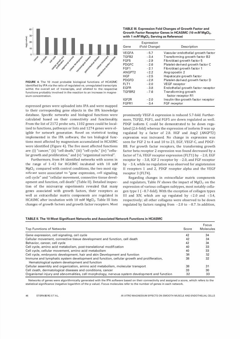
were identified (Figure 4). The five most affected functions
are: (i) ‘‘cancer,’’ (ii) ‘‘cell death,’’ (iii) ‘‘cell cycle,’’ (iv) ‘‘cellu-
lar growth and proliferation,’’ and (v) ‘‘organismal survival’’.
Furthermore, from 84 identified networks with scores in
the range of 1–42 for HCASMC incubated with 10 mM
MgCl2 compared with control conditions, the two most sig-
nificant were associated to ‘‘gene expression, cell signaling,
cell cycle’’ and ‘‘cellular movement, connective tissue devel-opment and function, cell death’’ (Table II). Detailed inspec-
tion of the microarray experiments revealed that many
genes associated with growth factors, their receptors as
well as extracellular matrix components are regulated in
HCASMC after incubation with 10 mM MgCl2. Table III lists
changes of growth factors and growth factor receptors. Most
prominently VEGF-A expression is reduced 5.7-fold. Further-
more, TGFb2, FGF1, and FGF5 are down regulated as well.PDGF isoform C could be demonstrated to be down regu-
lated (2.6-fold) whereas the expression of isoform D was up
regulated by a factor of 2.8. HGF and Ang2 (ANGPT2)
expression was increased. No change in expression was
seen for FGF 2 to 4 and 10 to 23, EGF, VEGF-C, and PDGF-
BB. For growth factor receptors, the transforming growth
factor beta receptor 2 expression was found decreased by a
factor of 7.6, VEGF receptor expression (FLT1) by À2.0, EGF
receptor by À3.8, IGF 2 receptor by À2.0, and FGF receptor
by À3.4, while no regulation was observed for angiotension
II receptors 1 and 2, PDGF receptor alpha and the VEGF
receptor 3 (FLT4).
Regarding changes in extracellular matrix componentsand regulators, Table IV shows the impact of MgCl2 on the
expression of various collagen subtypes, most notably colla-
gen type I (À8.7-fold). With the exception of collagen types
III and XIV, which are up regulated by þ2.0 and þ3.4,
respectively; all other collagens were observed to be down
regulated by factors ranging from À2.0 to À8.7. In addition,
FIGURE 4. The 10 most probable biological functions of HCASMC
identified by IPA via the ratio of regulated vs. unregulated transcripts
within the overall set of transcripts, and allotted to the respective
functions probably involved in the reaction to an increase in magne-
sium concentration.
TABLE III. Expression Fold Changes of Growth Factor and
Growth Factor Receptor Genes in HCASMC (10 mM MgCl2,
with 1 mM MgCl2 Serving as Reference)
Gene
Expression
(Fold Change) Description
VEGFA À5.7 Vascular endothelial growth factor
TGFB2 À3.4 Transforming growth factor B2
FGF5 À2.9 Fibroblast growth factor 5
PDGFC À2.6 Platelet-derived growth factor C
FGF1 À2.1 Fibroblast growth factor 1
ANGPT2 þ2.2 Angiopoetin 2
HGF þ2.5 Hepatocyte growth factor
PDGFD þ2.8 Platelet-derived growth factor D
FLT1 À2.0 VEGF receptor
EGFR À3.8 Endothelial growth factor receptor
TGFBR2 À7.6 Transforming growth
factor receptor R1
IGF2R À2.0 Insulin-like growth factor receptor
FGFR1 À3.4 FGF receptor
TABLE II. The 10 Most Significant Networks and Associated Network Functions in HCASMC
Top Functions of Networks Score
Focus
Molecules
Gene expression, cell signaling, cell cycle 42 34
Cellular movement, connective tissue development and function, cell death 42 34
Behavior, cancer, cell cycle 42 34
Cell cycle, amino acid metabolism, post-translational modification 40 33
Cell cycle, cellular movement, amino acid metabolism 40 33
Cell cycle, embryonic development, hair and skin Development and function 38 32
Immune and lymphatic system development and function, cellular growth and proliferation,
Hematological system development and function
38 32
Cellular assembly and organization, amino acid metabolism, molecular transport 38 31
Cell death, dermatological diseases and conditions, cancer 33 30
Organismal injury and abnormalities, cell morphology, nervous system development and function 32 33
Networks of genes were algorithmically generated with the IPA software based on their connectivity and assigned a score, which refers to the
statistical significance (negative logarithm of the p value). Focus molecules refer to the number of genes in each network.
46 ST ERN BE RG E T A L. IN VITRO MAGNESIUM EFFECTS ON SMOOTH MUSCLE AND ENDOTHELIAL CELLS

7/30/2019 bài báo cáo vật liệu y sinh( Vũ Văn Kỳ)2
http://slidepdf.com/reader/full/bai-bao-cao-vat-lieu-y-sinh-vu-van-ky2 7/10
our analysis revealed a number of integrin subunits which
appear to be differentially expressed, with subtypes A4,
A11, and B1 being down regulated by factors of À2.1, À3.0,
and À2.3, respectively. In contrast, up regulation was
observed for subunits A10 (þ2.4) and B8 (þ2.6). Expres-
sion of fibronectin (FN1) as well as expression of the matrix
metalloproteinases 3, 10, 12, and 14, all involved in woundrepair and cell migration, was also substantially down regu-
lated (À3.3 to À8.4).
Taken together, these findings suggest a comprehensive
reprogramming of HCASMC in the presence of 10 mM MgCl2most likely towards decreased abilities of proliferation, cell
growth and cellular movement.
Cell cycle and apoptosis analyses
Within the group of highly regulated networks and biologi-
cal functions (Figure 4) cell cycle genes are regulated.
Therefore we performed more detailed real time RT-PCR
based analyses of regulated genes involved in cell cycle con-trol. These analyses verified the down regulation of the
cyclin E2 (between 17 and 21%) and CDK2 (between 57
and 73%), both necessary for progression from G1- to S-
phase [Figure 5(B), diagram] for 10 and 4 mM MgCl2,
respectively. P21 mRNA is up regulated (between 261 and
305%) and exerts an inhibitory function on cyclin E2 fur-
ther enhancing the retarding effect on the cell cycle. In con-
trast, cyclin D2 mRNA is up regulated (between 276 and
304%) by 10 and 4 mM MgCl2, respectively, and should
have a stimulating effect on HCASMC division [Figure 5(B),
diagram]. There is no statistical significant difference
between 4 and 10 mM MgCl2 in up or down regulating
cyclin D2 and p21 or cyclin E2 and CDK2, respectively. The
data from real time RT-PCR experiments [Figure 5(B), dia-
gram] confirm the data from microarray hybridization
experiments [Figure 5(B), table]. Taken together there is no
effect on cell cycle progression [Figure 5(C)]. None of these
changes in expression was seen in HCAEC. In order to eval-
uate the effects of magnesium on the cell cycle itself, the
relative percentages of HCASMC in each phase were quanti-
fied by propidium iodide staining and subsequent FACS
analysis. The relative percentages of smooth muscle cells in
sub G0/G1, G0/G1, S, and G2/M at various magnesium con-
centrations are illustrated in Figure 5(C). Treatment with
magnesium did not result in significant changes of cell cycle
phases. However, we did observe a slight increase in apo-
ptosis of HCASMC with increasing magnesium concentra-
tions to 1.26 6 0.15 and 1.55 6 0.38-fold (4 and 10 mM ,
respectively) compared to 1 mM MgCl2, which reached sta-
tistical significance for 10 mM [Figure 5(D)]. For doxorubi-
cin, which was used as a positive control, a 3.1 6 0.24-fold
increase in apoptosis was observed.
DISCUSSION
Magnesium, the most abundant intracellular divalent cation,
is involved in fundamental biological functions. Supplemen-
tation of magnesium can convey cardiovascular protective
effects just as reduction of carotid intimal thickening29 and
improvement of endothelial function.30 Based on this, this
article investigated any direct biological effects of the mag-
nesium released from biodegradable magnesium alloy stents
on gene expression, viability, and proliferation of coronary
endothelial (HCAEC) and smooth muscle cells (HCASMC).
Results of the presented in vitro experiments demon-
strate that magnesium concentrations ranging from 6.25 to25 mM MgCl2 enhance HCAEC proliferation (Figure 1). Fur-
thermore, the gene expression analyses show distinct
changes in mRNA abundance in HCASMC, but only minor
changes in HCAEC, after incubation with 10 mM MgCl2 (Fig-
ure 3). In this context, it can be stated that magnesium
affects HCASMC gene expression in a way that indicates in-
terference with major elements of neointima formation, e.g.,
down regulation of growth factor receptors and of compo-
nents of the extracellular matrix (Tables III and IV).
For the gene expression studies the extracellular magne-
sium concentration of 10 mM was chosen according to the
following considerations: a minimum of about 1 mM , a max-
imum of about 200 mM , and a practical ex vivo result givinga twofold increase in tissue concentration following implan-
tation of a magnesium stent. The cited minimum is based
on the concentration of magnesium in human serum
(0.5–1.5 mM 31), the theoretical maximum is determined by
total amount of magnesium incorporated into such bioab-
sorbable stent ($4 mg, resulting in a theoretical maximum
of 197 mM local concentration [tissue volume 0.83 mL
(length: 1.25 mm, inner diameter: 3.0 mm, outer diameter:
5.5 mm)]), and the ex vivo result came from spot sample
measurements in porcine vascular tissue in presence or
absence of a magnesium stent, which indicated a twofold
increase in magnesium tissue concentration (Table I).
Further validity to the use of an extracellular magnesium
concentration of 10 mM was given by the observations that,
in accordance with results from the ex vivo stent implanta-
tion experiment, an incubation of HCASMC with 10 mM
MgCl2 resulted in an about 2.5-fold increase of intracellular
magnesium as compared with incubation at 1 mM MgCl2(Figure 2).
Interestingly, our in vitro experiments on HCAEC and
HCASMC proliferation and viability demonstrate that magne-
sium differently affects these cell types in a concentration
range of 6.25–25 mM MgCl2 (Figure 1). Under these condi-
tions proliferation and viability of HCASMC is lower than
TABLE IV. Expression Fold Changes of Collagens in HCASMC
(10 mM MgCl2, with 1 mM MgCl2 Serving as Reference)
Gene Expression (fold change)
COL1A1 À8.7
COL4A1 À2.7
COL4A2 À3.3
COL5A1 À2.0
COL6A1 À3.7
COL12A1 À2.0
COL15A1 À2.4
COL22A1 À3.7
COL3A1 þ2.0
COL14A1 þ3.4
ORIGINAL RESEARCH REPORT
JOURNAL OF BIOMEDICAL MATERIALS RESEARCH B: APPLIED BIOMATERIALS
|JAN 2012 VOL 100B, ISSUE 1 47

7/30/2019 bài báo cáo vật liệu y sinh( Vũ Văn Kỳ)2
http://slidepdf.com/reader/full/bai-bao-cao-vat-lieu-y-sinh-vu-van-ky2 8/10
proliferation and viability of HCAEC irrespective of an initial
cell cycle arrest by serum deprivation (data not shown).
This might in part be explained by the lack of increased in-
tracellular magnesium ions in endothelial cells after incuba-
tion of endothelial cells at 10 mM MgCl2, most likely reflect-
ing the tight regulation of magnesium levels of endothelial
cells via regulation of magnesium influx, efflux, intracellular
compartmentalization, or a combination thereof.32
Accordingly, whole genome gene expression analyses
revealed 69 differentially expressed transcripts for HCAEC,
but 2416 regulated HCASMC transcripts after 24 h of incu-
bation with 10 mM MgCl2 compared with control condi-
tions. Obviously, stimulation of HCAEC proliferation by
MgCl2 [Figure 1(b)] is not associated with substantial
changes in mRNA abundance [Figure 3(b)], but is most
likely due to a regulatory physiological function of magne-
sium, e.g., cofactor role of magnesium for numerous enzy-
matic reactions.33,34 Furthermore, magnesium is involved in
regulation of ion transport processes by carrier proteins
and channels and thereby modulates signal transduction
processes.35,36
For differentially expressed genes in the smooth muscle
cells, the automated pathway analysis identifies functions
including ‘‘cancer,’’ ‘‘cell death,’’ ‘‘cell cycle,’’ ‘‘cellular growth
and proliferation,’’ and ‘‘organismal survival’’ as most likely
being affected by MgCl2 (Figure 4). These functions cover
FIGURE 5. Influence of MgCl2 on cell cycle regulation. Effect of 10 mM MgCl2 on genes involved in G1/S phase cell cycle regulation based on
IPA. Down regulated genes are presented in green, up regulated genes in red (A). Quantification of mRNA abundance of genes involved in cell
cycle regulation using real-time PCR (B, diagram) and microarray hybridization data for these genes induced by 10 m M MgCl2 (B, table). Effect
of MgCl2 on cultured HCASMC cell cycle phases after a 24-h exposure to different MgCl2 concentrations. Data are from flow cytometric analyses
of cultured HCASMC labeled with propidium iodide. Data are presented as percentages of cells in sub-G0/G1, G0/G1, S, and G2/M phase of three
independent experiments (C). Caspase 3 activity of HCASMC exposed to different MgCl 2 concentrations as well as doxorubicin (1 lM Dox) for
24 h (D). Data are presented as mean (SD for n ¼ 4. Statistical analysis was performed using a one-way ANOVA followed by Dunnett’s post hoc
test (against 1 mM MgCl2). p < 0.05 was considered significant (*). [Color figure can be viewed in the online issue, which is available at
wileyonlinelibrary.com.]
48 ST ERN BE RG E T A L. IN VITRO MAGNESIUM EFFECTS ON SMOOTH MUSCLE AND ENDOTHELIAL CELLS

7/30/2019 bài báo cáo vật liệu y sinh( Vũ Văn Kỳ)2
http://slidepdf.com/reader/full/bai-bao-cao-vat-lieu-y-sinh-vu-van-ky2 9/10
genes involved in processes attributed to cell growth, cell
division, migration, and regulation of extracellular matrix—
all part of the current understanding behind neointimal
hyperplasia. In more detail, we monitored the down regula-
tion of several growth factor receptors and associated
growth factors along with reduced transcript levels of met-
alloproteinases, fibronectin and at least seven different
kinds of collagen as a direct adjustment on an increasedextracellular magnesium concentration in smooth muscle
cells (Tables III and IV). Considering the major contribution
of matrix metalloproteinases to cell migration37 and their
extensive down regulation, magnesium might lead to a net
reduction of smooth muscle cell migration.
Although numerous cell cycle regulatory genes and cell
proliferation associated genes in smooth muscle cells were
found down regulated by gene expression analyses [Figure
5(A)] these observations did translate into halting smooth
muscle cell proliferation [Figure 5(C)].
CONCLUSION
Magnesium at a concentration of 10 mM regulates HCASMCgene expression in such a way that migration and prolifera-
tion are down regulated resulting in a quiescent appearance
of HCASMC. In contrast, proliferation of HCAEC is stimulated
by 10 mM magnesium with minor effects on gene expres-
sion. Taken together, the differential effect of magnesium on
HCASMC and HCAEC might well explain the good in vivo
performance of the bioabsorbable magnesium stent.21–23
The observed stimulation of HCAEC proliferation in a con-
centration range of 6.25–25 mM magnesium emphasizes the
demand for controlled magnesium stent corrosion and the
related magnesium ion release. Potential approaches in this
direction may be magnesium stent coatings based on corro-
sion inhibiting ceramics or hydrophobic polymers, asrecently suggested by Lu et al.38,39
ACKNOWLEDGMENTS
The authors thank Martina Nerger and Babette Hummel for
their expert technical assistance. The authors are furthermore
grateful to Prof. Gerhard Hennighausen and Dr. Claus Harder
for helpful notes and suggestions. Additionally, thanks are
given to the Biotronik AG for the provision of the bioabsorb-
able magnesium stents.
REFERENCES
1. Dotter CT. Transluminally-placed coilspring endarterial tube
grafts. Long-term patency in canine popliteal artery. Invest Radiol
1969;4:329–332.
2. Hanawa T. Materials for metallic stents. J Artif Organs 2009;12:
73–79.
3. Fischman DL, Leon MB, Baim DS, Schatz RA, Savage MP, Penn I,
Detre K, Veltri L, Ricci D, Nobuyoshi M. A randomized comparison
of coronary-stent placement and balloon angioplasty in the treat-
ment of coronary artery disease. Stent Restenosis Study Investi-
gators. N Engl J Med 1994;331:496–501.
4. Mitra AK, Agrawal DK. In stent restenosis: Bane of the stent era.
J Clin Pathol 2006;59:232–239.
5. Ferns GA, Avades TY. The mechanisms of coronary restenosis:
Insights from experimental models. Int J Exp Pathol 2000;81:
63–88.
6. Kraitzer A, Kloog Y, Zilberman M. Approaches for prevention of
restenosis. J Biomed Mater Res B Appl Biomater 2008;85:
583–603.
7. Farb A, Burke AP, Kolodgie FD, Virmani R. Pathological mecha-
nisms of fatal late coronary stent thrombosis in humans. Circula-
tion 2003;108:1701–1706.
8. Lanzer P, Sternberg K, Schmitz KP, Kolodgie F, Nakazawa G,
Virmani R. Drug-eluting coronary stent very late thrombosis revis-
ited. Herz 2008;33:334–342.
9. Joner M, Finn AV, Farb A, Mont EK, Kolodgie FD, Ladich E, Kutys
R, Skorija K, Gold HK, Virmani R. Pathology of drug-eluting stents
in humans: Delayed healing and late thrombotic risk. J Am Coll
Cardiol 2006;48:193–202.
10. Nebeker JR, Virmani R, Bennett CL, Hoffman JM, Samore MH,
Alvarez J, Davidson CJ, McKoy JM, Raisch DW, Whisenant BK,
Yarnold PR, Belknap SM, West DP, Gage JE, Morse RE, Gligoric
G, Davidson L, Feldman MD. Hypersensitivity cases associated
with drug-eluting coronary stents: a review of available cases
from the Research on Adverse Drug Events and Reports (RADAR)
project. J Am Coll Cardiol 2006;47:175–181.
11. Virmani R, Guagliumi G, Farb A, Musumeci G, Grieco N, Motta T,
Mihalcsik L, Tespili M, Valsecchi O, Kolodgie FD. Localized hyper-
sensitivity and late coronary thrombosis secondary to a siroli-
mus-eluting stent: Should we be cautious? Circulation 2004;109:
701–705.
12. Colombo A, Karvouni E. Biodegradable stents: ‘‘Fulfilling the mis-
sion and stepping away’’. Circulation 2000;102:371–373.
13. Tamai H, Igaki K, Kyo E, Kosuga K, Kawashima A, Matsui S,
Komori H, Tsuji T, Motohara S, Uehata H. Initial and 6-month
results of biodegradable poly-l-lactic acid coronary stents in
humans. Circulation 2000;102:399–404.
14. Welch TR, Eberhart RC, Chuong CJ. The influence of thermal
treatment on the mechanical characteristics of a PLLA coiled
stent. J Biomed Mater Res B Appl Biomater 2009;90:302–311.
15. Vogt F, Stein A, Rettemeier G, Krott N, Hoffmann R, Bosserhoff
AK, Michaeli W, Hanrath P, Weber C, Blindt R. Long-term assess-
ment of a novel biodegradable paclitaxel-eluting coronary poly-
lactide stent. Eur Heart J 2004;25:1330–1340.
16. Uurto I, Juuti H, Parkkinen J, Kellomaki M, Keski-Nisula L,
Nevalainen T, Tormala P, Salenius JP. Biodegradable self-expand-
ing poly-L/D-lactic acid vascular stent: A pilot study in canine and
porcine iliac arteries. J Endovasc Ther 2004;11:712–718.
17. Venkatraman SS, Tan LP, Joso JF, Boey YC, Wang X. Biodegrad-able stents with elastic memory. Biomaterials 2006;27:1573–1578.
18. Buenger CM, Grabow N, Kroger C, Lorenzen B, Hauenstein K,
Goosmann M, Schmitz KP, Kreutzer HJ, Lootz D, Ince H, Nienaber
CA, Klar E, Schareck W, Sternberg K. Iliac anastomotic stenting
with a sirolimus-eluting biodegradable poly-L-lactide stent: A pre-
liminary study after 6 weeks. J Endovasc Ther 2006;13:630–639.
19. Peuster M, Wohlsein P, Brugmann M, Ehlerding M, Seidler K,
Fink C, Brauer H, Fischer A, Hausdorf G. A novel approach to tem-
porary stenting: degradable cardiovascular stents produced from
corrodible metal-results 6–18 months after implantation into New
Zealand white rabbits. Heart 2001;86:563–569.
20. Heublein B, Rohde R, Kaese V, Niemeyer M, Hartung W, Haverich
A. Biocorrosion of magnesium alloys: A new principle in cardio-
vascular implant technology? Heart 2003;89:651–656.
21. Di Mario C, Griffiths H, Goktekin O, Peeters N, Verbist J, Bosiers
M, Deloose K, Heublein B, Rohde R, Kasese V, Ilsley C, Erbel R.
Drug-eluting bioabsorbable magnesium stent. J Interv Cardiol2004;17:391–395.
22. Erbel R, Di Mario C, Bartunek J, Bonnier J, de Bruyne B, Eberli
FR, Erne P, Haude M, Heublein B, Horrigan M, Ilsley C, Bose D,
Koolen J, Luscher TF, Weissman N, Waksman R. Temporary scaf-
folding of coronary arteries with bioabsorbable magnesium
stents: A prospective, non-randomised multicentre trial. Lancet
2007;369:1869–1875.
23. Waksman R, Erbel R, Di Mario C, Bartunek J, de Bruyne B, Eberli
FR, Erne P, Haude M, Horrigan M, Ilsley C, Bose D, Bonnier H,
Koolen J, Luscher TF, Weissman NJ. Early- and long-term intra-
vascular ultrasound and angiographic findings after bioabsorb-
able magnesium stent implantation in human coronary arteries.
JACC Cardiovasc Interv 2009;2:312–320.
ORIGINAL RESEARCH REPORT
JOURNAL OF BIOMEDICAL MATERIALS RESEARCH B: APPLIED BIOMATERIALS
|JAN 2012 VOL 100B, ISSUE 1 49
7/30/2019 bài báo cáo vật liệu y sinh( Vũ Văn Kỳ)2
http://slidepdf.com/reader/full/bai-bao-cao-vat-lieu-y-sinh-vu-van-ky2 10/10
24. Drynda A, Deinet N, Braun N, Peuster M. Rare earth metals used
in biodegradable magnesium-based stents do not interfere with
proliferation of smooth muscle cells but do induce the upregula-
tion of inflammatory genes. J Biomed Mater Res A 2008;91:
360–369.
25. Raju B, Murphy E, Levy LA, Hall RD, London RE. A fluorescent in-
dicator for measuring cytosolic free magnesium. Am J Physiol
1989;256:C540–C548.
26. Chomczynski P, Sacchi N. Single-step method of RNA isolation
by acid guanidinium thiocyanate-phenol-chloroform extraction.Anal Biochem 1987;162:156–159.
27. Schroeder A, Mueller O, Stocker S, Salowsky R, Leiber M, Gass-
mann M, Lightfoot S, Menzel W, Granzow M, Ragg T. The RIN:
An RNA integrity number for assigning integrity values to RNA
measurements. BMC Mol Biol 2006;7:3.
28. Ritter CA, Sperker B, Grube M, Dressel D, Kunert-Keil C, Kroemer
HK. Overexpression of glutathione S-transferase A1–1 in ECV 304
cells protects against busulfan mediated G2-arrest and induces
tissue factor expression. Br J Pharmacol 2002;137:1100–1106.
29. Turgut F, Kanbay M, Metin MR, Uz E, Akcay A, Covic A. Magne-
sium supplementation helps to improve carotid intima media
thickness in patients on hemodialysis. Int Urol Nephrol 2008;40:
1075–1082.
30. Shechter M, Sharir M, Labrador MJ, Forrester J, Silver B, Bairey
Merz CN. Oral magnesium therapy improves endothelial function
in patients with coronary artery disease. Circulation 2000;102:
2353–2358.
31. Sasaki S, Oshima T, Matsuura H, Ozono R, Higashi Y, Sasaki N,
Matsumoto T, Nakano Y, Ueda A, Yoshimizu A, Kurisu S, Kambe
M, Kajiyama G. Abnormal magnesium status in patients with car-
diovascular diseases. Clin Sci (Lond) 2000;98:175–181.
32. Romani A. Regulation of magnesium homeostasis and transport
in mammalian cells. Arch Biochem Biophys 2007;458:90–102.
33. Billard JM. Ageing, hippocampal synaptic activity and magne-
sium. Magnes Res 2006;19:199–215.
34. Paolisso G, Scheen A, D’Onofrio F, Lefebvre P. Magnesium and
glucose homeostasis. Diabetologia 1990;33:511–514.
35. Agus ZS, Morad M. Modulation of cardiac ion channels by mag-
nesium. Annu Rev Physiol 1991;53:299–307.
36. Matsuda H. Magnesium gating of the inwardly rectifying Kþ
channel. Annu Rev Physiol 1991;53:289–298.
37. Newby AC. Matrix metalloproteinases regulate migration, prolifer-
ation, and death of vascular smooth muscle cells by degrading
matrix and non-matrix substrates. Cardiovasc Res 2006;69:
614–624.
38. Lu P, Cao L, Liu Y, Xu X, Wu X. Evaluation of magnesium ions
release, biocorrosion, and hemocompatibility of MAO/PLLA-modi-
fied magnesium alloy WE42. J Biomed Mater Res B Appl Bio-
mater 2011;96:101–109.
39. Lu P, Fan H, Liu Y, Cao L, Wu X, Xu X. Controllable biodegrad-
ability, drug release behavior and hemocompatibility of PTX-elut-
ing magnesium stents. Colloids Surf B Biointerfaces 2011;83:
23–28.
50 ST ERN BE RG E T A L. IN VITRO MAGNESIUM EFFECTS ON SMOOTH MUSCLE AND ENDOTHELIAL CELLS