Embed Size (px)
Citation preview

)bAieiican%Mlsdllm
1oxfittesPUBLISHED BY THE AMERICAN MUSEUM OF NATURAL HISTORYCENTRAL PARK WEST AT 79TH STREET, NEW YORK, N. Y. I0024
NUMBER 2424 JUNE 23, I970
The Morphology of Eodiscoglossus,A Complete Jurassic Frog
BY MAX K. HECHT1
INTRODUCTION
Almost perfectly preserved specimens of anurans from the Mesozoicare rare and merit thorough study. The frog described in the presentpaper was collected in the middle 1940's by L. Ferrer-Condal at thefamous locality of Monsech, near the village of Rubies, Province ofLerida, Spain. This locality is renowned for its beautifully preservedfishes, invertebrates (insects and crustaceans), and reptiles (including twogenera of lizards; Hoffstetter, 1966). It has also yielded another frog,Monsechobatrachus, but the specimen is poorly preserved (Hecht, 1963).The anuran specimen described here includes both a part and a coun-
terpart. The more complete portion, the part, is in the Museum of Sci-ence in Madrid, and the counterpart is in the private collection of Ferrer-Condal. The part was figured in Piveteau (1955); the same photographappeared in Mele'ndez (1957) under the name of Eodiscoglossus santonjaeVillalta. Although a brief summary of its features was listed by Hecht(1963), until now no detailed study of Eodiscoglossus has been published.In order to evaluate its phylogenetic significance, a complete descriptionof this specimen is necessary.The fossil material from Monsech fluoresces extremely well in ultra-
violet radiation (3660 A). Fluorescence photographs made by the meth-
lResearch Associate, Department of Vertebrate Paleontology, the American Museum ofNatural History; Professor of Biology, Queens College of the City University of New York.

AMERICAN MUSEUM NOVITATES
ods described in Hecht (1960), Colbert and Tarka (1960), and Rolfe(1964) show more detail than can be observed directly in incandescentlight. All the photographs for the present paper show the specimen underblack light.
ACKNOWLEDGMENTSI express my thanks to Prof. B. Melendez of the University of Madrid
and to Dr. J. Ferrer-Condal of Tirvia, Spain, for their patience and forlending me the material; to Mr. Chester Tarka for his excellent photo-graphs; and to the National Science Foundation (Grant GB-2452) forthe aid that made this study possible.
TAXONOMY
CLASS AMPHIBIA
SUPERORDER SALIENTIA
ORDER ANURA
FAMILY DISCOGLOSSIDAE
EODISCOGLOSSUS VILLALTA, 1957
TYPE SPECIES: Eodiscoglossus santonjae Villalta.DISTRIBUTION: Santa Maria de Meya Formation, Upper Jurassic
(Upper Kimmeridgian); Province of Lerida, Spain.REVISED GENERIC DIAGNOSIS: A small discoglossid frog characterized
by its toothless upper jaw, five digits on the forelimb, nuptial pad on thefirst digit with accessory nuptial pads on the second and third digits, andthe presence of a plectrum. It is distinguished from all discoglossid frogsby its toothless maxilla and premaxilla; from the fossil Scotiophryne, La-tonia and Zaphrissa by the lack of cranial ornamentation; from Bombinaby the presence of a plectrum; and from all other living forms by itssmaller size and secondary sexual ornamentation of the male.
Eodiscoglossus santonjae Villalta, 1957TYPE: Unnumbered specimen, Museum of Science, Madrid, Spain.HoRIZON AND LOCALITY: Upper Kimmeridgianl of the Santa Maria
de Meya Formation, Province of Lerida, Spain. The quarry from whichthe specimen comes is in the vicinity of the village of Rubies, about30 kilometers north of Balaguer.
SPECIFIC DIAGNOSIS: Same as for the genus.DESCRIPTION: The skeleton is divided between the part and the coun-
'Krusat (MS.) stated that the main quarry of this deposit is of Upper Portlandian age.
2 NO. 2424
HECHT: JURASSIC FROG
terpart. The part (fig. 1) is composed of an almost complete skull,vertebral column, pectoral girdle, right forelimb and right hind limb, aless complete left forelimb, an incomplete pelvic girdle, and a short pieceof the femur of the left hind limb. The outline of the general body shapeis clearly seen. Pigment probably representing the retina is indicated,and dark areas are also present in the region of the abdomen. Most ofthe heavily keratinized parts of the body are preserved, as, for example,the nuptial pads on the first three digits of the forelimb.The counterpart (fig. 2), which is less complete, includes fragments of
the skull, parts of the vertebral column, part of the right forelimb andpelvic girdle, and most of the right hind limb.MEASUREMENTS: Measurements are best determined from the part.
The specimen measures 27.3 mm. from the premaxilla to the ischium.The skull length, from the premaxilla to the occipital condyles, is about7.5 mm. The width of the skull across the quadratic-squamosal area is5.0 mm. Approximate lengths are given for the following: Humerus, 6.0mm.; urostyle, 9.0 mm.; femur, 12.4 mm.
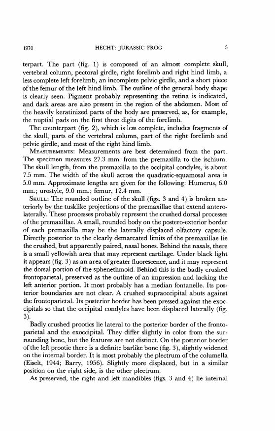
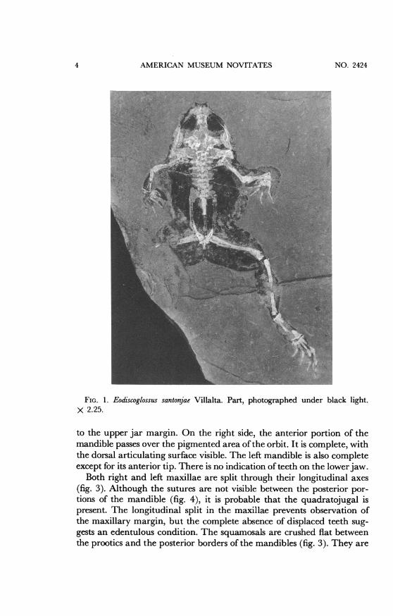
SKULL: The rounded outline of the skull (figs. 3 and 4) is broken an-teriorly by the tusklike projections of the premaxillae that extend antero-laterally. These processes probably represent the crushed dorsal processesof the premaxillae. A small, rounded body on the postero-exterior borderof each premaxilla may be the laterally displaced olfactory capsule.Directly posterior to the clearly demarcated limits of the premaxillae liethe crushed, but apparently paired, nasal bones. Behind the nasals, thereis a small yellowish area that may represent cartilage. Under black lightit appears (fig. 3) as an area of greater fluorescence, and it may representthe dorsal portion of the sphenethmoid. Behind this is the badly crushedfrontoparietal, preserved as the outline of an impression and lacking theleft anterior portion. It most probably has a median fontanelle. Its pos-terior boundaries are not clear. A crushed supraoccipital abuts againstthe frontoparietal. Its posterior border has been pressed against the exoc-cipitals so that the occipital condyles have been displaced laterally (fig.3).
Badly crushed prootics lie lateral to the posterior border of the fronto-parietal and the exoccipital. They differ slightly in color from the sur-rounding bone, but the features are not distinct. On the posterior borderof the left prootic there is a definite barlike bone (fig. 3), slightly widenedon the internal border. It is most probably the plectrum of the columella(Eiselt, 1944; Barry, 1956). Slightly more displaced, but in a similarposition on the right side, is the other plectrum.As preserved, the right and left mandibles (figs. 3 and 4) lie internal
31970
AMERICAN MUSEUM NOVITATES
FIG. 1. Eodiscoglossus santonjae Villalta. Part, photographed under black light.x 2.25.
to the upper jar margin. On the right side, the anterior portion of themandible passes over the pigmented area ofthe orbit. It is complete, withthe dorsal articulating surface visible. The left mandible is also completeexcept for its anterior tip. There is no indication of teeth on the lowerjaw.
Both right and left maxillae are split through their longitudinal axes(fig. 3). Although the sutures are not visible between the posterior por-tions of the mandible (fig. 4), it is probable that the quadratojugal ispresent. The longitudinal split in the maxillae prevents observation ofthe maxillary margin, but the complete absence of displaced teeth sug-gests an edentulous condition. The squamosals are crushed flat betweenthe prootics and the posterior borders of the mandibles (fig. 3). They are
4 NO. 2424

HECHT: JURASSIC FROG
FIG. 2. Eodiscoglossus santonjae Villalta. Counterpart, photographed under blacklight. x 2.25.
probably T-shaped. The pterygoids are visible beneath the squamosalson either side.VERTEBRAL COLUMN: There are eight presacral vertebrae (fig. 8). The
first seven are represented by crushed neural arches that bear low neuralspines. In the area of the vertebral column there is a light gray matrix.This contrasts with the black pigmented areas that may represent skinor other tissues. The eighth vertebra is represented by a bone-filled de-pression. The sacrum and a portion of the urostyle are also preserved.The first vertebra, or atlas, is badly crushed, but a low neural spine is
evident. The condylar facet can be seen on the right side, and directlyanterior to it is a remnant of the right occipital condyle.
1970 5
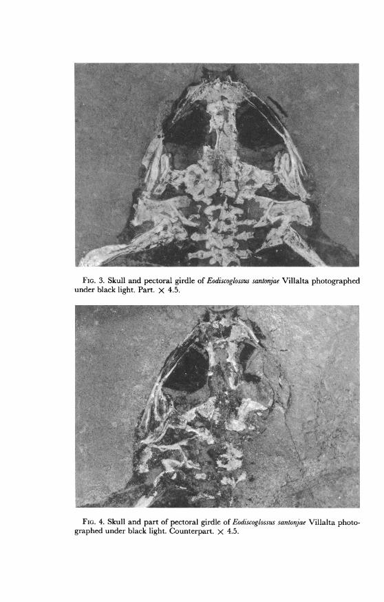
FIG. 3. Skull and pectoral girdle of Eodiscoglossus santonjae Villalta photographedunder black light. Part. X 4.5.
4,8-':W
FIG. 4. Skull and part of pectoral girdle of Eodiscoglossus santonjae Villalta photo-graphed under black light. Counterpart. X 4.5.

HECHT: JURASSIC FROG
The second vertebra also has an almost complete neural arch withan anteriorly placed transverse process. The right transverse processbears an articulating rib.The third vertebra is similar to the second, but it is more complete.
Its neural spine appears to be thick and low. The neural arch is longerand heavier than that of the anterior vertebrae. The right transverseprocess bears an articulating rib. The left transverse process is also welldeveloped, but the articulation with the rib is not clear. Both the trans-verse process and rib overlie a portion of what is probably the left cora-coid. The postzygopophyseal-prezygapophyseal contact with the pos-terior vertebra is well preserved.The well-preserved fourth vertebra is similar to the three preceding
ones. Its neural arch is crushed and the neural spine obscure. The trans-verse processes are wide; the left one clearly bears a rib. On the rightside the transverse process and rib overlie the internal end of the clavicle.The transverse processes on the fifth vertebra are narrow, and they
do not bear any ribs. They are directed laterally in a manner similar tothe more anterior vertebrae.The sixth vertebra appears to be shorter than the preceding one. Its
neural spine is not clearly preserved and its transverse processes, whichare thin and short, are directed slightly forward.The seventh vertebra is completely crushed. It bears, on the left side,
a distinct anteriorly projecting, thin transverse process. The eighth ver-tebra is represented by remnants of bone only. A sliver of bone on theleft side probably represents the transverse process. This was directedanteriorly, as indicated by both the sliver and a shallow impression. Asimilar anteriorly projected depression represents the right side.The ninth vertebra (figs. 6 and 7), or sacrum, is indicated by a bone-
line depression. A proximal fragment of the simple diapophysis extendsin the direction of the ilium on the right side. Its continuations to theilium is represented by a black pigmented imprint. On the left side ofthe sacrum there is a short stub and an impression representing the sacraldiapophysis. To the left of the sacrum is a gray hatchet-shaped arearesembling an expanded sacral hypophysis. This is in contact with theanterior end of the ilium. However, figures 2 and 7 clearly demonstratethat the sacral diapophyses are not expanded.UROSTYLE: Only the crushed anterior portion of the urostyle (figs. 8
and 9) is preserved. It is not possible to determine with certainty whetherit has a single or double condyle for articulation with the sacrum. Theevidence suggests a single condyle. The presence of a transverse processon the anterior portion of the urostyle, the usual condition in the Disco-
1970 7

AMERICAN MUSEUM NOVITATES
FIG. 5. Vertebral column, right pectoral girdle, right forelimb of Eodiscoglossussantonjae Villalta photographed under black light. Part. X 4.5.
glossidae, is not indicated. The urostyle is represented posteriorly by animpression in the matrix that extends to the pelvic symphysis. The uro-style is surrounded by black pigment that covers all the area between theanteriorly projecting arms of the ilia. Anteriorly on the left side of theurostyle there is a gap in the approximate region occupied by the trans-verse process in other discoglossids. On the right side there is a morelimited gray area, again in the appropriate region for the transverseprocess. However, there is no indication of bone, and therefore no con-clusive evidence for the transverse processes.
PECTORAL GIRDLE AND LIMBS: The pectoral girdle and limbs (figs. 5, 6,7, and 8) are preserved on both the right and left side, including thesuprascapula, scapula, coracoid. humerus, radio-ulna, carpus, meta-carpus and digits, but there are only indistinct remains of the clavicle.The entire pectoral girdle lies within the pigmented area of the bodyoutline.The details of the scapula are obscured by the overlying suprascapula.
The latter appears to be a broad thin bone, as it is in all frogs. On the leftside, the internal, or dorsal, process of the suprascapula points internally
8 NO. 2424
HECHT: JURASSIC FROG
-,,e!,-\Av1I,'^'>.X "', : v' #;d :, 8S,+w-,t ^;o v. -,$e..:v.*- ' ; s f~~~~~~~~~~~~~~~~~i '
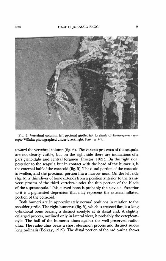
FIG. 6. Vertebral column, left pectoral girdle, left forelimb of Eodiscoglossus san-tonjae Villalta photographed under black light. Part. X 4.5.
toward the vertebral column (fig. 6). The various processes of the scapulaare not clearly visible, but on the right side there are indications of apars glenoidalis and central foramen (Proctor, 1921). On the right side,posterior to the scapula but in contact with the head of the humerus, isthe external half of the coracoid (fig. 5). The distal portion of the coracoidis swollen, and the proximal portion has a narrow neck. On the left side(fig. 6), a thin sliver of bone extends from a position anterior to the trans-verse process of the third vertebra under the thin portion of the bladeof the suprascapula. This curved bone is probably the clavicle. Posteriorto it is a pigmented depression that may represent the external inflatedportion of the coracoid.
Both humeri are in approximately normal positions in relation to theshoulder girdle. The right humerus (fig. 5), which is crushed flat, is a longcylindrical bone bearing a distinct condyle at its distal end. A slightlyenlarged process, outlined only in lateral view, is probably the ectepicon-dyle. The ball of the humerus abuts against the well-preserved radio-ulna. The radio-ulna bears a short olecranon process and distinct sulcuslongitudinalis (Bolkay, 1919). The distal portion of the radio-ulna shows
1970 9
AMERICAN MUSEUM NOVITATES
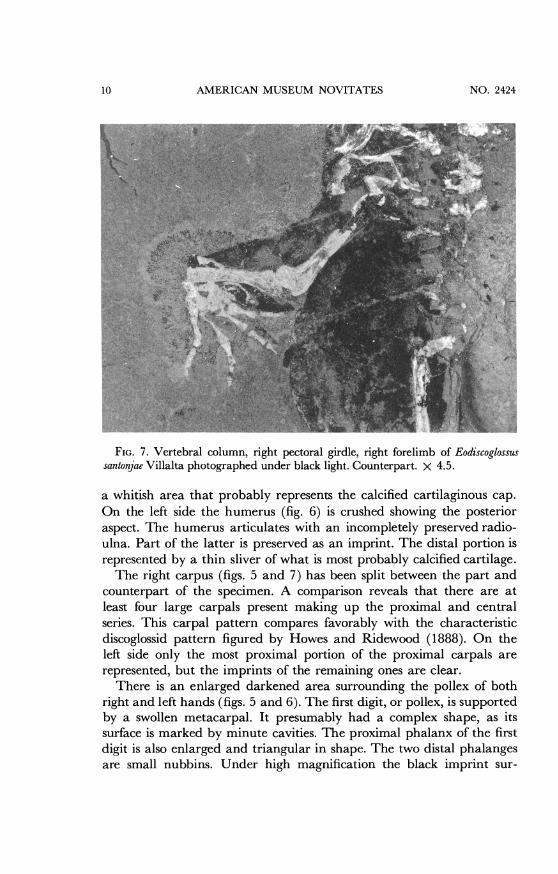
FIG. 7. Vertebral column, right pectoral girdle, right forelimb of Eodiscoglossussantonjae Villalta photographed under black light. Counterpart. X 4.5.
a whitish area that probably represents the calcified cartilaginous cap.On the left side the humerus (fig. 6) is crushed showing the posterioraspect. The humerus articulates with an incompletely preserved radio-ulna. Part of the latter is preserved as an imprint. The distal portion isrepresented by a thin sliver of what is most probably calcified cartilage.The right carpus (figs. 5 and 7) has been split between the part and
counterpart of the specimen. A comparison reveals that there are atleast four large carpals present making up the proximal and centralseries. This carpal pattern compares favorably with the characteristicdiscoglossid pattern figured by Howes and Ridewood (1888). On theleft side only the most proximal portion of the proximal carpals arerepresented, but the imprints of the remaining ones are clear.
There is an enlarged darkened area surrounding the pollex of bothright and left hands (figs. 5 and 6). The first digit, or pollex, is supportedby a swollen metacarpal. It presumably had a complex shape, as itssurface is marked by minute cavities. The proximal phalanx of the firstdigit is also enlarged and triangular in shape. The two distal phalangesare small nubbins. Under high magnification the black imprint sur-
10 NO. 2424
HECHT: JURASSIC FROG
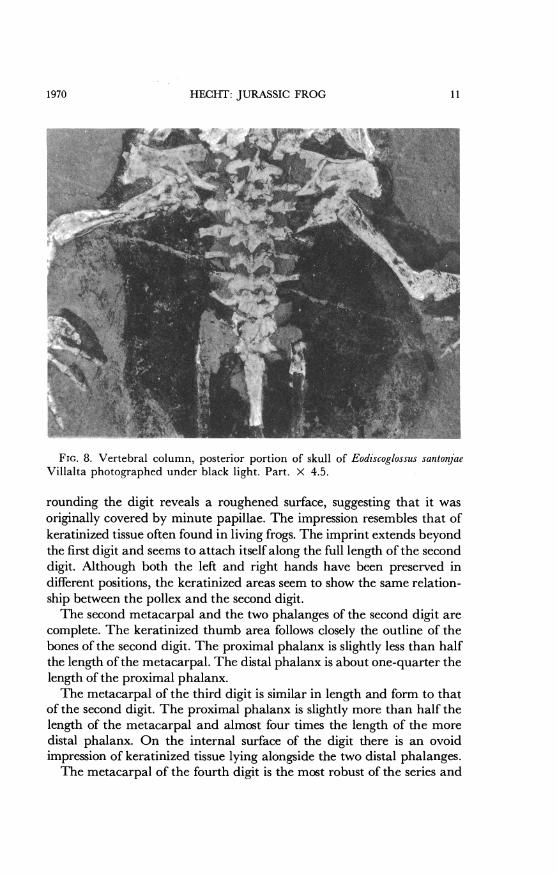
FIG. 8. Vertebral column, posterior portion of skull of Eodiscoglossus santonjaeVillalta photographed under black light. Part. X 4.5.
rounding the digit reveals a roughened surface, suggesting that it wasoriginally covered by minute papillae. The impression resembles that ofkeratinized tissue often found in living frogs. The imprint extends beyondthe first digit and seems to attach itself along the full length of the seconddigit. Although both the left and right hands have been preserved indifferent positions, the keratinized areas seem to show the same relation-ship between the pollex and the second digit.The second metacarpal and the two phalanges of the second digit are
complete. The keratinized thumb area follows closely the outline of thebones of the second digit. The proximal phalanx is slightly less than halfthe length of the metacarpal. The distal phalanx is about one-quarter thelength of the proximal phalanx.The metacarpal of the third digit is similar in length and form to that
of the second digit. The proximal phalanx is slightly more than half thelength of the metacarpal and almost four times the length of the moredistal phalanx. On the internal surface of the digit there is an ovoidimpression of keratinized tissue lying alongside the two distal phalanges.The metacarpal of the fourth digit is the most robust of the series and
1970 11
12 AMERICAN MUSEUM NUV11A 1 IN '. zIt4I
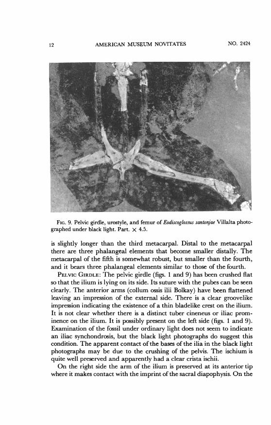
FIG. 9. Pelvic girdle, urostyle, and femur of Eodiscoglossus santonjae Villalta photo-graphed under black light. Part. X 4.5.
is slightly longer than the third metacarpal. Distal to the metacarpalthere are three phalangeal elements that become smaller distally. Themetacarpal of the fifth is somewhat robust, but smaller than the fourth,and it bears three phalangeal elements similar to those of the fourth.
PELvic GIRDLE: The pelvic girdle (figs. 1 and 9) has been crushed fiatso that the ilium is lying on its side. Its suture with the pubes can be seen
clearly. The anterior arms (collum ossis ilii Bolkay) have been flattenedleaving an impression of the external side. There is a clear groovelikeimpression indicating the existence of a thin bladelike crest on the ilium.It is not clear whether there is a distinct tuber cineneus or iliac prom-inence on the ilium. It is possibly present on the left side (figs. 1 and 9).Examination of the fossil under ordinary light does not seem to indicatean iliac synchondrosis, but the black light photographs do suggest thiscondition. The apparent contact of the bases of the ilia in the black lightphotographs may be due to the crushing of the pelvis. The ischium isquite well preserved and apparently had a clear crista ischii.On the right side the arm of the ilium is preserved at its anterior tip
where it makes contact with the imprint of the sacral diapophysis. On the
-11KT {) IA .
HECHT: JURASSIC FROG
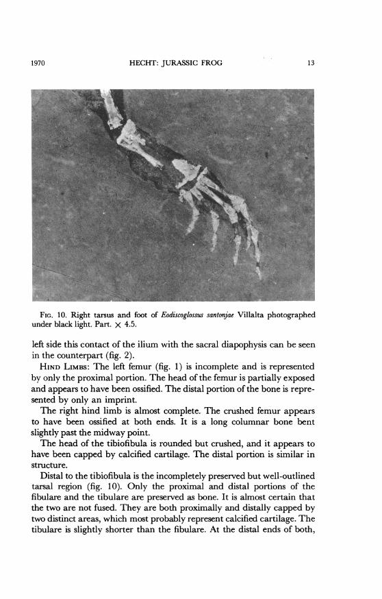
FIG. 10. Right tarsus and foot of Eodiscoglossus santonjae Villalta photographedunder black light. Part. X 4.5.
left side this contact of the ilium with the sacral diapophysis can be seenin the counterpart (fig. 2).HIND LIMBS: The left femur (fig. 1) is incomplete and is represented
by only the proximal portion. The head of the femur is partially exposedand appears to have been ossified. The distal portion of the bone is repre-sented by only an imprint.The right hind limb is almost complete. The crushed femur appears
to have been ossified at both ends. It is a long columnar bone bentslightly past the midway point.The head of the tibiofibula is rounded but crushed, and it appears to
have been capped by calcified cartilage. The distal portion is similar instructure.
Distal to the tibiofibula is the incompletely preserved but well-outlinedtarsal region (fig. 10). Only the proximal and distal portions of thefibulare and the tibulare are preserved as bone. It is almost certain thatthe two are not fused. They are both proximally and distally capped bytwo distinct areas, which most probably represent calcified cartilage. Thetibulare is slightly shorter than the fibulare. At the distal ends of both,
131970
AMERICAN MUSEUM NOVITATES
which represent the proximal row of primitive tetrapod tarsalia, there isa large gap. This gap separates the two proximal bones and the row ofcompletely preserved metatarsals. The remaining tarsalia, which werecartilaginous and not preserved, must have been in this gap. Directlyposterior to the first digit the two elements of the prehallux are indicated(figs. 9 and 10). These two elements are represented on the specimen bysplit bone and are more evident in the photograph than in the actualmaterial. Adjacent to the prehallux are the split elements of the firstmetatarsal and the first digit. The first metatarsal is expanded on eitherend and narrow at the waist. Distal to it lies the similarly shaped firstphalanx. The distal phalanx is triangular and comes to an attenuatedpoint.The second digit is represented by an imprint of the second metatarsal
and the split bone of the first and terminal phalanges. The first phalanxis expanded at either end, but the second is triangular in form, with anattenuated point.The third digit is represented by an elongated metatarsal and three
phalanges. The metatarsal is incompletely preserved, but the bone is splitin the distal and proximal areas. The proximal phalangeal element is thelongest and is similar in shape to the previously described proximal ele-ments of the other digits. It is represented proximally and distally bysplit bone, and in the middle by an imprint. The first phalanx is abouttwo-thirds the length of the metatarsal. The second distal phalanx isabout two-thirds the length of the first. The distal phalanx is triangularin form and less than half the length of the more proximal phalanx.The fourth digit is the longest. Its metatarsal is damaged on both ends
but is intact in the middle. The first phalanx is about one-third shorterthan the metatarsal. The second phalanx is about one-third shorter thanthe first, and the third is the shortest one of the series. The terminalphalanx is triangular in form.The fifth digit is folded under the fourth, and its three distal phalanges
are visible. A thin black imprint, representing the web between the digits,is evident at the level of the metacarpals of the proximal portions of thephalanges. The position of the web is probably not natural, but a resultof preservation. On the median surface of the foot, at the level of theproximal tarsalia, there is an imprint of keratinized integument (fig. 10)that probably represents a secondary sexual male adornment.
PHYLOGENETIC SIGNIFICANCEThe above description of the Kimmeridgian genus Eodiscoglossus dem-
onstrates that the basic osteology of the Discoglossidae was established by
14 NO. 2424
HECHT: JURASSIC FROG
the Late Jurassic. The skull and vertebral column of Eodiscoglossus exhibitmajor characters that can be used to demonstrate its affinity with livinggenera of this family. The presence of a plectrum distinguishes it fromBombina, which lacks this structure (Maree, 1945; Slabbert, 1945). Thepresence of a simple non-expanded sacral diapophysis in Eodiscoglossus in-dicates its similarity to Discoglossus and Barbourula. This feature, alongwith the absence of dermal ornamentation on the skull, distinguish itfrom the Tertiary Latonia and Pelophilus (Friant, 1960) and from theCretaceous Scotiophryne (Estes, 1969). A unique feature of Eodiscoglossusmay be the absence of teeth in the upper jaw. The apparent lack ofrudimentary coccygeal transverse processes is remarkable. This conditionmay be an artifact of preservation, but as the study of Madej (1965) hasdemonstrated, there is great variation in this character. However, it isalways present in Bombina.The light-colored matrix surrounding the vertebral column (evident
in the photographs) may be the remains of the calcareous secretions ofthe endolymphatic system (Dempster, 1930), unless it is an accident ofpreservation. Perhaps the unique aspect of this anuran specimen is thepreservation of the keratinized integument. The presence of the nuptialpad borne on the enlarged pollex, along with secondary pads on thesecond and third digits, and the presence of ornamentation on the hindlimb is similar to the condition found in breeding males of such genera asBombina and Discoglossus (Knoepffler, 1961).
It may be inferred that this specimen represents a breeding male.Therefore, not only was the discoglossid morphology established by theLate Jurassic, but apparently amplexial behavior is also at least thisancient.
REFERENCES
BARRY, T. H.1956. The ontogenesis of the sound-conducting apparatus of Bufo angusticeps
Smith. Morph. Jahrb., Bd. 97, Heft 4, pp. 477-544.BOLKAY, ST. J.
1919. Elements of the comparative osteology of the tailless Batrachia. GlasnikaZemal. Mus. Bosni i. Hercegovini, vol. 31, pp. 278-356.
COLBERT, E. H., AND CHESTER TARKA1960. Illustration of fossil vertebrates. Medical and Biol. Illus., vol. 10, pp.
237-246.DEMPSTER, WILFRID T.
1930. The morphology of the amphibian endolymphatic organ. Jour. Morph.and Physiol., vol. 50, no. 1, pp. 71-126.
EISELT, J.1944. Die Muscularis opercularis und die mittlere Ohrspare der Anuran Am-
1970 15

AMERICAN MUSEUM NOVITATES
phibien. Arch. Naturgesch., N.F., vol. 10, pp. 179-230.ESTES, RICHARD
1969. A new fossil discoglossid frog from Montana and Wyoming. Breviora, no.328, pp. 1-7.
FRIANT, MADELEINE1944. Caracteres anatomiques d'un Batracien oligocene de la Limagne, le
Prodiscoglossus vertaizoni nov. gen. nov. sp. Compt. Rendus Acad. Sci.Paris, vol. 219, pp. 561-562.
1960. Les Batraciens anoures. Caracteres osteologiques des Discoglossidaed'Europe. Acta Zool., vol. 41, pp. 113-139, 12 figs.
HECHT, MAX K.1960. A new frog from an Eocene oil-well core in Nevada. Amer. Mus. Novi-
tates, no. 2096, pp. 1-14.1963. A reevaluation of the early history of the frogs. Part II. Syst. Zool., vol.
12, no. 1, pp. 20-35.HOFFSTETTER, ROBERT
1965. Les Sauria (= Lacertilia) du Jurassique Superieur de Montsech(Espagne). Bull. Soc. Geol. France, 7th ser., vol. 7, pp. 549-557.
HOWEs, G. B., AND W. RIDEWOOD1888. On the carpus and tarsus of the Anura. Proc. Zool. Soc., no. XI, pp.
141-149.KNOEPFFLER, LOUIS-PHILLIPPE
1961. Contribution 'a l'etude du genre Discoglossus (Amphibiens, Anoures).Theses Fac. Sci. Univ. Paris, ser. A, no. 932, 94 pp.
KRUSAT, GEORGE[MS.] Beitrag zur Geologie und Palaontologie der Sierre del Monsech (Provin-
cia de Lerida, Spanien). Diplom-Arbeit, Freie Universitat Berlin, 1966.MADEJ, ZDZISLAW
1965. Variations in the sacral region ofthe spine in Bombina bombina and Bombinavariegata. Acta Biologica Cracoviensia, Series Zoologia, vol. VIII, pp.186-197.
MAREE, W. A.1945. Contributions to the cranial morphology of the European anura Alytes
obstrecans (Laurenti). Ann. Univ. Stellenbosch, vol. XXIII, sect. A.,pp. 43-65.
MELENDEZ, BERMUDO1957. La evolucion biologica. [Adaptation of Leonardi, Piero, 1950. L'evolu-
zione dei viventi, Brescia, Morcelliana, 360 pp.] Madrid, Ediciones Fax,405 pp.
PIVETEAU, JEAN (ED.)1937. Traite de Paleontologie. Tome V. Paris, Masson et Cie., 1110 pp.
PROCTOR, JOAN B.1921. On the variation of the scapula in the batrachian groups Aglossa and
Arcifera. Proc. Zool. Soc. London, pp. 197-214.ROLFE, W. D. IAN
1965. Use of ultraviolet rays. In Kummel, B., and D. Raup (eds.), Handbook ofpaleontological techniques. San Francisco and London, W. H. Freemanand Company, pp. 350-360.
NO. 242416
1970 HECHT: JURASSIC FROG 17
SLABBERT, G. K.1945. Contributions to the cranial morphology of the European anuran
Bombina variegata (Linne). Ann. Univ. Stellenbosch, vol. XXIII, sect. A,pp. 67-89.
VILLALTA COMELLA, JOSE F. DE1957. Eodiscoglossus santonjae Vill., la Rana mas antigua conocida, que ha sido
encontrada en el Jurfasico superior de Santa Maria de Mey'a (Lerida).In Melendez, Bermudo, La evoluci6n biologica. Madrid, Ediciones Fax,p. 146, fig. 80. [Adaptation of Leonardi, Piero, 1950. L'evoluzione deiviventi. Brescia, Morcelliana, 360 pp.]