Embed Size (px)
Citation preview
CASE REPORT
Bailout technique to rescue the abruptly occluded side branchwith collapsed true lumen after main vessel stenting
Atsushi Funatsu1 • Ryo Hirokawa1 • Shigeru Nakamura1
Received: 15 December 2015 / Accepted: 30 December 2015 / Published online: 11 January 2016
� The Author(s) 2016. This article is published with open access at Springerlink.com
Abstract Guidewire recrossing into the abruptly occlu-
ded side branch (SB) after main vessel (MV) stenting in the
coronary bifurcation is difficult, particularly if the SB has a
dissection because the true lumen of SB is collapsed by a
hematoma and the second guidewire easily goes into the
false lumen. This paper reports a bailout technique to
rescue the occluded SB that was complicated by a hema-
toma because of an unsuccessful guidewire recrossing after
MV stenting using a small balloon dilation in the collapsed
SB true lumen behind the stent strut and wire penetration.
Keywords Bifurcation lesion � Side branch �Percutaneous coronary intervention
Introduction
Bifurcation lesion is one of the complex lesions in the
percutaneous coronary intervention (PCI) because it is
necessary not only to recanalize the main vessel (MV) but
also to avoid the side branch (SB) occlusion. SB occlusion
has been reported in 6–9 % of bifurcation stenting [1–3].
Therefore, before stent implantation in the MV, a protec-
tive guidewire is usually placed into the SB, and after stent
implantation, the SB is recrossed by another guidewire
from inside of the stent. When the SB is completely
occluded, another guidewire must be manipulated to
recross the SB only by referring to the jailed guidewire as a
marker. However, in some cases, the second guidewire
unintentionally enters into subintimal space of the SB,
resulting in a dissection and hematoma at the SB. When
this occurs, it becomes more difficult to recross into the
true lumen of the SB because that is collapsed by the
hematoma. If the SB is a small vessel and the patient has no
symptoms, it can be left. However, if the SB is a relatively
large vessel and the patient has acute chest pain, it might be
necessary to reopen the SB.
Here, we report a bailout technique to rescue the
occluded right ventricular (RV) branch complicated dis-
section using a balloon dilation on the first jailed guidewire
and penetration by a tapered-tip guidewire (ASAHI Gaia
second guidewire; ASAHI Intecc, Japan) from inside the
stent strut to pick up an SB true lumen.
Case
A 58-year-old male was admitted to our hospital because of
an acute inferior myocardial infarction. An emergency
coronary angiogram revealed the right coronary artery
(RCA) total occlusion just distal of RV branch bifurcation.
PCI was performed for the culprit lesion. After crossing the
guidewire to the RCA distally and to the RV branch, we
used an intravascular ultrasound (IVUS) for examination
following a thrombectomy by aspiration catheter. IVUS
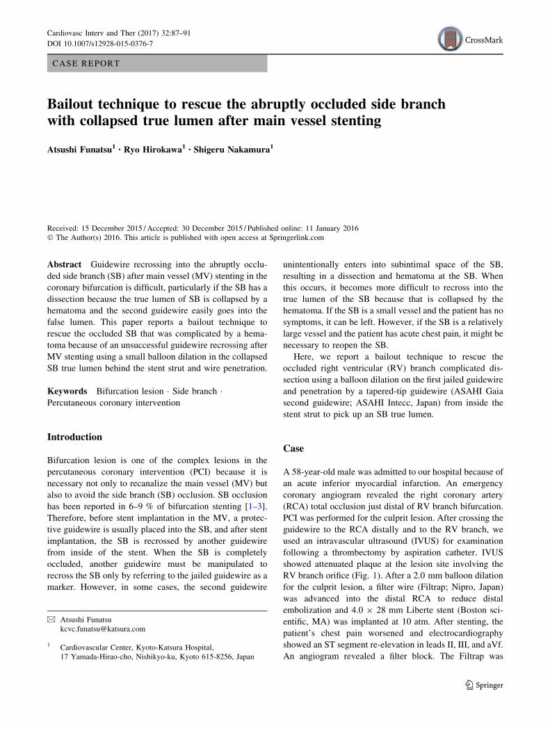
showed attenuated plaque at the lesion site involving the
RV branch orifice (Fig. 1). After a 2.0 mm balloon dilation
for the culprit lesion, a filter wire (Filtrap; Nipro, Japan)
was advanced into the distal RCA to reduce distal
embolization and 4.0 9 28 mm Liberte stent (Boston sci-
entific, MA) was implanted at 10 atm. After stenting, the
patient’s chest pain worsened and electrocardiography
showed an ST segment re-elevation in leads II, III, and aVf.
An angiogram revealed a filter block. The Filtrap was
& Atsushi Funatsu
1 Cardiovascular Center, Kyoto-Katsura Hospital,
17 Yamada-Hirao-cho, Nishikyo-ku, Kyoto 615-8256, Japan
123
Cardiovasc Interv and Ther (2017) 32:87–91
DOI 10.1007/s12928-015-0376-7
removed and injected with nicorandil, and the main coro-
nary flow was improved. However, the patient continued to
experience chest pain and ST elevation in leads from V1 to
V3. The angiogram showed that the RV branch was com-
pletely occluded (Fig. 2). Because the occluded RV branch
was the only major RV branch and an RV infarction might
have occured if the RV branch was lost, we attempted
rewiring to the RV branch using ASAHI SION blue
guidewire (ASAHI Intecc, Japan) with a Crusade catheter
(Kaneka, Japan). The second guidewire entered into the SB
but the tip did not go distally enough that the second
guidewire got into the subintimal space of the RV branch.
Although we tried to recross several times, the results were
the same. IVUS image at the RV branch bifurcation from
the RCA MV revealed a jailed first guidewire in the true
lumen that was collapsed by an expanded false lumen
presented as a high echogenic lumen (Fig. 2). The RV
orifice was also sealed by shifted plaque. In this situation,
the second guidewire was likely to enter into the expanded
false lumen, and it was impossible to recross into the
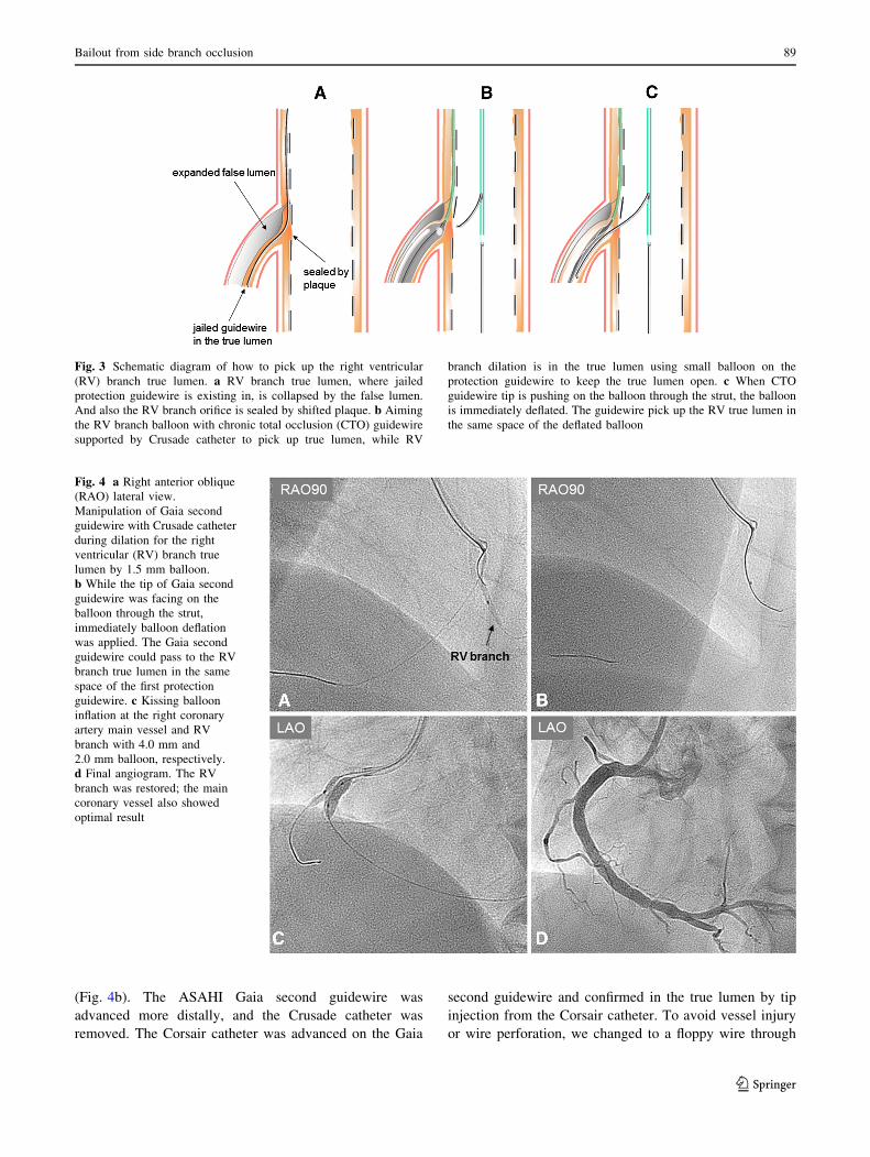
collapsed true lumen. In order to pick up the true lumen,
enlarging the true lumen was a key point. We decided to
dilate the RV branch behind the stent strut and pick it up
from inside the stent (Fig. 3). A 1.5 mm balloon could not
enter the vessel behind the deployed stent struts. First, a
Corsair 135 cm catheter (ASAHI Intecc, Japan) was
advanced into the RV branch on the protection jailed
guidewire behind the stent struts. The Corsair catheter
successfully reached the RV branch. Then, we removed the
Corsair catheter and inserted 1.5 mm balloon on the same
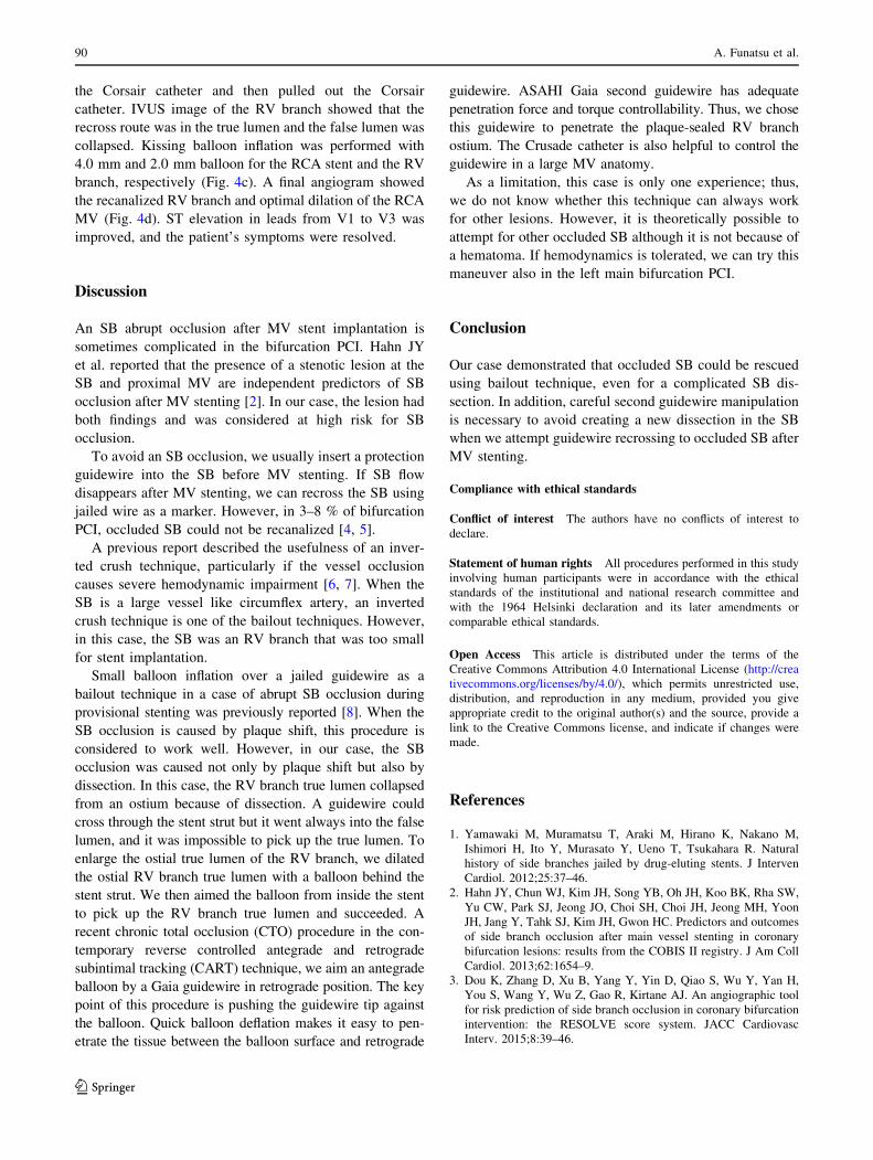
guidewire. While the RV branch balloon dilation was in the
true lumen, we aimed the RV branch balloon with the
ASAHI Gaia second guidewire, supported by Crusade
catheter, on the MV guidewire (Fig. 4a). While the ASAHI
Gaia second guidewire tip was pushing on the balloon
through the strut, the balloon was immediately deflated.
The wire picked up the RV true lumen in the same space of
the first protection guidewire without hard resistance
Fig. 1 Angiogram and
intravascular ultrasound (IVUS)
image (Opticross; Boston
scientific, Japan) at the right
ventricular (RV) branch
bifurcation after guidewire
crossing and suction. IVUS
image revealed that culprit
lesion included attenuated
plaque, and the RV branch was
running through the plaque
(white arrow). A small branch
was also seen at the opposite
side of RV branch by the
angiogram and IVUS
Fig. 2 Angiogram after
stenting showed that the right
ventricular (RV) branch
remained completely occluded.
Intravascular ultrasound image
from the right coronary artery
main vessel at the RV branch
bifurcation revealed that the RV
branch was detected at 9 o’clock
position. The true lumen of the
RV branch (white arrow) was
collapsed by an expanded false
lumen that was presented as a
high echogenic lumen (white
arrowhead)
88 A. Funatsu et al.
123
(Fig. 4b). The ASAHI Gaia second guidewire was
advanced more distally, and the Crusade catheter was
removed. The Corsair catheter was advanced on the Gaia
second guidewire and confirmed in the true lumen by tip
injection from the Corsair catheter. To avoid vessel injury
or wire perforation, we changed to a floppy wire through
Fig. 3 Schematic diagram of how to pick up the right ventricular
(RV) branch true lumen. a RV branch true lumen, where jailed
protection guidewire is existing in, is collapsed by the false lumen.
And also the RV branch orifice is sealed by shifted plaque. b Aiming
the RV branch balloon with chronic total occlusion (CTO) guidewire
supported by Crusade catheter to pick up true lumen, while RV
branch dilation is in the true lumen using small balloon on the
protection guidewire to keep the true lumen open. c When CTO
guidewire tip is pushing on the balloon through the strut, the balloon
is immediately deflated. The guidewire pick up the RV true lumen in
the same space of the deflated balloon
Fig. 4 a Right anterior oblique
(RAO) lateral view.
Manipulation of Gaia second
guidewire with Crusade catheter
during dilation for the right
ventricular (RV) branch true
lumen by 1.5 mm balloon.
b While the tip of Gaia second
guidewire was facing on the
balloon through the strut,
immediately balloon deflation
was applied. The Gaia second
guidewire could pass to the RV
branch true lumen in the same
space of the first protection
guidewire. c Kissing balloon
inflation at the right coronary
artery main vessel and RV
branch with 4.0 mm and
2.0 mm balloon, respectively.
d Final angiogram. The RV
branch was restored; the main
coronary vessel also showed
optimal result
Bailout from side branch occlusion 89
123
the Corsair catheter and then pulled out the Corsair
catheter. IVUS image of the RV branch showed that the
recross route was in the true lumen and the false lumen was
collapsed. Kissing balloon inflation was performed with
4.0 mm and 2.0 mm balloon for the RCA stent and the RV
branch, respectively (Fig. 4c). A final angiogram showed
the recanalized RV branch and optimal dilation of the RCA
MV (Fig. 4d). ST elevation in leads from V1 to V3 was
improved, and the patient’s symptoms were resolved.
Discussion
An SB abrupt occlusion after MV stent implantation is
sometimes complicated in the bifurcation PCI. Hahn JY
et al. reported that the presence of a stenotic lesion at the
SB and proximal MV are independent predictors of SB
occlusion after MV stenting [2]. In our case, the lesion had
both findings and was considered at high risk for SB
occlusion.
To avoid an SB occlusion, we usually insert a protection
guidewire into the SB before MV stenting. If SB flow
disappears after MV stenting, we can recross the SB using
jailed wire as a marker. However, in 3–8 % of bifurcation
PCI, occluded SB could not be recanalized [4, 5].
A previous report described the usefulness of an inver-
ted crush technique, particularly if the vessel occlusion
causes severe hemodynamic impairment [6, 7]. When the
SB is a large vessel like circumflex artery, an inverted
crush technique is one of the bailout techniques. However,
in this case, the SB was an RV branch that was too small
for stent implantation.
Small balloon inflation over a jailed guidewire as a
bailout technique in a case of abrupt SB occlusion during
provisional stenting was previously reported [8]. When the
SB occlusion is caused by plaque shift, this procedure is
considered to work well. However, in our case, the SB
occlusion was caused not only by plaque shift but also by
dissection. In this case, the RV branch true lumen collapsed
from an ostium because of dissection. A guidewire could
cross through the stent strut but it went always into the false
lumen, and it was impossible to pick up the true lumen. To
enlarge the ostial true lumen of the RV branch, we dilated
the ostial RV branch true lumen with a balloon behind the
stent strut. We then aimed the balloon from inside the stent
to pick up the RV branch true lumen and succeeded. A
recent chronic total occlusion (CTO) procedure in the con-
temporary reverse controlled antegrade and retrograde
subintimal tracking (CART) technique, we aim an antegrade
balloon by a Gaia guidewire in retrograde position. The key
point of this procedure is pushing the guidewire tip against
the balloon. Quick balloon deflation makes it easy to pen-
etrate the tissue between the balloon surface and retrograde
guidewire. ASAHI Gaia second guidewire has adequate
penetration force and torque controllability. Thus, we chose
this guidewire to penetrate the plaque-sealed RV branch
ostium. The Crusade catheter is also helpful to control the
guidewire in a large MV anatomy.
As a limitation, this case is only one experience; thus,
we do not know whether this technique can always work
for other lesions. However, it is theoretically possible to
attempt for other occluded SB although it is not because of
a hematoma. If hemodynamics is tolerated, we can try this
maneuver also in the left main bifurcation PCI.
Conclusion
Our case demonstrated that occluded SB could be rescued
using bailout technique, even for a complicated SB dis-
section. In addition, careful second guidewire manipulation
is necessary to avoid creating a new dissection in the SB
when we attempt guidewire recrossing to occluded SB after
MV stenting.
Compliance with ethical standards
Conflict of interest The authors have no conflicts of interest to
declare.
Statement of human rights All procedures performed in this study
involving human participants were in accordance with the ethical
standards of the institutional and national research committee and
with the 1964 Helsinki declaration and its later amendments or
comparable ethical standards.
Open Access This article is distributed under the terms of the
Creative Commons Attribution 4.0 International License (http://crea
tivecommons.org/licenses/by/4.0/), which permits unrestricted use,
distribution, and reproduction in any medium, provided you give
appropriate credit to the original author(s) and the source, provide a
link to the Creative Commons license, and indicate if changes were
made.
References
1. Yamawaki M, Muramatsu T, Araki M, Hirano K, Nakano M,
Ishimori H, Ito Y, Murasato Y, Ueno T, Tsukahara R. Natural
history of side branches jailed by drug-eluting stents. J Interven
Cardiol. 2012;25:37–46.
2. Hahn JY, Chun WJ, Kim JH, Song YB, Oh JH, Koo BK, Rha SW,
Yu CW, Park SJ, Jeong JO, Choi SH, Choi JH, Jeong MH, Yoon
JH, Jang Y, Tahk SJ, Kim JH, Gwon HC. Predictors and outcomes
of side branch occlusion after main vessel stenting in coronary
bifurcation lesions: results from the COBIS II registry. J Am Coll
Cardiol. 2013;62:1654–9.
3. Dou K, Zhang D, Xu B, Yang Y, Yin D, Qiao S, Wu Y, Yan H,
You S, Wang Y, Wu Z, Gao R, Kirtane AJ. An angiographic tool
for risk prediction of side branch occlusion in coronary bifurcation
intervention: the RESOLVE score system. JACC Cardiovasc
Interv. 2015;8:39–46.
90 A. Funatsu et al.
123
4. Pan M, Suarez de Lezo J, Medina A, Romero M, Segura J,
Ramirez A, Pavlovic D, Hernandez E, Ojeda S, Adomuz C. A
stepwise strategy for the stent treatment of bifurcated coronary
lesions. Catheter Cardiovasc Interv. 2002;55:50–7.
5. Pan M, Suarez de Lezo J, Medina A, Romero M, Delgado A,
Segura J, Ojeda S, Mazuelos F, Hernandez E, Melian F, Pavlovic
D, Esteban F, Herrador J. Drug-eluting stents for the treatment of
bifurcation lesions: a randomized comparison between paclitaxel
and sirolimus eluting stents. Am Heart J. 2007;153:15–7.
6. Furuichi S, Airoldi F, Colombo A. Reverse inverse crush: a way to
get out of trouble. Catheter Cardiovasc Interv. 2007;70:708–12.
7. PanM, RomeroM, Ojeda S, Segura J, Mazuelos F, Suarez de Lezo J,
MedinaA, Suarez deLezo J. Inverted crush technique for uncrossable
side branch occlusion during provisional side branch stenting: a new
role for jailed wire. Rev Esp Cardiol. 2011;64:718–22.
8. Aminian A, Dolatabadi D, Lalmand J. Small balloon inflation over
a jailed wire as a bailout technique in a case of abrupt side branch
occlusion during provisional stenting. J Invasive Cardiol.
2010;22:449–52.
Bailout from side branch occlusion 91
123