Embed Size (px)
Citation preview

AMWAY
1
CORNEOTHERAPY
The information contained in this technical bulletin is, to the best of our knowledge, true and accurate. No warranty, expressed or implied is made or intended. The use should be based upon the customer’s own investigations and appraisal. No recommendation should be construed as an inducement to use a material in infringement of patents or applicable government regulations. January 2016
CALIFORNIA SCC JANUARY 24, 2016HERVE OFFREDO, CSO
BARNET

2
WHO CREATED THE WORD CORNEOTHERAPY?
First used by the American dermatologist Pr. Albert Kligman in the mid1960’s.
Treatment centred on renovation of the corneal layer to correct the entireskin: “OUTSIDE‐IN THERAPY”.
“Long term effects of therapeutic treatment of the corneallayer could assist repair of the underlying structures of the skinsuch as the epidermis and dermis”.
Dr. Kligman understood that cells could communicate.
Barnet adopted this word and approach in December 2013.

3
Whether it be from the aging process or some other factor, occasionally the stratum corneum fails in places. When weakened, viruses and bacteria penetrate the skin, and water escapes the skin at a faster rate. Like a dam that's sprung a leak, a failing stratum corneum demands immediate attention.
Corneotherapy aims at the maintenance or the recovery of the stratum corneum to improve the function of the skin barrier homeostasis (balance) of the skin.
However, the undesirable corneocyte was not necessarily seen as a key cell.
No nucleus = no brain!
4
ABRASION30% GLYCOLIC
1% RETINOIC ACID

5
Today, the star corneocytes are protected and considered for :
REDUCING DULLNESS
REDUCING STINGING
REDUCING FINE LINES
REDUCING VISIBLE PORES
REDUCING YELLOWING
REDUCING DRYNESS

GLM‐DS ‐ Time release technology of AHAs to gently remove dead cells. A glycolic/lactic/malic acid in a mineral delivery system.
Furcellaran– To bind water on the skin, to increase lipids and filaggrin in the top layers of the skin, and fence off ROS
AWL Complex – To double water in corneocytes in 2 weeks. An apple/watermelon/lentil extract complex
Silver vine extract– To reduce the grey color of the skin
Ume extract– To reduce the yellow color of the skin
Saccharide Isomerate– To do a perfect differentiation for a resistant stratum corneum to stingers with less visible pores and fence off ROS and bacteria
Lysophosphatidic acid– To improve differentiation with less visible pores for skin using biochemistry
Lavanda oil‐ To bind corneocytes and to restore lipids6

7
THE REMOVAL OF DEAD CELLS
Alpha Hydroxy Acids in skin care are back. AHAs were one of the earlier and broadly embraced approaches for a visible clinical effect. In the 1990’s though, “irritation potential” was a concern. The use level of AHAs was a numbers game: 5% to 15% or even more. The cosmetic industry moved toward “exfoliation at the skin’s pH” with a glucosamine‐based complex.
In 2016, we offer GLM‐DS. It is designed to diffuse AHAs in a progressive and controlled way. Exfoliation performance equals conventional AHAs within 2 weeks without any stinging sensation.
A new tool for gently resurfacing the stratum corneum, GLM‐DS is a lamellar gel acting as a cutaneous reservoir of AHA.

8
EFFECT OF 2% GLM‐DS ON ACNE SCARS4 weeks, 24 people
DAY 0
DAY 14
DAY 28
25% less visible scars, 80% less blackheads

9
STRATUM CORNEUM OF AHAS FOR NEW SKIN
GLM‐DS (Lamellar Water Gel) was designed to diffuse AHAs in aprogressive and controlled way into the skin and to be easy to incorporateinto formulations with a non‐acid pH.
Lamellar gel with sodium magnesium silicate
AHAs between the layers = reservoir of AHA.
Glycolic acid (15%)
Lactic acid (7%)
Citric acid (6%)
Total AHA = 28%

10
CONTROLLED DIFFUSION OF AHA
To demonstrate the benefits of LamellarWater Gel (LWG) technology, wecompared its diffusion properties with aconventional Carboxy Methyl Cellulose(CMC) gel.
CMC does not have any special reservoirproperties, similar to a formulationcontaining free AHAs.
The LWG and CMC structures both contain28% of the same AHAs (same ratios oflactic, glycolic and citric). They wereapplied to the surface of an agarose gel inwhich AHA diffusion is measured withinfrared spectroscopy. This approachenables the measure of AHA diffusionkinetics.
Agar (H2O)
ZnSe Crystal

1111
GLM‐DS: CONTROLLED DIFFUSION OF AHA
GLM‐DSGel CMC
Time (hours)
The AHAs of GLM‐DS are released in a more progressive and continuous manner.
We can therefore expect 2 types of benefits:‐Extended contact time giving high performance exfoliation of the skin.‐A more progressive supply of AHA which is therefore less aggressive for the skin.
0.30
0.25
0.20
0.15
0.0
AHA (g%)
Kinetics of AHA release (time to reach a plateau):‐5h 25 min for the CMC technology.‐9h 12 min for the LWG technology.
0 2 4 6 8 10 12

12
PERFORMANCE INDEX (P.I.) DEFINITION AND CALCULATION METHOD
AHAs are evaluated with two major parameters: exfoliation and inflammation.
The P.I. of an AHA or combination of AHAs is the ratio of its exfoliating performance to theinflammation it generates (1).
A P.I. greater than 1 is characteristic of a good exfoliation with a minimum of inflammation.
It is important to maintain a balance between both effects. A cocktail of AHAs cannot be used forpowerful exfoliation if it is going to generate severe irritation or even burns.
(1) Smith WP; Hydroxy acids and skin aging. Cosmetics & Toiletries 109 (9) 41‐44, 46‐48 (1994)
ProtocolHuman skin explants (39 year old donor). Application of creams at Day 0, Day 1 and Day 2. Analysis of desquaming effectiveness at Day 5 by stripping and counting of scales obtained on the strip.
Creams pH % Exfoliation
GLM‐DS 2% (=0.56 AHA) 3.68 + 50%
AHA 8% 1.90 + 59%
AHA 15% 1.62 + 75%

13
EVALUATION OF INFLAMMATION LEVEL
ProtocolHuman skin explants (39 year old donor). Application of creams at D0, D1 and D2 after stripping. Analysis of inflammation‐inducing effect by quantificationof COX2 (purple‐pink) by immunohistochemistry.
The quantification of the enzyme COX2 (greenband), responsible for the production ofProstaglandin mediators, is first performed inthe epidermis.An additional analysis showed that the 8% and15% AHA formulations also caused significantinflammation of the dermis, characteristic of anaggressive effect.
While GLM‐DS did not generate any significantinflammation, the AHA 8 and 15 formulationsactivate a significant inflammatory response ofmore than 60%.
CREAM GLM‐DS 2%(0.56% AHA)Good epidermal cohesion.15% inflammation
CREAM AHA‐8 (8% AHA)
Damaged cohesion68%***inflammation
CREAM AHA‐15 (15% AHA)
Damaged cohesion82%***inflammation
*** p<0.001 Student t Test

14
EXFOLIATION BENEFIT / INFLAMMATION
CREAMGLM‐DS 2% (0.56%
AHA)
+50%* of scales
15% Non Aggressive Inflammation
CREAM AHA‐8 (8% AHA)
+59% of scales
68%***Aggressive
Inflammation
CREAM AHA‐15(15% AHA)
+75%* of scales
82%*** Aggressive
Inflammation
EXFOLIATING PERFORMANCE
INFLAMMATION LEVEL
PERFORMANCE INDEX 3.33 0.86 0.91
**p<0.05; ***p<0.001 Student t TestThe P.I. index of GLM‐DS is 3 or 4 times higher than the P.I. of AHA formulas.

15
QUALITY OF THE EXFOLIATION
The heterogeneity index of the scales
An aggressive treatment causes deepdesquamation and eliminates a clump ofepidermal cells. This phenomenon isvisible on the strips with the AHA 8 and15 creams. The exfoliation then damagesthe cohesion of the skin below thestratum corneum and may beaccompanied by burns.
Only GLM‐DS provides uniformexfoliation which means its action is moretargeted and optimized than the AHA 8and AHA 15 formulations.
Heterogeneity Index
487
CREAM GLM‐DS 2%(0.56% AHA)
Heterogeneity Index
956
CREAM AHA‐8(8% AHA)
Heterogeneity Index
1181
CREAM AHA‐15(15% AHA)
*p<0.05 Student t Test
ProtocolHuman skin explants (39 year old donor). Application of preparations at D0, D1 and D2. Analysis of desquamating effectiveness at D5 by stripping andcounting of scales obtained on the strip.
A higher number is a sign of aggressive and anarchic exfoliation.

16
TEST ON SENSITIVE SKIN
Ten volunteers with sensitive skin A solution of water with 10% GLM‐DS is applied under an occlusive patch for 48 hours.
Skin is observed 30 minutes and 24 hours after removing the patch.
The Median Irritation Index (MII) is 0.00 out of 4.00.

17
GLM‐DS
INCI Name: Water (and) Glycolic Acid (and) Lactic Acid (and) Sodium Magnesium Silicate (and) Citric Acid (and) Xanthan Gum
REACH: All components are pre‐registered or exempt.Canada: Low volume exemption.
China: All components are listed in the Inventory of Existing Cosmetic Ingredients in China (IECIC).
Use Level: 2%
Sunburn Alert: This product contains an alpha hydroxy acid (AHA) that may increase your skin’s sensitivity to the sun and particularly the possibility of sunburn. Use a sunscreen, wear protective clothing and limit sun exposure while using this product and for a week afterwards.

18
Application: Topical application of 1.5% Furcellaran for one week on human skin explants.
Explants treated with Furcellaran have an homogenous and smoothed surface with only a few desquaming cells.
Furcellaran’s RESTRUCTURING EFFECT
Untreated explant Explant treated with Furcellaran
T 0 After 1 week

19
Subjects: 5 volunteers
Samples: 100% Furcellaranand 100% Hyaluronic Acid (HA)
Application: Put in cells with a relative humidity that will vary from 0 to 95%
Measurement: For each condition of relative humidity, the polymers capture water molecules to reach a maximum called “gain mass sorption equilibrium”.The value of gain mass reflects the ability of each polymer to trap water molecules.
HYALURONIC‐LIKE ACTION – HYDROSCOPY COMPARATIVE STUDY
Polysaccharide obtained via an original depolymerization process. Economic Process.

20
HYALURONIC‐LIKE ACTION – HYDROSCOPY COMPARATIVE STUDY
Results
Furcellaran is able to trap atmospheric water molecules following a kinetic comparable to that of hyaluronic acid.
Furcellaran is able to trap nearly its own weight in water, and to maintain a moisture film on the surface of the skin.
Hyaluronic Acid
Furcellaran
Relative Mass G
ain
% of Relative Humidity
Furcellaran is HA‐like.

21
Subjects: 30 volunteers
Samples: solution containing 1.5% Furcellaran (sol.)(equivalent to 0.009% Furcellaran)solution containing 0.025% Hyaluronic Acid (MW = 1.3 to 1.8 MD)
Application: 15 volunteers apply Furcellaran solution15 volunteers apply Hyaluronic AcidApplied twice daily for 2 weeksSkin hydration level measured using corneometer
HA is a high molecular weight: 2,000,000 DFurcellaran is 200,000 D
Furcellaran vs. HA – IN VIVO TEST

22
FURCELLARAN VS. HYALURONIC ACID
Its hydrating action is faster and more efficient than Hyaluronic Acid.
Furcellaran increases the skin hydration level 4 hours after only a single application.
‐2
0
2
4
6
8
10
12
4H 6H 24H
Varia
tion % of skin hydration level versus T0
Furcellaran Hyaluronic Acid
p<0.05 student test

23
Effect of Furcellaran on Gene Expression of Proteins Involved in Corneocyte Formation, Adhesion and Integrity
FURCELLARAN AND STRATUM CORNEUM STRENGTH
Corneodesmosin (CDSN) – adhesion protein of the extracellular portion of corneodesmosomes, involved in the cohesion of corneocytes
Calmodulin_like 5 (CALML5) – forming part of the family of calcium‐binding protein, a key enzyme in the terminal differentiation of keratinocytes
Cornuline (CRNN) – protein which plays a role in epidermal differentiation Small proline‐rich protein 2A (SPRR2A) – protein important for the barrier function of the epidermis Loricrine (LOR) – protein which plays an important role in the structure of corneocytes in cornified envelope and
junctions between corneocytes formation
65%
0%
10%
20%
30%
40%
50%
60%
70%
Furcellaran 24H (1%)
Furcellaran increases by 41% the expression of CDSN after one application (24H). This protein is located at the superficial layer of the epidermis.
SHORT TERM EFFECT
CDSN
65%
98%
48%
69%
0%
20%
40%
60%
80%
100%
120%
DEFB4 RNASE SPRR2A LOR
Furcellaran 72H (1%)
Furcellaran increases the expression of CALM5, CRNN, SPRR2A and LOR by respectively 86%, 54%, 48% and 69% after 3 applications (72H). These proteins are located deeper.
LONG TERM EFFECT
Relativ
eexpression
(in
%of
control)
Relativ
eexpression
(in
%of
control)

24
Primary Use: MoisturizerINCI Name: Water (and) Sodium Carrageenan (and)
Sea SaltREACH: All components are pre‐registered or
exempt.Canada DSL: All components are listed or sold in
commerceChina Registration: All components are listed on the Inventory
of Existing Cosmetic Ingredients in China (IECIC).
Suggested Use Level: 1% ‐ 3%
FURCELLARAN

25
AWL COMPLEX IN 2 HOURS
Before AWL Complex After 2 hours, with 3% AWL Complex:
Fine lines are significantly diminished.

26
AWL Complex is a natural complex combining watermelon rind extract, Lens Esculenta (Lentil) Fruit Extract and unripened apple and apple skin in an optimized delivery system.
Watermelon rind is one of nature’s few materials to contain citrulline. Citrulline is essential to the functioning of filaggrin which forms a critical part of the skin’s own water based moisturizing complex.
The lentil extract contains vitamin B5 and trisaccharides.
The apple starch is a source of polysaccharides, sodium lactate and sodium PCA.

27
AWL COMPLEX– IMMEDIATE SKIN HYDRATION (15 MIN.)
11
33
47
0
5
10
15
20
25
30
35
40
45
50
Placebo 1% AWL Complex 3% AWL Complex
Skin im
pedance DP
M value
s
ProtocolA gel containing 3% AWL Complex was applied to the skin. Measurements taken at various time periods.
AWL Complex is shown to hydrate the skin after 15 minutes in a dose dependent manner.

28
AWL COMPLEX – SKIN HYDRATION (24 HOURS)SINGLE APPLICATION
1
7
12
0
2
4
6
8
10
12
14
Placebo 1% AWL Complex 3% AWL Complex
Skin im
pedance DP
M value
s
ProtocolA gel containing 3% AWL Complex was applied to the skin. Measurements taken at various time periods.
AWL Complex continues to hydrate the skin after 24 hours.

29
AWL COMPLEX – INCREASING HYDRATION IN 2 WEEKS
Protocol20 subjects, 60 or older with dry and scaly, whitish looking skin used 3% AWL Complex twice daily for two weeks on legs. Cells were recovered from shave biopsies.
After 2 weeks of AWL Complex use at 3%, cells were recovered by biopsies and weighed. There was 85% more water in cells treated by AWL Complex than those not treated.
The cells were then applied onto paper to see how long it would take to lose 90% of water.
Control
AWL Complex Treated
It was more difficult for cells treated with AWL Complex to lose their water. It took 4 times longer than the control cells.
30 Minutes
2 Hours X4

30
AWL COMPLEX – MOISTURIZING FOR 10 DAYS
Dermatologist Assessment of Dryness on Face
1 ‐ Slight Flaking2 – Moderate Flaking3 – Marked Scaling4 – Severe Scaling
Test material Conc BL Day 1 Day 2 Day 3 Day 5 Day 10
Placebo gel 0% 2.7 2.74 2.71 2.6 2.77 2.66
AWL Complex 3% 2.74 2.42 2.37 1.75 1.4 1.05
% Improvement 12% 13% 35% 50% 61%
*Data presented are averages of at least 20 subjects and based on a 0‐4 point scale described above. Data highlighted in yellow are statistically significant
Results show a complete reduction of excessive scaling in 10 days, while the placebo has no effect.

31
AWL COMPLEX – REDUCING IRRITATION FOR 10 DAYS
Dermatologist Assessment of Redness, Skin Irritation on Face
1 ‐Minimal2 – Moderate 3 – Severe4 – Fiery Red
Test material Conc BL Day 1 Day 2 Day 3 Day 5 Day 10
Placebo gel 0% 1.8 1.78 1.82 1.87 1.92 1.89
AWL Complex 3% 1.82 1.7 1.64 1.58 1.47 1.32
% Improvement 5% 10% 16% 25% 30%
*Data presented are averages of at least 20 subjects and based on a 0‐4 point scale described above. Data highlighted in yellow are statistically significant
Results show a gradual improvement during the 10 days of treatment with AWL Complex, while the placebo has no effect.
32
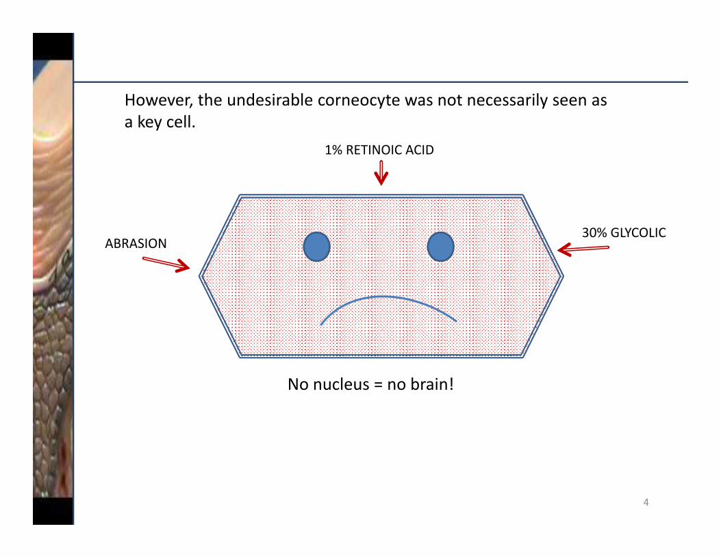
AWL COMPLEX– SKIN COHESION IN 2 WEEKS
Before AWL Complex
Cells are not cohesive, they are easy to remove.
After 2 weeks with AWL Complex
Tape strips fewer cells as they are more cohesive.
0
10
20
30
40
50
60
70
Day 0 Day 1 Day 3 Day 14
Untreated with 3% AWL Complex
Light transmission –LED Units
33
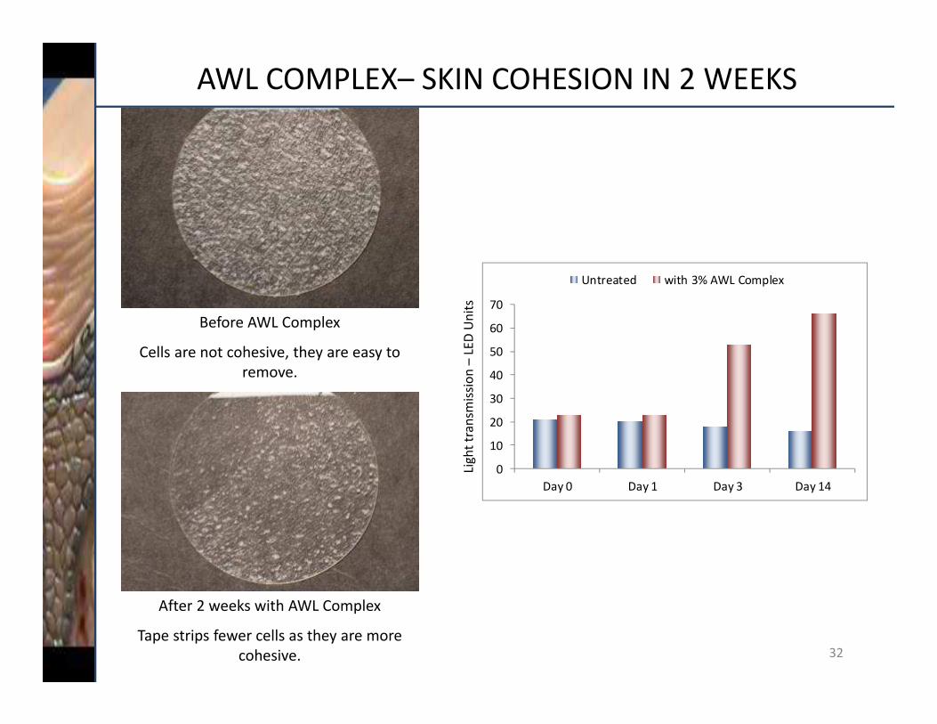
AWL COMPLEX
INCI Name: Water (and) Glycerin (and) Citrullus lanatus (Watermelon) Fruit Extract (and) Pyrus malus (Apple) Fruit Extract (and) Lens Esculenta (Lentil) Fruit Extract (and) Sodium PCA (and) Sodium Lactate
REACH Status: Low volume exemptionCanada DSL: Components are listed on the DSL, RICL or exemptECOCERT Status: In compliance with the ECOCERT Standard
for natural ingredientsChina Registration: All components are listed in the Inventory of
Existing Cosmetic Ingredients in China (IECIC).
Suggested Use Level: 3%

3434
CONCEPT: TRANSLUCENT SKIN
In all age groups in Japan, “skin translucency” is a top priority.
% of group
con
sidering
translu
cency a top priority
Age in years
If we average all of the age groups, 55% of 1077 women interviewed that “ideal skin” is translucent skin – more than any other criteria (pore size, dryness, etc.)
Ref. JMA Research Institute.Inc.http://www.jmar.biz/hot/html/w_dai07_2.html

35
Before carbonylation
20µmol/L sodium hypochlorite & Silver Vine.
Silver Vine solid10 50 1000Conc.
Stratum corneum collected from the upper arm
SILVER VINE EXT. (1%) BLOCKS CARBONYLATION INDUCED BY A CHEMICAL
Carbonylation treatmentfor 16 hours
(µg/mL)
Sodium hypochlorite induces carbonylation.

36
Silver Vine. solid
0
25
50
75
100
125
150
175
No treatment
0 10 50
Carbon
ylation level (AU
)
Carbonylation treatment
SILVER VINE EXTRACT showed a very strong inhibition effect on protein carbonylation in the stratum corneum.
50 µg/mL = 1% as product
Almost 100%Inhibition with 1% Silver Vine
AU: Arbitrary unit
(µg/mL)
SILVER VINE EXT. (1%) BLOCKS CARBONYLATION INDUCED BY A CHEMICAL

37
React with car exhaust gas or tobacco smoke in sealed plastic containers for 24 hrs
EXHAUST GAS INCREASES CARBONYLATION
Collection of stratum corneum by tape stripping
Fluorescence labeling with carbonyl group
Protein carbonylation in the stratum corneum increased when exposed to car exhaust gas.
Control(Stored in normal air)
Reacted withcar exhaust gas
SC
pollutant gasin container
exhaust gas
24 Hours

38
Protocol1) Stratum corneum was collected from volunteer’s upper arm.2) The stratum corneum was soaked in solution containing 50 µg/mL of Silver Vine Extract for 24hrs and dried.3) The stratum corneum were placed in a plastic bag. 4) Car exhaust gas was put into the bag and sealed and stored for 24hrs. 5) Evaluation
Silver Vine solid0 500
+‐ +
0
Carbon
ylation level (AU
)
0
20
40
60
80
100
120
140
gas
Silver Vine solid0 500
+‐ +
Carbon
ylation level (AU
)
****
n=3**: p<0.01
AU: Arbitrary unit
SILVER VINE EXTRACT (1%) DECREASES CAR EXHAUST CARBONYLATION
(µg/mL)
50 µg/mL = 1% Silver Vine Extract
SILVER VINE EXTRACT at 1% inhibited protein carbonylation in the stratum corneum induced by the exposure to car exhaust gas by 100 %.

39
SILVER VINE EXT. (2%) REDUCES TOBACCO CARBONYLATION
Protocol1) Stratum corneum was collected from volunteer’s upper arm. 2) The stratum corneum was soaked in solution containing 100 µg/mL of
Silver Vine Extract for 24hrs and dried.3) The stratum corneum were placed in a plastic bottle. 4) Tobacco smoke (mainstream smoke) was put into the bottle and sealed
and stored for 24hrs. 5) Evaluation
Carbon
ylation level (AU
)
0
20
40
60
80
100
120
gas
Silver Vine solid
0 1000+‐ +
****
Corneoclear solid
0 1000
+‐ +(µg/mL)
n=3**: p<0.01
Silver Vine Extract at 2% totally inhibited protein carbonylation in the SC induced by the exposure to tobacco smoke.
Carbon
ylation level (AU
)
100 µg/mL = 2% Silver Vine Extract

40
Carbon
ylation level (AU
)0
20406080
100120140160
Protocol1) Stratum corneum was collected from volunteer’s upper arm. 2) The stratum corneum were placed in a plastic bag. 3) Car exhaust gas was put into the bag and sealed and stored for 24hrs. 4) The carbonylated stratum corneum was soaked in solution
containing 50 µg/mL of Silver Vine Extract for 24hrs. 5) Evaluation
Silver Vine solid0 500
+‐ +(µg/mL)
SILVER VINE EXTRACT (1%) REVERSES POLLUTION‐INDUCED CARBONYLATION
gas
Silver Vine solid
0 500
+‐ +
Carbon
ylation level (AU
)
****
n=3* : p<0.05***: p<0.001
38.8%degradation
50 µg/mL = 1% Silver Vine Extract

41
SILVER VINE EXTRACT
INCI Name: Water (and) Butylene Glycol (and) Actinidia polygama Fruit Extract
REACH Status: Natural / Low Volume ExemptionCanada DSL: Natural / Low Volume ExemptionChina Registration: All components are listed in the Inventory of
Existing Cosmetic Ingredients in China (IECIC). Japan: Approved as a QD additiveSuggested Use Level: 0.2% ‐ 1.0%Solubility: Water

42
WHEN THE COLOR OF THE SKIN REVEALS YOUR AGE
Apparent age: 38.9. This picture was artificially darkened (at right).
Apparent age: 42.4. People interviewed then guess that this
person is older than 42.

43
WHY THE SKIN GETS DARKER
Age (years)
Melanin indexAGEs index (A.U)
r=0.678, p < 0.0001
1.0
0.9
0.8
0.7
0.6
0.5
0.4
0.315 25 35 45 55 65
AGEs index (A.U)
b*r=0.629, p < 0.0001
24
22
20
18
16
14
12
101.0 1.5 2.0 2.5 3.0 3.5
24
22
20
18
16
14
12
100.3 0.5 0.7 0.9 1.1
b*
r=0.533, p < 0.0005
The accumulation of AGEs and melanin make the skin look darker.
Ohshima H et al., Skin Res Technol., 15, 496‐502 (2009)
AGE = Advanced Glycated Elements
and MELANIZATION
A combination of GLYCATION

44
1. INHIBITORY EFFECT ON CROSS‐LINK REACTION
ProtocolMix ribose (reduction sugar), lysozyme (protein) and test sample. Keep at 37° C for one week. Electrophorese the mixture, then measure the amount of cross‐linked protein by Coomassie dye
Ume Extract Freeze Dried (µg/ml)**: 1000 µg/ml = 5% liquid product
Protein
Sugar
Cross‐linked
compoundAGEs+
Prod
uctio
n of cross‐link
(%)
The chart shows that 1% Ume Extract is good at reducing the cross‐link.

45
ProtocolMix α‐diketone and test sample. Keep at 37° C for 10 hours. Measure decomposition (benzoic acid) produced by HPLC
Protein
Sugar
Cross‐linked
compoundAGEs+
2. CUTOFF EFFECT ON CROSS‐LINK REACTIONCross‐link cutoff (%
)
Ume Extract Freeze Dried (µg/ml)**: 2000 µg/ml = 10% liquid product
The chart shows that 1% Ume Extract is good
at cutting the cross‐link.

46
Protein
Sugar
Cross‐linked
compoundAGEs+
ProtocolMix ribose, type 1 collagen and test sample. Keep at 37° for 2 weeks. Measure the amount of produced AGEs using AGEs antibody
3. INHIBITORY EFFECT ON AGEs FORMATIONAG
Es Formation (%
)
***
***
******
Ume Extract Freeze Dried (µg/ml)**: 2000 µg/ml = 10% liquid product
***: p<0.001
The chart shows that 1% Ume Extract is good at reducing AGE.

47
Protein
Sugar
Cross‐linked
compoundAGEs+
4. ACCELERATION OF AGEs DECOMPOSITION
ProtocolMix ribose, type 1 collagen. Keep at 37° for 2 weeks. Add test sample. Measure the amount of remaining AGEs using AGEs antibody
AGEs decom
position acceleratio
n (%
)
Ume Extract Freeze Dried (µg/ml)1000 µg/ml = 5% liquid product
*: p<0.05 **
The chart shows that 1% Ume Extract has a slight effect
on AGE decomposition.

48
UME EXTRACT
INCI Name: Water (and) Butylene Glycol (and) Prunus mume Fruit Extract
REACH Status: ExemptCanada DSL: Natural exemptionECOCERT Status: In compliance with the ECOCERT Standard
for natural ingredientsChina Registration: All components are listed in the Inventory of
Existing Cosmetic Ingredients in China (IECIC).
Suggested Use Level: 3%

49
TWO SOLUTIONS FOR PORE MINIMIZATION
Acting on the critical genes for perfect differentiation
Acting on the biological pathway for perfect differentiation

50
CLINICAL TEST: OVERALL PERFECTING ACTION OF SACCHARIDE ISOMERATE ON ONE VOLUNTEER (OF 20)
Day 0 Day 0 Day 0
Day 28 Day 28 Day 28
Porphyrine: ‐56% Roughness: ‐38% Number of pores: ‐22%
After 4 weeks using Saccharide Isomerate, this woman founds her skin smoother, softer, healthier, and regenerated.

51
CONCEPT
+
n n
Galactose N‐acetyl‐glucosamin
BENEFITSSaccharide Isomerate is produced by culture in bioreactor to obtain a pure, natural and characterized molecule (GALACTOSE and N‐ACETYL‐GLUCOSAMIN), which has no land‐based equivalent.
Saccharide Isomerate moisturizes and exfoliates the skin. Benoiderm helps new corneocytes to be healthy, which results in less visible pores and more resistance to stingers.
Saccharide Isomerate stimulates the skin’s defense against P. acnes.

52
PROPERTIES RELATED TO ACETYL GLUCOSAMINE
N‐acetyl‐glucosamine
J. Cosmet. Sci., 60, 423‐428 (July/August 2009)
Glucosamine HCl: Alternative to AHA in Skin Care
Glucosamine competes with a lectin called CD44. This CD44 is a cell adhesion molecule. Glucosamine competitively binds to CD44 and reduces cell adhesion. Glucosamine disrupts corneocyte bonds.

53
SACCHARIDE ISOMERATE IMPROVES SKIN RENEWAL
Cell renewal rate is doubled in just 1 week. By improving the skin’s natural renewal process, Saccharide Isomerate will help to eliminate dead cells on the skin’s surface and avoid the formation of a too thick and rough stratum corneum.
+61%
+82% +88%
0
+30%
+58%
+69%
0
10
20
30
40
50
60
70
80
90
0 5 10
Duration of treatment (in days)
15
%of
cellu
larrenew
al
Saccharide Isomerate
Untreated
Protocol:
‐ 17 volunteers aged between 18 and 59‐ Application of a colored cream containing 5% DHA, 1 day before start of treatment.‐ From Day 0, lotion containing 1% Saccharide Isomerate applied twice a day for 2 weeks.
Skin pigmentation by the DHA enables monitoring of cell renewal; pigmentation will be eliminated faster if cell renewal activated.

54
SMOOTHING EFFECT OF 1% SACCHARIDE ISOMERATE
**
Average variation
Maximum variation
% variatio
n versus D
ay 0
p<0.01 student test
Day 28
Day 0
Improving skin renewal and reactivating water memory assists in obtaining an increase in skin smoothness.
Protocol:
•20 volunteers aged between 35 and 45
•Lotion containing 1% Saccharide Isomerate applied twice a day for 28 days
•Use of the VISIA tool to visualize skin texture smoothing effect

55
EPIDERMAL DIFFERENTIATION
The primary function of the epidermis is to produce the stratum corneum. It is formed through the differentiation of the keratinocytes from the basal layer to the skin’s surface layer. Many proteins are involved at each stage of this differentiation process.
Chronology of differentiation protein expression
Differentiation proteins:
INVOLUCRIN (INV) : involved in forming the cornified envelop
TRANSGLUTAMINASE 1 (TGM1): ensures assembling of proteins that make up the cornified envelope
SMALL PROLINE RICH PROTEIN (SPRP): precursor proteins in the formation of the cornified envelope
LATE CORNIFIED ENVELOP (LCE): precursor proteins in the formation of the cornified envelope
CORNEODESMOSINE (CDSN): major role in cohesion of the cornified layer
NICE 1: involved in terminal differentiation of keratinocytes properly turn into corneocytes
An ingredient upregulating the gene coding for these different proteins would be a prime candidate in corneotherapy to create healthy corneocytes. Saccharide Isomerate is tested on these genes.

56
SACCHARIDE ISOMERATE INCREASES THE SYNTHESIS OF DIFFERENTIATION PROTEINS ACTING ON 9 GENES
The increase in differentiation proteins will contribute to the formation of a higher quality physical barrier, and activation of stratum corneum renewal. The gene NICE is especially increased.
Increase in
gen
e expressio
n (%
)

57
SACCHARIDE ISOMERATE DECREASES SKIN SENSITIVITY
Principle of stinging test: the test consists of applying lactic acid at 10% (pH 2) on the nasal fold, and physiologic serum on theother one. The stinging sensation triggered by lactic acid is evaluated by volunteers themselves, on a scale from 0 to 3(no severe sensation to severe sensation). The soothing effect of a product is assessed according to the variation of stingingsensation generated by lactic acid
After 1 week treatment only, 76% of volunteers observed a decrease in their skin’s sensitivity due to a stronger stratum corneum.
Protocol:•30 volunteers aged 30 +/‐ 2•Application of a gel containing 1% Saccharide Isomerate , twice daily for 1 week.
**
p<0.01 student test% variatio
n versus D
ay 0
Average variation
Maximum variation

58
OVERALL PERFECTING ACTION OF SACCHARIDE ISOMERATE AT 1%PORES ARE LESS VISIBLE
Astringent effect on pores
Variation of number and total surface area of visible pores after 28 days.
Varia
tion vs. T 0 (%
)
Total surface area of poresNumber of pores
p<0.01 student test
BEFORE
AFTERPores are less visible

59
THE MEDIATORS OF INNATE IMMUNITY AND INFLAMMATORY REACTION
Peptides of innate immunity:Defensin beta (DEFB103) : deconstructs the membrane of exogenous bacteria ; known for its efficacy against Staphylococcus aureus Secretory Leucocyte Peptidase Inhibitor (SLPI): inhibits proteases, mainly elastases activated by bacteria to better penetrate the tissues Ribonuclease Rnase 7 (RB RNASE 7): destroys bacterial and viral RNAS100 Calcium Binding Protein A10 (S100A10): inhibits bacterial growth by interacting with their cellular cycle
Mediators of inflammation:S100 Calcium Binding Protein A7 (S100A7): also called psoriasin, promotes the activity of collagenases and elastases during inflammation Toll‐like Receptor 2 (TLR2) recognizes bacterial LipoPolySaccharides (high allergen potential) and activates TNFaTNFa: activates the chemokins CXCL5 and CXCL10CXC Ligand 5 et 10 (CXCL5 et CXCL10): chemokins involved in the migration of neutrophils

60
S100 A7 TLR2 TNFα CXCL5 CXCL10
SACCHARIDE ISOMERATE REINFORCES INNATE IMMUNITY WHILE DECREASING PRO‐INFLAMMATORY MEDIATORS
Protocol: assessment of gene expression by human reconstituted epidermis treated with 1% Saccharide Isomerate applied topically.
Relativ
e expressio
n (%
of the
con
trol)
By acting on both innate immunity and inflammatory mediators, Saccharide Isomerate improves skin health while decreasing its reactivity.
Pro‐inflammatory Mediators
Relativ
e expressio
n (%
of the
con
trol)

61
SACCHARIDE ISOMERATE DECREASES P. ACNES GROWTH
P. Acnes have bacterial excretions, known as porphyrins, that lodge in the pores and are a contributing factor in acne. It is possible to visualize and quantify theamount of porphyrin on the surface of the skin since this molecule is fluorescent under UV light.
Day 0
Day 28
**
p<0.01 student test
After 28 Days Treatment
% variatio
n of porph
yrin vs. Day 0
Protocol:20 volunteers age 35 to 45 applied a lotion with 1% Saccharide Isomerate, twice daily for 4 weeks. Effect of bacterial growth evaluated by quantification of porphyrin thanks to UV light.
Average decrease Maximum decrease
On average, the porphyrin amount is decreased by 54%. This means that the P. acnes population is reduced.

62
SACCHARIDE ISOMERATE
INCI Name: Water (and) Saccharide IsomerateREACH Status: ExemptCanada DSL: Listed RICL (Revised In Commerce List)China Registration: All components are listed in the Inventory of
Existing Cosmetic Ingredients in China (IECIC).Suggested Use Level: 1.0% ‐ 2.0%Solubility: Water
* Saccharide Isomerate is available in two versions: with phenoxyethanol (as shown in this presentation) and with phenethyl alcohol. The phenethyl alcohol version is in compliance with the ECOCERT Standard for natural ingredients, and is available on a special order basis.

63
LYSOPHOSPHATIDIC ACID (LPA) MINIMIZES PORE SIZE
Placebo 0.1% Lysophosphatidic Acid
Before
After 4 weeks
Actual image Processed image Actual image Processed image
0.1% Lysophosphatidic Acid reduces by 20% the visible size and number of pores (tested on 11 volunteers, twice daily).
Similar before/after Smaller spots after

64
RELATIONSHIP BETWEEN DIFFERENTIATION AND PORE SIZE
Presence of nucleated cells (parakeratosis) around pores (in cheek area).
Pore
Large pore surrounded by nucleated cells. Photo by Nikko Chemicals
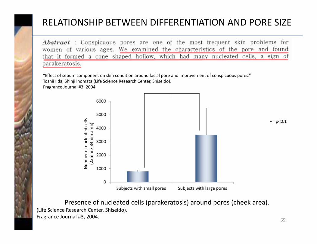
RELATIONSHIP BETWEEN DIFFERENTIATION AND PORE SIZE
“Effect of sebum component on skin condition around facial pore and improvement of conspicuous pores.” Toshii Iida, Shinji lnomata (Life Science Research Center, Shiseido).Fragrance Journal #3, 2004.
Num
ber o
f nucleated
cells
(23m
m x 34m
m area)
+
+ : p<0.1
Presence of nucleated cells (parakeratosis) around pores (cheek area).(Life Science Research Center, Shiseido).Fragrance Journal #3, 2004.
65

66
DIFFERENTIATION
(1) Calcium helps for activation of PKC and CaMK.
P
P
Transcription
?Ca2+ (1)Ca2+ (1)PPKCPKC
CaMKCaMK
DG
P
ATP
ADP
Lysophosphatidic AcidLysophosphatidic AcidLPA Receptors
When Lysophosphatidic Acid attaches to the receptors, Ca2+ intake channels become wider letting in more Ca2+. Ca2+ acts as a major differentiation regulator with DG.
Cell membrane
Regulation of Transcription
Calcium Moduline
Protein Kinase
Differentiation
Ca2+Ca2+
Diglyceride (activation of PKC)
Acts as a signaling molecule
Ca2+ (1)Ca2+ (1)
Nuclear

67
LYSOPHOSPHATIDIC ACID TRIGGERS A CALCIUM FLASH
80
100
120
140
160
Control
Ca2+ free
Ca2+ and LPA (x µg/ml)
Intensity
(% of con
trol)
****
**** **
0.03 mM Ca2+;100 µg/ml LPA
(Light blue: Ca2+) –Calcium flash (seen
as blue fluorescence)
0.03 mM Ca2+;No LPA(HEPES)
Fluorescence microscope
An influx of calcium initiates differentiation.
6.25 µg
12.25 µg
25 µg
50 µg 100 µg

68
EFFECT OF LYSOPHOSPHATIDIC ACID ON THE PHOSPHORYLATION OF PKCα
LPA induces phosphorylation of PKCα and increases CA2+ intake.
20.5 120.5 1 (hr)
100 µg/ml LPAVehicle (PBS(‐))
75
80
85
90
95
100
Densito
metric
value
PKCα P
A Larger spot means increased phosphylated PKCα.

69
IMPROVEMENT OF STRATUM CORNEUM: IN VIVO @ 0.2%
Placebo
0.2% LPA
Before Treatment After 6 weeks
IMPROVEMENTCorneocytes are packed in thin layers, show hexagonal shape and no nucleated cells are observed: stratum corneum is healthy.
No change
Before Treatment After 6 weeks

70
PORE SIZE REDUCTION: IN VIVO @ 0.1%
‐40%
‐20%
0
20%
Before After 4 weeks
Size of visible poresNumber of visible pores
Before After 4 weeks
Placebo
0.1% LPA
Placebo
0.1% LPA
‐40%
‐20%
0
20%
0.1% LPA reduced visible size and number of pores on human skin by 20%.
ProtocolSubjects: 11 people.Cream with 0.1% LPA and placebo were applied twice daily for 4 weeks (split face).

71
LYSOPHOSPHATIDIC ACID (LPA)
INCI Name: Lecithin (and) Lysophosphatidic Acid (and) Lysolecithin
REACH Status: Low volume exemptionCanada DSL: Listed DSL as BARPORE 42*China Registration: All components are listed in the Inventory of Existing
Chemical Substances in China (IECSC) and the Inventory of Existing Cosmetic Ingredients in China (IECIC) as BARPORE 42*
ECOCERT Status: Compliant with the ECOCERT Standard for natural ingredients
Suggested Use Level: 0.1% ‐ 0.2%%Solubility: Water
* BARPORE 42 is the same material as Disapore 20, but with a China‐friendly INCI name (Lecithin).

72
SMOOTHING EFFECT OF 1% LAVANDA OIL
Young skin
Examples of MDN
Old skin
Lavanda Oil action
Analysis of the “micro‐depression network” (MDN) in the epidermis
Protocol
• 10 volunteers• Twice daily application for 28 days• Lavanda Oil1%
Varia
tion in M
DN Value
Placebo 1% Lavanda Oil
Lavanda Oil significantly decreases (p<0.10) the micro‐depression index
and smoothes the skin.

73
WHAT IS A CORNEODESMOSOME?
1. Cytoskeleton filaments2. Desmosome3. Hemidesmosome4. Basement membrane
Inter membrane space
DesmocollinDesmoplakinCytokeratinCatenin
DESMOSOME
Elements of Corneodesmosomes
Desmoplakin: linked to cytokeratin and catenin
Catenin: bridge between desmoplakin and desmocollin
Desmocollin: transmembranous protein, linked together thanks to calcium; proteolysis during desquamation.

Organelles of storage and secretion in the extracellular domain of lipids (ceramide, cholesterol)
Their synthesis occurs in the cornified layer and is responsible for barrier properties 74
WHAT IS A KERATINOSOME (AKA LAMELLAR BODIES)?Lamellar Bodies = Odland Bodies = Keratinosomes
Intercellular Lamellae
Dense Band
Keratohyalin
Lamellar Bodies
Cornified Layer
Granular Layer
Spinous Layer
Basal Layer
Basal Lamina
Differentiation of epidermal cell layers and formation of lamellar bodies and intercellular hydrophobic barrier of the skin. (With permission of McGraw‐Hill, New York. F.B. Fitzpatrick et al., eds. 1987, Dermatology in General Medicine.)

75
ROLE OF EXTRACELLULAR LIPIDS IN STRATUM CORNEUM HEALTH
Normal stratum corneum
Stripped stratum corneum
Presence of extracellular lipids
Lipids removed by acetone

76
IN VITRO TEST: IMPROVING CORNIFIED LAYER COHESION
Placebo Lavanda Oil
Keratinosomes
By increasing the number of keratinosomes Lavanda Oil1% increases the synthesis of lipidic cement and therefore improves the sealing of stratum
corneum and retains water in the stratum corneum.
Effect on the number of keratinosomes responsible for the synthesis of lipids involved in the formation of lipidic cement
Lavanda Oil 1%

77
IN VITRO TEST: IMPROVING CORNIFIED LAYER COHESION
Placebo
Formation of inter‐corneocytes bonds in a culture of corneocytes
Lavanda Oil 0.01%
Gen
e ex
pres
sion
(% c
ontro
l)
Effect on the synthesis of adhesion proteins responsible for corneocytes adhesion
Lavanda Oil 1%
Lavanda Oil at 1% increases the expression of the adhesion proteins and improves the cohesion between corneocytes

78
LAVANDA OIL
INCI Name: Caprylic/Capric triglyceride (and) Hydrogenated vegetable oil (and) Lavandula stoechas extract
REACH Status: Pre‐RegisteredCanada DSL: Listed DSL/ Natural ExemptionChina Registration: All components are listed in the Inventory
of Existing Cosmetic Ingredients in China (IECIC).
ECOCERT Status: Compliant with the ECOCERT Standard for Natural Ingredients
Suggested Use Level: 1%
79
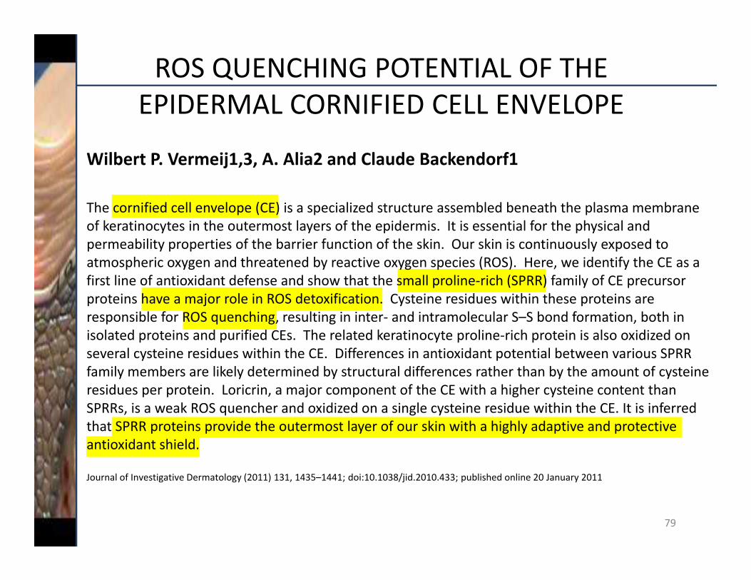
ROS QUENCHING POTENTIAL OF THE EPIDERMAL CORNIFIED CELL ENVELOPE
Wilbert P. Vermeij1,3, A. Alia2 and Claude Backendorf1
The cornified cell envelope (CE) is a specialized structure assembled beneath the plasma membrane of keratinocytes in the outermost layers of the epidermis. It is essential for the physical and permeability properties of the barrier function of the skin. Our skin is continuously exposed to atmospheric oxygen and threatened by reactive oxygen species (ROS). Here, we identify the CE as a first line of antioxidant defense and show that the small proline‐rich (SPRR) family of CE precursor proteins have a major role in ROS detoxification. Cysteine residues within these proteins are responsible for ROS quenching, resulting in inter‐ and intramolecular S–S bond formation, both in isolated proteins and purified CEs. The related keratinocyte proline‐rich protein is also oxidized on several cysteine residues within the CE. Differences in antioxidant potential between various SPRR family members are likely determined by structural differences rather than by the amount of cysteine residues per protein. Loricrin, a major component of the CE with a higher cysteine content than SPRRs, is a weak ROS quencher and oxidized on a single cysteine residue within the CE. It is inferred that SPRR proteins provide the outermost layer of our skin with a highly adaptive and protective antioxidant shield.
Journal of Investigative Dermatology (2011) 131, 1435–1441; doi:10.1038/jid.2010.433; published online 20 January 2011

80
ROS ARE ALL OVER

81
SPRRSACTIVES / SACCHARIDE ISOMERATE
SPRR1A SPRR2ASPRR1B
+ 510%+ 490%+ 750%

82
SPRSACTIVES / FURCELLARAN
SPRR2A
+ 125%
+ 60%
SPRR1A

83
CORNEOTHERAPY: “A BARRIER AGAINST UNWANTED INFLUENCES FROM THE ENVIRONMENT”
Pore MinimizationSaccharide IsomerateLPA
CarbonylationSilver Vine
GlycationUme Extract
Bacteria
Differentiation Gene NICE1Saccharide Isomerate
Cornified EnvelopeSaccharide IsomerateFurcellaran
CorneodesmosomeLavanda OilSaccharide Isomerate N
N
N N
Beta DefensineSaccharide Isomerate
STRATUM CORNEUM
EPIDERMIS
LipidsWater
N = NucleusR = Receptors
HygroscopyNMFFurcellaran
WaterAWL Complex
Lipids
Odland BodiesLavanda Oil
Detergent(strips out lipids)
NN
RR RDead Cell
ExfoliationGLM‐DS
83