Embed Size (px)
Citation preview
Basal Forebrain Cholinergic System Is Involved in Rapid Nerve GrowthFactor (NGF)-Induced Plasticity in the Barrel Cortex of Adult Rats
Neal Prakash,1 Susana Cohen-Cory,1 Silke Penschuck,1 and Ron D. Frostig1–3
Departments of 1Neurobiology and Behavior, 2Biomedical Engineering, and the 3Center for the Neurobiology of Learningand Memory, University of California, Irvine, California, 92697-4550
Submitted 21 May 2003; accepted in final form 15 September 2003
Prakash, Neal, Susana Cohen-Cory, Silke Penschuck, and Ron D.Frostig. Basal forebrain cholingergic system is involved in rapidnerve growth factor (NGF)-induced plasticity in the barrel cortex ofadult rats. J Neurophysiol 91: 424–437, 2004. First published Sep-tember 24, 2003; 10.1152/jn.00489.2003. We have previously re-ported that topical application of nerve growth factor (NGF) to thebarrel cortex of an adult rat rapidly augmented a whisker functionalrepresentation (WFR) by increasing its area and height within minutesafter NGF application. In addition, we found that TrkA, the high-affinity NGF receptor, was only found on fibers projecting into thebarrel cortex. Here we use a combination of techniques includingchronic intrinsic signal optical imaging, neuronal fiber tracking andimmunohistological techniques, to test the hypothesis that NGF-in-duced rapid cortical plasticity is mediated by the cortical projectionsof the basal forebrain cholinergic system (BFCS). Our studies localizethe source of the cells in the BFCS that project to a single WFR andalso demonstrate that TrkA-immunoreactive fibers in the cortex arealso cholinergic and likely arise from the BFCS. In addition, byselectively lesioning the BFCS cortical fibers with the immunotoxin192 IgG-saporin, we show that NGF-induced WFR-cortical plasticityis eliminated. These results, taken together with our previously re-ported imaging results that demonstrated that agonists of the cholin-ergic system (particularly nicotine) showed transient NGF-like aug-mentations of a WFR, implicate the BFCS cortical projections asnecessary for NGF’s rapid plasticity in the adult rat somatosensorycortex.
I N T R O D U C T I O N
Previously we reported that neurotrophins, such as nervegrowth factor (NGF) and brain-derived neurotrophic factor(BDNF), can rapidly induce large-scale plasticity in the adultsomatosensory cortex (Prakash et al. 1996a,b). For example,within minutes after topical application to the cortex, NGFcaused a transient, large-scale expansion in the area of awhisker functional representation (WFR) coupled with an in-crease in the height of the WFR. The current study attempts todescribe potential underlying mechanisms that mediate thisNGF-plasticity. One possibility may be that NGF is actingdirectly on cortical neurons to induce such plasticity. However,most evidence indicates that in the adult rat, NGF receptors arenot expressed by cortical neurons but rather in neurons thatproject to the cortex (Merlio et al. 1992) and on projectionfibers to the somatosensory cortex (Holtzman et al. 1995;Prakash et al. 1996b). This evidence thus suggests that NGF-induced adult cortical plasticity should involve neuronal pro-jections into the cortex that are presynaptically responsive to
NGF. Here we present evidence that indicates that the basalforebrain cholinergic system (BFCS) is the source of thesecortical projection fibers and thus implicates the BFCS in theNGF-induced cortical plasticity.
The basal forebrain (BF) in the rat, known as the magno-cellular basal forebrain nuclei, includes cells located in thenucleus basalis of Meynert and the septal nucleus of the diag-onal band of Broca (Fibiger 1982; Sofroniew et al. 1982; Wenket al. 1980). Cholinergic cells within this system project to theentire rat cortex (Lehmann et al. 1980) in a loose topographi-cally organized manner (Jimenez-Capdeville et al. 1997; La-mour et al. 1982) and constitute the only known cholinergicprojection to the adult cortex (Mesulam et al. 1983). Afterrelease, acetylcholine (ACh) can influence target cells in thecortex through two broad classes of cholinergic receptors:muscarinic and nicotinic receptors.
It has been demonstrated that NGF is synthesized in thecortex by the target cells of BF projections in the cortex andthat it binds to receptors located on the BF nerve terminals andis retrogradely transported to cell bodies of BF neurons, whereit regulates their survival and function as predicted by theneurotrophic hypothesis (Cuello et al. 1992; Hefti et al. 1989).While extensive evidence exists for the neurotrophic hypoth-esis in the developing brain, evidence also exists to support thenotion that NGF continues to play a maintenance function forthe BFCS after the brain matures. Exogenous application ofNGF can rescue axotomized BF cholinergic neurons, reversethe decline in cortical function, and restore memory (Dekker etal. 1992; Gage et al. 1988; Jacobs et al. 1994; Koliatsos et al.1990; Kromer 1987; Will and Hefti 1985; Williams et al.1986). More direct experimental proof for the role of endoge-nous cortical NGF in the maintenance of the BFCS (Mingerand Davies 1992a,b) was obtained when local injections ofanti-NGF antibodies were delivered into the cortex of the adultrat (Gutierrez et al. 1997). These rats demonstrated a lack ofACh release in the cortex, a disruption of connectivity betweencortex and BF, and a disruption of learning and memory. Theimportance of endogenous cortical NGF was also demonstratedby the use of mutant mice (Chen et al. 1997). Heterozygousmutant mice for the NGF gene showing a reduced level of bothNGF-mRNA and protein within the cortex had significantlearning and memory problems that were accompanied by aloss and shrinkage of BF cells. Infusion of NGF in these adultmutants abolished the memory deficits and corrected the def-
Address for reprint requests and other correspondence: R. D. Frostig, Dept.of Neurobiology and Behavior, University of California, Irvine CA, 92697-4550 (E-mail: [email protected]).
The costs of publication of this article were defrayed in part by the paymentof page charges. The article must therefore be hereby marked ‘‘advertisement’’in accordance with 18 U.S.C. Section 1734 solely to indicate this fact.
J Neurophysiol 91: 424–437, 2004.First published September 24, 2003; 10.1152/jn.00489.2003.
424 0022-3077/04 $5.00 Copyright © 2004 The American Physiological Society www.jn.org
icits in both area and projection density of BF cells to thecortex.
It is clear that NGF plays a role in maintaining the BFcholinergic projections to the cortex. Two key findings havealso suggested the possibility that NGF can also induce imme-diate release of ACh. Knipper and collaborators obtained invitro evidence for NGF-induced rapid release of ACh in thecortex (Knipper et al. 1994a,b). The authors demonstrated sucheffects in synaptosomes prepared from the rat hippocampusthat included BF projection fibers. They showed enhancedACh release from these synaptosomes starting within 1 minafter NGF application. Anti-NGF antibodies blocked the NGF-induced rapid release of ACh. The rapid induction of AChrelease cannot be readily explained by NGF’s known role ofregulating the survival and function of BF cholinergic systemas this role requires a longer window of time (hours to days) forthese effects. In addition, the rapid release of ACh supports theexistence of NGF receptors on the terminals BF projectionfibers. Rapid induction of ACh release by NGF was alsodemonstrated in synaptosomes prepared from the visual cortexof rats and further demonstrated that such induction of AChrelease involves the activation of the high-affinity TrkA recep-tor and the p75 low-affinity neurotrophin receptor (p75) (Salaet al. 1998). Further, we obtained in vivo evidence for NGF’sability to rapidly increase (within minutes after application) theheight and areal extent of whisker evoked activity in thesomatosensory cortex (Prakash et al. 1996b). Our previoushistological results suggested that NGF’s site of action is notdirectly on the neurons within the somatosensory cortex butrather on projections originating outside it. Given that BFCSprojection neurons are the only type of neurons with bothprojections to the cortex and NGF receptors (Holtzman et al.1995), our findings (Prakash et al. 1996b) suggested an in vivolink for NGF’s rapid in vitro effects that may be mediated bythe BFCS.
One possible mechanism for NGF’s induced plasticity is thatunder behavioral conditions when cortical representations needto be rapidly augmented by increasing cortical excitability andby recruiting more neurons, cortical neurons release NGF in anactivity-dependent manner in addition to the basal release ofNGF needed for the survival and maintenance of the BF. Thereleased NGF binds to its receptors (TrkA and p75) located onthe BFCS projection fibers in the cortex. The binding of NGFthen enhances the release of ACh from BFCS projectionswithin the cortex. The release of ACh, in turn, further enhancescortical activity through ACh receptors and potentially gluta-mate release enhancement by ACh. These actions, in turn, leadto an increase in the excitability of neurons within a WFR andthe recruitment of additional neurons to the WFR, leading anareal expansion and increased height of the WFR as observedwith the imaging results (Prakash et al. 1996b). Elsewhere wereported, in support of this hypothesis, that activation of mus-carinic or nicotinic receptors in the somatosensory cortex re-sults in a WFR plasticity that is similar to the NGF-inducedplasticity (Penschuck et al. 2002). Here we present data thattested three other critical aspects of our hypothesis: we dem-onstrate that projections from BFCS reach the level of a singleWFR in barrel cortex; we demonstrate that TrkA-immunore-active fibers present in the somatosensory cortex are alsocholinergic fibers that likely arise from the BFCS; and we showthat specifically lesioning the BFCS fibers in the barrel cortex
results in a loss of the ability of NGF to rapidly augment aWFR. Some of these data have been published previously inabstract form (Prakash et al. 2000a).
M E T H O D S
Imaging
We have followed a procedure described in Prakash et al. (1996b)and is described briefly here. Adult male Sprague-Dawley rats (300–550 g) were initially anesthetized with pentobarbital (50 mg/kg ip).Throughout the rest of the experiment, pentobarbital was injected asneeded to maintain the rat in a consistent anesthetic state such that therespiration rate was 1.1 � 0.2 (range) breaths/s, rectal temperaturewas 37.0 � 0.5°C, ear color was pinkish-pale, and the corneal reflexin response to a saline drop was weak to mild. Lidocaine (0.1 ml) wasinjected under the scalp, and 5 min later a single incision was madewith a scalpel roughly along the midline of the scalp. Fascia andmuscle overlying the parietal cortex were cleared, and the bone wascleaned and dried. Next, the skull overlying the somatosensory cortexwas removed. However, unlike the previous protocol (Prakash et al.1996b) in which the dura and arachnoid mater were removed (durec-tomy), in this set of experiments, the dura remained in place but smallslits or holes were made in it (durotomy) using a 30-gauge needle.This change in protocol was instituted to decrease the probability thatrats would have significant brain herniation. A petroleum-jelly wellwas then built around the partially exposed cortex, filled with artificialcerebrospinal fluid [ACSF, containing (in g) 7.250 NaCl, 0.370 KCl,0.170 KH2PO4, 0.277 CaCl2, 0.290 MgSO4, 2.160 NaHCO3, and1.800 glucose (all per 1 l H20), pH 7.30, 37°C], and sealed with acoverslip.
A 12-bit slow-scan CCD camera (Photometrics, Tucson, AZ) im-aged a 6.8 � 5.1 mm (192 � 144 pixels; 35.5-�m pixel width) regionof the cortex that was illuminated with a stabilized 100-W tungsten-halogen light source (Zeiss Instruments) passed through an opticalfilter (630 nm band-pass 30 nm; Omega Optics, Brattleboro, VT). Toreduce potential surface artifacts induced by pulsations of the brainand vessels, the camera was focused 300 �m below the surface of thecortex. The light reflectance was measured for each pixel (9 consec-utive frames, 500 ms each) before, during, and after stimulation of thecontralateral whisker(s). Whisker stimulation was given with a com-puter-controlled mechanical stimulator (Bakin Systems II, Irvine, CA)at 5 Hz for 1 s. The interstimulus interval between trials was 16 s.Data files were created by the summation of 64 trials and thus hadduration of �20 min. The right C2 whisker was deflected 2° rostro-caudally (0.5 mm at 1.5 cm from the snout) with an average velocityof 30°/s. Eight imaging data files were collected per rat. The first fourdata files served as baseline reference. Additionally as an internalcontrol to determine the affects of change the ACSF in the petroleum-jelly well, the ACSF was replaced between data files 2 and 3. Nosignificant difference in the measured parameters was ever noted afterthis ACSF change. After the fourth data file, the ACSF was replacedwith recombinant human NGF dissolved in ACSF (15 �g in 500 �l).
Quantitative analysis of a whisker functional representation
Analysis methods have been described in detail elsewhere (Chen-Bee et al. 1996; Masino and Frostig 1996; Masino et al. 1993; Prakashet al. 1996b, 2000b). Briefly, data files were created from the sum-mation of 64 imaging trials. Ratio values for a data file were com-puter-calculated for each pixel by dividing the reflectance values ofthe images 0.5–1.5 s poststimulation by the images 1.0–0.0 s beforestimulation. Before analysis, a Gaussian filter (half-width 5) wasapplied to the raw ratio values to filter out high-frequency noise. Thepeak location of a WFR is defined as the pixel with the most negativeratio value, and this ratio value minus the median ratio value is thepeak height of a WFR, which indicates the maximal magnitude of
425MECHANISMS UNDERLYING NGF CORTICAL PLASTICITY
J Neurophysiol • VOL 91 • JANUARY 2004 • www.jn.org
evoked activity. The area at half-height of a WFR is the area boundedby the threshold that contains pixels 50% of the peak height value.
Relationship between imaging and underlying neuronalactivity
We focused our analysis on reflectance changes of 630-nm lightoccurring between 0.5 and 1.5 s poststimulus onset, an epoch ofactivity predominantly consisting of localized oxygen consumption inthe capillary bed serving activated neurons (Ances et al. 2001; Frostiget al. 1990; Kim et al. 2000; Malonek and Grinvald 1996; Malonek etal. 1997; Thompson et al. 2003) as the area of imaged activitygenerated from this epoch has been previously found to be wellcorrelated with the underlying area of evoked neuronal activity (Brett-Green et al. 2001; Frostig et al. 1990; Masino 2003; Polley et al.1999). We also have repeatedly found a strong correspondence be-tween the spatial distribution of intrinsic signals and the underlyingsingle-unit responses such that the location of the strongest single-unitresponse for each rat coincided with the location of that rat’s strongestintrinsic signal response; evoked single-unit responses were present atall locations containing evoked intrinsic signal responses; and bothresponses decayed similarly with increasing distance from the loca-tion of peak activity (Brett-Green et al. 2001; Masino 2003; Polley etal. 1999). Furthermore, postimaging single-unit recordings demon-strated that plasticity of a WFR (both in its areal extent and height)was identical to the one obtained with intrinsic signal optical imaging(Polley et al. 1999). Thus for both the normal functional organizationof cortex and its plasticity, intrinsic signal optical imaging that isbased on localized oxygen consumption has been shown to be areliable indicator of underlying physiological neuronal activity pat-terns and their plasticity.
Retrograde labeling of basal forebrain cellsprojecting to barrel cortex
Fifteen male Sprague-Dawley adult rats (300–600 g) were imaged,and the exact location of the peak of activity for the C2 WFR wasdetermined using stereotactic methods. The location of Bregma wasdefined as the origin, and C2 WFR location was measured in relationto the origin using its peak activity as obtained by imaging through thethinned skull (�150 �m thickness). The average location of C2 wasuseful in other experiments, such as the 192-IgG-saporin injectionsdescribed in the following text, and in localizing the C2 barrel incoronal sections. A small burr hole was drilled through the cortex overthe peak activity of the WFR. Approximately 1 �l Fast-DiI oil(1,1�-dilinoleyl-3,3,3�,3�-tetramethylindocarbocyanine perchlorate,Molecular Probes, Eugene, OR) dissolved in dimethylsulfoxide wasthen pressure injected (PicoSpritzer II, General Valve, Brookshire,TX) through a glass pipette that was lowered 500 �m below andperpendicular to the pial surface. Fast-DiI is a fluorescent moleculethat travels along lipid membranes, thus staining axons (anterogradelyand retrogradely) and cell bodies (predominantly retrogradely). Afterwaiting 3 min, the pipette was slowly retracted, and the burr hole wassealed with sterile bone wax. The scalp incision was closed withwound clips, and prophylactic antibiotics were administered (Panalog(nystatin, neomycin sulfate, thiostrepton, triamcinolone acetonide) onthe eyes, Terramycin over the wound, and Ampicillin subcutaneouslyin the back). Rats were survived for 6–10 days and then given anoverdose of sodium pentobarbital (Nembutal, 100 mg). When com-pletely areflexive, rats were perfused transcardially with 0.1 M PBS at4°C (�150 ml) followed by fixation with a solution containing 4%paraformaldehyde and 0.25% gluteraldehyde in 0.1 M Sorensen’sbuffer (�150 ml). The brain was removed and postfixed in the samesolution overnight, followed by immersion for 2–3 days in 30%sucrose dissolved in 0.1 M PB. On sinking, the brain was cut coro-nally on a cryostat at –20°C; each section was thaw-mounted seriallydirectly onto gelatin-coated slides. As DiI is prone to dissolution after
sectioning, sections were viewed either dry or with 0.1 M PBS and acoverslip. The locations of DiI-positive cell bodies throughout thebasal forebrain were noted and photographed using a Rhodamine filteron a fluorescent microscope (Nikon).
Techniques to detect cholinergic neurons and fibers
Cholinergic neurons in the nervous system produce the neurotrans-mitter acetylcholine by the enzyme choline acetyl transferase (ChAT).ACh is then transported into synaptic vesicles by the vesicular ace-tylcholine transporter (VAChT). After acetylcholine is released at thesynapse, it is rapidly degraded by acetylcholine esterase (AChE).Based on these three enzymes, different histochemical methods havebeen developed to visualize cholinergic neurons. AChE visualizationtechniques are the oldest and best-characterized staining techniquesfor cholinergic neurons and fibers. These techniques are the most-sensitive visualization techniques but are not specific enough becausenorepinephrinergic and dopaminergic neurons may also stain positivefor AChE (Albanese and Butcher 1979; Henderson 1989; Tago et al.1986). Immunohistochemical techniques for staining neurons andfibers for either ChAT of VAChT have proven to be more specific forcholinergic neurons but are generally less sensitive at elucidatingcholinergic fibers than AChE staining (Armstrong et al. 1983; Eck-enstein and Sofroniew 1983; Holler et al. 1996; Mizukawa et al. 1986;Satoh et al. 1983; Weihe et al. 1996). Therefore depending on whetherwe required sensitivity or specificity, all three staining methods werechosen for different experiments described in the following text.
Co-localizing TrkA and ChAT
We required maximal specificity to demonstrate that cholinergicfibers were also expressing TrkA. Therefore ChAT was chosen forthese experiments. Initial studies were conducted to optimize thesingle staining protocols for TrkA and ChAT, and in few rats, doublestaining with VAChT and TrkA was also performed. A rat was givenan overdose of pentobarbital and then perfused transcardially with4°C PBS (0.1 M PBS, �150 ml) and fixed with 4% paraformaldehyde(�150 ml). The brain was removed and placed in the same fixativeovernight, then transferred to 30% sucrose in 0.1M PB until it sank(�3 days). The brains were frozen (�20°C) and sectioned to 35-�mslices. For double staining, 20- to 35-�m slices were incubatedsimultaneously with goat anti-ChAT antibody (Chemicon Interna-tional, Temecula, CA) and rabbit anti-TrkA antibody (Holtzman et al.1995) in 5% donkey serum overnight at 4°C. After rinse in 0.1 MTris-buffered saline (TBS), sections were incubated for 1 h at roomtemperature in darkness in Cy2-conjugated donkey anti-rabbit anti-body and Cy3-conjugated donkey anti-goat antibody (both from Jack-son ImmunoResearch Labs). Four controls were used that had iden-tical treatment as the other slices except for the omission of one of thefollowing antibodies: anti-ChAT, anti-TrkA, anti-rabbit, or anti-goat.After rinse in 0.1 M TB, sections were mounted on gelatin-coatedslides and dried for �15 min. Sections were treated with Prolongmounting media (Molecular Probes) and coverslipped and stored inthe dark at 4°C. One to 2 days later sections were visualized andimaged under a fluorescent microscope (Nikon or Olympus) with aFITC (for Cy2) or rhodamine (for Cy3) filter.
Cholinergic depletion of a cortical whisker functionalrepresentation
IgG-saporin (192) is a cytotoxin that was developed by R.G. Wileyand colleagues and has been demonstrated to be extremely effectiveand specific at eliminating cholinergic basal forebrain cells (Book etal. 1992, 1994; Sachdev et al. 1998; Wenk 1997; Wiley 1992; Wileyet al. 1991) (Baskerville et al. 1997; Zhu and Waite 1998). IgG-saporin (192) works by binding to p75 receptors on cholinergic fibers,where it is internalized and transported back to the nucleus. Saporin
426 N. PRAKASH, S. COHEN-CORY, S. PENSCHUCK, AND R. D. FROSTIG
J Neurophysiol • VOL 91 • JANUARY 2004 • www.jn.org
then binds to ribosomes and irreversibly blocks protein synthesis,which leads to cell death. One effective method of depleting the barrelcortex of cholinergic inputs is by ventricular injection of 192 IgG-saporin (Baskerville et al. 1997; Zhu and Waite 1998; reviewed bySchliebs et al. 1996; Wiley 1996). However, even more precise,localized depletions of cholinergic inputs are possible by injection ofa small amount of 192 IgG-saporin directly into the cortex (Bucci etal. 1998; Sachdev et al. 1998). Data obtained from such localizedinjections is not confounded by interpretations related to possiblesecondary effects from the complete cholinergic depletion of a hemi-sphere(s) and parts of the cerebellum, as obtained with intracerebro-ventricular injections (Wiley et al. 1991). To test the hypothesis thatcholinergic fibers are involved in NGF’s induced plasticity of a WFR,we locally depleted cholinergic fibers within the C2-WFR, using asimilar protocol as Sachdev and collaborators who were also using192 IgG-saporin to test cortical plasticity in the adult barrel cortex(Sachdev et al. 1998). These authors reported that local injection of192 IgG-saporin into the barrel cortex results in a loss of the normalcortical plasticity that occurs between two-whiskers when all the otherwhiskers are trimmed from a rat’s snout (Sachdev et al. 1998).
Combining chronic imaging with cholinergic depletion
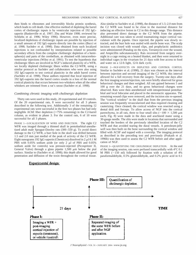
Thirty rats were used in this study, 20 experimental and 10 controls.Of the 20 experimental rats, 8 were successful for all 3 phasesdescribed in the following text. Additionally 3 of the remaining 12experimental rats were successful in the first two phases but had onlynegligible AChE fiber depletion (�75% remaining) in the C2-barrelcolumn, as evident in phase 3. For the control rats, 6 of 10 weresuccessful for all 3 phases.
PHASE 1—LOCALIZATION OF WFRS AND INJECTION. The right C2WFR was imaged through a thinned skull in pentobarbital-anesthe-tized adult male Sprague-Dawley rats (300–550 g). To avoid directdamage to the C2 WFR, a burr hole in the skull was drilled between1.5 and 2.0 mm just medial of the peak of activity of the C2 WFR.Eight nanograms 192 IgG-saporin (Chemicon) dissolved in 2 �l 0.1 MPBS with 0.05% sodium azide (or only 2 �l of PBS and 0.05%sodium azide for controls) was pressure-injected (Picospritzer II,General Valve) through a glass pipette 1,500 �m below the pialsurface. Similar to (Sachdev et al. 1998); this depth allowed for goodpenetration and diffusion of the toxin throughout the cortical tissue.
Also similar to Sachdev et al. (1998), the distance of 1.5–2.0 mm fromthe C2 WFR was found to be close to the maximal distance forinducing an effective lesion in C2 in this dosage range. This distancealso prevented direct damage to the C2 WFR from the pipette.Additional care was taken to avoid traumatizing major cortical vas-culature with the pipette. Once injected, the pipette was slowly re-tracted, and the burr hole was sealed with sterile bone wax. The scalpincision was closed with wound clips, and prophylactic antibioticswere administered (Panalog on the eyes, Terramycin over the wound,and Ampicillin subcutaneously). Rats recovered from surgery over-night in a cage resting on a heated blanket, and then were returned toindividual cages in the vivarium for 21 days with free access to foodand water on a 12-h light, 12-h dark cycle.
PHASE 2—NGF-EFFECTS ON DEPLETED AND CONTROL CORTEX.Similar to Sachdev et al. (1998), 21 days was chosen as the intervalbetween injection and second imaging of the C2 WFR; this intervalallowed for a full recovery from the surgery. Twenty-one days afterthe first imaging session/injection, rats were briefly observed for grossbehavioral changes and then weighed. All rats gained between 5 and100 g over the 21 days, and no gross behavioral changes wereobserved. Rats were then anesthetized with intraperitoneal pentobar-bital and local lidocaine and placed in the stereotactic apparatus. Theremaining wound clips were removed, and the incision site re-opened.The “cortical window” in the skull that from the previous imagingsession was frequently revascularized and thus required cleaning andcauterizing. Once cleaned, the cortical window was resected using adental drill and forceps. To allow access of NGF into the corticalparenchyma, in all rats, three to four small slits (�100 � 1,500 �meach; Fig. 8) were made in the dura and arachnoid mater using a30-gauge needle. The slits were made in locations that surrounded andtouched the borders of the previously identified location of the C2WFR and that avoided tearing the dural vessels. A petroleum-jellywell was then built on the bone surrounding the cortical window andfilled with ACSF and topped with a coverslip. The imaging protocolas described in the preceding text and previously (Prakash et al.1996b) was then used to assess the C2 WFR before and after appli-cation of NGF.PHASE 3—QUANTIFYING THE CHOLINERGIC DEPLETION. At the endof the imaging session, rats were perfused transcardially with 4°C 0.1M PBS (�150 ml) followed by fixation with a solution of 4%paraformaldehyde, 0.5% gluteraldehyde, and 0.2% picric acid in 0.1
427MECHANISMS UNDERLYING NGF CORTICAL PLASTICITY
J Neurophysiol • VOL 91 • JANUARY 2004 • www.jn.org

M Sorensen’s buffer (�150 ml). The brain was removed and placedin 30% sucrose in 0.1 M PB until it sank. (Approximately half thebrains were prepared for tangential cutting: the cortex was dissectedfrom the rest of the brain and flattened to 2.0 mm between 2 slides andthen the slides and brain were placed in 30% sucrose). The brainswere then frozen to –20°C, and serial sections were cut coronally ortangentially at 40-�m thickness on a cryostat (Zeiss). Alternatingsections were stained for cytochrome oxidase (CO) (Wong-Riley1979) to visual barrels and layer IV. The other alternating sectionswere stained for AChE, using Tago’s modifications (Tago et al. 1986).Additionally, slices were obtained from several rats and stained forrecombinant human NGF-immunoreactivity to ascertain the penetra-tion of NGF through dural tears. This protocol was similar to aspreviously described (Prakash et al. 1996b). Sections were mountedonto gelatin-coated slides and allowed to dry, then covered withmounting media followed by coverslips. Sections were analyzed andimaged under a microscope (Olympus), and the location of the left andright C2 barrels and their relations to the vascular patterns wereascertained from the CO sections. The vascular patterns from theadjacent AChE section were then compared with CO section vascularpatterns and used to determine the location of the C2-barrels in thatsection. Photomicrographs (�20 or �40) were then taken in eachC2-barrel and in layer I/II directly above the barrel. Photomicrographswere digitized, and in PhotoShop 5.0 (Adobe, San-Jose, CA), a100-�m line was then drawn through the barrel in layer IV and abovethe barrel in layer I/II. The line was drawn perpendicular to the pialsurface for coronal sections or horizontal for tangential sections.“Fiber crossings” were calculated by adding up the number of inter-sections the two 100-�m lines made with AChE-positive fibers. AChEfiber depletion was defined as {1 � [(total fiber crossings on injectedside)/(total fiber crossings on control side)] � 100%}. Although therewere no obvious differences across the different lamina, we quantifiedthe fiber depletion in layers IV and layer I/II as these layers arepresumably the primary loci relevant to NGF’s effects. However,there were differences in the fiber depletion in the C2 barrel acrossdifferent rats. This was most likely from the 1.5–2.0 mm variability ininjection site distance from the C2 barrel, which was introduced toavoid damaging major blood vessels (see Phase 1 in the precedingtext). However, this variability across rats proved to be advantageousbecause it allowed for a correlation of NGF’s induced plasticity withthe fiber depletion ratio (Fig. 4).
R E S U L T S
Regional topography of basal forebrain projection to a WFR
To determine whether the general pattern of BFCS projec-tion cells to the somatosensory cortex holds even for a singleWFR, we injected Fast-DiI, a retrograde tracer, into the corticalC2-whisker functional representation that resulted in transportof tracer to several areas of the brain, such as the thalamus,zona incerta, and basal forebrain, in similar patterns as wereported in mice (Prakash et al. 2000b). Previous studies havesuggested that the somatosensory cortex of the rat receivesbasal forebrain projections predominantly from the nucleusbasalis of Meynert [nBM; which consists of the ventral part ofthe globus pallidus (vGP), the substantia innominata (S.I.),some cells in the internal capsule, and a few cells scattered inother nuclei (Bigl et al. 1982; McKinney et al. 1983)]. Ourprimary interest in this study was to determine the preciseregions of the basal forebrain that were labeled. First wewanted to verify the identities of the basal forebrain nuclei thatproject to the adult rat barrel cortex. Second, we wanted todetermine whether an individual WFR had a more specificbasal forebrain projection locus than reported for the rest of the
somatosensory cortex (Bigl et al. 1982; Kiss and Patel 1992;McKinney et al. 1983). Consistent with these previous reports,which examined the innervation of the entire somatosensorycortex, we found that the C2-WFR was innervated by a scat-tered group of large cells throughout the nBM (Fig. 2). Othercells were found scattered throughout the basal forebrain butnever in as high a concentration as the nBM. Overall, ourresults suggest that the pattern of projections to a single WFRis not more localized than the general pattern of BF cells thatproject to the sensory cortex. These results were helpful for theinterpretation of the intracortical 192 IgG-Saporin injectionsdescribed in the following text.
Cytochemistry of basal forebrain projection cells and TrkAcortical fibers
The basal forebrain contains at least two major classes ofneurons that project fibers to the cortex: parvalbumin-immu-noreactive (-IR; GABAergic) and ChAT-IR (cholinergic). Thecholinergic fibers are the best candidates for also expressingTrkA because previous work demonstrated that most of theabundant ChAT-IR cell bodies in the basal forebrain are alsop75-IR/TrkA-IR (Sobreviela et al. 1994), whereas GABAergiccells in the basal forebrain are p75-IR negative (Kiss et al.1993). Although there is no question that cholinergic basalforebrain cells also express TrkA, there was no direct evidencethat cholinergic projection fibers in the cortex co-expressChAT and TrkA.
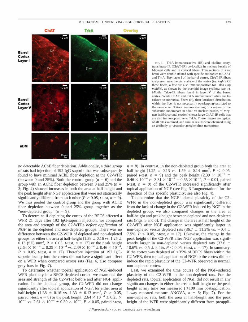
To assess whether TrkA-IR fibers observed in the cortex arealso cholinergic, co-localization studies with TrkA-IR andChAT-IR were performed. We found that TrkA fibers wereless abundant than ChAT fibers (�1:4 ratio), but that allTrkA-IR fibers in the barrel cortex were also ChAT-IR (Fig. 1).Furthermore, the TrkA fibers were sparsely distributed, butpresent in all cortical layers (I–VI). To further verify thisfinding, double-labeling studies with TrkA-IR and the vesicu-lar acetylcholine transporter (VAChT)-IR were performed (notshown). These experiments demonstrated similar types ofstaining patterns as the ones presented in Fig. 1. In summary,we found TrkA-IR only on fibers in the adult rat barrel cortex,and all these fibers also were cholinergic (either ChAT-IR orVAChT-IR).
Necessity of the bfcs for the expression of ngf’s rapidplasticity
Confirming our observation that TrkA expression is foundexclusively in projection fibers into barrel cortex and that allTrkA-IR fibers are cholinergic, we next analyzed the results ofdepletion of the BFCS fibers in the barrel cortex (Fig. 2). Thisdepletion was accomplished by injecting 192 IgG-saporin intothe cortex near the C2-WFR and then re-imaging of the sameC2-WFR 3 wk later. Because of our histological studies usingFast-DiI, we knew that the pattern of BF projection cells to asingle WFR is similar to the general patterns of BF projectioncells to the somatosensory cortex. Thus imaging-based local-ized injections of 192 IgG-saporin to the somatosensory cortexnear a single WFR would result in confined, yet representativeand thus interpretable, pattern of lesions in the BF. In these ratsthe depletion was between 35 and 95%. A control group hadsaline injections into the cortex near the C2-WFR, which had
428 N. PRAKASH, S. COHEN-CORY, S. PENSCHUCK, AND R. D. FROSTIG
J Neurophysiol • VOL 91 • JANUARY 2004 • www.jn.org
no detectable AChE fiber depletion. Additionally, a third groupof rats had injection of 192 IgG-saporin that was subsequentlyfound to have minimal AChE fiber depletion at the C2-WFR(between 0 and 25%). Both the control group (n � 6) and thegroup with an AChE fiber depletion between 0 and 25% (n �3; Fig. 4) showed increases in both the area at half-height andthe peak height after NGF application that were not statisticallysignificantly different from each other (P � 0.05, t-test, n � 9).We thus pooled the control group and the group with AChEfiber depletion between 0 and 25% group together as the“non-depleted group” (n � 9).
To determine if depleting the cortex of the BFCS affected aWFR 21 days after 192 IgG-saporin injection, we comparedthe area and strength of the C2-WFRs before application ofNGF in the depleted and non-depleted groups. There was nodifference between the C2-WFR of depleted and non-depletedgroups for either the area at half-height [1.38 � 0.16 vs. 1.25 �0.13 (SE) mm2, P � 0.05, t-test, n � 17] or the peak height(2.64 � 10�4 � 0.25 � 10�4 vs. 2.39 � 10�4 � 0.46 � 10�4,P � 0.05, t-test, n � 17). Therefore injection of 192 IgG-saporin locally into the cortex did not have a significant effecton a WFR when compared across rats (Fig. 6, also comparegray bars in Fig. 7).
To determine whether topical application of NGF-inducedWFR plasticity in a BFCS-depleted cortex, we examined thearea and strength of the C2-WFR before and after NGF appli-cation. In the depleted group, the C2-WFR did not changesignificantly after topical application of NGF, for either area athalf-height (1.38 � 0.16 vs. 1.33 � 0.13 mm2, P � 0.05,paired t-test, n � 8) or the peak height (2.64 � 10�4 � 0.25 �10�4 vs. 2.61 � 10�4 � 0.30 � 10�4, P � 0.05, paired t-test,
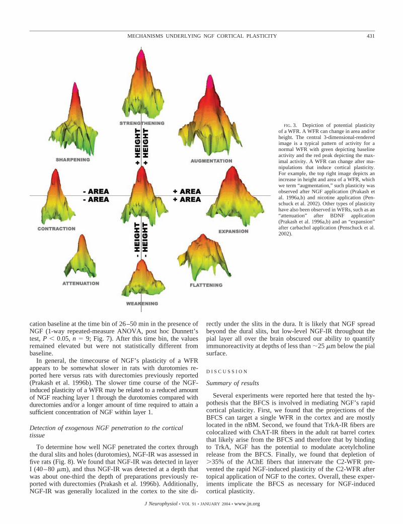
n � 8). In contrast, in the non-depleted group both the area athalf-height (1.25 � 0.13 vs. 1.59 � 0.14 mm2, P � 0.05,paired t-test, n � 9) and the peak height (2.39 � 10�4 �0.46 � 10�4 vs. 3.31 � 10�4 � 0.55 � 10�4, P � 0.01, pairedt-test, n � 9) of the C2-WFR increased significantly aftertopical application of NGF (see Fig. 3 “augmentation” for thedepiction of this specific plasticity; see also Fig. 4).
To determine that the NGF-induced plasticity of the C2-WFR in the non-depleted group was significantly differentfrom the lack of change in the C2-WFR induced by NGF in thedepleted group, we also compared changes in the area athalf-height and peak height between depleted and non-depletedrats (Figs. 5 and 6). The change in the area at half height of theC2-WFR after NGF application was significantly larger innon-depleted versus depleted rats (36.7 � 11.2% vs. �0.4 �7.5%, P � 0.05, t-test, n � 17). Likewise, the change in thepeak height of the C2-WFR after NGF application was signif-icantly larger in non-depleted versus depleted rats (37.6 �10.6% vs. 0.5 � 8.4%, P � 0.05, t-test, n � 17). In summary,if the cortex was depleted of �35% of BFCS fibers within theC2-WFR, then topical application of NGF to the cortex did notinduce the rapid plasticity of the C2-WFR observed in normal,non-depleted cortex.
Last, we examined the time course of the NGF-inducedplasticity of the C2-WFR in the non-depleted rats. For thedepleted rats, topical application of NGF did not result in anysignificant changes in either the area at half-height or the peakheight at any time bin measured (�100 min postapplication,1-way repeated-measure ANOVA, n � 8; Fig. 7). For thenon-depleted rats, both the area at half-height and the peakheight of the WFR were significantly different from preappli-
FIG. 1. TrkA-immunoreactive (IR) and choline acetyltransferase-IR (ChAT-IR) co-localize in nucleus basalis ofMeynert cells and in cortical fibers. Thin sections of a ratbrain were double stained with specific antibodies to ChATand TrkA. Top: layer I of the barrel cortex. ChAT-IR fibersare present near the pial surface of the cortex (top right). Ofthese fibers, a few are also immunopositive for TrkA (topmiddle), as shown by the overlaid image (yellow; see �).Middle: TrkA-IR fibers found in layer V of the barrelcortex. While ChAT and TrkA immunoreactivities are lo-calized to individual fibers (�), their localized distributionwithin the fiber is not necessarily overlapping/restricted tothe same area. Bottom: immunostaining of a region of thesubstantia innominata in adult rat nucleus basalis of Mey-nert (nBM; coronal section) shows large ChAT-IR cells thatare also immunopositive to TrkA. These images are typicalof all rats examined, and similar results were obtained usingan antibody to vesicular acetylcholine transporter.
429MECHANISMS UNDERLYING NGF CORTICAL PLASTICITY
J Neurophysiol • VOL 91 • JANUARY 2004 • www.jn.org
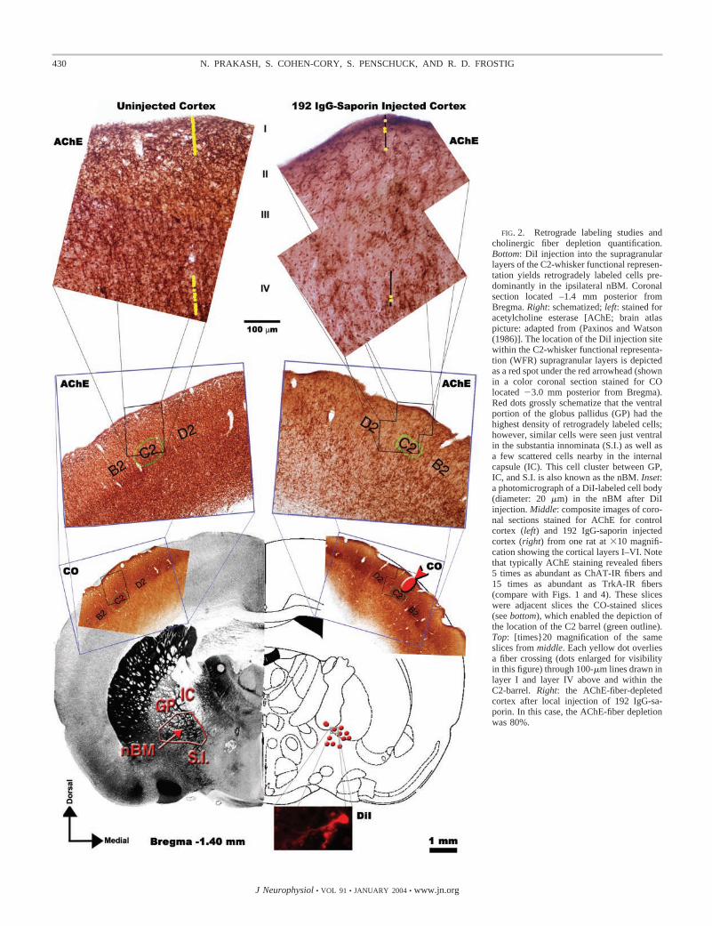
FIG. 2. Retrograde labeling studies andcholinergic fiber depletion quantification.Bottom: DiI injection into the supragranularlayers of the C2-whisker functional represen-tation yields retrogradely labeled cells pre-dominantly in the ipsilateral nBM. Coronalsection located –1.4 mm posterior fromBregma. Right: schematized; left: stained foracetylcholine esterase [AChE; brain atlaspicture: adapted from (Paxinos and Watson(1986)]. The location of the DiI injection sitewithin the C2-whisker functional representa-tion (WFR) supragranular layers is depictedas a red spot under the red arrowhead (shownin a color coronal section stained for COlocated �3.0 mm posterior from Bregma).Red dots grossly schematize that the ventralportion of the globus pallidus (GP) had thehighest density of retrogradely labeled cells;however, similar cells were seen just ventralin the substantia innominata (S.I.) as well asa few scattered cells nearby in the internalcapsule (IC). This cell cluster between GP,IC, and S.I. is also known as the nBM. Inset:a photomicrograph of a DiI-labeled cell body(diameter: 20 �m) in the nBM after DiIinjection. Middle: composite images of coro-nal sections stained for AChE for controlcortex (left) and 192 IgG-saporin injectedcortex (right) from one rat at �10 magnifi-cation showing the cortical layers I–VI. Notethat typically AChE staining revealed fibers5 times as abundant as ChAT-IR fibers and15 times as abundant as TrkA-IR fibers(compare with Figs. 1 and 4). These sliceswere adjacent slices the CO-stained slices(see bottom), which enabled the depiction ofthe location of the C2 barrel (green outline).Top: [times}20 magnification of the sameslices from middle. Each yellow dot overliesa fiber crossing (dots enlarged for visibilityin this figure) through 100-�m lines drawn inlayer I and layer IV above and within theC2-barrel. Right: the AChE-fiber-depletedcortex after local injection of 192 IgG-sa-porin. In this case, the AChE-fiber depletionwas 80%.
430 N. PRAKASH, S. COHEN-CORY, S. PENSCHUCK, AND R. D. FROSTIG
J Neurophysiol • VOL 91 • JANUARY 2004 • www.jn.org
cation baseline at the time bin of 26–50 min in the presence ofNGF (1-way repeated-measure ANOVA, post hoc Dunnett’stest, P � 0.05, n � 9; Fig. 7). After this time bin, the valuesremained elevated but were not statistically different frombaseline.
In general, the timecourse of NGF’s plasticity of a WFRappears to be somewhat slower in rats with durotomies re-ported here versus rats with durectomies previously reported(Prakash et al. 1996b). The slower time course of the NGF-induced plasticity of a WFR may be related to a reduced amountof NGF reaching layer 1 through the durotomies compared withdurectomies and/or a longer amount of time required to attain asufficient concentration of NGF within layer 1.
Detection of exogenous NGF penetration to the corticaltissue
To determine how well NGF penetrated the cortex throughthe dural slits and holes (durotomies), NGF-IR was assessed infive rats (Fig. 8). We found that NGF-IR was detected in layerI (40–80 �m), and thus NGF-IR was detected at a depth thatwas about one-third the depth of preparations previously re-ported with durectomies (Prakash et al. 1996b). Additionally,NGF-IR was generally localized in the cortex to the site di-
rectly under the slits in the dura. It is likely that NGF spreadbeyond the dural slits, but low-level NGF-IR throughout thepial layer all over the brain obscured our ability to quantifyimmunoreactivity at depths of less than �25 �m below the pialsurface.
D I S C U S S I O N
Summary of results
Several experiments were reported here that tested the hy-pothesis that the BFCS is involved in mediating NGF’s rapidcortical plasticity. First, we found that the projections of theBFCS can target a single WFR in the cortex and are mostlylocated in the nBM. Second, we found that TrkA-IR fibers arecolocalized with ChAT-IR fibers in the adult rat barrel cortexthat likely arise from the BFCS and therefore that by bindingto TrkA, NGF has the potential to modulate acetylcholinerelease from the BFCS. Finally, we found that depletion of�35% of the AChE fibers that innervate the C2-WFR pre-vented the rapid NGF-induced plasticity of the C2-WFR aftertopical application of NGF to the cortex. Overall, these exper-iments implicate the BFCS as necessary for NGF-inducedcortical plasticity.
FIG. 3. Depiction of potential plasticityof a WFR. A WFR can change in area and/orheight. The central 3-dimensional-renderedimage is a typical pattern of activity for anormal WFR with green depicting baselineactivity and the red peak depicting the max-imal activity. A WFR can change after ma-nipulations that induce cortical plasticity.For example, the top right image depicts anincrease in height and area of a WFR, whichwe term “augmentation,” such plasticity wasobserved after NGF application (Prakash etal. 1996a,b) and nicotine application (Pen-schuck et al. 2002). Other types of plasticityhave also been observed in WFRs, such as an“attenuation” after BDNF application(Prakash et al. 1996a,b) and an “expansion”after carbachol application (Penschuck et al.2002).
431MECHANISMS UNDERLYING NGF CORTICAL PLASTICITY
J Neurophysiol • VOL 91 • JANUARY 2004 • www.jn.org
Cytochemistry of basal forebrain projection cellsand TrkA cortical fibers
The basal forebrain contains at least two major projectionneurons that have fibers in the cortex: IR (GABAergic) andChAT-IR (cholinergic). The cholinergic fibers are the bestcandidates for also being the TrkA-IR fibers we observed in thebarrel cortex, because previous work has shown that most ofthe abundant ChAT-IR cell bodies in the basal forebrain arealso p75-IR/TrkA-IR (Sobreviela et al. 1994). While there is noquestion that BFCS cells also express TrkA, there is still nodirect evidence that their projection fibers in the cortex co-express ChAT and TrkA. This lack of direct evidence may berelated to a low sensitivity of TrkA antibodies (Sobreviela et al.1994) and even some ChAT antibodies at detecting antigen inthe relatively thin cholinergic fibers. Nevertheless as we wereable to detect TrkA fibers in the cortex, co-localization studieswith TrkA and ChAT (or VAChT) were then performed. Wefound that while TrkA-IR fibers were less abundant than ChATfibers, they were also ChAT-IR (or VAChT; Fig. 1). BecauseTrkA-IR fibers were found to be ChAT-IR (or VAChT-IR),this provided direct anatomical evidence that NGF can act oncholinergic fibers in the barrel cortex. It is interesting that a
recent study on the role of NGF in plasticity of the developingvisual cortex led to the same conclusions (Rossi et al. 2002). Inthis study, cholinergic afferents to the visual cortex weredestroyed by application of the excitotoxic drug quisqualic acidto the BF. Afterward, using Western blot, TrkA and p75expression was dramatically but not completely reduced in thecortex, suggesting that in the developing rat these receptors arelocated on predominantly cholinergic terminals. Taken to-gether, these results generalize the anatomical connectionsbetween NGF and the BFCS to both the developing and theadult cortex.
NGF/basal forebrain cholinergic system functionalinteractions
One way to test whether the BFCS is implicated in NGF’splasticity of a WFR is to directly lesion the BFCS with aspecific toxin, such as 192 IgG-saporin, to detect whether NGFcan still augment a WFR in the lesioned rat. An alternative wayis to pharmacologically block the cholinergic receptors locatedon the cortical BFCS projections by topical application of ageneral cholinergic antagonist and detect whether NGF canstill induce rapid plasticity of a WFR in such a pharmacolog-ically blocked cortex. Unfortunately, there is no single generalantagonist for both muscarinic and nicotinic cholinergic recep-tors. Therefore to perform properly such an experiment, at leasttwo general antagonists are needed prior to the application ofNGF. This is a complicated experiment because the timecourses of the antagonists actions after topical application tothe cortex is different as we previously demonstrated for thetopical application of muscarinic and nicotinic receptor ago-nists (Penschuck et al. 2002). Thus a general cholinergic block-ade of the cortex is challenging, and therefore the interpretationof the effect of NGF application is questionable under suchconditions (Penschuck et al. 1999).
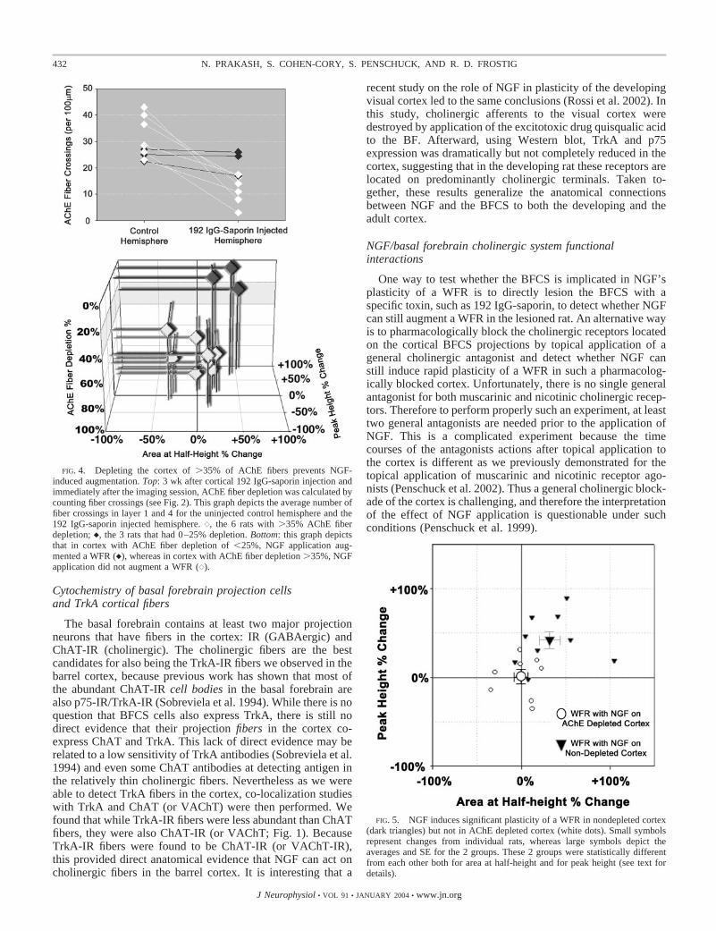
FIG. 4. Depleting the cortex of �35% of AChE fibers prevents NGF-induced augmentation. Top: 3 wk after cortical 192 IgG-saporin injection andimmediately after the imaging session, AChE fiber depletion was calculated bycounting fiber crossings (see Fig. 2). This graph depicts the average number offiber crossings in layer 1 and 4 for the uninjected control hemisphere and the192 IgG-saporin injected hemisphere. {, the 6 rats with �35% AChE fiberdepletion; }, the 3 rats that had 0–25% depletion. Bottom: this graph depictsthat in cortex with AChE fiber depletion of �25%, NGF application aug-mented a WFR (}), whereas in cortex with AChE fiber depletion �35%, NGFapplication did not augment a WFR ({).
FIG. 5. NGF induces significant plasticity of a WFR in nondepleted cortex(dark triangles) but not in AChE depleted cortex (white dots). Small symbolsrepresent changes from individual rats, whereas large symbols depict theaverages and SE for the 2 groups. These 2 groups were statistically differentfrom each other both for area at half-height and for peak height (see text fordetails).
432 N. PRAKASH, S. COHEN-CORY, S. PENSCHUCK, AND R. D. FROSTIG
J Neurophysiol • VOL 91 • JANUARY 2004 • www.jn.org
Using the 192 IgG-saporin lesion method described in thepreceding text, we found that in rats with depletion of �35%of the AChE fibers that innervate the C2-WFR, topical NGFapplication did not induce WFR plasticity, thus supporting the
hypothesis that the BFCS is necessary for the mediation ofNGF-induced rapid plasticity of a WFR. Additional support forfunctional interactions between NGF and the BFCS has beensuggested from research on the developing visual cortex of therat. Pesavento and colleagues have demonstrated in the devel-oping rat visual cortex that NGF modulation of LTP is medi-ated by the cholinergic system (Pesavento et al. 2000). Inaddition, NGF-induced increase of ACh and glutamate releasefrom cortical synaptosomes was strongly impaired after elim-ination of the cholinergic afferents to the visual cortex (Rossiet al. 2002). Taken together, these results generalize the func-tional interactions between NGF and BFCS as part of anunderlying system important for cortical plasticity in both thedeveloping and the adult cortex. This conclusion, however,seem at odds with a recent study that used transplants ofNGF-secreting fibroblasts to restore a reduction stimulus-evoked activity in barrel cortex of the adult rat (Rahimi andJuliano 2001). These authors demonstrate that NGF secretedfrom the grafted fibroblasts could restore a reduction ofevoked-activity in a barrel cortex depleted from cholinergicprojections from the BFCS. The restoration of stimulus-evokedactivity happened in spite of the fact that the cortex wasdepleted of �80% of AChE. These observations led the au-thors to conclude that NGF can directly affect cortical activitywithout the presence of the cholinergic system. The explana-tion for the apparent difference in conclusions may relate tomany technical differences on how the experiments were per-formed and analyzed. Moreover, there are major differences inwhat was studied in each experiment. Rahimi and Julianostudied the ability of chronic NGF delivery to restore normalcortical activity in a cortex with �80% AChE depletion, whilewe demonstrated the lack of ability of acute NGF applicationto induce transient plasticity in a cortex devoid of �35% of its
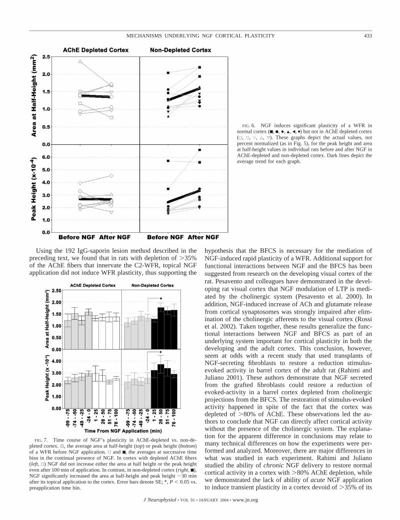
FIG. 6. NGF induces significant plasticity of a WFR innormal cortex (■ , F, }, Œ, Š, �) but not in AChE depleted cortex(�, E, {, ‚, ƒ). These graphs depict the actual values, notpercent normalized (as in Fig. 5), for the peak height and areaat half-height values in individual rats before and after NGF inAChE-depleted and non-depleted cortex. Dark lines depict theaverage trend for each graph.
FIG. 7. Time course of NGF’s plasticity in AChE-depleted vs. non-de-pleted cortex. 1, the average area at half-height (top) or peak height (bottom)of a WFR before NGF application. � and ■ , the averages at successive timebins in the continual presence of NGF. In cortex with depleted AChE fibers(left, �) NGF did not increase either the area at half height or the peak heighteven after 100 min of application. In contrast, in non-depleted cortex (right, ■ ),NGF significantly increased the area at half-height and peak height �30 minafter its topical application to the cortex. Error bars denote SE; *, P � 0.05 vs.preapplication time bin.
433MECHANISMS UNDERLYING NGF CORTICAL PLASTICITY
J Neurophysiol • VOL 91 • JANUARY 2004 • www.jn.org
AChE fibers. Finally, a recent study using a novel antibody toTrkA reported the existence of TrkA-IR on ChAT-IR neuronsin layers II-III and layers V–VI in the rat visual cortex (Tropeaet al. 2002). If these finding could be replicated in the somato-sensory cortex of the adult rat and the specificity of this novelantibody further verified, it may help in clarifying the differ-ences between ours and Rahimi and Juliano’s studies. In par-ticular, if there are TrkA-IR and ChAT-IR neurons in somato-sensory cortex in different cortical layers, there may be differ-ent layer-dependent effects. In Rahimi and Juliano’s study, thefibroblast grafts were placed in the deep layers of the cortex(1.1-mm depth), while our findings where occurring as NGFwas penetrating layer I (see following text).
There are at least two other potential ways to complementour studies to obtain a better insight for the role of the BFCSsystem in NGF’s ability to induce rapid plasticity of a WFR.The first method would be to stimulate the BFCS and observewhether WFR’s plasticity is identical to NGF’s induced plas-ticity. However, it is currently not possible to specificallystimulate only the BFCS within the BF. Nevertheless, stimu-lation of the BF (which includes cholinergic, GABAergic, andother components) (Dykes 1997) in the rat typically inducesenhancements of neuronal activity (Jimenez-Capdeville et al.1997; Metherate and Ashe 1991, 1993). Although it remains tobe shown, such enhancements at the neuronal level could, inprinciple, underlie the NGF-induced plasticity of a WFR. Thesecond way to test whether activation of the BFCS is impli-cated in NGF’s-induced plasticity of a WFR is to pharmaco-logically increase acetylcholine receptor activity. Such exper-iments are reported elsewhere (Penschuck et al. 2002) and useda protocol similar to the protocol reported here, but instead ofNGF, cholinergic agonists such as carbachol or nicotine weretopically applied to the durotomized cortex. In these experi-ments, both nicotine and carbachol caused a transient increasein the area of WFR similarly to NGF. However, NGF andnicotine had identical time courses for this increase, whereascarbachol had a slower time course. In addition, nicotine andNGF both augmented a WFR by increasing the height of theWFR in addition to the expansion of area, whereas carbacholonly expanded the area of a WFR without the increase in height(compare augmentation to expansion in Fig. 3 for the depiction
of both cases). Overall, these results are consistent with thehypothesis that activation of acetylcholine receptors could ex-plain WFR plasticity. They also suggest that NGF’s rapidplasticity could be mediated predominantly through nicotinicreceptors.
Is ngf rapidly inducing plasticity by activating nicotinicreceptors in cortical layer I?
Previously, we showed that at the time point when theNGF-induced plasticity of a WFR was maximal, NGF pene-trated layer I of the somatosensory cortex (Prakash et al.1996b). In the current study, the more limited penetration NGFfurther suggests that topical application of NGF can affectevoked cortical activity by inducing acetylcholine release fromBFCS projection axons within layer I. Assuming that thesensitivity for the detection of the NGF using immunohisto-logical techniques is optimal, the correlation between the pen-etration and plasticity could be interpreted in the followingway. NGF binding to BFCS projections within layer I causesenhancement of ACh release. Indeed layer I contains the high-est laminar densities of ACh axons and varicosities originatingfrom the BFCS (Mechawar et al. 2000), thus providing a richsubstrate for NGF action. The target of the cholinergic fibers inlayer I could be the apical dendrites of pyramidal and bipolarcells from layer II to V that extend to layer I (reviewed byNieuwenhuys 1994) or direct activation of neurons locatedwithin layer I.
One clue to the type of cholinergic target of NGF is its timecourse. NGF’s plasticity time course and its characteristics(augmentation) seem identical to nicotine’s (Penschuck et al.2002). These findings suggest that NGF’s-induced rapid plas-ticity acts predominantly through activation of nicotinic corti-cal receptors within layer I. This suggestion is supported byrecent findings that demonstrate that although nicotinic recep-tors can be found in all layers of the adult rat somatosensorycortex, they are heavily concentrated in layer I (Levy and Aoki2002). Indeed, recent evidence demonstrates directly that AChaffects neuronal activity in layer I through nicotinic receptorsby showing that almost all of layer I interneurons can beexcited by ACh nicotinic agonists. Furthermore, by activating
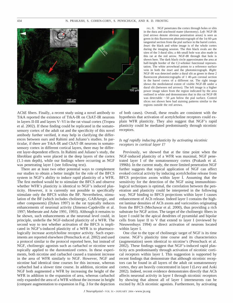
FIG. 8. NGF penetrates the cortex through holes or slitsin the dura and arachnoid mater (durotomy). Left: NGF-IR(red arrows denote obvious penetration areas) is seen asgreen in this fluorescent photomicrograph of the 1st 60-�mtangential section from the pial surface in the barrel cortex.Inset: the black and white image is of the whole cortexduring the imaging session. The thin black ovals are thesites of the 3 dural slits; a 4th small hole was also made inthis rat at the red arrow. NGF-IR through that hole isshown here. The dark black circle approximates the area athalf-height border of the C2-whisker functional represen-tation. The white arrowhead points to a reference surfacevein in both the inset and the photomicrograph. Right:NGF-IR was detected under a dural slit as green in these 2fluorescent photomicrographs of 1 40-�m coronal sectionin the barrel cortex of a different rat. The right imageshows the mediolateral extent of visible NGF-IR under adural slit (between red arrows). The left image is a higherpower image taken from the region indicated by the areaoutlined in white and demonstrates that in this rat NGF-IRwas detectable �50 �m below the pial surface. Controlslices not shown here had staining patterns similar to theregions outside the red arrows.
434 N. PRAKASH, S. COHEN-CORY, S. PENSCHUCK, AND R. D. FROSTIG
J Neurophysiol • VOL 91 • JANUARY 2004 • www.jn.org

selectively the nicotinic receptors of layer I interneurons, it wasdemonstrated that the predominant role of these interneurons isto inhibit layer II and III nonpyramidal cells, most likelyGABAergic interneurons, and thus probably cause a disinhibi-tion of cortical networks (Christophe et al. 2002). These au-thors further demonstrate that some of layer I interneuronshave indeed direct axonal projections to cortical layers II andIII. Indirect activation of layers II and III from layer I has alsobeen demonstrated. Stimulating layer I has been found toinduce LTP in layers II/III of the cortex indirectly via thecholinergic processes that also reach layer I (Hess and Dono-ghue 1999). Taken together, cholinergic activation of layer I byNGF can cause large-scale cortical plasticity in deeper corticallayers through direct and indirect mechanisms that could act inconcert and seem to be the result of activation of layer Inicotinic receptors.
Modulatory versus trophic effects of NGF
The presence of NGF in the adult cortex is thought to servelong-term processes, such as survival and maintenance ofneurons, as an extension of its role during cortical develop-ment. These processes occur after NGF binds to TrkA and/orp75 (reviewed in Bothwell 1995; Kaplan and Miller 1997).NGF is rapidly internalized on binding to p75 (Kiss et al. 1993)and/or TrkA (Grimes et al. 1997; Zapf-Colby and Olefsky1998) into a vesicle that is transported back to the cell bodywhere its trophic actions occur via second-messenger cascadeswithin the cell body and nucleus (recently reviewed in So-froniew et al. 2001). However, initially on binding, andthroughout the transport process, within this vesicle, the NGF/receptor complex actively induces second-messenger path-ways. Moreover, recently evidence has accumulated to suggestthat the signals induced by NGF may play other roles withinthe cortex that result in more immediate effects such as in-creasing synaptic efficacy. For example, in slices of rat visualcortex NGF induced a rapid increase in the height of impulse-evoked excitatory postsynaptic currents through AMPA andNMDA receptors, within 5–15 min after NGF applicationlinking NGF action to fast modulation via glutamate release(Carmignoto et al. 1997). The authors speculate that the NGFaction is mediated presynaptically through severed cholinergicnerve projections in the slice. In addition, adult cortical cellcultures showed rapid (within 10 min) activation of calcium-dependent potassium channels by NGF (Holm et al. 1997).Taken together, these results illustrate that NGF is capable ofinducing plasticity of adult neurons on several levels. First,initially on binding to TrkA, NGF induces acute changes incortical activity through changes in synaptic efficacy; suchchanges may continue to be induced while the NGF/receptorcomplex is transmitted back to the cell body. Once the complexreaches the cell body, there are also induced changes in proteinexpression. Hence in adult rat brain, NGF can rapidly augmentevoked sensory activity by modulating activity within theBFCS axons, as well as maintaining the survival of the BFCScells.
NGF-induced plasticity: a proposed model for corticalmodulation of a diffuse modulatory projection system
The cortex is generally portrayed as a passive recipient ofmodulatory systems projections including cholinergic, dopa-
minergic, noradrenergic, serotonergic, and histaminergic dif-fuse systems. Our findings implicate the cortical projections ofthe BFCS in the plasticity induced by NGF and therefore showthat under certain conditions, the sensory cortex is not just apassive recipient of the cholinergic projections but an activeparticipant in modulation of their activity, a modulation result-ing in a large-scale plasticity of a WFR. Therefore by enhanc-ing release of NGF, the cortex can transiently augment sensoryrepresentations like WFRs by recruiting additional neurons torespond to a stimulus, resulting in the expansion of the area ofa representation, and by enhancing the strength of their re-sponses, resulting in the increased height of the representation.It will be interesting to find out whether such cortical modu-lation of a diffuse modulatory system is unique to the cholin-ergic system or whether the cortex can also similarly modulateother diffuse modulatory systems. An interesting prediction ofsuch a model is that the cortical outcome of NGF-inducedplasticity should be different from the outcome expected fromdirectly enhancing BF activity (i.e., a direct BF stimulationexperiment). In the case of NGF-induced plasticity, the cortexmodulates only on the BFCS projection fibers within the cor-tex. In contrast, directly stimulating the BF enhances unselec-tively the activity of all the different systems that reside withinthe BF (including the BFCS) and therefore would result in adifferent cortical activation pattern compared with the selectiveenhancement of the cholinergic projections inside the cortexitself.
A C K N O W L E D G M E N T S
We thank J. Lee, and J. Hsu for technical assistance, Drs. R. Metherate, D.Polley, and Cynthia Chen-Bee for helpful discussions, and Genentech, Inc., forthe generous gifts of NGF and NGF-antibodies.
Present addresses: N. Prakash, Dept. of Neurology, University of California,Los Angeles, California 90095-6975; S. Penschuck, NeuropharmacologyDept., H. Lundbeck A/S, Ottiliavej 9, DK-2500 Valby, Denmark.
G R A N T S
This work was supported by the fellowships from the University of Cali-fornia Irvine Medical Scientist Training Program and the American HeartAssociation (N. Prakash) as well as National Institute of Neurological Disor-ders and Stroke Grants NS-39760 and NS-34519 and National Science Foun-dation Grant IBN-9507936 (R. D. Frostig).
R E F E R E N C E S
Albanese A and Butcher LL. Locus ceruleus somata contain both acetylcho-lin esterase and norepinephrine: direct histochemical demonstration on thesame tissue section. Neurosci Lett 14: 101–104, 1979.
Ances BM, Buerk DG, Greenberg JH, and Detre JA. Temporal dynamics ofthe partial pressure of brain tissue oxygen during functional forepaw stim-ulation in rats. Neurosci Lett 306: 106–110, 2001.
Armstrong DM, Saper CB, Levey AI, Wainer BH, and Terry RD. Distri-bution of cholinergic neurons in rat brain: demonstrated by the immunocy-tochemical localization of choline acetyltransferase. J Comp Neurol 216:53–68, 1983.
Baskerville KA, Schweitzer JB, and Herron P. Effects of cholinergic de-pletion on experience-dependent plasticity in the cortex of the rat. Neuro-science 80: 1159–1169, 1997.
Bigl V, Woolf NJ, and Butcher LL. Cholinergic projections from the basalforebrain to frontal, parietal, temporal, occipital, and cingulate cortices: acombined fluorescent tracer and acetylcholinesterase analysis. Brain ResBull 8: 727–749, 1982.
Book AA, Wiley RG, and Schweitzer JB. Specificity of 192 IgG-saporin forNGF receptor-positive cholinergic basal forebrain neurons in the rat. BrainResearch 590: 350–355, 1992.
Book AA, Wiley RG, and Schweitzer JB. 192 IgG-saporin. I. Specificlethality for cholinergic neurons in the basal forebrain of the rat. J Neuro-pathol Exp Neurol 53: 95–102, 1994.
435MECHANISMS UNDERLYING NGF CORTICAL PLASTICITY
J Neurophysiol • VOL 91 • JANUARY 2004 • www.jn.org
Bothwell M. Functional interactions of neurotrophins and neurotrophin recep-tors. Annu Rev Neurosci 18: 223–253, 1995.
Brett-Green BA, Chen-Bee CH, and Frostig RD. Comparing the functionalrepresentations of central and border whiskers in rat primary somatosensorycortex. J Neurosci 21: 9944–9954, 2001.
Bucci DJ, Holland PC, and Gallagher M. Removal of cholinergic input to ratposterior parietal cortex disrupts incremental processing of conditionedstimuli. J Neurosci 18: 8038–8046, 1998.
Carmignoto G, Pizzorusso T, Tia S, and Vicini S. Brain-derived neurotro-phic factor and nerve growth factor potentiate excitatory synaptic transmis-sion in the rat visual cortex. J Physiol 498: 153–164, 1997.
Chen KS, Nishimura MC, Armanini MP, Crowley C, Spencer SD, andPhillips HS. Disruption of a single allele of the nerve growth factor generesults in atrophy of basal forebrain cholinergic neurons and memorydeficits. J Neurosci 17: 7288–7296, 1997.
Chen-Bee CH, Kwon MC, Masino SA, and Frostig RD. Areal extentquantification of functional representations using intrinsic signal opticalimaging. J Neurosci Methods 68: 27–37, 1996.
Christophe E, Roebuck A, Staiger JF, Lavery DJ, Charpak S, and Audi-nat E. Two types of nicotinic receptors mediate an excitation of neocorticallayer I interneurons. J Neurophysiol 88: 1318–1327, 2002.
Cuello AC, Maysinger D, and Garofalo L. Trophic factor effects on cho-linergic innervation in the cerebral cortex of the adult rat brain. MolNeurobiol 6: 451–461, 1992.
Dekker AJ, Gage FH, and Thal LJ. Delayed treatment with nerve growthfactor improves acquisition of a spatial task in rats with lesions of thenucleus basalis magnocellularis: evaluation of the involvement of differentneurotransmitter systems. Neuroscience 48: 111–119, 1992.
Dykes RW. Mechanisms controlling neuronal plasticity in somatosensorycortex. Can J Physiol Pharmacol 75: 535–545, 1997.
Eckenstein F and Sofroniew MV. Identification of central cholinergic neu-rons containing both choline acetyltransferase and acetylcholinesterase andof central neurons containing only acetylcholinesterase. J Neurosci 3: 2286–2291, 1983.
Fibiger HC. The organization and some projections of cholinergic neurons ofthe mammalian forebrain. Brain Research 257: 327–388, 1982.
Frostig RD, Lieke EE, Ts’o DY, and Grinvald A. Cortical functionalarchitecture and local coupling between neuronal activity and the microcir-culation revealed by in vivo high-resolution optical imaging of intrinsicsignals. Proc Natl Acad Sci USA 87: 6082–6086, 1990.
Gage FH, Armstrong DM, Williams LR, and Varon S. Morphologicalresponse of axotomized septal neurons to nerve growth factor. J CompNeurol 269: 147–155, 1988.
Grimes ML, Beattie E, and Mobley WC. A signaling organelle containingthe nerve growth factor-activated receptor tyrosine kinase, TrkA. Proc NatlAcad Sci USA 94: 9909–9914, 1997.
Gutierrez H, Miranda MI, and Bermudez-Rattoni F. Learning impairmentand cholinergic deafferentation after cortical nerve growth factor depriva-tion. J Neurosci 17: 3796–3803, 1997.
Hefti F, Hartikka J, and Knusel B. Function of neurotrophic factors in theadult and aging brain and their possible use in the treatment of neurodegen-erative diseases. Neurobiol Aging 10: 515–533, 1989.
Henderson Z. Acetylcholinesterase on the dendrites of central cholinergicneurons: an electron microscopical study in the ferret. Neuroscience 28:95–108, 1989.
Hess G and Donoghue JP. Facilitation of long-term potentiation in layer II/IIIhorizontal connections of rat motor cortex following layer I stimulation:route of effect and cholinergic contributions. Exp Brain Res 127: 279–290,1999.
Holler T, Berse B, Cermak JM, Diebler MF, and Blusztajn JK. Differencesin the developmental expression of the vesicular acetylcholine transporterand choline acetyltransferase in the rat brain. Neurosci Lett 212: 107–110,1996.
Holm NR, Christophersen P, Olesen SP, and Gammeltoft S. Activation ofcalcium-dependent potassium channels in mouse [correction of rat] brainneurons by neurotrophin-3 and nerve growth factor. Proc Natl Acad Sci USA94: 1002–1006, 1997.
Holtzman DM, Kilbridge J, Li Y, Cunningham ETJ, Lenn NJ, Clary DO,Reichardt LF, and Mobley WC. TrkA expression in the CNS: evidence forthe existence of several novel NGF-responsive CNS neurons. J Neurosci 15:1567–1576, 1995.
Jacobs SE, Fine A and Juliano SL. Cholinergic basal forebrain transplantsrestore diminished metabolic activity in the somatosensory cortex of rats
with acetylcholine depletion [published erratum appears in J Neurosci 14: 3,1994]. J Neurosci 14: 697–711, 1994.
Jimenez-Capdeville ME, Dykes RW, and Myasnikov AA. Differential con-trol of cortical activity by the basal forebrain in rats: a role for bothcholinergic and inhibitory influences. J Comp Neurol 381: 53–67, 1997.
Kaplan DR and Miller FD. Signal transduction by the neurotrophin receptors.Curr Opin Cell Biol 9: 213–221, 1997.
Kim DS, Duong TQ, and Kim SG. High-resolution mapping of iso-orienta-tion columns by fMRI. Nat Neurosci 3: 164–169, 2000.
Kiss J and Patel AJ. Development of the cholinergic fibers innervating thecerebral cortex of the rat. Int J Dev Neurosci 10: 153–170, 1992.
Kiss J, Shooter EM and Patel AJ. A low-affinity nerve growth factorreceptor antibody is internalized and retrogradely transported selectivelyinto cholinergic neurons of the rat basal forebrain. Neuroscience 57: 297–305, 1993.
Knipper M, da Penha Berzaghi M, Blochl A, Breer H, Thoenen H, andLindholm D. Positive feedback between acetylcholine and the neurotro-phins nerve growth factor and brain-derived neurotrophic factor in the rathippocampus. Eur J Neurosci 6: 668–671, 1994a.
Knipper M, Leung LS, Zhao D, and Rylett RJ. Short-term modulation ofglutamatergic synapses in adult rat hippocampus by NGF. Neuroreport 5:2433–2436, 1994b.
Koliatsos VE, Nauta HJ, Clatterbuck RE, Holtzman DM, Mobley WC,and Price DL. Mouse nerve growth factor prevents degeneration of axoto-mized basal forebrain cholinergic neurons in the monkey. J Neurosci 10:3801–3813, 1990.
Kromer LF. Nerve growth factor treatment after brain injury prevents neu-ronal death. Science 235: 214–216, 1987.
Lamour Y, Dutar P, and Jobert A. Topographic organization of basalforebrain neurons projecting to the rat cerebral cortex. Neurosci Lett 34:117–122, 1982.
Lehmann J, Nagy JI, Atmadia S, and Fibiger HC. The nucleus basalismagnocellularis: the origin of a cholinergic projection to the neocortex ofthe rat. Neuroscience 5: 1161–1174, 1980.
Levy RB and Aoki C. Alpha7 nicotinic acetylcholine receptors occur atpostsynaptic densities of AMPA receptor-positive and -negative excitatorysynapses in rat sensory cortex. J Neurosci 22: 5001–5015, 2002.
Malonek D, Dirnagl U, Lindauer U, Yamada K, Kanno I, and Grinvald A.Vascular imprints of neuronal activity: relationships between the dynamicsof cortical blood flow, oxygenation, and volume changes following sensorystimulation. Proc Natl Acad Sci USA 94: 14826–14831, 1997.
Malonek D and Grinvald A. Interactions between electrical activity andcortical microcirculation revealed by imaging spectroscopy: implications forfunctional brain mapping. Science 272: 551–554, 1996.
Masino SA. Quantitative comparison between functional imaging and single-unit spiking in rat somatosensory cortex. J Neurophysiol 89: 1702–1712,2003.
Masino SA and Frostig RD. Quantitative long-term imaging of the functionalrepresentation of a whisker in rat barrel cortex. Proc Natl Acad Sci USA 93:4942–4947, 1996.
Masino SA, Kwon MC, Dory Y, and Frostig RD. Characterization offunctional organization within rat barrel cortex using intrinsic signal opticalimaging through a thinned skull. Proc Natl Acad Sci USA 90: 9998–10002,1993.
McKinney M, Coyle JT, and Hedreen JC. Topographic analysis of theinnervation of the rat neocortex and hippocampus by the basal forebraincholinergic system. J Comp Neurol 217: 103–121, 1983.
Mechawar N, Cozzari C, and Descarries L. Cholinergic innervation in adultrat cerebral cortex: a quantitative immunocytochemical description. J CompNeurol 428: 305–318, 2000.
Merlio JP, Ernfors P, Jaber M and Persson H. Molecular cloning of rat trkCand distribution of cells expressing messenger RNAs for members of the trkfamily in the rat central nervous system. Neuroscience 51: 513–532, 1992.
Mesulam MM, Mufson EJ, Levey AI, and Wainer BH. Cholinergic inner-vation of cortex by the basal forebrain: cytochemistry and cortical connec-tions of the septal area, diagonal band nuclei, nucleus basalis (substantiainnominata), and hypothalamus in the rhesus monkey. J Comp Neurol 214:170–197, 1983.
Metherate R and Ashe JH. Basal forebrain stimulation modifies auditorycortex responsiveness by an action at muscarinic receptors. Brain Res 559:163–167, 1991.
Metherate R and Ashe JH. Nucleus basalis stimulation facilitates thalamo-cortical synaptic transmission in the rat auditory cortex. Synapse 14: 132–143, 1993.
436 N. PRAKASH, S. COHEN-CORY, S. PENSCHUCK, AND R. D. FROSTIG
J Neurophysiol • VOL 91 • JANUARY 2004 • www.jn.org
Minger SL and Davies P. Persistent innervation of the rat neocortex by basalforebrain cholinergic neurons despite the massive reduction of cortical targetneurons. I. Morphometric analysis. Exp Neurol 117: 124–138, 1992a.
Minger SL and Davies P. Persistent innervation of the rat neocortex by basalforebrain cholinergic neurons despite the massive reduction of cortical targetneurons. II. Neurochemical analysis. Exp Neurol 117: 139–150, 1992b.
Mizukawa K, McGeer PL, Tago H, Peng JH, McGeer EG, and Kimura H.The cholinergic system of the human hindbrain studied by choline acetyl-transferase immunohistochemistry and acetylcholinesterase histochemistry.Brain Res 379: 39–55, 1986.
Nieuwenhuys R. The neocortex. An overview of its evolutionary develop-ment, structural organization and synaptology. Anat Embryol 190: 307–337,1994.
Paxinos G and Watson C. The Rat Brain in Stereotaxic Coordinates. SanDiego, CA: Academic, 1986.
Penschuck S, Chen-Bee CH, Prakash N, and Frostig RD. In vivo modula-tion of a cortical functional sensory representation shortly after topicalcholinergic agent application. J Comp Neurol 452: 38–50, 2002.
Penschuck S, Prakash N, Cohen-Cory S, and Frostig RD. Interaction ofNGF with the cholinergic basal forebrain system during NGF-induced rapidcortical plasticity in the rat primary somatosensory cortex. Proceedings ofthe 1st Goettingen Conference of the German Neuroscience Society 1999;27th Goettingen Neurobiology Conference, Stuttgart, New York: Thieme, p.733.
Pesavento E, Margotti E, Righi M, Cattaneo A, and Domenici L. Blockingthe NGF-TrkA interaction rescues the developmental loss of LTP in the ratvisual cortex: role of the cholinergic system. Neuron 25: 165–175, 2000.
Polley DB, Chen-Bee CH, and Frostig RD. Two directions of plasticity in thesensory-deprived adult cortex. Neuron 24: 623–637, 1999.
Prakash N, Cohen-Cory S, and Frostig RD. Differential effects of neuro-trophins on the functional organization of the adult barrel cortex in vivo. SocNeurosc Abstr 22: 997, 1996a.
Prakash N, Cohen-Cory S, and Frostig RD. Rapid and opposite effects ofBDNF and NGF on the functional organization of the adult cortex in vivo.Nature 381: 702–706, 1996b.
Prakash N, Cohen-Cory S, and Frostig RD. NGF-induced rapid functionalplasticity in the adult rat somatosensory cortex is mediated by fibers origi-nating in the basal forebrain cholinergic system. Soc Neurosc Abstr 26:1931, 2000a.
Prakash N, Vanderhaeghen P, Cohen-Cory S, Frisen J, Flanagan JG, andFrostig RD. Malformation of the functional organization of somatosensorycortex in adult ephrin-A5 knock-out mice revealed by in vivo functionalimaging. J Neurosci 20: 5841–5847, 2000b.
Rahimi O and Juliano SL. Transplants of NGF-secreting fibroblasts restorestimulus-evoked activity in barrel cortex of basal-forebrain-lesioned rats.J Neurophysiol 86: 2081–2096, 2001.
Rossi FM, Sala R, and Maffei L. Expression of the nerve growth factorreceptors TrkA and p75NTR in the visual cortex of the rat: development andregulation by the cholinergic input. J Neurosci 22: 912–919, 2002.
Sachdev RN, Lu SM, Wiley RG, and Ebner FF. Role of the basal forebraincholinergic projection in somatosensory cortical plasticity. J Neurophysiol79: 3216–3228, 1998.
Sala R, Viegi A, Rossi FM, Pizzorusso T, Bonanno G, Raiteri M, andMaffei L. Nerve growth factor and brain-derived neurotrophic factor in-crease neurotransmitter release in the rat visual cortex. Eur J Neurosci 10:2185–2191, 1998.
Satoh K, Armstrong DM, and Fibiger HC. A comparison of the distributionof central cholinergic neurons as demonstrated by acetylcholinesterase phar-macohistochemistry and choline acetyltransferase immunohistochemistry.Brain Res Bull 11: 693–720, 1983.
Schliebs R, Rossner S, and Bigl V. Immunolesion by 192IgG-saporin of ratbasal forebrain cholinergic system: a useful tool to produce cortical cholin-ergic dysfunction. Prog Brain Res 109: 253–264, 1996.
Sobreviela T, O’Clary D, Reichardt LF, Brandabur MM, Kordower JH,and Mufson EJ. TrkA-immunoreactive profiles in the central nervoussystem: colocalization with neurons containing p75 nerve growth factorreceptor, choline acetyltransferase, and serotonin. J Comp Neurol 350:587–611, 1994.
Sofroniew MV, Eckenstein F, Thoenen H, and Cuello AC. Topography ofcholine acetyltransferase-containing neurons in the forebrain of the rat.Neurosci Lett 33: 7–12, 1982.
Sofroniew MV, Howe CL, and Mobley WC. Nerve growth factor signaling,neuroprotection, and neural repair. Annu Rev Neurosci 24: 1217–1281,2001.
Tago H, Kimura H, and Maeda T. Visualization of detailed acetylcholines-terase fiber and neuron staining in rat brain by a sensitive histochemicalprocedure. J Histochem Cytochem 34: 1431–1438, 1986.
Thompson JK, Peterson MR, and Freeman RD. Single-neuron activity andtissue oxygenation in the cerebral cortex. Science 299: 1070–1072, 2003.
Tropea D, Capsoni S, Covaceuszach S, Domenici L, and Cattaneo A. Ratvisual cortical neurones express TrkA NGF receptor. Neuroreport 13:1369–1373, 2002.
Weihe E, Tao-Cheng JH, Schafer MK, Erickson JD, and Eiden LE.Visualization of the vesicular acetylcholine transporter in cholinergic nerveterminals and its targeting to a specific population of small synaptic vesicles.Proc Natl Acad Sci USA 93: 3547–3552, 1996.
Wenk GL. The nucleus basalis magnocellularis cholinergic system: one hun-dred years of progress. Neurobiol Learn Mem 67: 85–95, 1997.
Wenk H, Bigl V, and Meyer U. Cholinergic projections from magnocellularnuclei of the basal forebrain to cortical areas in rats. Brain Res 2: 295–316,1980.
Wiley RG. Neural lesioning with ribosome-inactivating proteins: suicidetransport and immunolesioning. Trends Neurosci 15: 285–290, 1992.
Wiley RG. Targeting toxins to neural antigens and receptors. Semin CancerBiol 7: 71–77, 1996.
Wiley RG, Oeltmann TN, and Lappi DA. Immunolesioning: selective de-struction of neurons using immunotoxin to rat NGF receptor. Brain Res 562:149–153, 1991.
Will B and Hefti F. Behavioural and neurochemical effects of chronicintraventricular injections of nerve growth factor in adult rats with fimbrialesions. Behav Brain Res 17: 17–24, 1985.
Williams LR, Varon S, Peterson GM, Wictorin K, Fischer W, BjorklundA, and Gage FH. Continuous infusion of nerve growth factor prevents basalforebrain neuronal death after fimbria fornix transection. Proc Natl Acad SciUSA 83: 9231–9235, 1986.
Wong-Riley M. Changes in the visual system of monocularly sutured orenucleated cats demonstrable with cytochrome oxidase histochemistry.Brain Res 171: 11–28, 1979.
Zapf-Colby A and Olefsky JM. Nerve growth factor processing and traffick-ing events following TrkA-mediated endocytosis. Endocrinology 139:3232–3240, 1998.
Zhu XO and Waite PM. Cholinergic depletion reduces plasticity of barrelfield cortex. Cereb Cortex 8: 63–72, 1998.
437MECHANISMS UNDERLYING NGF CORTICAL PLASTICITY
J Neurophysiol • VOL 91 • JANUARY 2004 • www.jn.org