Embed Size (px)
Citation preview

814 © 2017 Neurology India, Neurological Society of India | Published by Wolters Kluwer - Medknow
Basal ganglia: Their role in complex cognitive procedures in experimental models and in clinical practiceRita Moretti, Paola Caruso, Elena Crisman, Silvia Gazzin1
Abstract: Apart from the well known role of the basal ganglia (BG) in motor control, their important role in regulating the cognitive functions is emerging. This article traces the scientific work that explores this role of BG in reinforcement learning, perceptual decision making, and other nonmotor pathways (speech fluency, cognition, attention and behaviour). It also highlights the important role played by the BG networks in determining the development of a child’s brain. It retraces the various pathways and connections of the BG with the cerebral cortex, cerebellum and other regions that may be utilized in the establishment of complex cognitive procedures. Various diseases that may be the direct result of disruption of these basal ganglionic networks and interconnections are also recounted.
Key Words:Basal ganglion, behaviour, brain development, cognition, networks, speech
The role of the basal ganglia (BG) in motor control has been extensively studied and
is now well known. In addition, the role of BG in reinforcement learning, perceptual decision making, and other nonmotor pathways (speech fluency, cognition, and behaviour) has become an arena of new interest for researchers.[1‑6]
Converging evidence f rom s ingle‑ce l l recordings, lesion studies in humans and animals, and brain imaging studies in humans have made it clear that the BG has several important roles outside the motor sphere. Data from literature showed that the range of contributions of the BG spans many different cognitive faculties.[1‑7] These include the procedural memory,[7] habit and skill learning,[8] attention,[9,10] perception,[1] and language.[11‑13] BG circuitry may be a key component of a specialized memory subsystem that involves the acquisition of stimulus related response associations.[14,15] Experimental
evidence suggests that the BG contributes to even higher levels of cognitive functions, such as planning,[16,17] syllogistic reasoning,[18] and mathematical problem solving.[19]
Different studies in rats[20] and monkeys[21,22] have shown that the BG lesions may influence the acquisition of motor responses that occur as a conditional response to a discriminating stimuli. These mechanisms seem to be involved in the transmission of cortical signals that may subserve to influence several cognitive functions of the BG.
Main Cortical Basal Ganglia Functional Networks and Introduction
Historically, the projections from the cerebral cortex to the BG are recognized as highly organized topographic projection systems.[6,23‑26]
Anterior cortical areas project to anterior striatal regions; posterior cortical areas project to
Address for correspondence: Dr. Paola Caruso,
Neurological Clinic, Department of Internal
Medicine and Neurology, University of Trieste,
Cattinara Hospital, Strada di Fiume, 447,
34149 Trieste, Italy. E-mail: caruso.
Department of Internal Medicine and
Neurology, Neurological Clinic, 1FIF Science Park,
University of Trieste, Cattinara Hospital,
Trieste, Italy
How to cite this article: Moretti R, Caruso P, Crisman E, Gazzin S. Basal ganglia: Their role in complex cognitive procedures in experimental models and in clinical practice. Neurol India 2017;65:814-25.
Access this article onlineQuick Response Code:
Website:www.neurologyindia.com
DOI:10.4103/neuroindia.NI_850_16
PMID:xxxx
Key Message:Basal ganglia have an important role in determining complex cognitive behavioural patterns and in influencing the development of a child’s brain. Several primary developmental cognitive, learning, memory, language, behavioural and speech disorders related to extrapyramidal syndromes directly owe their genesis to disruption of specific basal ganglionic networks.
NI Feature: The Quest
This is an open access article distributed under the terms of the Creative Commons Attribution‑NonCommercial‑ShareAlike 3.0 License, which allows others to remix, tweak, and build upon the work non‑commercially, as long as the author is credited and the new creations are licensed under the identical terms.
For reprints contact: [email protected]
COMMENTARY
[PDF Purchased from http://www.neurologyindia.com on Thursday, July 6, 2017]abce

Moretti, et al.: Basal ganglia and cognition
Neurology India | Volume 65 | Issue 4 | July-August 2017 815
posterior striatal regions;ventromedial regions of cortex project to ventromedial striatal regions; and, dorsolateral areas of cortex project to dorsolateral striatal regions.[6] Almost all areas of the cerebral cortex send cortical inputs to the striatum.
Striatum, considered to be the entry point of the BG main circuits, receives glutamatergic inputs from the cerebral cortex and thalamus,[27,28] dopaminergic inputs from the substantia nigra (SNpr) [pars compacta], and serotonergic and noradrenergic inputs from the brain stem.
The sensory and motor cortical areas project to the putamen; the frontal–parietal–temporal association areas project to the putamen and caudate nucleus. Similarly, the caudate nucleus, nucleus accumbens, and anterior ventral part of the putamen also receive information from the limbic cortex.[27‑29]
The SNr and internal globus pallidum (GPi) constitute the output nuclei of the BG. Their main targets are the ventral anterior, ventral lateral, and the medial dorsal nuclei of the thalamus.[30‑32] In turn, thalamic nuclei mainly project to the frontal lobe,[33,34] and in a minor proportion of subjects, also to the temporal and parietal areas.[35‑37]
Basal Ganglia Behavior and Learning
BG were thought to be having the ability to integrate sensory, limbic, and cognitive information with the commands for movement. Alexander et al.,[25,26] suggested that, rather than serving as a tunnel for information from widespread cortical areas, the BG actually participated in multiple parallel segregated circuits with different regions of the frontal lobe. These regions included cortical areas concerned with skeleton‑motor and oculomotor control, as well as three regions of the prefrontal cortex involved in cognitive and limbic functions. More recently,[38,39] it has been suggested that the original ‘five circuit’ scheme, proposed by Alexander et al., should be broadened to include seven general categories of circuits, namely, the skeletomotor, oculomotor, dorsolateral prefrontal, lateral orbitofrontal, medial orbitofrontal, cingulate, and inferotemporal‑posterior parietal categories of circuits.
Within each of these categories, anatomical evidence supports the existence of multiple parallel cortical‑basal ganglia circuits.
Imaging studies have shown that the activity of the putamen is associated with repetitive and well‑learned movements. Involvement of the supplementary motor area, anterior striatal areas, and the caudate nucleus has been recorded during the learning of sequential movements.
The frontal eye field sends projections to the striatum and to the body of the caudate nucleus (areas that are referred as the supplementary eye fields), and is also involved in learning and acquisition of oculomotor behaviour, for example, the rostral and caudal motor regions are programmed to learn the behavioural sequences of hand movements.[40‑43]
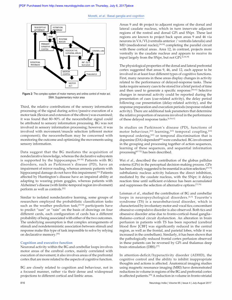
Inputs from posterior cortical areas are integrated in the BG circuits and influence regions of the frontal lobe. Results from positron emission tomography (PET) studies (3 series) of regional cerebral blood flow (CBF) show a double dissociation between selection of movements, which induces differential effects in the BG, but not the cerebellum; and, sensory information processing, which involves the cerebellum but not the BG [Figures 1 and 2].[44,45] Regarding motor learning of a sequence of finger movements, there was a shift of activation in the anterior–posterior direction of the BG, which paralleled changes in the motor areas of the frontal cortex. During new learning, the dorsolateral prefrontal cortex and striatum were activated; for selected movements, the premotor cortex and mid‑putamen were activated; whereas for automatic movements, the sensorimotor cortex and posterior putamen were activated. When participants paid attention to overlearned actions, the activation shifted back to the dorsolateral prefrontal cortex and striatum. The cerebellum was not activated when participants made new decisions and attended to their actions or selected movements.[44]
Second, a visuomotor coordination task was examined. In the absence of visual control over arm movements, participants were required to use a computer mouse to either generate new lines or to retrace the lines on a computer screen.[44] The neocerebellum, not the BG, was more engaged when lines were retraced (compared with new line generation). Animal experiments have shown that error detection and correction occur during line retracing but not during line generation. These data suggest that the neocerebellum (not the BG) is involved in monitoring and optimizing movements using sensory (proprioceptive) feedback.[44]
Prefrontal Dorsolateral cortex
Caudate (DL)
GP (Lateralis Dorsomedialis)
Thalamus (VA, MD)
Lateral-Orbital- cortex
Caudate (VM)
GP (Medialis Dorsalis)
Thalamus (VA, MD)
Anterior Cingulate cortex
N. Accubens
GP (Rostro Lateralis)
Thalamus (MD)
Figure 1: Direct frontal subcortical loops; schematic representation of the three direct frontal‑subcortical loops. Caudate DL: Dorsolateral, Caudate VM: Ventro medialis, GP: Globus pallidus, Thalamus VA MD: Ventral anterior and medialis
[PDF Purchased from http://www.neurologyindia.com on Thursday, July 6, 2017]abce
Moretti, et al.: Basal ganglia and cognition
816 Neurology India | Volume 65 | Issue 4 | July-August 2017
Third, the relative contributions of the sensory information processing of the signal during active/passive execution of a motor task (flexion and extension of the elbow) was examined; it was found that 80–90% of the neocerebellar signal could be attributed to sensory information processing. BG was not involved in sensory information processing; however, it was involved with movement/muscle selection (efferent motor component); the neocerebellum may be concerned with monitoring the outcome and optimizing the movements using sensory information.
Data suggest that the BG mediates the acquisition of nondeclarative knowledge, whereas the declarative subsystem is supported by the hippocampus.[46‑50] Patients with BG disorders, such as Parkinson’s disease (PD), have an impairment of mirror reading, whereas amnesic patients with hippocampal damage do not have this impairment.[51] Patients affected by Huntington’s disease have an impaired ability at adapting to wearing prism goggles, whereas patients with Alzheimer’s disease (with limbic‑temporal region involvement) perform as well as controls.[52]
Similar to isolated nondeclarative learning, some groups of researchers employed the probabilistic classification tasks such as the weather prediction task;[53,54] participants have to predict “sun” or “rain” on the basis of drawings on four different cards, each configuration of cards has a different probability of being associated with either of the two outcomes. The underlying assumption is that complex arrangements of stimuli and nondeterministic association between stimuli and response make this type of task impossible to solve by relying on declarative memory.[55]
Cognition and executive functionNeuronal activity within the BG and cerebellar loops involves motor areas of the cerebral cortex, mainly correlated with execution of movement; it also involves areas of the prefrontal cortex that are more related to the aspects of cognitive function.
BG are closely related to cognition and behaviour, not in a focused manner, rather via their dense and widespread projections to different cortical and limbic areas.
Areas 9 and 46 project to adjacent regions of the dorsal and lateral caudate nucleus, which in turn innervate adjacent regions of the rostral and dorsal GPi and SNpr. These last regions are known to project back upon areas 9 and 46 via neurons in VA/VL(ventralis anterior / ventralis lateralis) and MD (mediodorsal nuclei),[38,39] completing the parallel circuit with these cortical areas. Area 12, in contrast, projects more ventrally in the caudate nucleus and appears to receive its input largely from the SNpr, but not GPi.[6,38,39]
The physiological properties of the dorsal and lateral prefrontal cortex suggested that areas 9, 46, and 12, each appear to be involved in at least four different types of cognitive functions. First, many neurons in these areas display changes in activity related to the performance of delayed‑response tasks. These tasks require sensory cues to be stored for a brief period of time and then used to generate a specific response.[56‑61] Selective changes in neuronal activity could be reported during the presentation of cues (cue‑related activity), the delay period following cue presentation (delay‑related activity), and the response preparation and execution periods (response‑related activity). There are additional task parameters that determine the relative proportion of neurons involved in the performance of these delayed response tasks.[6,56,61]
In studies on Parkinson’s disease (PD), functions of motor behaviour,[62] learning,[63] temporal coupling,[64] temporal ordering,[65] or temporal discrimination that is dopamine (DA)‑dependent[66] were conducted. BG involvement in the grouping and processing together of action sequences, learning of these sequences, and sequential information processing[67‑72] has been described.
Wei et al., described the contribution of the globus pallidus externa (GPe) in the perceptual decision‑making process. GPe has been already suggested to be essential in action selection;[73‑75] subthalamic nucleus activity balances the direct inhibition, mediated by the caudate nucleus, with the SNpr; it delays reaction time until sufficient evidence has been accumulated and suppresses the selection of alternative options.[73,76]
Leisman et al., studied the contribution of BG and cerebellar loops in neuropsychological disorders.[4,5] Tourette’s syndrome (TS) is a neurobehavioral disorder, which is characterized by involuntary motor and vocal tics; concomitant obsessive–compulsive disorder is also observed. Both tics and obsessive disorder arise due to fronto‑cortical–basal ganglia–thalamo–cortical circuit dysfunction. An alteration in brain perfusion in patients with TS has been reported (cerebral blood flow [CBF] was significantly reduced in the central region, as well as the frontal, and parietal lobes, while it was increased in the cerebellum). Similarly, it has been shown that the pathologically reduced frontal cortex perfusion observed in these patients can be reversed by GPi and thalamus deep brain stimulation (DBS).[77]
In attention‑deficit/hyperactivity disorder (ADHD), the cognitive control and the ability to inhibit inappropriate thoughts and actions is affected. Anatomical imaging studies using magnetic resonance imaging (MRI) have demonstrated reductions in volume in regions of the BG and prefrontal cortex in affected patients.[78] A reduction in volume in fronto‑striatal
Figure 2: The complex system of motor memory and online control of motor act. SMA: Supplementary motor area
[PDF Purchased from http://www.neurologyindia.com on Thursday, July 6, 2017]abce
Moretti, et al.: Basal ganglia and cognition
Neurology India | Volume 65 | Issue 4 | July-August 2017 817
regions has been observed in ADHD patients compared to controls; moreover, these regions are less activated during tasks that require cognitive control.[79]
BehaviourHouk proposed that the BG is involved in the selection and/or initiation of cortical patterns of activation for both planned behaviour and thoughts.[80] Within areas 9 and 46, many neurons have cue‑ and delay‑related activity that is tuned to the spatial position of stimuli (spatially‑tuned activity) or the order in which the stimuli are present (sequentially‑tuned activity). Area 12 appears to be much less involved in spatial and sequential information and more involved in remembering the identity of particular objects (object‑tuned activity).
Throughout areas 9, 46, and 12, many neurons appear to be more involved in the learning and remembering of specific rules or associations used in the performance of conditional response (forced choice) tasks (rule‑related activity).[6] A relatively small but consistent proportion of neurons in areas 9, 46, and 12 also display changes in activity that coincide with the expectation or anticipation of primary reinforcement or food rewards (reward‑related activity).[81] Bilateral damage to area 9 in monkeys produces severe and long‑lasting impairments in order monitoring and in the identification of different objects (but not spatial position), in particular, when the sequence of cues to be remembered is self‑ordered.[6,82] Similarly, as evident from animal studies, BG lesions impair the acquisition of motor responses that are a conditional response to the discriminating stimuli.[21,22,83] Similar damage is seen in humans with dorsal prefrontal lesions,[84] leading to deficits in tasks that require the categorization and sorting of different stimuli,[85] or the planning and monitoring of script events or sequential actions.[86] These types of tasks are considered to impose significant demands on planning and short‑term (or working) memory for objects or sequential actions. Experimental lesions of area 46 result in an inability of animals to perform spatial delayed‑response tasks, spatial delayed‑alternation tasks, and go/no‑go tasks.[6,87]
Lesions of area 12 in monkeys produce an inability of animals to make switches in behavioural set. This leads to perseverative responses on delayed‑response tasks, in which the identity of different objects or colors must be remembered (object matching, object reversal, or object alternation tasks), or to inappropriate responses in auditory‑cued go/no‑go tasks.[88] Large lesions of area 12 produce deficits in the learning and performance of visual discrimination tasks that do not involve delayed‑responses.[6,89]
Widespread connectivity differences between healthy controls, precocious schizophrenic patients and patients with major depression has been seen. Decreased connectivity was found between the medial prefrontal area and a ventral mood processing region in major depressed patients compared to controls.[90]
A decreased connection network starting from the medial prefrontal area and ending in the orbital frontal cortex in schizophrenic patients has been shown. The dorsal anterior cingulate cortex is overactive early in schizophrenia, which might lead to decreased activity in the ventral anterior cingulate cortex, leading to affective symptoms and poor motivation.[91,92]
The role of BG in neuropsychiatric symptoms exhibited in Huntington’s disease (that usually shows hyperactive disorders with agitation, euphoria, or anxiety) and PD (hypokinetic disorder with apathy) is well known. In the first case, the behavioural disorder may be caused by excitatory subcortical output through the medial and orbitofrontal circuits to the pallidum, thalamus, and cortex as well as premotor and motor cortex; whereas in PD, apathy results from hypostimulation of frontal subcortical circuits resulting from damage to putamen, striatum, and globus pallidus.
Memory and attentionTasks that require sequential planning, monitoring of bilateral fine sequences, or learning of new movement sequences[6,93,94] produce sites of peak activations in lateral portions of area 9, such as for verbal working memory (including word generation tasks).[6,95] Further, area 46 is particularly active during spatial working memory tasks, difficult planning tasks, go/no‑go tasks, some nonspatial object and verbal working memory tasks, and verb generation tasks,[96] during the generation and monitoring of multiple movement sequences involving the hand or fingers.[97,98] Prefrontal activations in areas 9 and 46 are related to the elaboration or learning of novel sequences compared to when the task is well learned.[6] The human equivalent of area 12 (area 47) has been shown to be active during tasks that employ verbal working memory, including word generation tasks,[6,99] but not during spatial working memory tasks. Brown and Marsden studied BG involvement on attention focus. The study was conducted among patients with PD who were incapable of carrying out dual tasks or self‑monitoring,[100] as well as in carrying out simultaneous actions.[101] Patients with PD also have problems with covert[102] and overt attentional priming,[103] with set‑shifting.[104,105] Hayes reports that the treatment of motor‑symptoms with L‑dopa medication results not only in the improvement of motor behavior, but also in attentional set‑shifting, suggesting a regulatory role of dopamine in motor and attentional control. Loss of striatal dopamine also impairs predictive control (ability to use current information to adapt future behaviour).[106] Saint‑Cyr speculates that the deficit in attentional control may result from two disrupted pathways, namely, the thalamo‑cortical pathway and the thalamic nuclei under the control of the pallido‑nigral projections. In addition, direct projections from the GPe to thalamic reticular shell nuclei may be essential because this mechanism modulates the signal‑to‑noise ratio of information processing.[107‑110]
DBS has been showed to decrease “off” motor symptoms and motor fluctuations in PD; however, it may also induce amelioration of cognitive performances.[111‑113]
LanguageAbdullaev et al., examined the role of caudate nucleus during reading, naming, recognition memory tasks, categorization, and lexical decision making tasks. They found that caudate nucleus cells exhibited excitatory responses related to both semantic and phonological‑articulatory encoding and that the delay‑related firing of cells was increased whenever semantic processing was required.[6,114] Comparing the properties of striatal cells with those seen in the prefrontal (Broca’s) area as well as the temporal and parietal lobe areas, the authors found that the caudate nucleus cells had properties that were “strikingly similar” to those in Broca’s area.
[PDF Purchased from http://www.neurologyindia.com on Thursday, July 6, 2017]abce

Moretti, et al.: Basal ganglia and cognition
818 Neurology India | Volume 65 | Issue 4 | July-August 2017
In monkeys, striatal cells are involved during sequential working memory test. Many of these cells had visual‑related responses that varied according to the order of a given target; many of the caudate nucleus cells seemed to anticipate the fixation of specific targets. Shuvaev and Shefer[115] found that the task‑related striatal cells could be separated into two distinct groups, with one group of cells clearly tuned to response execution and another group more involved in the instructional decision‑making process.[6]
Ford et al., studied the connectivity between Broca’s area and the BG, suggesting the involvement of BG in primary language functions.[116‑121] BG are involved in language processing, mostly enhancing cortical signals for selected items and suppressing cortical signals for competing items.
Direct connectivity between the Broca’s area and the putamen has been demonstrated, suggesting an involvement of both these structures in the articulatory process and initiation of phonological responses; a direct electrode stimulation of the anterior putamen area leading to a temporary speech deficit was found.[122‑125]
The dorsal and ventral lateral prefrontal cortex of macaques projecting to the anterior putamen, are implicated in different language processing domains, such as phonological processing,[126,127] reading,[128] semantic processing, and semantic priming.[129] In monkeys, part of the caudate nucleus responds specifically to the sight of food or food rewards.[130‑131]
Large portions of the GPi and SNpr contain neurons whose activity is not modulated by simple skeletomotor or oculomotor tasks. Many of these regions fall within the regions that innervate areas of the prefrontal cortex involved in cognitive processing. Hikosaka et al., recorded the activity of neurons in the SNpr of monkeys trained to perform an oculomotor spatial‑delayed response task.[132,133]
More recently Ford et al., studied the Broca’s area and striatal thalamic connections through a tractography study. The results suggested a correlation between the BG and the Broca’s area, particularly, an input from two adjacent cortical areas subserving different but closely related language functions converging on the same region of the anterior putamen.[134]
Newer Aspects of the Role of Basal Ganglia: Their Possible Involvement in the Development of a
Child’s Brain
It has been known for a while that individuals who are markedly late in achieving developmental milestones are at a high risk of developing subsequent cognitive impairment.[135,136] The mechanisms underlying infantile motor and adult cognitive associations remain poorly characterized.[137] One possibility is that the neural systems that subserve motor development in infancy also contribute to the development and operation of specific cognitive processes later in life. These are not well defined but can include different higher cognitive functions, such as language or motor development or executive procedural networks.[138]
Murray et al.,[139] examined these questions in a large British general population birth cohort study, in which
measurements were available for development in language and motor domains in infancy, general intellectual functions in childhood and adolescence, and specific neuropsychological functions (e.g., verbal fluency, a test of executive/frontal lobe function, etc.) in adulthood. These authors noted that[138] faster attainment of motor developmental milestones is related to better adult cognitive performance in some domains, such as executive function.[137]
The complete realization of complex acts and movements[140‑143] is played out in a group of collectively functioning components, i.e., the sensory, motor, and anterior cingulate areas of the cortex, the intralaminar thalamic nucleus (in conjunction with the reticular nucleus), the amygdala, and the striatum. In mammals, the latter has a heterogeneous structure,[144] in which the continuous matrix is inter‑digitated with the isolated striosomes. The input to the striatum appears to be more intimately connected to the components just identified. Given that striatal output reaches the primary motor cortex (M1) via the GPi, whereas the matrix output does not, it seems that the striatum may be more essentially related to consciousness and is much like the individual motor elements of the infant.[137‑142] Likewise, the pars intermedia appears to be have more intimate associations with consciousness‑related part of the cerebellum because it has analogous projections. Moreover, the threshold for overt movements may be exceeded only when both the feeding components are dispatching signals concurrently.[137] The matrix, conversely, appears to serve already‑established motor patterns because its output ultimately reaches the PMA/SMA (premotor area/supplementary motor area) and the prefrontal area. Its cerebellar partner is clearly the hemispherical region.[145]
The focus of competition for attention appears to be the PMA/SMA because it receives inputs from all the thalamic nuclei handling basal ganglia/cerebellum, in which remote regions might influence attention.[137] The inferior olive seems to play a complementary role to the cerebellum, sending signals through the climbing fibers when something unexpected occurs.[146]
The periodic shifting of attention, as when we simultaneously converse (or merely think) and drive in a busy thoroughfare, must be making considerable demands on the putative differential clutch mechanism, and this could be the dual responsibility of the substantia nigra pc and the subthalamic nucleus, which appear to serve as gain control for the striosome‑related and matrix‑related routes, respectively. This situation is exemplified by our ability to think of one thing while overtly performing another act.[136]
For a given set of synaptic coupling between the premotor and supplementary motor areas, and the primary motor area, a specific pattern of output signals from the former will produce a specific sequence of muscular movements. Efferent copies of these output signals, dispatched through axon collaterals, will carry the full information sent to the muscles, via the motor area; however, they will not directly produce movement because their target neurons are not immediately concerned with motor output.[137,139,144] The duality of routes, and the fact that these overlap in the premotor and supplementary motor region, could well underlie the interplay between the
[PDF Purchased from http://www.neurologyindia.com on Thursday, July 6, 2017]abce
Moretti, et al.: Basal ganglia and cognition
Neurology India | Volume 65 | Issue 4 | July-August 2017 819
explicit and implicit in brain function. The parallel loops from the frontal cortex traverse via the striatum to basal ganglia and thalamus, and again towards the frontal cortex.[147] The complex regulating system through the striatal neurons serves to disinhibit the thalamic neurons.[148]
This disinhibition produces a gating function enabling other functions to occur, but does not directly causing them to occur, so that the activation of striatal neurons enables, but does not directly cause, subsequent motor movements.[137]
It has been suggested that whatever information is encoded by striatal neurons, it must be vastly compressed or eliminated on its way up to the frontal cortex. This constraint coincides with the gating hypothesis, i.e., the basal ganglia do not need to convey detailed information to the frontal cortex; instead, they simply need to inform different regions of the frontal cortex to update themselves when the need arises.[137]
Another constraint to consider concerns the number of different subregions of the frontal cortex, for which the basal ganglia can plausibly provide separate gating control. Leisman et al.,[137] suggested that the fine‑grained gating is important for mitigating conflicts where two representations require separate gating control and yet fall within one gating region. The number of neurons in the GPi/SNr provides an upper limit estimate, which is roughly 320,000 in the human. This suggests that the gating signal operates on a region of frontal neurons, instead of individually controlling specific neurons.
An interesting possible candidate that may be responsible for determining the regions of the frontal cortex that is independently controlled by the BG is a distinctive anatomical structure called stripe, consisting of interconnected groups of neurons.[149] It is plausible that each stripe or cluster of stripes constitutes a separately controlled group of neurons; each stripe can be separately updated by the BG system,[136] which might extend the functional circuits described by Alexander et al.,[147] to a much finer grained level.[150] Thus, it is possible to maintain some information in one set of stripes, while selectively updating other stripes.[137]
As Leisman et al.,[137] emphatically pointed out, intelligence, in general, should be considered as the ability to consolidate already‑learned motor patterns into more complex composites. This type of consolidation is sometimes merely a covert operation, rather than an overt one. This definition was discussed in the context of autism.[151,152] A normal child, lying on its back and wanting to roll over onto its front, soon learns that this can be readily accomplished if at first, the head, then the shoulders, and finally the hips are swiveled in the same direction. If the timing of this sequence is correct, the supine‑prone transition requires minimum effort. Autistic infants appear to experience considerable difficulty in learning this simple motor sequence. Indeed, the sequence does not even occur in their failed attempts. Instead, they awkwardly arch their backs and ultimately fall into the desired position.[137]
The most spectacular feature to evolve thus far has been seen in mammals. This permitted acquisition, during a subject’s own lifetime, of novel context‑specific reflexes, especially
those relying on the sequences of muscular movements. This mechanism makes heavy demands on the neural circuitry because it requires an attentional mechanism. As attention is an active process, there has to be feedback from the muscles, carrying information about their current state, including their current rate of change of state, which is where the action of the BG is required. Without such information, anticipation would be impossible, and there could be no meaningful decision on the most appropriate way of continuing an ongoing movement. Without such a mechanism, novel context‑specific reflexes could not be acquired, when the the brake from BG to the frontal cortex is released.[137,153]
Abnormal motor development can accurately be used as a marker to predict autism and other developmental disorders.[154] Many authors have noted a relationship between incoordination and clumsiness, especially of posture and gait, and autism, as well as with other neurodevelopmental disorders. The type of gait and motor disturbances have been compared mostly in subjects where these appear to be either basal ganglionic or, more commonly, cerebellar in origin.[155] The most common of all comorbidities in practically all neurobehavioral disorders of childhood is the developmental coordination disorder (DCD) or more simply put “clumsiness” or motor incoordination. In fact, practically all children in this spectrum have some degree of motor incoordination. The type of incoordination is also usually of the same type, primarily involving the muscles that control gait and posture or gross motor activity (many times, cerebellar alterations have been implicated as the causative factor)[144] sometimes, to a lesser degree, fine motor coordination has also been affected.[136]
Parkinson’s disease is an excellent model that is influenced by the BG control, and also strongly on the intimate relationship between the basal ganglia and the frontal cortex.[156‑159]
Children with developmental disabilities including autism spectrum disorders and attention deficit/hyperactivity disorder (ADHD) demonstrate locomotor difficulties. ADHD and autistic spectrum individuals have reported significant motor difficulties, both fine and gross.[152,160]
Although it has been fairly well known that attention deficit disorders also have coincidental motor and balance disorders, what is lesser known and is more significant is the association between ADD/ADHD and motor controlled dysfunction (developmental coordination disorder [DCD]; clumsiness). Motor control issues were first noted in what were then called ‘minimal brain dysfunction syndromes’ (MBD). MBD was the term used to describe children of normal intelligence, with attention deficit and/or motor dysfunction (that is, suffering form “soft” neurological signs). Several studies by Denckla and others[161,162] have shown that association exists between ADHD, dyscoordination, and/or motor perceptual dysfunction.
In Asperger’s syndrome, it has been noted that individuals have significant degrees of motor incoordination, and sometimes, executive problems affect writing and drawing skills,[163,164] as well as posture, gait, and gesture incoordination.[165,166]
What emerges from neuroimaging studies is astonishing; although the literature on fiber tracts is limited in ADHD,[167] Makris et al., noted that gray matter abnormalities suggest
[PDF Purchased from http://www.neurologyindia.com on Thursday, July 6, 2017]abce
Moretti, et al.: Basal ganglia and cognition
820 Neurology India | Volume 65 | Issue 4 | July-August 2017
that white matter connections may be altered selectively in neural systems. This finding is in confirmation with the findings of a prior study,[168] that using diffusion tensor magnetic resonance imaging (DT‑MRI), showed alterations within the frontal and cerebellar white matter in children and adolescents with ADHD. To this end, the cingulum bundle (CB) and superior longitudinal fascicle II (SLF II) were investigated in vivo in 12 adults with childhood ADHD using DT‑MRI. Relative to controls, the fractional anisotropy (FA) values were significantly smaller in both regions of interest in the right hemisphere, in contrast to a control region (the fornix). This indicated the presence of an alteration of anatomical connections within the attention and EF (Executive Function) cerebral systems in adults with childhood ADHD. The demonstration of FA abnormalities in the CB and SLF II in adults with childhood ADHD provides further support for persistent structural abnormalities that persist into adulthood.[137]
Other works have observed that, in children with ADHD,[164] frontal‑subcortical connections are disrupted by subcortical dysfunction showing decreased glucose consumption in the frontal cortex, along with a decrease in the nigrostriatal D2 receptor uptake ratios. When boys suffering from ADHD were tested, there appeared to be a clear difference in the activity of the BG. These children have less activity in that area than the control children. After administering methylphenidate, boys with ADHD had increased activity in the BG whereas normal boys had decreased activity in the BG.[137]
A similar finding was noted when PET scans were perfomed in patients with hyperactivity disorder, where a normal appearing frontal metabolism existed with decreased caudate and putamen metabolism.[165,169] Methylphenidate, a dopamine reuptake inhibitor, may increase the functioning in a previously dysfunctional BG, whereas raising dopamine levels in normal individuals would most likely result in decreased activity of the basal ganglia in order to prevent overproduction of dopamine. Increasing dopamine levels may increase frontal metabolism due to an increased activity of the striatum. This would lead to a decreased firing of the globus pallidus, thereby inhibiting the thalamo‑cortical firing, which in turn decreased the hyperkinetic behavior.[136]
Anatomical imaging studies using MRI have demonstrated subtle reductions in volume in regions of the BG and prefrontal cortex.[170‑173]
Quiu et al.[174] employed large deformation diffeomorphic metric mapping (LDDMM) to examine the effects of ADHD, gender, and their interaction on the BG shapes. The BG (caudate nucleus, putamen, globus pallidus) were manually delineated on MRI from normally developing children and children with ADHD. It has been found that boys with ADHD showed significantly smaller BG volumes compared with normally developing boys. The LDDMM also revealed that the groups remarkably differed in the basal ganglia shapes. Volume compression was seen bilaterally in the caudate head and body and anterior putamen as well as in the left anterior globus pallidus and right ventral putamen in patients with ADHD. Volume expansion was most pronounced in the posterior putamen. They concluded that the shape compression pattern
of BG in ADHD suggests an atypical brain development involving multiple frontal‑subcortical control loops, including circuits with premotor, oculomotor, and prefrontal cortices.[175]
Moreover, also considering the necessity of voluntary control of motor action, it is important not only to detect the motor areas, which start action and release it, but also the area where motor act can be stopped; the 'go' process is likely to be generated by the premotor areas that project via the direct pathways of the BG (through striatum, pallidum, and thalamus), eventually exciting the primary motor cortex and generating corticospinal volleys to the relevant effectors, each interacting with the globus pallidus.[175] Aaron et al.,[176] brilliantly outlined the nature of inhibition in the fronto‑basal‑ganglia networks related to cognition. They collected evidence indicating that the right inferior frontal cortex (IFC) is the critical region for 'stop' signal response inhibition,[177] with the most critical portion likely being the pars opercularis (Brodmann area 44) in humans. The right IFC can send a 'stop' command to intercept the 'go' process via the activation of the globus pallidus through a projection from the STN.[175] Thus, once the 'stop' command is generated in the frontal cortex, it could be rapidly conveyed to the BG via the so‑called “hyperdirect pathway” to intercept the 'go' process in the final stages of the race.[137]
In summary, BG, through their control on motor act and its refinement, can modulate the intelligence process to permit an interface of the human beings with the environment; this way 'go/no go' strategies determine our reaction times and their destruction or their impairment may be responsible for various cognitive manifestations of autism, ADHD, mental retardation, and Asperger syndrome.
Consideration and Conclusions
The physiological properties of many neurons in the striatum and pallidum of humans, as well as primates appear to be similar in many aspects such as those reported in studies of the physiology of the prefrontal cortex.
In addition to BG circuits, another system appears to be operating across circuit boundaries that integrates information about the rewarding value of a behavioral act. Shultz proposed that the neural substrate for this reward system is the phasic activity of dopamine synthesizing cells in the SNpc, which convey information regarding primary reinforcement and behavioural state to cells in the striatum. As such influences exist throughout the striatum, it is quite likely that many, if not all, BG circuits utilize the reward‑related information to modify the properties of cells within them to carry out meaningful behavioral acts.[9,178,179] Owen et al., compared the activity of normal individuals and PD patients during the performance of a difficult planning task, a spatial working memory task, and a simple visually guided movement task.[180,181] PD patients had very little GPi activation during the cognitive tasks, compared to normal individuals; the greatest differences occurred in tasks with most cognitive demands; and, no differences were found in GPi activation in the simple motor task.[182‑185]
The connection of BG with the prefrontal cortex suggests that it has different roles in cognition, memory, and emotion; this connection appears to be disrupted in psychiatric and
[PDF Purchased from http://www.neurologyindia.com on Thursday, July 6, 2017]abce
Moretti, et al.: Basal ganglia and cognition
Neurology India | Volume 65 | Issue 4 | July-August 2017 821
neurodegenerative diseases, in which disconnection with major feedback pathways to the neuraxis is also seen.
Financial support and sponsorshipNil.
Conflicts of interestThere are no conflicts of interest.
References
1. Brown LL, Schneider JS, Lidsky TI. Sensory and cognitive functions of the basal ganglia. Curr Opin Neurobiol 1997;7:157‑63.
2. Brown RG, Marsden CD. Cognitive function in Parkinson’s disease: From description to theory. Trends Neurosci 1990;13:21‑9.
3. Leisman G, Melillo R. The development of the frontal lobes in infancy and childhood: Asymmetry and the nature of temperament and adjustment. In: Cavanna AE, editor. Frontal Lobe: Anatomy, Functions and Injuries. Hauppauge, NY: Nova Scientific Publishers; 2012. p. 1‑48.
4. Leisman G, Melillo R. The basal ganglia: Motor and cognitive relationships in a clinical neurobehavioral context. Rev Neurosci 2013;24:9‑25.
5. Leisman G, Braun‑Benjamin O, Melillo M. Cognitive‑motor interactions of the basal ganglia in development. Front Syst Neurosci 2014;8:16.
6. Middleton FA, Strick PL. Basal ganglia output and cognition: Evidence from anatomical, behavioral, and clinical studies. Brain Cogn 2003;42:183‑200.
7. Packard MG, Knowlton BJ. Learning and memory functions of the basal ganglia. Ann Rev Neurosci 2002;25:563‑93.
8. Squire LR, Knowlton BJ. The medial temporal lobe, the hippocampus and the memory systems of the brain. In: Gazzaniga M, editor. The new cognitive neurosciences. Cambridge, MA: MIT Press; 2000. p. 765‑79.
9. Ravizza SM, Ivry RB. Comparison of the basal ganglia and cerebellum in shifting attention. J Cogn Neurosci 2001;13:285‑97.
10. Teicher MH, Anderson CM, Polcari A, Glod CA, Maas LC, Renshaw PF. Functional deficits in basal ganglia of children with attention‑deficit/hyperactivity disorder shown with functional magnetic resonance imaging relaxometry. Nat Med 2000;6:470‑3.
11. Prat CS, Keller TA, Just MA. Individual differences in sentence comprehension: A functional magnetic resonance imaging investigation of syntactic and lexical processing demands. J Cogn Neurosci 2007;19:1950‑63.
12. Teichmann M, Dupoux E, Kouider S, Bachoud‑Lévi AC. The role of the striatum in processing language rules: Evidence from word perception in Huntington’s disease. J Cogn Neurosci 2006;18:1555‑69.
13. Ullman MT. Contributions of memory circuits to language: The declarative/procedural model. Cognition 2004;92:231‑70.
14. Packard MG, Hirsh R, White NM. Differential effects of fornix and caudate nucleus lesions on two radial maze tasks: Evidence for multiple memory systems. J Neurosci 1989;9:1465‑72.
15. Packard MG, Knowlton BJ. Learning and memory functions of the basal ganglia. Annu Rev Neurosci 2002;25:563‑93.
16. Anderson JR, Albert MV, Fincham JM. Tracing problem solving in real time: fMRI analysis of the subject‑paced Tower of Hanoi. J Cogn Neurosci 2005;17:1261‑74.
17. Dagher A, Owen AM, Boecker H, Brooks DJ. The role of the striatum and hippocampus in planning: A PET activation study in Parkinson’s disease. Brain 2001;124:1020‑32.
18. Goel V, Buchel C, Frith C, Dolan RJ. Dissociation of mechanisms underlying syllogistic reasoning. Neuroimage 2000;12:504‑14.
19. Stocco A, Anderson JR. Endogenous control and task representation: An fMRI study in algebraic problem‑solving. J Cogn Neurosci 2008;20:1300‑14.
20. Kesner RP, Bolland BL, Dakis M. Memory for spatial locations, motor responses, and objects: Triple dissociation among the hippocampus, caudate nucleus, and extrastriate visual cortex. Exp Brain Res 1993;93:462‑70.
21. Fernandez‑Ruiz J, Wang J, Aigner TG, Mishkin M. Visual habit formation in monkeys with neurotoxic lesions of the ventrocaudal neostriatum. Proc Natl Acad Sci U S A 2001;98:4196‑201.
22. Teng E, Stefanacci L, Squire LR, Zola SM. Contrasting effects on discrimination learning after hippocampal lesions and conjoint hippocampal‑caudate lesions in monkeys. J Neurosci 2000;20:3853‑63.
23. Chesselet MF, Delfs JM. Basal ganglia and movement disorders: An update. Trends Neurosci 1996;19:417‑22.
24. Parent A, Hazrati LN. Functional anatomy of the basal ganglia. II. The place of subthalamic nucleus and external pallidum in basal ganglia circuitry. Brain Res Brain Res Rev 1995;20:128‑54
25. Alexander GE, DeLong MR, Strick PL. Parallel organization of functionally segregated circuits linking basal ganglia and cortex. Annu Rev Neurosci 1986;9:357‑81.
26. Alexander M. Clinical‑anatomical correlations of aphasia following predominantely subcortical lesions. In: Boller F, Grafman J, editors. Handbook of neuropsychology. Vol. 2. Amsterdam: Elsevier; 1989. p. 47‑66.
27. Albin RL, Young AB, Penney JB. The functional anatomy of basal ganglia disorders. Trends Neurosci 1989;12:366‑75.
28. Smith AD, Bolam JP. The neural network of the basal ganglia as revealed by the study of synaptic connections of identified neurones. Trends Neurosci 1990;13:259‑65.
29. Alexander GE, Crutcher MD. Functional architecture of basal ganglia circuits: Neural substrates of parallel processing. Trends Neurosci 1990;13:266‑71.
30. Bolam JP, Hanley JJ, Booth PA, Bevan MD. Synaptic organisation of the basal ganglia. J Anat 2000;196:527‑42.
31. McFarland NR, Haber SN. Thalamic relay nuclei of the basal ganglia form both reciprocal and nonreciprocal cortical connections, linking multiple frontal cortical areas. J Neurosci 2002;22:8117‑32.
32. Smith AD, Bolam JP. The neural network of the basal ganglia as revealed by the study of synaptic connections of identified neurones. Trends Neurosci 1990;13:259‑65.
33. Hoover JE, Strick PL. Multiple output channels in the basal ganglia. Science 1993;259:819‑21.
34. Middleton FA, Strick PL. Anatomical evidence for cerebellar and basal ganglia involvement in higher cognitive function. Science 1994;266:458‑61.
35. Middleton FA Strick PL. Basal ganglia and cerebellar output influences non‑motor function. Mol Psychiatry 1996;1:429‑33.
36. Middleton FA, Strick PL. The temporal lobe is a target of output from the basal ganglia. Proc Natl Acad Sci U S A 1996;93:8683‑7.
37. Clower DM, Dum RP, Strick PL. Basal ganglia and cerebellar inputs to ‘AIP’. Cereb Cortex 2005;15:913‑20.
38. Middleton FA, Strick PL. Basal ganglia and cerebellar loops: Motor and cognitive circuits. Brain Res Brain Res Rev 2000;31:236‑50.
39. Middleton FA, Strick PL. Basal ganglia output and cognition: Evidence from anatomical, behavioral, and clinical studies. Brain Cogn 2000;42:183‑200.
40. Yin HH, Mulcare SP, Hilario MR. Dynamic reorganization of striatal circuits during the acquisition and consolidation of a skill. Nat Neurosci 2009;12:333‑41.
[PDF Purchased from http://www.neurologyindia.com on Thursday, July 6, 2017]abce
Moretti, et al.: Basal ganglia and cognition
822 Neurology India | Volume 65 | Issue 4 | July-August 2017
41. Boecker H, Dagher A, Ceballos‑Baumann AO. Role of the human rostral supplementary motor area and the basal ganglia in motor sequence control: Investigations with H2 15O PET. J Neurophysiol 1998;79:1070‑80.
42. Kermadi I, Joseph JP. Activity in the caudate nucleus of monkey during spatial sequencing. J Neurophysiol 1995;74:911‑33.
43. Chen LL, Wise SP. Supplementary eye field contrasted with the frontal eye field during acquisition of conditional oculomotor associations. J Neurophysiol 1995;73:1122‑34
44. Jueptner M, Weiller C. A review of differences between basal ganglia and cerebellar control of movements as revealed by functional imaging studies. Brain 1998;121:1437‑49.
45. Jueptner M, Krukenberg M. Motor system: Cortex, basal ganglia, and cerebellum. Neuroimaging Clin N Am 2001;11:203‑19.
46. Packard MG, Knowlton BJ. Learning and memory functions of the basal ganglia. Annu Rev Neurosci 2002;25:563‑93.
47. Yin HH, Knowlton BJ. The role of the basal ganglia in habit formation. Nat Rev Neurosci 2006;7:464‑76.
48. Squire LR. Declarative and nondeclarative memory: Multiple brain systems supporting learning and memory. J Cogn Neurosci 1992;4:232‑43.
49. Squire LR, Zola SM. Structure and function of declarative and nondeclarative memory systems. Proc Natl Acad Sci U S A 1996;93:13515‑22.
50. Reder LM, Park H, Kieffaber PD. Memory systems do not divide on consciousness: Reinterpreting memory in terms of activation and binding. Psychol Bull 2009;135:23‑49.
51. Cohen NJ, Squire LR. Preserved learning and retention of pattern‑analyzing skill in amnesia: Dissociation of knowing how and knowing that. Science 1980;210:207‑10.
52. Chan AS, Butters N, Paulsen JS, Salmon DP, Swenson MR, Maloney LT. An assessment of the semantic network in patients with Alzheimer’s disease. J Cogn Neurosci 1993;5:254‑61.
53. Gluck MA, Bower GH. From conditioning to category learning: An adaptive network model. J Exp Psychol Gen 1988;117:227‑47.
54. Knowlton BJ, Squire LR, Gluck MA. Probabilistic classification learning in amnesia. Learn Mem 1994;1:106‑20.
55. Gluck MA, Shohamy D, Myers C. How do people solve the “weather prediction” task? Individual variability in strategies for probabilistic category learning. Learn Mem 2002;9:408‑18.
56. Rosenkilde CE. Functional heterogeneity of the prefrontal cortex in the monkey: A review. Behav Neural Biol 1979;25:301‑45.
57. Goldman‑Rakic PS. Development of cortical circuitry and cognitive function. Child Dev 1987;58:601‑22.
58. Goldman‑Rakic PS, Selemon LD. New frontiers in basal ganglia research. Introduction. Trends Neurosci 1990;13:241‑4.
59. Goldman‑Rakic PS, Funahashi S, Bruce CJ. Neocortical memory circuits. Cold Spring Harb Symp Quant Biol 1990;55:1025‑38.
60. Playford ED, Jenkins IH, Passingham RE, Frackowiak RS, Brooks DJ. Impaired activation of frontal areas during movement in Parkinson’s disease: A PET study. Adv Neurol 1993;60:506‑10.
61. Fuster JM1; Network memory. Trends Neurosci 1997;20:451‑9.62. Harrington DL, Haaland KY, Hermanowicz N. Temporal processing
in the basal ganglia. Neuropsychology 1998;12:3‑12.63. Martin P, Tewesmeier M, Albers M, Schmid G, Scharfetter C.
Investigation of gestural and pantomime performance in chronic schizophrenic inpatients. Eur Arch Psychiatry Clin Neurosci 1994;244:59‑64.
64. Malapani C, Rakitin B, Levy R, Meck WH, Deweer B, Dubois B, Gibbon J. Coupled temporal memories in Parkinson’s disease: A dopamine‑related dysfunction. J Cogn Neurosci 1998;10:316‑31.
65. Sagar HJ, Sullivan EV, Gabrieli JD, Corkin S, Growdon JH.
Temporal ordering and short‑term memory deficits in Parkinson’s disease. Brain 1988;111:525‑39.
66. Rammsayer T, Classen W. Impaired temporal discrimination in Parkinson’s disease: Temporal processing of brief durations as an indicator of degeneration of dopaminergic neurons in the basal ganglia. Int J Neurosci 1997;91:45‑55.
67. Graybiel AM. The basal ganglia and chunking of action repertoires. Neurobiol Learn Mem 1998;70:119‑36.
68. Dominey PF. Complex sensory‑motor sequence learning based on recurrent state representation and reinforcement learning. Biol Cybern 1995;73:265‑74.
69. Dominey PF, Jeannerod M. Contribution of frontostriatal function to sequence learning in Parkinson’s disease: Evidence for dissociable systems. Neuroreport 1997;8:iii‑ix.
70. Beiser DG, Houk JC. Model of cortical‑basal ganglionic processing: Encoding the serial order of sensory events. J Neurophysiol 1998;79:3168‑88.
71. Berns GS, Sejnowski TJ. A computational model of how the basal ganglia produce sequences. J Cogn Neurosci 1998;10:108‑21.
72. Hikosaka O, Nakahara H, Rand MK, Sakai K, Lu X, Nakamura K, et al. Parallel neural networks for learning sequential procedures. Trends Neurosci 1999;22:464‑71.
73. Gurney K, Prescott TJ, Redgrave P. A computational model of action selection in the basal ganglia. I. A new functional anatomy. Biol Cybern 2001;84:401‑10.
74. Gurney K, Prescott TJ, Redgrave P. A computational model of action selection in the basal ganglia. II. Analysis and simulation of behaviour. Biol Cybern 2001;84:411‑23.
75. Frank MJ, Claus ED. Anatomy of a decision: Striato‑orbitofrontal interactions in reinforcement learning, decision making, and reversal. Psychol Rev 2006;113:300‑26.
76. Kotz SA, Schwartze M, Kassow MS. Non‑motor basal ganglia functions: A review and proposal for a model of sensory predictability in auditory language perception. Corte×2009;45:982‑90.
77. Haense C, Müller‑Vahl KR, Wilke F, Schrader C, Capelle HH, Geworski L, et al. Effect of deep brain stimulation on regional cerebral blood flow in patients with medically refractory Tourette syndrome. Front Psychiatry 2016;7:118.
78. Castellanos FX, Lee PP, Sharp W, Jeffries NO, Greenstein DK, Clasen LS, et al. Developmental trajectories of brain volume abnormalities in children and adolescents with attention‑deficit/hyperactivity disorder. JAMA 2000;288:1740‑8.
79. Semrud‑Clikeman M, Steingard RJ, Filipek P, Biederman J, Bekken K, Renshaw PF. Using MRI to examine brain‑behavior relationships in males with attention deficit disorder with hyperactivity. J Am Acad Child Adolesc Psychiatry 2000;39:477‑84.
80. Booth JR, Wood L, Lu D, Houk JC, Bitan T. The role of the basal ganglia and cerebellum in language processing. Brain Res 2007;1133:136‑44.
81. Yamatani K, Ono T, Nishijo H, Takaku A. Activity and distribution of learning‑related neurons in monkey (Macaca fuscata) prefrontal cortex. Behav Neurosci 1990;104:503‑31.
82. Petrides M. Functional organization of the human frontal cortex for mnemonic processing. Evidence from neuroimaging studies. Ann N Y Acad Sci 1995;769:85‑96.
83. Kesner RP, Bolland BL, Dakis M. Memory for spatial locations, motor responses, and objects: Triple dissociation among the hippocampus, caudate nucleus, and extrastriate visual cortex. Exp Brain Res 1993;93:462‑70.
84. Petrides M, Milner B. Deficits on subject‑ordered tasks after frontal‑ and temporal‑lobe lesions in man. Neuropsychologia 1982;20:249‑62.
[PDF Purchased from http://www.neurologyindia.com on Thursday, July 6, 2017]abce
Moretti, et al.: Basal ganglia and cognition
Neurology India | Volume 65 | Issue 4 | July-August 2017 823
85. Milner B. Effects of different brain lesions on card sorting. Arch Neurol 1963;9:90‑100.
86. Pascual‑Leone A, Grafman L, Hallett M. Procedural learning and prefrontal cortex. Ann N Y Acad Sci 1995;769:61‑70.
87. Sirigu A, ZaIla T, Pi1lon B, Grafman L, Dubois B, Agid Y. Planning and script analysis foIlowing prefrontal lobe lesions. Ann N Y Acad Sci 1995;769:277‑88.
88. Málková L, Mishkin M, Bachevalier J. Long‑term effects of selective neonatal temporal lobe lesions on learning and memory in monkeys. Behav Neurosci 1995;109:212‑26.
89. Rushworth MF, Nixon PD, Eacott MI, Passingham RE. Ventral prefrontal cortex is not essential for working memory. J Neurosurg 1997;17:4829‑38.
90. Penner J, Ford KA, Taylor R, Schaefer B, Théberge J, Neufeld RWJ, et al. Medial prefrontal and anterior insular connectivity in early schizophrenia and major depressive disorder: A resting functional MRI evaluation of large‑scale brain network models. Front Hum Neurosci 2016;10:132.
91. Williamson P. Are anticorrelated networks in the brain relevant to schizophrenia? Schizophr Bull 2007;33:994‑1003.
92. Kaiser RH, Andrews‑Hanna JR, Wager TD, Pizzagalli DA. Large‑scale network dysfunction in major depressive disorder: A meta‑analysis of resting‑state functional connectivity. JAMA Psychiatry 2015;72:603‑11.
93. Sakai K, Hikosaka O, Miyauchi S, Takino R, Sasaki Y, Putz B. Transition of brain activation from frontal to parietal areas in visuomotor sequence learning. J Neurosci 1998;18:1827‑40.
94. Sakai ST, Stepniewska I, Qi HX, Kaas JH. Pallidal and cerebellar afferents to pre‑supplementary motor area thalamocortical neurons in the owl monkey: A multiple labelling study. J Comp Neurol 2000;417:164‑80.
95. Paulesu E, Frith CD, Frackowiak RSJ. The neural correlates of the verbal component of working memory. Nature 1993;362:342‑5.
96. Grasby PM, Frith CD, Friston KJ, Bench C, Frackowiak RSJ, Dolan RJ. Functional mapping of brain areas implicated in auditory‑verbal memory function. Brain 1993;116:1‑2.
97. Jueptner M, Stephan KM, Frith CD, Brooks DJ, Frackowiak RS, Passingham RE. Anatomy of motor learning. I. Frontal cortex and attention to action. J Neurophysiol 1997;77:1313‑24.
98. Jueptner M, Frith CD, Brooks DJ, Frackowiak RS, Passingham RE. Anatomy of motor learning. II. Subcortical structures and learning by trial and error. J Neurophysiol 1997;77:1325‑37.
99. Fiez JA, Raife EA, Balota DA, Schwarz IP, Raichle MB, Petersen SE. A positron emission tomography study of the short term maintenance of verbal information. J Neurosci 1996;16:808‑22.
100. Brown P, Marsden CD. What do the basal ganglia do? Lancet 1998;351:1801‑4.
101. Taylor AE, Saint‑Cyr JA, Lang AE. Frontal lobe dysfunction in Parkinson’s disease. The cortical focus of neostriatal outflow. Brain 1986;109:845‑83.
102. Downes JJ, Roberts AC, Sahakian BJ, Evenden JL, Morris RG, Robbins TW. Impaired extra‑dimensional shift performance in medicated and unmedicated Parkinson’s disease: Evidence for a specific attentional dysfunction. Neuropsychologia 1989;27:1329‑43.
103. Bennett KM, Castiello U. Three‑dimensional covert attentional functions in Parkinson’s disease subjects. Exp Brain Res 1996;112:277‑88.
104. Hayes AE, Davidson MC, Keele SW, Rafal RD. Toward a functional analysis of the basal ganglia. J Cogn Neurosci 1998;10:178‑98.
105. Owen AM, James M, Leigh PN, Summers BA, Marsden CD, Quinn NP, et al. Fronto‑striatal cognitive deficits at different stages of Parkinson’s disease. Brain 1992;115:1727‑51.
106. Graybiel AM, Rauch SL. Toward a neurobiology of OCD. Neuron
2000;28:343‑7.107. Destexhe A, Contreras D, Steriade M. Mechanisms underlying the
synchronizing action of corticothalamic feedback through inhibition of thalamic relay cells. J Neurophysiol 1998;999‑1016.
108. Destexhe A, Contreras D, Steriade M. CorticalIy‑induced coherence of a thalamic‑generated oscillation. Neuroscience 1999;92:427‑43.
109. Moretti R, Torre P, Antonello RM, Cattaruzza T, Cazzato G, Bava A. Frontal lobe dementia and subcortical vascular dementia: A neuropsychological comparison. Drugs Aging 2004;21:931‑7.
110. Moretti R, Torre P, Antonello RM, Cazzato G, Bava A. Subcortical‑cortical lesions and two‑step aplasia in a bilingual patient. In: Shohov SP, editor. Advances in psychology research. Vol. 23. New York: Novascience Publisher; 2003. p. 33‑44.
111. Moretti R, Torre P, Antonello RM, Capus L, Gioulis M, Zambito Marsala S, et al. Cognitive changes following subthalamic nucleus stimulation in two patients with Parkinson disease. Percept Motor Skills 2002;95:477‑86.
112. Moretti R, Torre P, Antonello RM, Capus L, Gioulis M, Zambito Marsala S, et al. “Speech start‑hesitation” following subthalamic nucleus stimulation in Parkinson Disease. Eur Neurol 2003;49:251‑3.
113. Moretti R, Torre P, Antonello RM, Capus L, Zambito Marsala S, Cattaruzza T, et al. Neuropsychological changes after subthalamic nucleus stimulation: A 12‑month follow‑up in nine patients with Parkinson Disease. Parkinson Relat Disord 2003;10:73‑9.
114. Abdullaev YG, Bechtereva NP, Melnichuk KV. Neuronal activity of human caudate nucleus and prefrontal cortex in cognitive tasks. Behav Brain Res 1998;97:159‑77.
115. Shuvaev VT, Shefer VI. Structure of neuronal activity of the caudate nucleus of monkeys during decision‑making and the realization of the motor program in different variants of a delayed spatial choice task. Neurosci Behav Physiol 1995;25:63‑70.
116. Mink JW. The basal ganglia: Focused selection and inhibition of competing motor programs. Prog Neurobiol 1996;50:381‑425.
117. Nambu A, Tokuno H, Hamada I, Kita H, Imanishi M, Akazawa T, et al. Excitatory cortical inputs to pallidal neurons via the subthalamic nucleus in the monkey. J Neurophysiol 2000;84:289‑300.
118. Crosson B1, Benefield H, Cato MA, Sadek JR, Moore AB, Wierenga CE, et al. Left and right basal ganglia and frontal activity during language generation: Cotributions to lexical, semantic, and phonological processes. J Intern Neuropsych Soc 2003;9:1061‑77.
119. Crosson B. Subcortical functions in language and memory. New York: Guilford; 1992.
120. Crosson B. Subcortical mechanisms in language: Lexical‑semantic mechanisms and the thalamus. Brain Cogn 1999;40:414‑38.
121. Crosson B, Zawacki T, Brinson G, Lu L, Sadek J. Models of subcortical functions in language: Current status. J Neurolinguistics 1997;4:277‑300.
122. Wauquier A, Rolls ET. The neurophysiological basis of brain‑stimulation reward. In: Wauquier A, Rolls ET, editors. Brain stimulation rewards. Amsterdam: North Holland; 1976. p. 65‑87.
123. Rolls ET, Hornak J, Wade D, McGrath J. Emotion‑related learning in patients with social and emotional changes associated with frontal lobe damage. J Neurol Neurosurg Psychiatry 1994;57:1518‑24.
124. Rolls ET, Sienkiewicz ZJ, Yaxley S. Hunger modulates the responses to gustatory stimuli of single neurons in the caudolateral orbitofrontal cortex of the macaque monkey. Eur J Neurosci 1989;1:53‑60.
125. Nishino H, Ono T, Fukuda M, Sasaki K, Muramoto KI. Single unit activity in monkey caudate nucleus during operant bar pressing feeding behavior. Neurosci Lett 1981;21:105‑10.
126. Tettamanti M, Moro A, Messa C, Moresco RM, Rizzo G, Carpinelli A, et al. Basal ganglia and language: Phonology modulates dopaminergic release. Neuroreport 2005;16:397‑401.
[PDF Purchased from http://www.neurologyindia.com on Thursday, July 6, 2017]abce
Moretti, et al.: Basal ganglia and cognition
824 Neurology India | Volume 65 | Issue 4 | July-August 2017
127. Seghier ML, Price CJ. Reading aloud boosts connectivity through the putamen. Cereb Cortex 2010;20:570‑82.
128. Devlin JT, Matthews PM, Rushworth MF. Semantic processing in the left inferior prefrontal cortex: A combined functional magnetic resonance imaging and transcranial magnetic stimulation study. J Cogn Neurosci 2003;15:71‑84.
129. Rolls E, Johnstone S. Neuropsychological analysis of striatal function. In: Vallar G, Cappa SF, Wallesch CW, editors. Neuropsychological disorders associated with subcortical lesions. Oxford: Oxford Associated Press; 1992.
130. Hassani OK, Cromwell HC, Schultz W. Influence of expectation of different rewards on behaviour related neuronal activity in the striatum. J Neurophysiol 2001;85:2477‑89.
131. Hikosaka O. Basal ganglia‑ possible role in motor coordination and learning. Curr Opin Neurobiol 1991;1:638‑43.
132. Hikosaka O, Wurtz RH. Visual and oculomotor functions of monkey substantia nigra pars reticulata. Relation of visual and auditory responses to saccades. J Neurophysiol 1983;49:1230‑53.
133. Hikosaka O, Sakamoto M, Miyashita N. Effects of caudate nucleus stimulation on substantia nigra cell activity in monkey. Exp Brain Res 1993;95:457‑72.
134. Ford AA, Triplett W, Sudhyadhom A, Gullett J, McGregor K, Fitzgerald DB, et al. Broca’s area and its striatal and thalamic connections: A diffusion‑MRI tractography study. Front Neuroanat 2013;7:8.
135. Von Wendt L, Makinenm H, Rantakallio P. Psychomotor development in the first year and mental retardation: A prospective study. J Ment Defic Res 1984;28:219‑25.
136. Melillo R. Primitive reflexes and their relationship to delayed cortical maturation, underconnectivity and functional disconnection in childhood neurobehavioral disorders. Funct Neurol Rehabil Ergon 2011;1:279‑314.
137. Leisman G, Braun‑Benjamin O, Melillo R. Cognitive‑motor interactions of the basal ganglia in development. Front Syst Neurosci 2014;8:16.
138. Murray GK, Veijola J, Moilanen K, Miettunen J, Glahn DC, Cannon TD, et al. Infant motor development is associated with adult cognitive categorisation in a longitudinal birth cohort study. J Child Psychol Psychiatry 2006;47:25‑9.
139. Murray GK, Jones PB, Kuh D, Richards M. Infant developmental milestones and subsequent cognitive function. Ann Neurol 2007;62:128‑36.
140. Jeannerod M. To act or not to act: Perspectives on the representation of actions. Q J Exp Psychol 1999;52A:1‑29.
141. Posner MI, Rothbart MK. Attention, self‑regulation and consciousness. Philos Trans R Soc Lond B 1998;353:1915‑27.
142. Leisman G, Machado C, Melillo R, Mualem R. Intentionality and “free‑will” from a neurodevelopmental perspective. Front Integr Neurosci 2012;6:36.
143. Leisman G, Melillo R. The basal ganglia: Motor and cognitive relationships in a clinical neurobehavioral context. Rev Neurosci 2013;24:9‑25.
144. Graybiel AM, Ragsdale CW. Histochemically distinct compartments in the striatum of human, monkeys, and cat demonstrated by acetylcholinesterase staining. Proc Natl Acad Sci U S A 1978;75:5723‑6.
145. Moretti R, Torre P, Antonello RM, Cazzato G, Bava A. The cerebellum and reading process. New York: Nova Science Publishers; 2003.
146. De Zeeuw CI, Simpson JI, Hoogenraad CC, Galjart N, Koekkoek SKE, Ruigrok TJH. Microcircuitry and function of the inferior olive. Trends Neurosci 1998;21:391‑400.
147. Alexander GE, DeLong MR, Strick PL. Parallel organization of
functionally segregated circuits linking basal ganglia and cortex. Annu Rev Neurosci 1986;9:357‑81.
148. Chevalier G, Deniau JM. Disinhibition as a basic process in the expression of striatal functions. Trends Neurosci 1990;13:277‑80.
149. Pucak ML, Levitt JB, Lund JS, Lewis DA. Patterns of intrinsic and associational circuitry in monkey prefrontal cortex. J Comp Neurol 1996;376:614‑30.
150. Beiser DG, Houk JC. Model of cortical‑basal ganglionic processing: Encoding the serial order of sensory events. J Neurophysiol 1998;79:3168‑88.
151. Cotterill R. M. J. Enchanted Looms Conscious Networks in Brains and Computers. Cambridge: Cambridge University Press; 1998.
152. Melillo R, Leisman G. Neurobehavioral Disorders of Childhood: An Evolutionary Perspective. New York, NY: Springer Science; 2009.
153. Moretti R, Torre P, Antonello RM, Ukmar M, Longo R, Bava A. Learned movements in a left‑handed pianist: An f‑MRI evaluation. J Clin Neurosci 2002;9:680‑4.
154. Leisman G. Brain networks, plasticity, and functional connectivities inform current directions in functional neurology and rehabilitation. Funct Neurol Rehabil Ergon 2011:1:315‑56.
155. Nayate A, Bradshaw JL, Rinehart NJ. Autism and Asperger’s disorder: Are they movement disorders involving the cerebellum and/or basal ganglia? Brain Res Bull 2005;67:327‑34.
156. Fitzpatrick RC, Taylor JL, McCloskey DI. Ankle stiffness of standing humans in response to imperceptible perturbation: Reflex and task‑dependent components. J Physiol 1992;454:533‑47.
157. Gatev P, Thomas S, Kepple T, Hallett M. Feed forward ankle strategy of balance during quiet stance in adults. J Physiol 1999;514:915‑28.
158. Masani K, Sayenko DG, Vette AH. What triggers the continuous muscle activity during upright standing? Gait Posture 2013;37:72‑7.
159. Masani K, Vette AH, Popovic MR. Controlling balance during quiet standing: Proportional and derivative controller generates preceding motor command to body sway position observed in experiments. Gait Posture 2006;23:164‑72.
160. Przysucha EP, Taylor MJ. Control of stance and developmental coordination disorder: The role of visual information. Adapt Physical Activ Quart 2004;21:19‑33.
161. Denckla M, Rudel R, Chapman C, Krieger J. Motor proficiency in dyslexic children with and without attention disorders. Arch Neurol 1985;42:228‑31.
162. Kadesjö B, Gillberg C. Attention deficits and clumsiness in Swedish 7‑year‑olds. Dev Med Child Neurol 1998;40:796‑804.
163. Wing L. Autism: Possible clues to the underlying pathology: Clinical facts. In: Wing L, editor. Aspects of Autism: Biological Research. London: Gaskell/National Autistic Society; 1988. p. 1‑10.
164. Wing L, Attwood A. Syndromes of autism and atypical development. In: Cohen DJ, Donnellan AM, editors. Handbook of Autism and Pervasive Developmental Disorders. New York, NY: Wiley; 1988. p. 3‑19.
165. Gillberg C, Gillberg IC. Aspergers syndrome some epidemiological aspects: A research note. J Child Psychol Psychiatry 1989;30:631‑8.
166. Gillberg C, Kadesjö B. Why bother about clumsiness? The implications of having developmental coordination disorder (DCD). Neural Plast 2003;10:59‑68.
167. Makris N, Buka SL, Biederman J, Papadimitriou GM, Hodge SM, Valera EM. Attention and executive systems abnormalities in adults with childhood ADHD: A DT‑MRI study of connections. Cereb Cortex 2008;18:1210‑20.
168. Ashtari M, Kumra S, Bhaskar SL, Clarke T, Thaden E, Cervellione KL. Attention‑deficit/hyperactivity disorder: A preliminary diffusion tensor imaging study. Biol Psychiatry 2005;57:448‑55.
169. Teicher MH, Anderson CM, Polcari A, Glod CA, Maas LC, Renshaw PF. Functional deficits in basal ganglia of children
[PDF Purchased from http://www.neurologyindia.com on Thursday, July 6, 2017]abce
Moretti, et al.: Basal ganglia and cognition
Neurology India | Volume 65 | Issue 4 | July-August 2017 825
with attention‑deficit/hyperactivity disorder shown with functional magnetic resonance imaging relaxometry. Nat Med 2000;6:470‑3.
170. Castellanos FX, Lee PP, Sharp W, Jeffries NO, Greenstein DK, Clasen LS. Developmental trajectories of brain volume abnormalities in children and adolescents with attention‑deficit/hyperactivity disorder. JAMA 2002;288:1740‑8.
171. Sergeant JA, Geurts H, Oosterlaan J. How specific is a deficit of executive functioning for attention‑deficit/hyperactivity disorder? Behav Brain Res 2002;130:3‑28.
172. Seymour B, O’Doherty JP, Dayan P, Koltzenburg M, Jones AK, Dolan RJ. Temporal difference models describe higher‑order learning in humans. Nature 2004;429:664‑7.
173. Durston S, Thomas KM, Worden MS, Yang Y, Casey BJ. The effect of preceding context on inhibition: An event‑related fMRI study. Neuroimage 2002;16:449‑53.
174. Qiu A, Crocetti D, Adler M, Mahone EM, Denckla MB, Miller MI. Basal ganglia volume and shape in children with attention deficit hyperactivity disorder. Am J Psychiatry 2009;166:74‑82.
175. Aaron AR, Poldrack RA. Cortical and subcortical contributions to stop signal response inhibition: Role of the subthalamic nucleus. J Neurosci 2006;26:2424‑33.
176. Aaron AR, Durston S, Eagle DM, Logan GD, Stinear CM, Stuphorn V. Converging evidence for a fronto‑basal‑ganglia network for inhibitory control of action and cognition. J Neurosci 2007;27:11860‑4.
177. Chambers CD, Bellgrove MA, Stokes MG, Henderson TR, Garavan H, Robertson IH. Executive “brake failure” following deactivation of human frontal lobe. J Cogn Neurosci 2006;18:444‑55.
178. Schultz W, Dayan P, Montague PR. A neural substrate of prediction and reward. Science 1997;275:1593‑9.
179. Schultz W. Predictive reward signal of dopamine neurons. J Neurophysiol 1998;80:1‑27.
180. Owen AM, Doyon J, Dagher A, Sadikot A, Evans AC. Abnormal basal ganglia outflow in Parkinson’ s disease identified with PET. Implications for higher cortical functions. Brain 1998;121:949‑65.
181. Owen AM, Doyon J, Petrides M, Evans AC. Planning and spatial working memory: A positron emission tomography study in humans. Eur J Neurosci 1996;8:353‑64.
182. Owen AM. Cognitive planning in humans: Neuropsychological, neuroanatomical and neuropharmacologicaI perspectives. Prog Neurobiol 1997;53:431‑50.
183. Delgado MR, Nystrom LE, Fissell C, Noll DC, Fiez JA. Tracking the hemodynamic responses to reward and punishment in the striatum. J Neurophysiol 2000;84:3072‑7.
184. Elliott R, Newman JL, Longe OA, Deakin JF Differential response patterns in the striatum and orbitofrontal cortex to financial reward in humans: A parametric functional magnetic resonance imaging study. J Neurosci 2003;23:303‑7.
185. Elliott R, Friston KJ, Dolan RJ. Dissociable neural responses in human reward systems. J Neurosci 2000;20:6159‑65
[PDF Purchased from http://www.neurologyindia.com on Thursday, July 6, 2017]abce