Embed Size (px)
Citation preview
Basic Echo in ICU
WINFOCUS: World Interactive Network FOcused on
Critical Ultrasound
www.winfocus.org
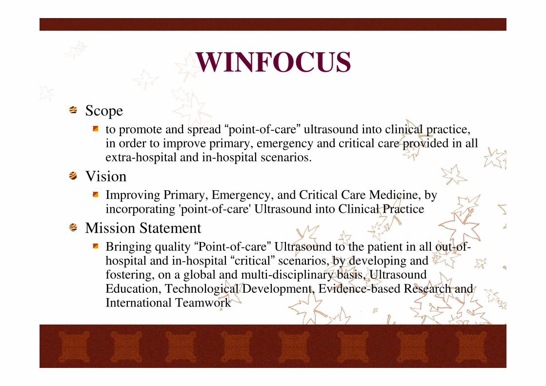
WINFOCUS
Scope
to promote and spread “point-of-care” ultrasound into clinical practice, in order to improve primary, emergency and critical care provided in all extra-hospital and in-hospital scenarios.
VisionImproving Primary, Emergency, and Critical Care Medicine, by incorporating 'point-of-care' Ultrasound into Clinical Practice
Mission Statement
Bringing quality “Point-of-care” Ultrasound to the patient in all out-of-hospital and in-hospital “critical” scenarios, by developing and fostering, on a global and multi-disciplinary basis, Ultrasound Education, Technological Development, Evidence-based Research and International Teamwork
Echocardiography practice, training
and accreditation in the intensive
care: document for the World
Interactive Network Focused on
Critical Ultrasound (WINFOCUS)
http://www.cardiovascularultrasound.co
m/content/6/1/49
2008
Scope of uses in ICU
Following cardiac surgery
Diagnosis
Monitoring
Advantages
Cheap
Portable
Widely available
Echocardiography should be incorporated into
ICU training programs due to its ability to
positively impact on patient management
Rationale of use in ICU
Point-of care echocardiography in the management of the critically ill patient
Providing rapid assessment of cardiac function and physiology
Complements data available from standard invasive hemodynamic monitoring
Both a monitoring and diagnostic tool for rapid bedside assessment of cardiovascular pathophysiology in the critically ill
in the UK > 90% of TEE studies are now
undertaken by anaesthetists
Focused echocardiography as an adjunct in the
peri-arrest period is likely to become a core
competency for acute medicine trainees

Intensivist vs. Cardiologist
Cardiology training programmes
excludes formal exposure to general ICU
the specific conditions most relevant to the ICU
are not generally addressed
the use of echocardiography as a monitoring tool is
not taught
Intensivist vs. Cardiologist
Appropriately trained consultant cardiologist-
echocardiographers for the repeated
examinations required in the ICU is not always
available
The practicing intensivist may not have the
necessary echocardiographic skills
Intensivist vs. Cardiologist
Development of an appropriate program will
require extensive cooperation and support
form a hospital's cardiology service
initial training
support for quality assurance
maintenance of competency among the ICU
backup in the case of a difficult diagnosis
Training
Theoretical Training
Practical training
gained under the guidance of a named supervisor
trained in echocardiography within a department
Winfocus Critical Care
Echocardiography
Winfocus Basic Echo
Winfocus Advanced Echo
Growing support
ICU echocardiography- should we use it in a
heartbeat? Chest 2002
Portable echocardiography- is essential for the
treatment of acutely ill patients BMJ 2006
Echocardiography for the intensivists Care of the
Critically Ill 2003
Beside Ultrasonography in the ICU Chest 2005
Echo in ICU- time for widespread use ICM 2006
Potential scope in ICU
systolic function and RWMA
tamponade and pericardial effusion
hypovolemia and volume responsiveness
acute cor pulmonale
hypoxemia
complication of AMI
chest trauma
assessment of shock
Jones et al CCM 2004
"Incorporation of goal directed ultrasound in the
evaluation of non-traumatic, symptomatic,
undifferentiated hypotension in adult patients
results in fewer viable dignostic aetiologies and a
more accurate physician impression of final
diagnosis"

Echo Examination
The transducers:
Frequency of transducer: 3.5-5Mhz
Phased array of pizeo-electric crystals in the
transducer
The patient
Undress the top
Supine position
Turn slightly left lateral
Adequate Sonic Gel
Poor ECHO images in:
Obese
COAD/hyperinflated chest
Chest wall deformity
Edema
Emergency Echo
FATE and FEEL
Goals
acquire standard TTE views in ACLS compliant
manner
recognise major causes of arrest/ shock
recognise when referral for second opinion
FEEL: focused echocardiography evaluation
in life support
FATE: focused assessed transthoracic echo
Normal FATE view
Subcostal 4 chamber
Apical 4 chamber
Parasternal long axis
Parasternal LV short axis
Pleural scanning
Extended FATE
Normal FATE +
Subcostal vena cava
Apical 2 chamber
Apical long axis
Apical 5 chamber
Parasternal short axis mitral plane
Parasternal aorta short axis
FATE
Hypvolemia
Myocardial dysfunction
Pericardial effusion
Pulmonary embolism
Papillary muscle rupture
VSD in AMI
valve dysfunction
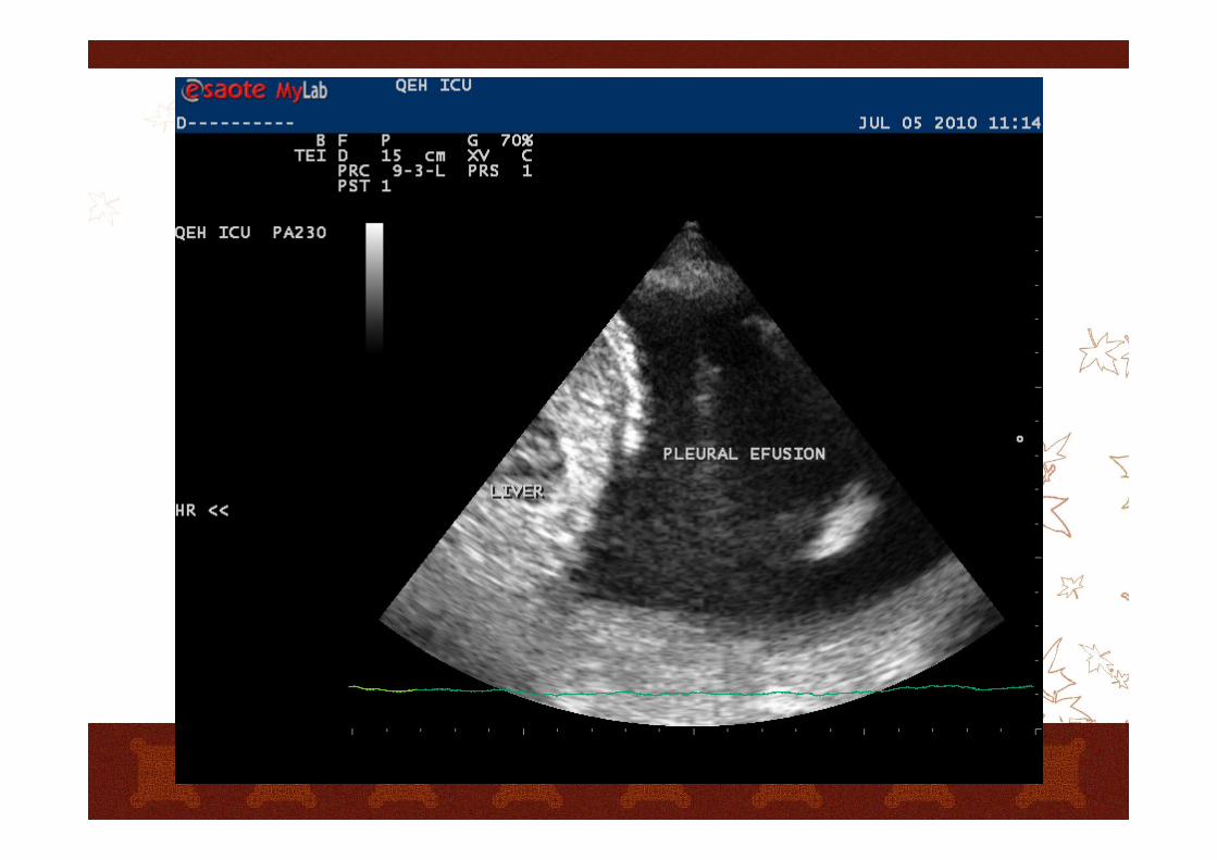
Pleural effusion/ pneumothorax
Pathologies to be considered
Pericardial effusionpost cardiac surgery, post cardiac catheterization, trauma, renal failure, infection
Dilated RA + RVpulmonary embolism, RV infarction, pulmonary hypertension, volume overload
Dilated LA + LVischemic heart disease, dilated cardiomyopathy, sepsis, volume overload, aortic insufficiency
LV hypertrophy
Aortic stenosis, arterial hypertension, left
ventricular outflow tract obstruction, hypertrophic
cardiomyopathy, myocardial deposits diseases
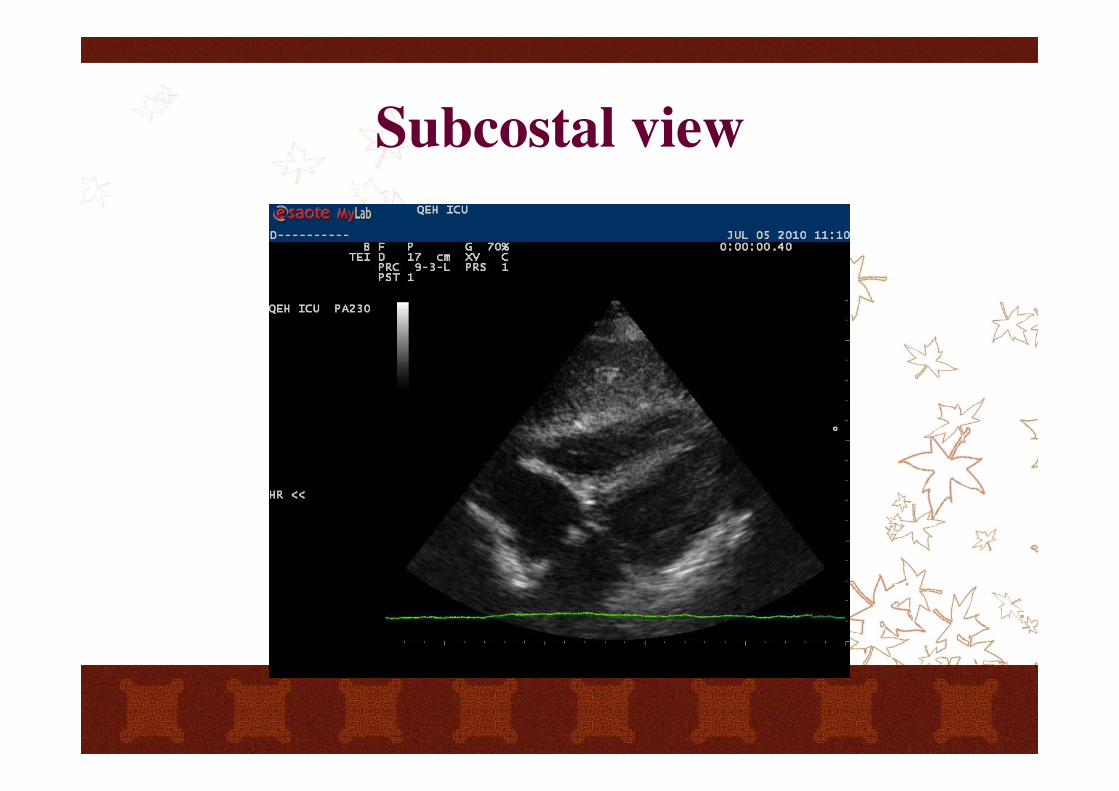
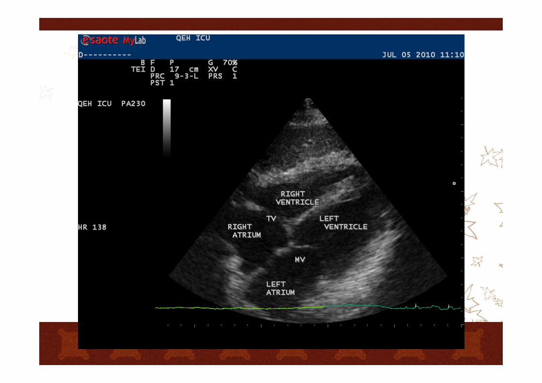
Subcostal view
4 Chamber:
Place transducer below the xyphoid process
The index marker is rotated to 3 o’clock position
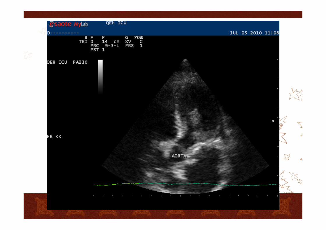
Subcostal view
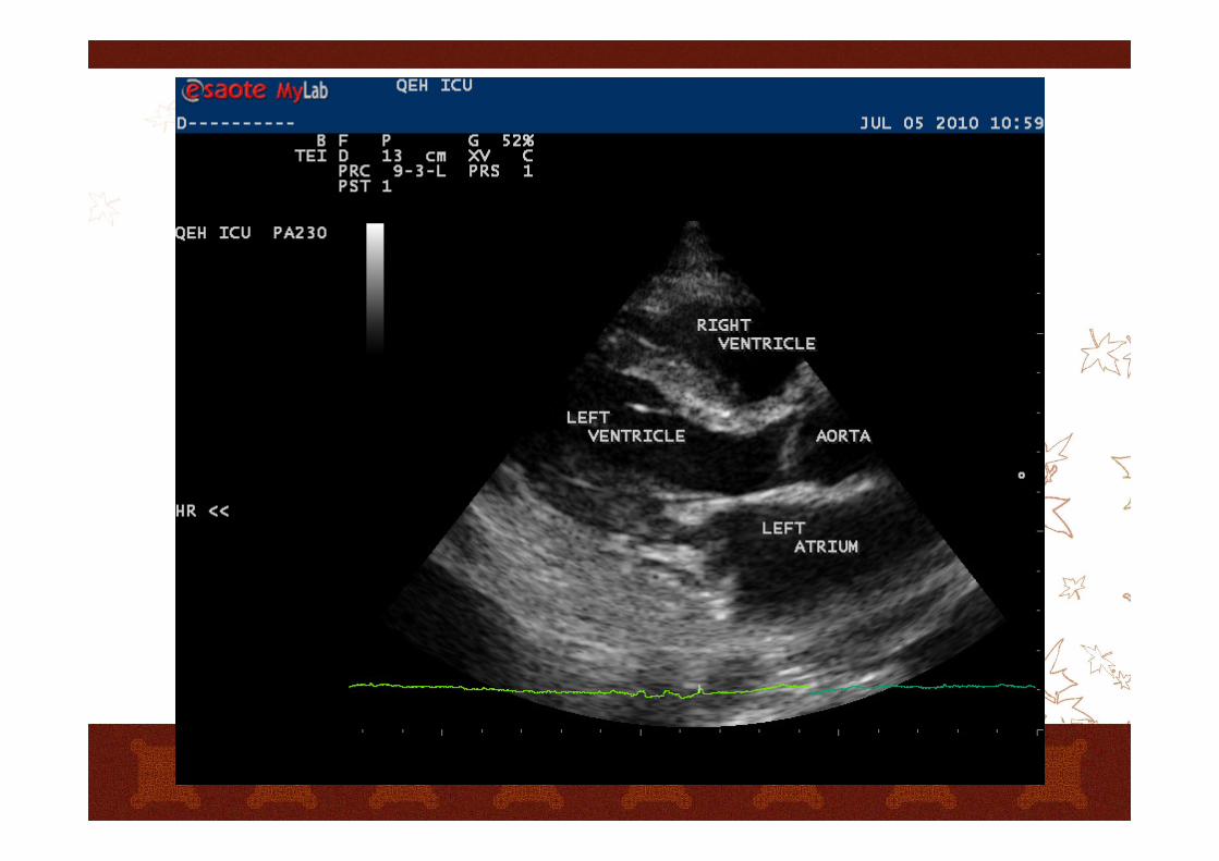
Parasternal view
Long axis
index marker of transducer pointing to the right
shoulder
Short axis
turn 90 degree clockwise where a long axis view is
taken
the marker pointing to left shoulder
By tilting and shifting along the line of the long
axis, a series of views from apex to the pulmonary
artery can be obtained
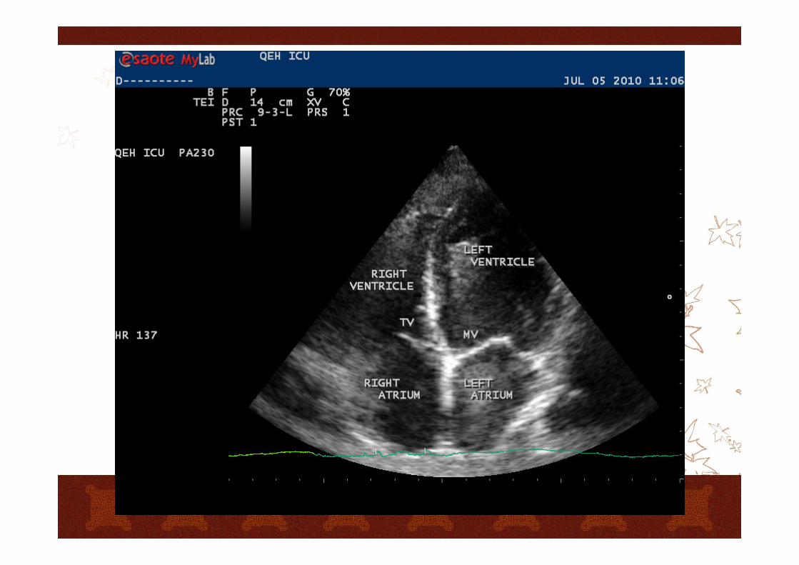
Apical view
4 chamber:
Palpate the apex beat. Place the transducer at the apex
towards the patient’s head
Index marker is rotated to approximately 3 o’clock position
5 Chamber:
Fanning of the transducer at apex to open up the LVOT and
aortic valve [the 5th chamber]
2 chamber:
Rotate the transducer 45 clockwise
Echo examination
2D image:
anatomical assessment, valvular movement, RWMA
M mode:
motion assessment over time, distance or depth measurement
CW and PW:
haemodynamic assessment, calculate velocity, then pressure gradients
CFM:
both haemodynamic and anatomical information
Pre-existing cardiac disease?
RV dilatation = acute or chronic
LV dilatation = almost always chronic
Biventricular dilatation = chronic failure
Atrial dilatation = chronic pressure or volume
overload
RV or LV hypertrophy = chronic pressure
overload
LV shape / chamber dimension / thickness
RV shape / chamber dimension / thickness
atrial dimension
IV septum position
LA measurement
Select M-mode
Align cursor to give horizontal IVS at the level
of aortic valve cusps
Freeze the image
Measure with caliper LA diameter at end
systole
Upper limit of LA: 40mm
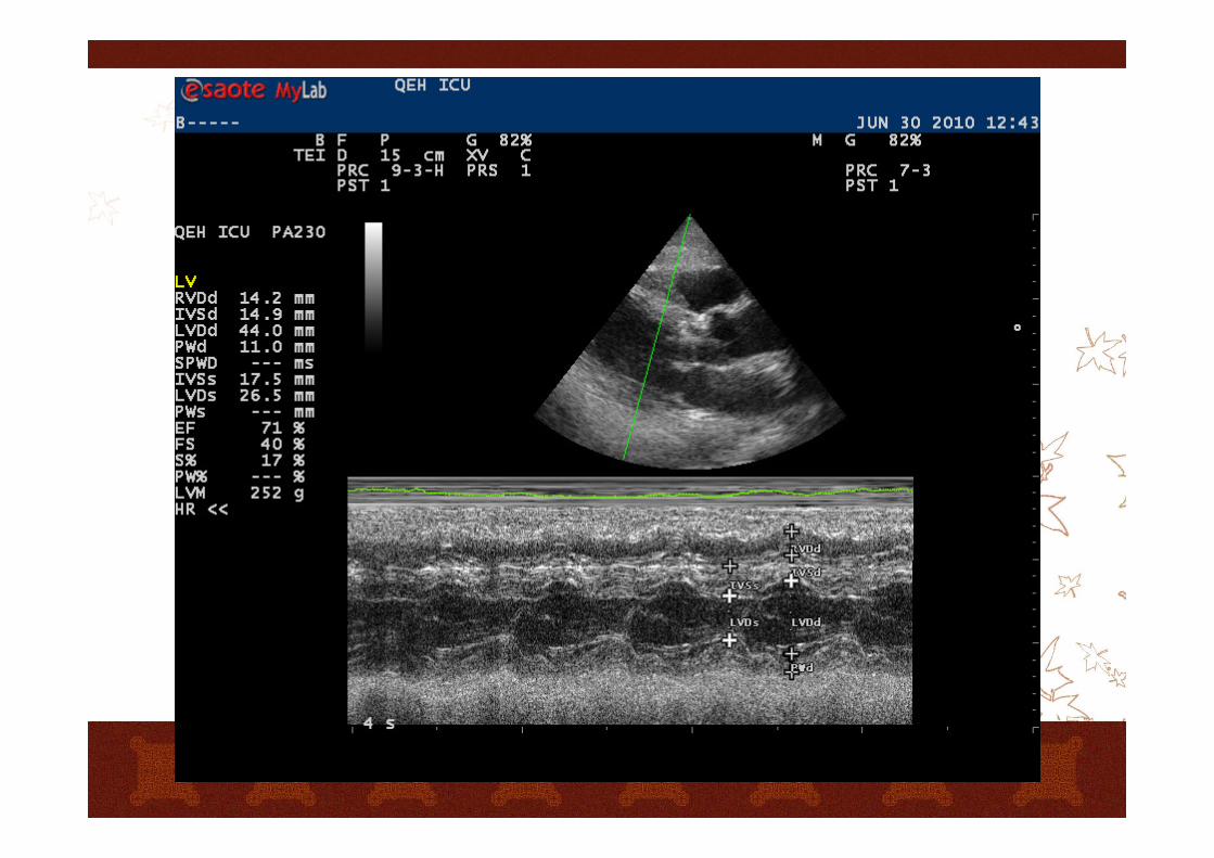
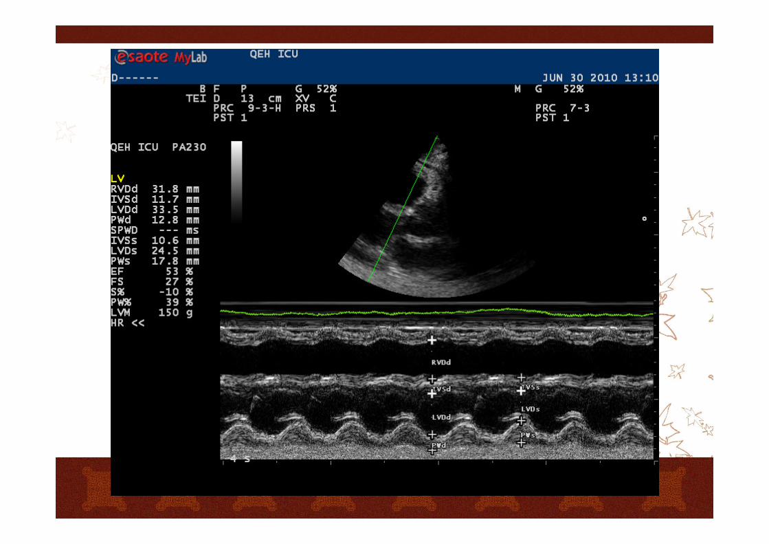
LV Dimension
LV systolic Fx Evaluation
M mode Measurement of LV dimension
LVEDD [normal=3.5-5.6 cm] measured at Q wave
LVESD [normal=2.0-4.0 cm] measured at end of T
wave
Basic volume assessment
Severe hypovolemia
Volume overload
Pitfalls
Severe hypovolemia
End systolic LV obliteration (kissing walls)
LV end diastolic area (LVEDA)
< 5.5 cm/m2 BSA
LVEDA variation with loading
Severe hypovolemia
IVC diameterspontaneous respiration: EED < 9 mm
mechanical ventilation: EED < 15 mm
IVC respiratory variationspontaneous respiration: > 50%
mechanical ventilation: > 18%
IVC variation with loading
* EED end expiratory diameter
Volume overload
Dilated, fixed IVC
Pitfall
cardiac tamponade
constrictive pericarditis
Basic volume status assessment
Easy in severe hypovolemia
Easy in clear volume overload
Difficult in less severe hypovolemia/
significant cardiac disease
Consider pre-existing cardiac disease
Consider respiratory status
Ventricular dysfunction assessment
LV global systolic function
fractional shortening (FS)
visual ejection fraction (eyeballing)
LV regional function
RV systolic function
Fractional shortening
Parasternal Long Axis View with M-mode
cursor just below the MV leaftlets,
perpendicular to the IVS
FS% = (LVEDD-LVESD)/LVEDD x 100%
Fractional shortening
Normal
> 30%
Severely impaired
< 20%
Fractional shortening
RWMA
unreliable
Simplication of Teicholz method
EF % = FS % x 2
Ejection fraction
Normal > 55%
mild dysfunction: 45-55%
moderate dysfunction: 30-44%
severe dysfunction: < 30%
Ejection Fraction
2D Echo
Simpson’s Method
Apical 2 or 4 chamber view
Divide the LV into different slides of known
thickness
Volume size=Slice area X Slice thickness
EF = (LVEDV-LVESV)/LVEDV x 100%
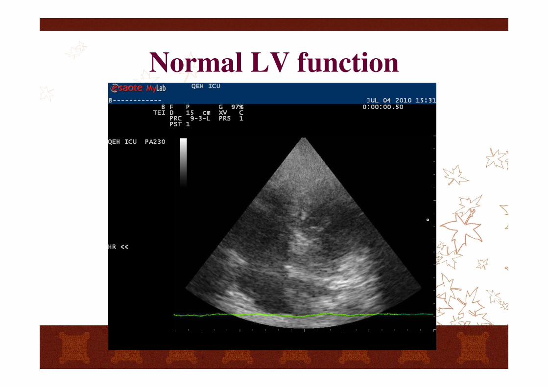
Normal LV function
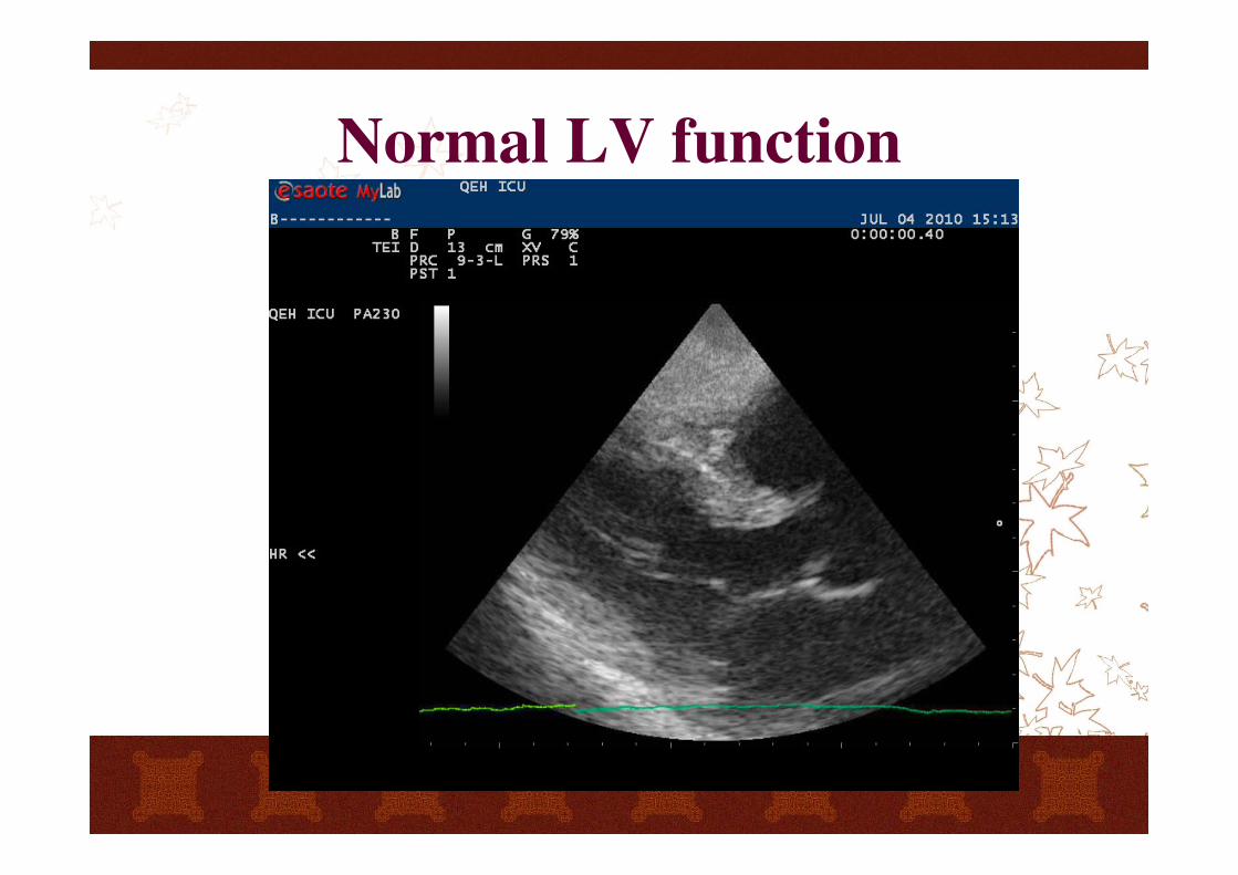
Normal LV function
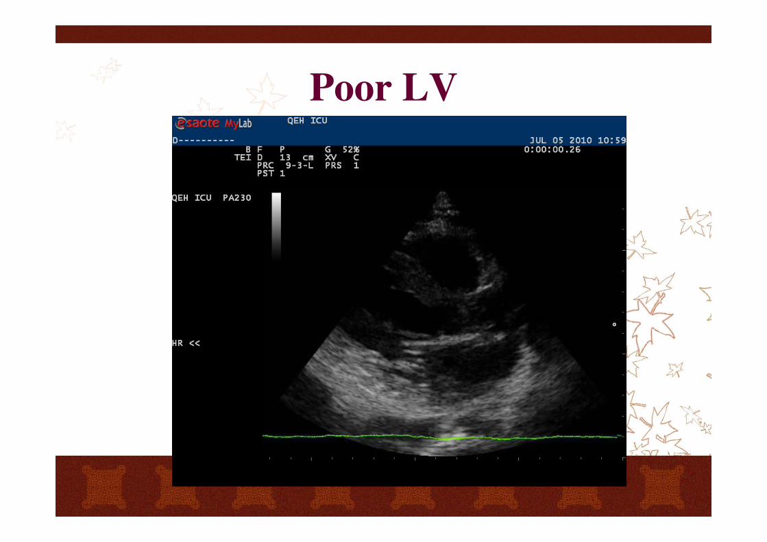
Poor LV
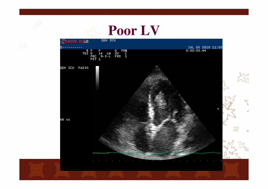
Poor LV
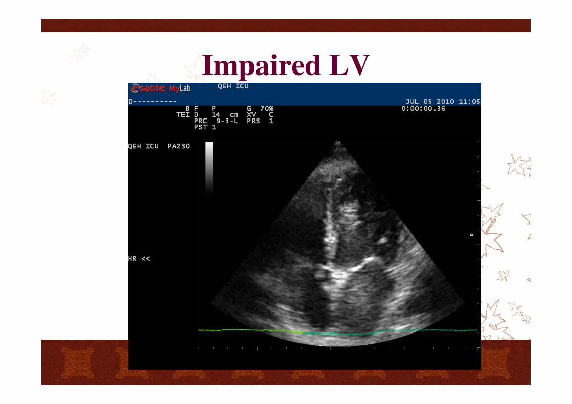
Impaired LV
LV function
Qualitative assessment of LV function
tend to be underestimated in a dilated LV
overestimated in small cavity LV
LV function
Interpret findings in the context of drugs and in
the context of actual preload (volume status)
Repeated echo assessment of LV function
Interpret findings considering inotropic/
mechanical support
marked tachycardia/atrial fibrillation may
underestimate of LV systolic function
Right ventricle
RVEDA / LVEDA
> 0.6 moderate dysfunction
> 1 severe dysfunction
Paradoxical septal movement
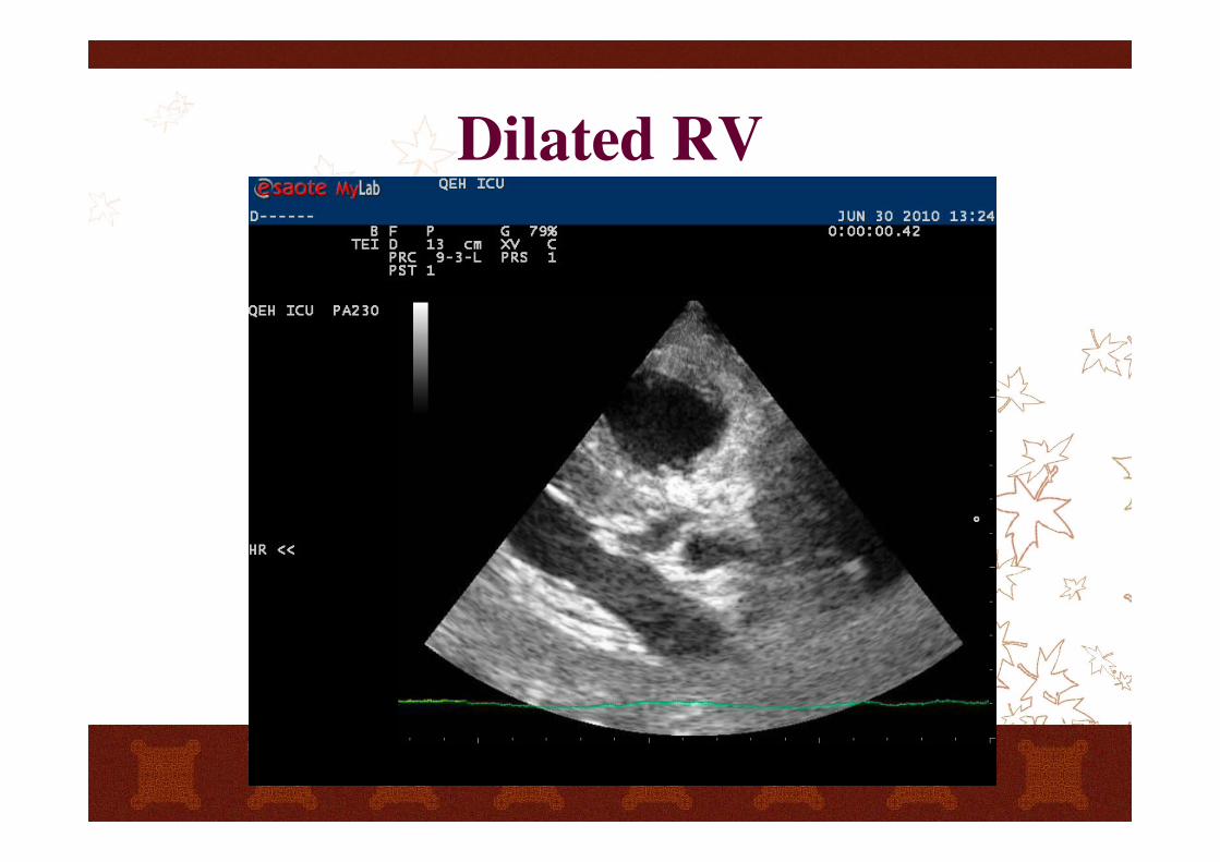
Dilated RV
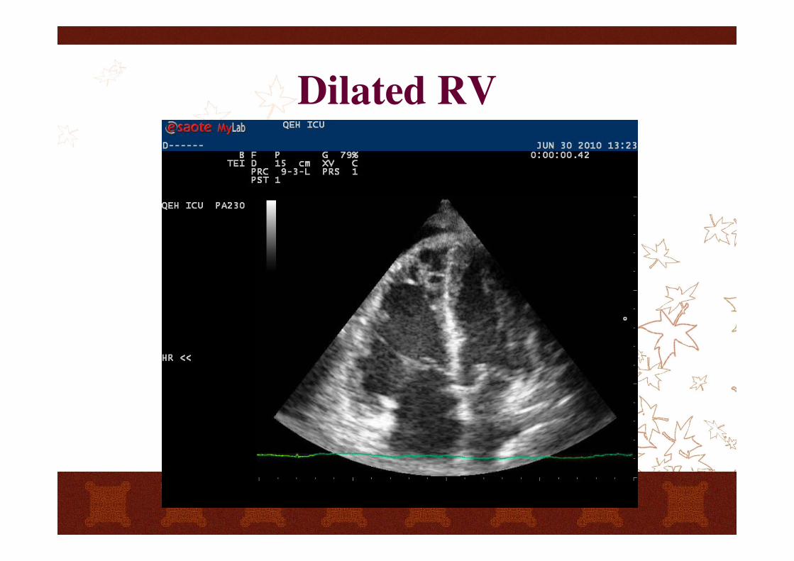
Dilated RV
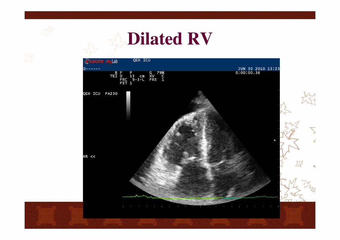
Dilated RV
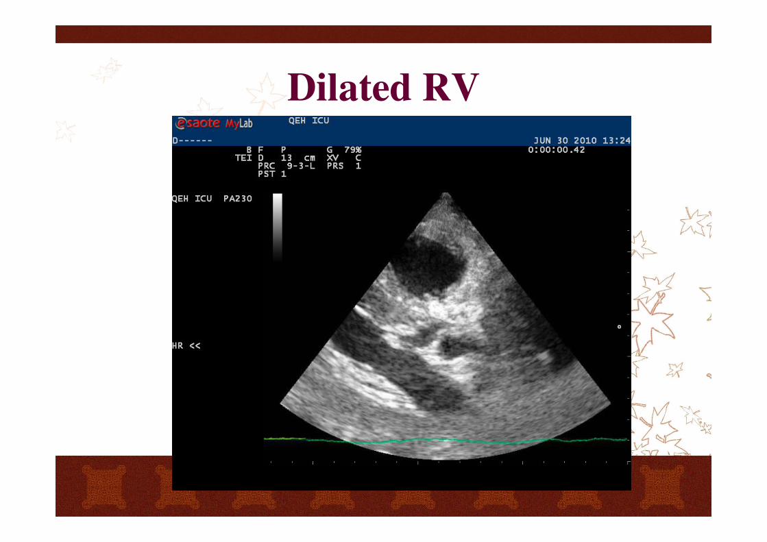
Dilated RV
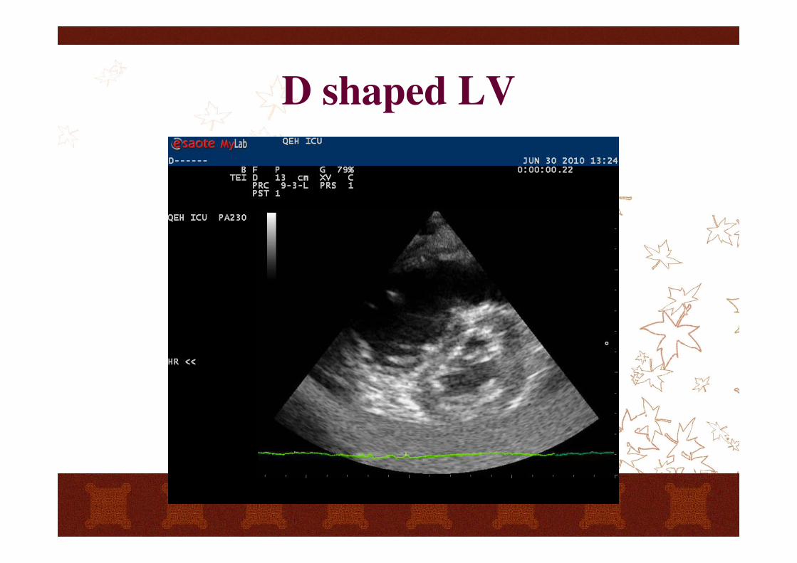
D shaped LV
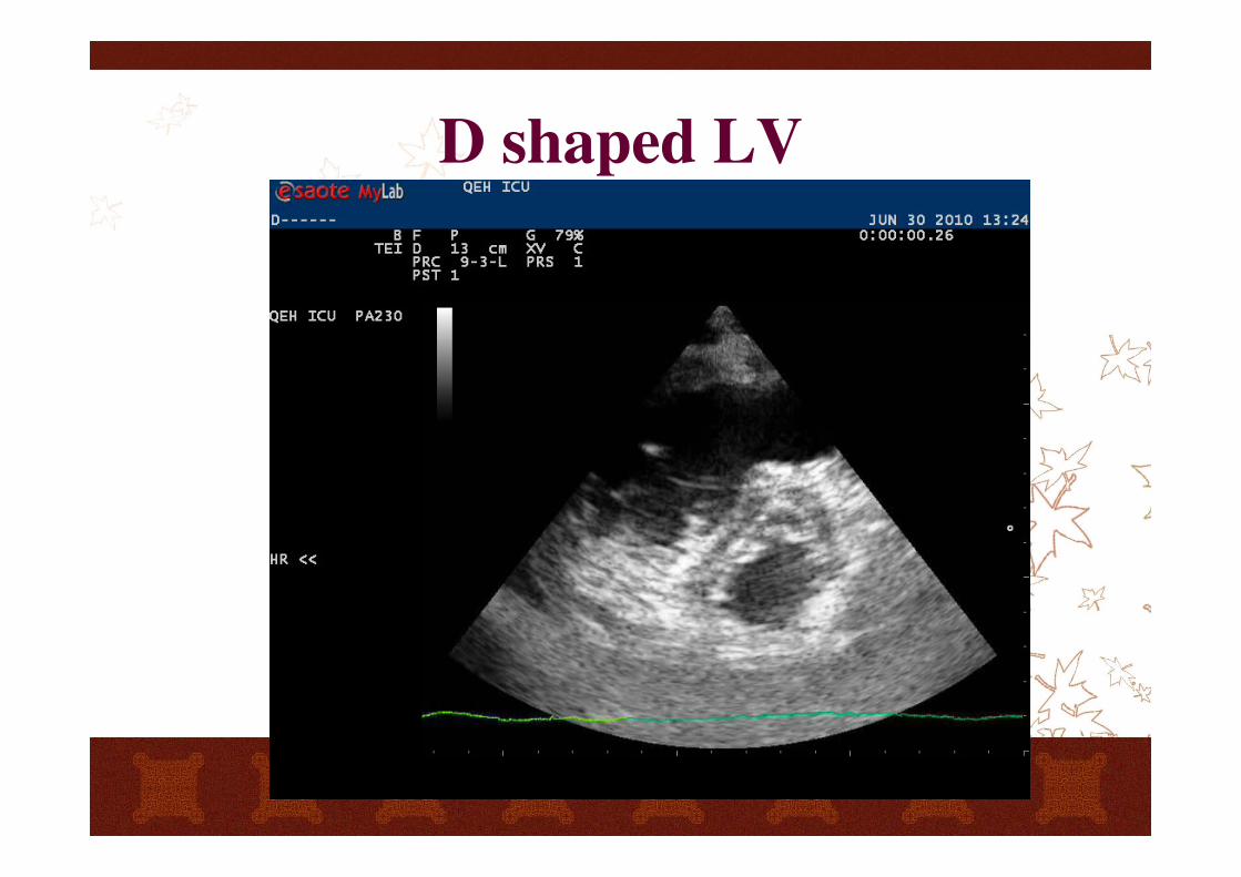
D shaped LV
Mitral valve
Excessive leaflet mobility
myxomatous valve disease, torn chordae, prolapse, flail
myocardial infarction, torn papillary muscle
Restrictive leaflet mobility
rheumatic disease
calcific disease
dilated cardiomyopathy
acute ischemic disease
Interpretation guide
Look for obvious pathology
Assess wall thickness and chamber dimensions
Assess myocardial function
image pleura on both side
relate the information to the clinical context
FEEL
to assess the function of the heart and identify
treatable conditions in peri-resuscitation care
FEEL
to differentiate "true" PEA from "pseudo-PEA"
early detection of ROSC
identify 4 treatable causes of cardiac arrest
cardiac tamponade
hypovolemia
pulmonary embolism
severe LV dysfunction
FEEL
high quality CPR with minimal interruptions to
reduce the no-flow intervals
FEEL < 10 sec
Approach to Shock
Any pre-existing cardiac disease?
IVC
RV
LV
Any pre-existing cardiac disease?
LV and LA dilatation: DCM (postischemic/
valvular/ idiopathic)
Marked LV hypertrophic: HOCM, severe AS,
Hypertension
RA dilatation: chronic cor pulmonale
IVC
Differentiate between hypovolemia and other
causes of shock
Spontaneous breathing: EDD < 9mm and
respiratory variation > 50%
Mechanical ventiltation: EDD < 15mm and
respirtaory variation > 18%
Right Ventricle
Small and hyperkinetic RV: tamponade
Dilated and hypokinetic RV: RV failure
LV dysfunction
RV AMI
Acute cor pulmonale: pulmonary embolism
Left Ventricle
Severe LV hypokinesia
LV AMI, Sepsis related, myocarditis, DCM
LV hyperkinesia
Acute valvular dysfunction (AR, MR), HOCM,
diastolic dysfunction
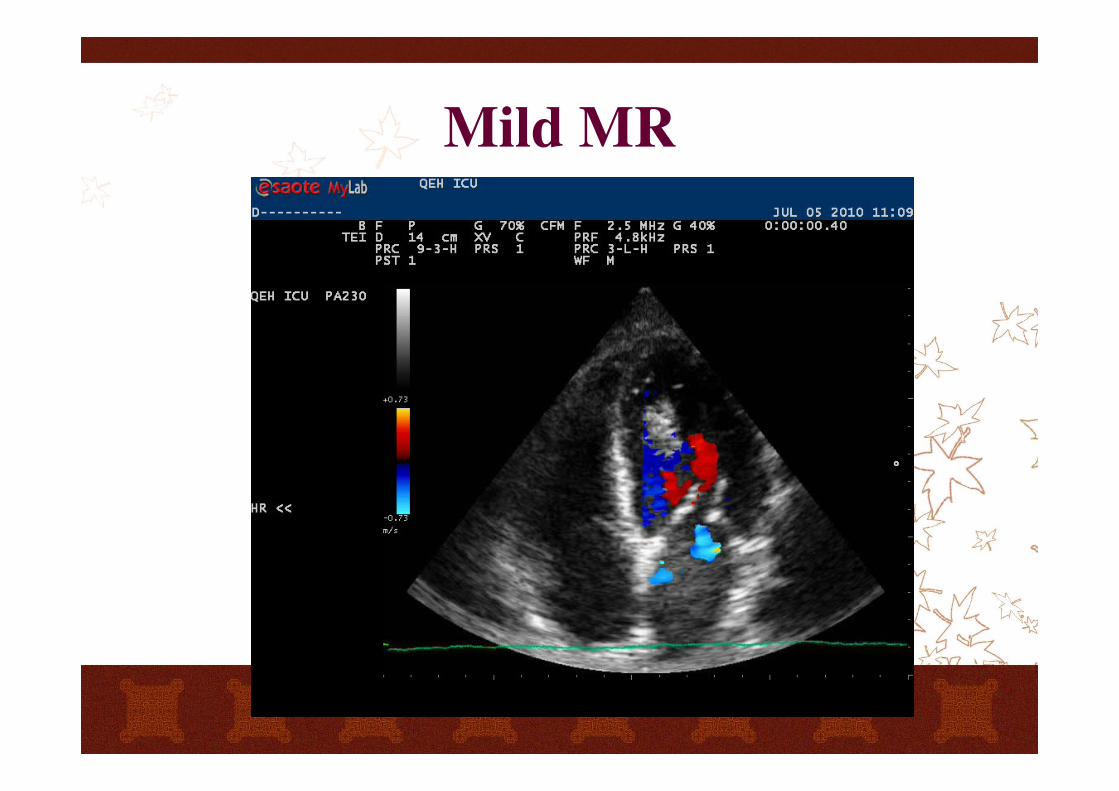
Mild MR
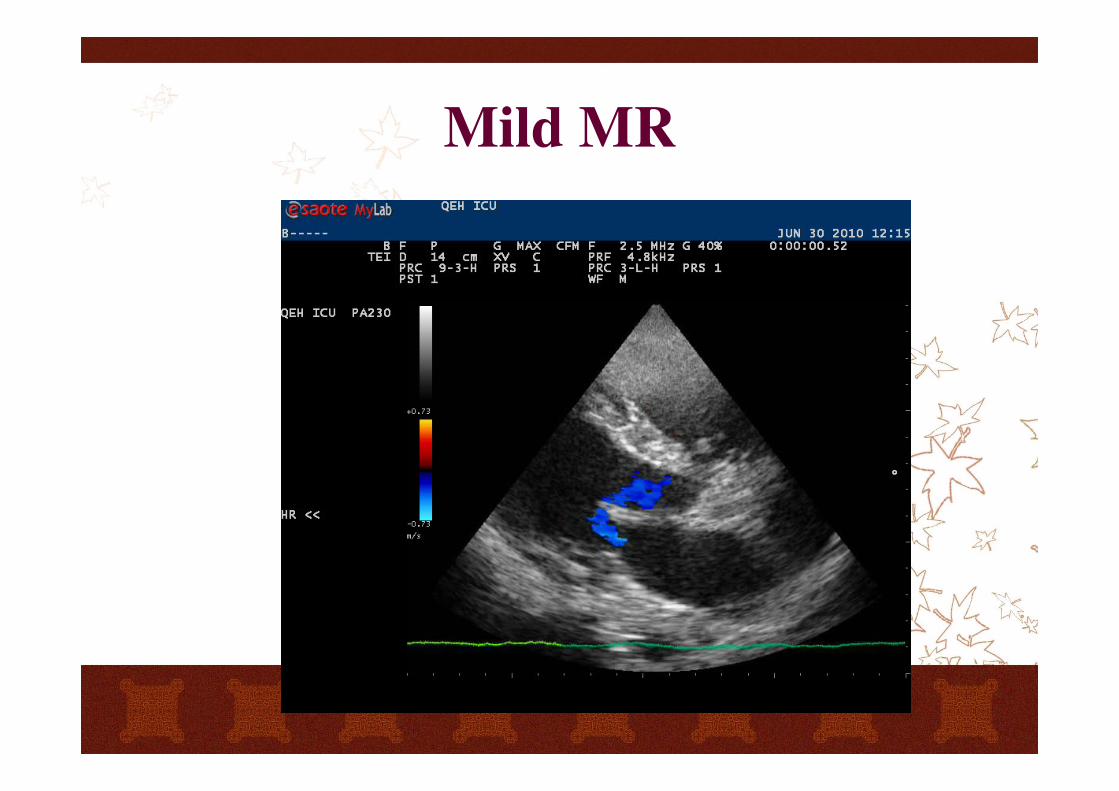
Mild MR
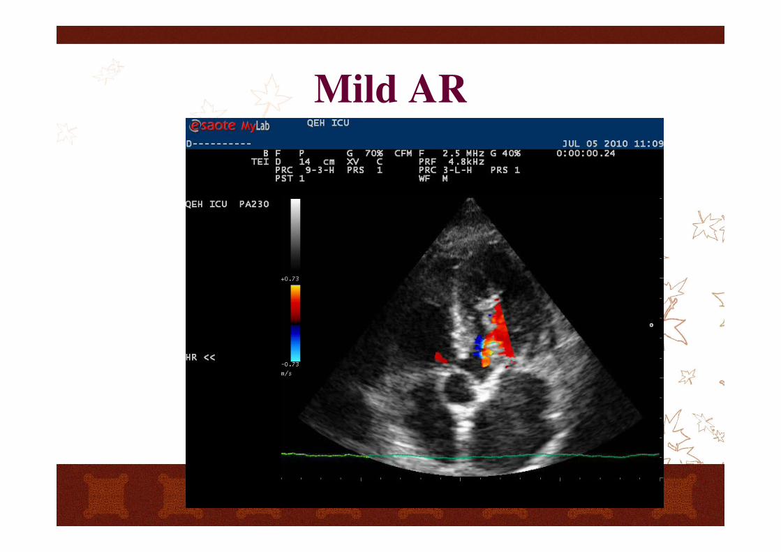
Mild AR
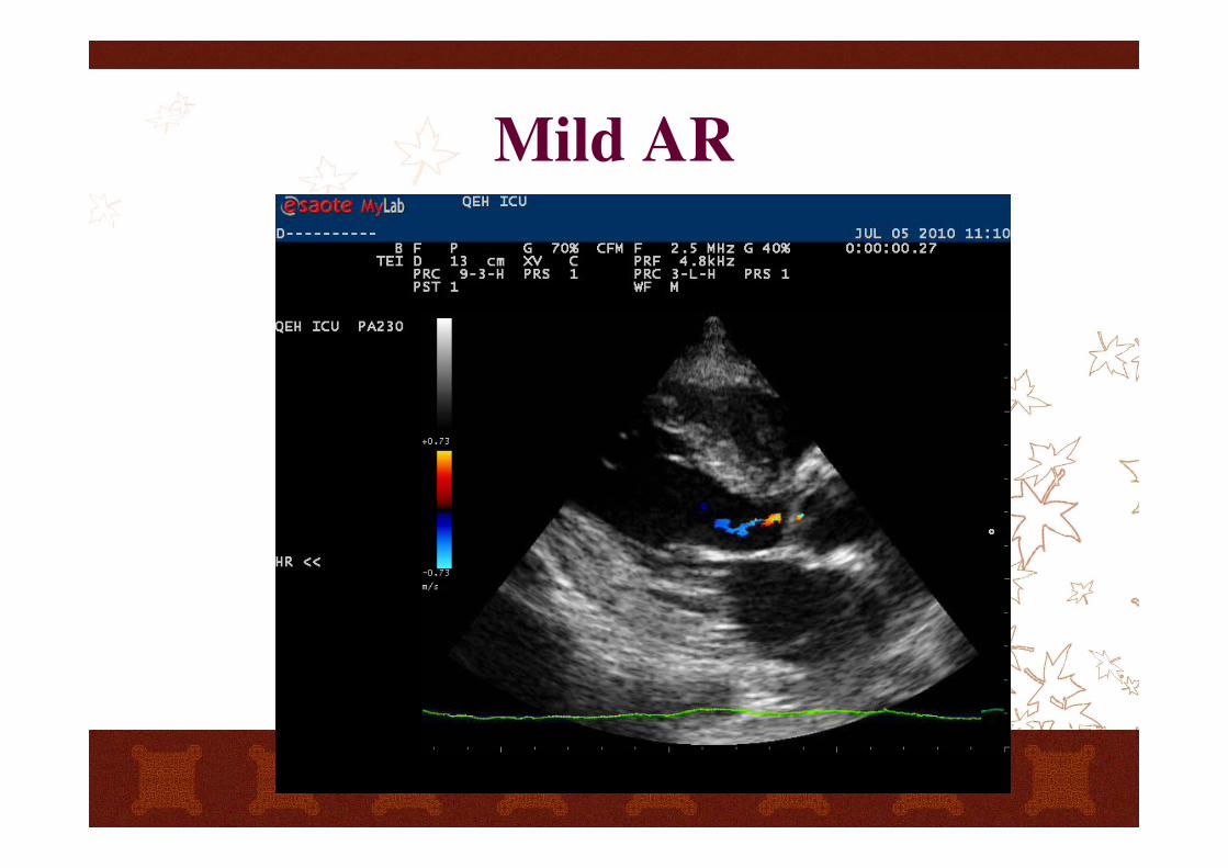
Mild AR
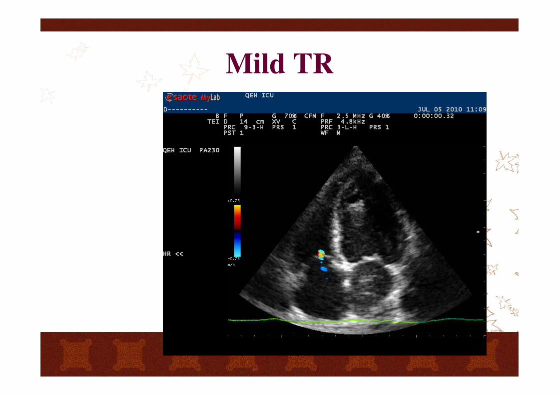
Mild TR
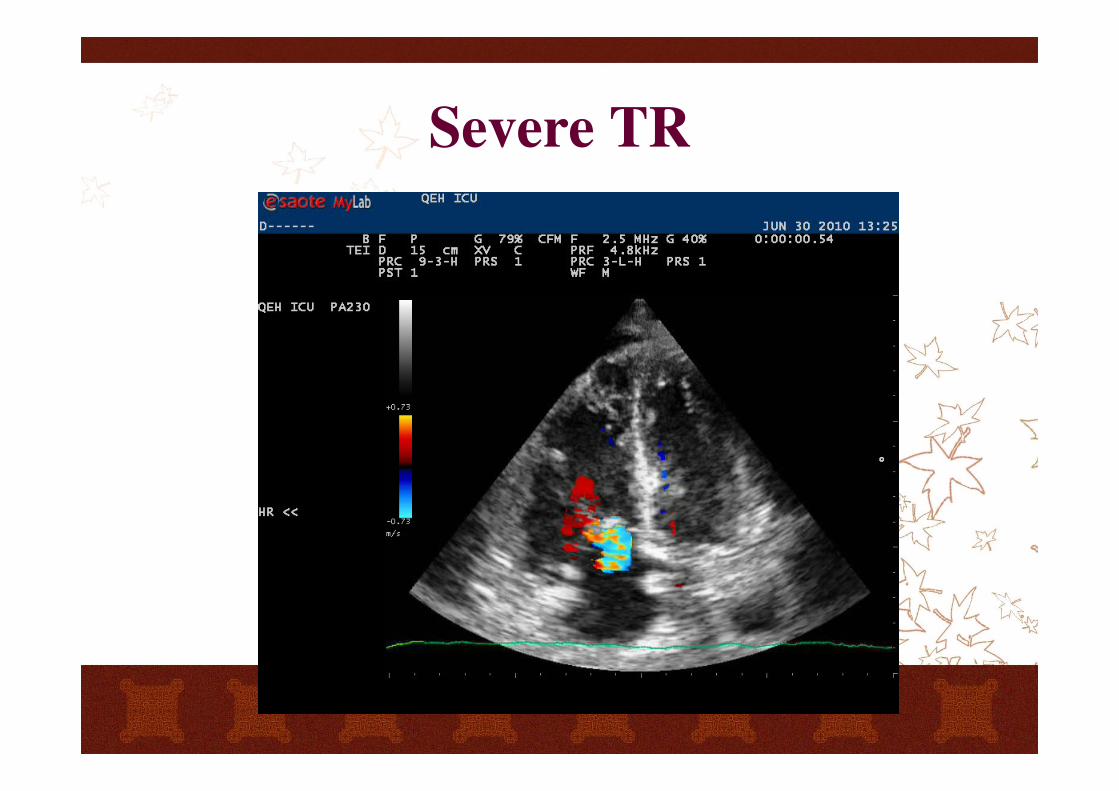
Severe TR
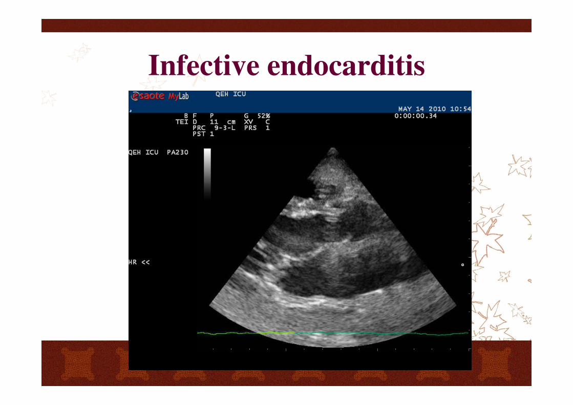
Infective endocarditis
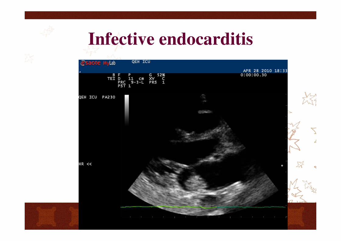
Infective endocarditis

Infective endocarditis
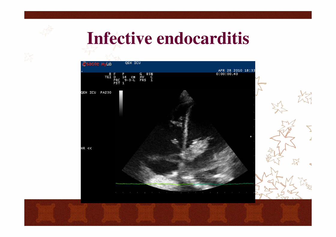
Infective endocarditis
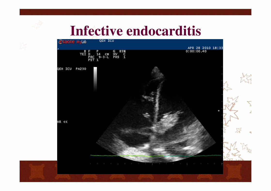
Infective endocarditis
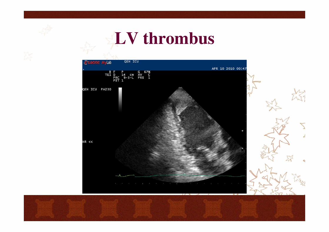
LV thrombus
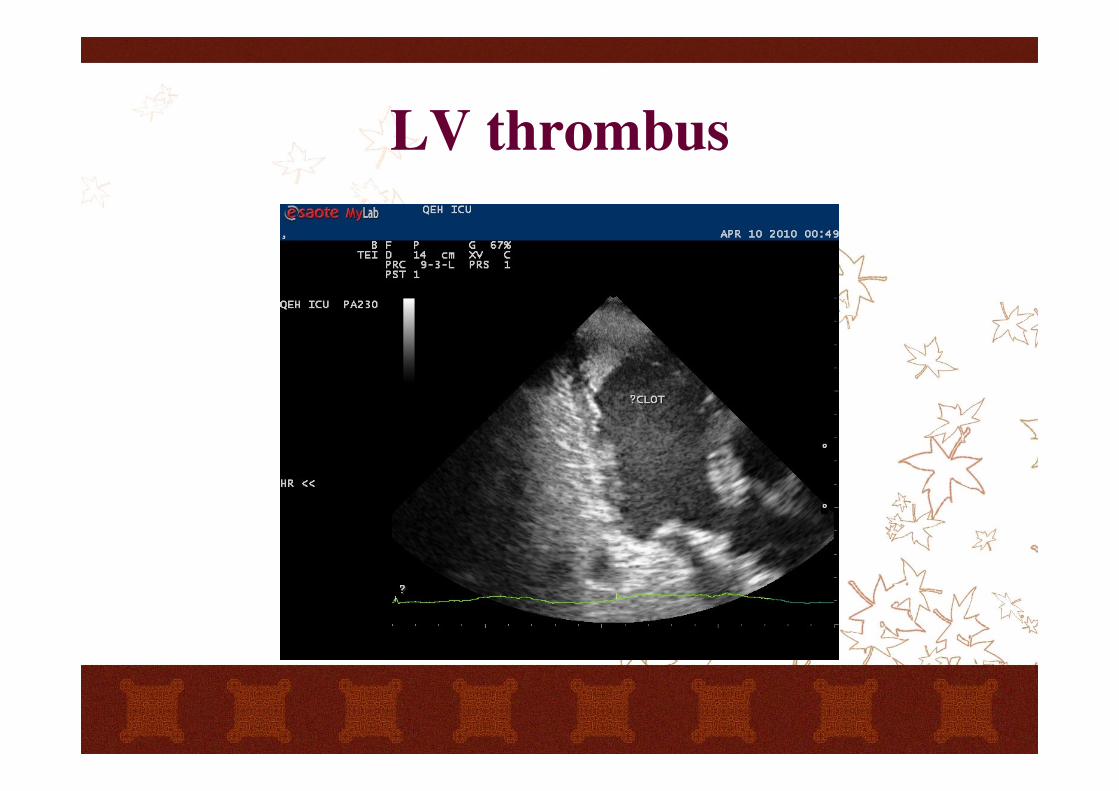
LV thrombus
References
Course material of Winfocus basic echo
Echocardiography practice, training and
accreditation in the intensive care: document
for the World Interactive Network Focused on
Critical Ultrasound
http://www.cardiovascularultrasound.com/cont
ent/6/1/49
Useful links
www.winfocus.org
www.fate-protocol.com
http://www.hkcem.com/html/courses/usg/
Upcoming events
Winfocus courseOrganisers: HKCEM and WINFOCUS
Date: 13-17, Nov, 2012
http://www.hkcem.com/html/courses/usg/
8th Winfocus World CongressBarcelona, Spain
20-22, October, 2012
http://www.winfocus.org/world/barcelona2012/registration
www.winfocus.org
www.winfocus.org/world/echo
http://www.hkcem.com/html/courses/usg/

Thank you