Embed Size (px)
Citation preview
7/25/2019
1
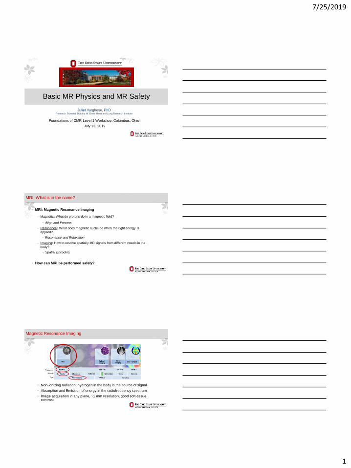
Basic MR Physics and MR Safety
Juliet Varghese, PhDResearch Scientist, Dorothy M. Davis Heart and Lung Research Institute
Foundations of CMR Level 1 Workshop, Columbus, Ohio
July 13, 2019
MRI: What is in the name?
• MRI: Magnetic Resonance Imaging
‒ Magnetic: What do protons do in a magnetic field?
• Align and Precess
‒ Resonance: What does magnetic nuclei do when the right energy is
applied?
• Resonance and Relaxation
‒ Imaging: How to resolve spatially MR signals from different voxels in the
body?
• Spatial Encoding
• How can MRI be performed safely?
Magnetic Resonance Imaging
• Non-ionizing radiation, hydrogen in the body is the source of signal
• Absorption and Emission of energy in the radiofrequency spectrum
• Image acquisition in any plane, ~1 mm resolution, good soft-tissue contrast
7/25/2019
2
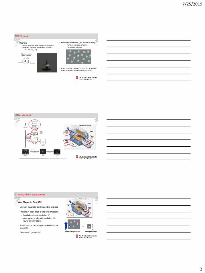
• Magnetic
‒ Nuclei with odd total number of protons + neutrons possess a magnetic moment
• 1H, 31P, 23Na, 13C
MR Physics
Hydrogen atom1 proton + 1 electron
Nucleus
+-
Electron +m
Hydrogen protonSometimes referred to as “a spin”
Magnetic
field directionN
S • A very strong magnet is needed to induce even a weak magnetization in tissue.
• Normal Conditions (No external field)• Random orientation of spins
• No net magnetization
MRI in a Nutshell
Cardiovascular MRI : physical principles to practical protocols / Vivian S. Lee ; Lippincott Williams & Wilkins, c2006
Z
X
Y
B0
Main Magnetic Field (B0)
• Uniform magnetic field inside the cylinder
• Protons in body align along two directions
‒ Parallel and antiparallel to B0
‒ More protons aligned parallel to B0
(lower energy state)
• Equilibrium or net magnetization in tissue
along B0
• Greater B0, greater M0
Creating Net Magnetization
Z
X
Y
B0
External magnetic field
B0
Net Magnetization
=M0
7/25/2019
3
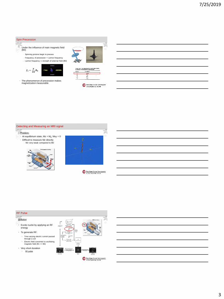
Spin Precession
• Under the influence of main magnetic field (B0)
‒ Spinning protons begin to precess
‒ Frequency of precession = Larmor frequency
‒ Larmor frequency ∝ strength of external field (B0)
• The phenomenon of precession makes magnetization measurable
𝒇𝑳 =𝜸
𝟐𝝅𝑩𝟎
Problem:
• At equilibrium state, Mz = M0; Mxy = 0
• Difficult to measure Mz directly
‒ Mz very weak compared to B0
Detecting and Measuring an MRI signal
Z
X
Y
B0
M0
Solution
• Excite nuclei by applying an RF
energy
• To generate RF:
• Time varying electric current passed
through a coil
• Electric field converted to oscillating
magnetic field (B1 << B0)
• Very short duration
• RF pulse
RF Pulse
7/25/2019
4
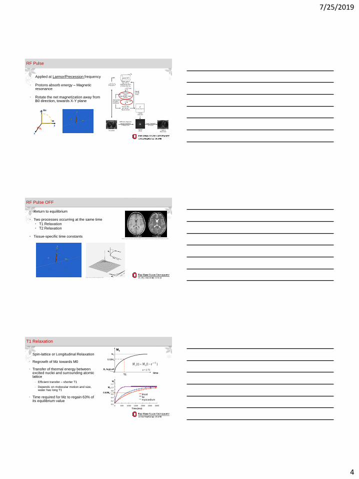
• Applied at Larmor/Precession frequency
• Protons absorb energy – Magnetic resonance
• Rotate the net magnetization away from B0 direction, towards X-Y plane
RF Pulse
y
x
z
B1
Bo
M
RF Pulse OFF
• Return to equilibrium
• Two processes occurring at the same time
• T1 Relaxation
• T2 Relaxation
• Tissue-specific time constantshttps://casemed.case.edu/clerkships/neurology/Web%20Neurorad/MRI%20Basics.htm
https://mri-q.com/bloch-equations.html
• Spin-lattice or Longitudinal Relaxation
• Regrowth of Mz towards M0
• Transfer of thermal energy between excited nuclei and surrounding atomic lattice
‒ Efficient transfer – shorter T1
‒ Depends on molecular motion and size, water has long T1
• Time required for Mz to regain 63% of its equilibrium value
T1 Relaxation
Mo
time
Mz
0.63Mo
B1 field off
T1
)1()( 1/
0
Tt
z eMtM −−=
0.0
0.2
0.4
0.6
0.8
1.0
1.2
0 500 1000 1500 2000 2500 3000
Time (ms)
Mz
bloodfatmyocardium
0.63Mo
Mo
e = 2.71

7/25/2019
5
• Spin-spin or transverse relaxation
• Transverse magnetization decays back to zero
• Interaction between magnetic field and individual protons
• Time required for 63% of the initial magnetization to dissipate
• Effective transverse relaxation time (T2*) –static inhomogeneities in local magnetic field
T2 Relaxation
Mxy
0.63Mo
Mo
0.37Mo
B1 field off
T2time
2/
0)(Tt
xy eMtM −=
T2*
Ridgway. JCMR 2010 12:71
• Emission of RF energy can be measured before equilibrium is obtained
• Change of Mxy detected by a coil
• Free induction decay
• For cardiac imaging, a separate RF receiver coil is used to maximize signal detection
Signal Detection
M
z
B1
y
x
Detector coil
https://www.siemens-healthineers.com/magnetic-resonance-imaging/options-and-
upgrades/coils/body-18
Ridgway. JCMR 2010 12:71
Spatial Encoding
Cardiovascular MRI : physical principles to practical protocols / Vivian S. Lee ; Lippincott Williams & Wilkins, c2006
7/25/2019
6
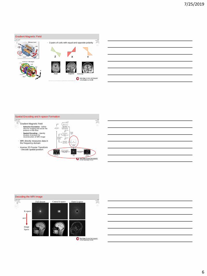
• 3 pairs of coils with equal and opposite polarity
Gradient Magnetic Field
X
Y
https://casemed.case.edu/clerkships/neurology/Web%20Neurorad/MRI%20Basics.htm
Z X Y
ZCoronalSagittalAxial
Z
X
Y
B0
• Gradient Magnetic Field
‒ Selective Excitation - Define slice for imaging and excite the protons in that slice
‒ Spatial Encoding – Identify location of protons for reconstruction of MRI image
• MRI directly measures data in the frequency domain
• Inverse 2D Fourier Transform - Decode spatial position
Spatial Encoding and k-space Formation
K-space
Image
Space
Full dataset Central k-space Outer k-space
IFFT
Decoding the MRI Image
7/25/2019
7
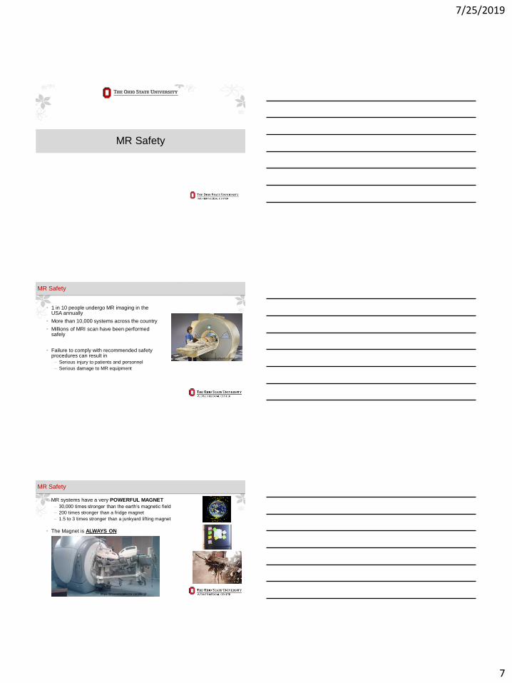
MR Safety
• 1 in 10 people undergo MR imaging in the USA annually
• More than 10,000 systems across the country
• Millions of MRI scan have been performed safely
• Failure to comply with recommended safety procedures can result in
‒ Serious injury to patients and personnel
‒ Serious damage to MR equipment
MR Safety
https://science.howstuffworks.com/mri.htm
• MR systems have a very POWERFUL MAGNET
‒ 30,000 times stronger than the earth’s magnetic field
‒ 200 times stronger than a fridge magnet
‒ 1.5 to 3 times stronger than a junkyard lifting magnet
• The Magnet is ALWAYS ON
MR Safety
https://mrimetaldetector.com/blog/
7/25/2019
8
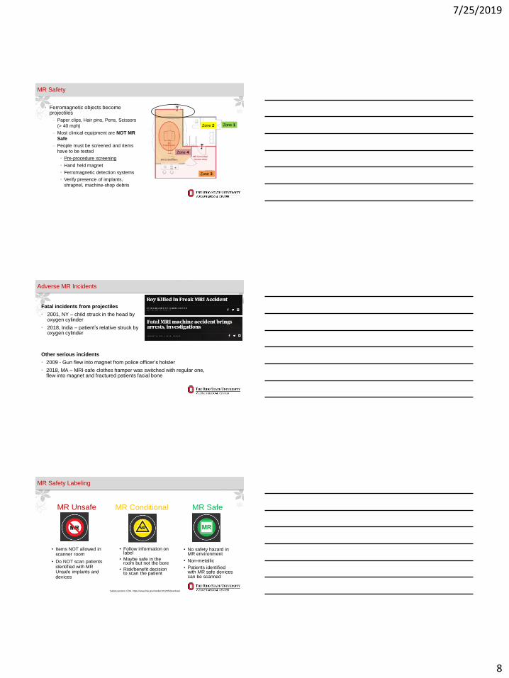
• Ferromagnetic objects become projectiles
‒ Paper clips, Hair pins, Pens, Scissors
(> 40 mph)
‒ Most clinical equipment are NOT MR
Safe
‒ People must be screened and items
have to be tested
• Pre-procedure screening
• Hand held magnet
• Ferromagnetic detection systems
• Verify presence of implants,
shrapnel, machine-shop debris
MR Safety
Zone 1Zone 2
Zone 3
Zone 4
Other serious incidents
• 2009 - Gun flew into magnet from police officer’s holster
• 2018, MA – MRI-safe clothes hamper was switched with regular one, flew into magnet and fractured patients facial bone
Adverse MR Incidents
Fatal incidents from projectiles
• 2001, NY – child struck in the head by oxygen cylinder
• 2018, India – patient’s relative struck by oxygen cylinder
MR Safety Labeling
MR Unsafe MR Conditional MR Safe
• Follow information on label
• Maybe safe in the room but not the bore
• Risk/benefit decision to scan the patient
• No safety hazard in MR environment
• Non-metallic
• Patients identified with MR safe devices can be scanned
Safety posters: FDA https://www.fda.gov/media/101205/download
• Items NOT allowed in scanner room
• Do NOT scan patients identified with MR Unsafe implants and devices
7/25/2019
9
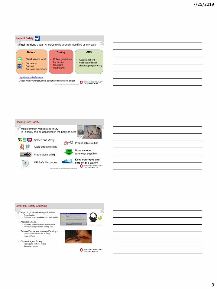
Implant Safety
Safety posters: FDA https://www.fda.gov/media/101205/download
Before During After
• Check device label
• Document
• Consult
• Pre-scan procedure
• Follow guidelines
for device
• Constant
monitoring
• Assess patient
• Post-scan device
check/reprogramming
Fatal incident, 1992 - Aneurysm clip wrongly identified as MR safe
http://www.mrisafety.com
Check with your institution’s designated MR safety officer
Heating/Burn Safety
Screen and Verify
Avoid street clothing
Proper positioning
Proper cable routing
Normal mode,
whenever possible
Keep your eyes and
ears on the patient
• Most common MRI related injury
• RF energy can be deposited in the body as heat
Safety posters: FDA https://www.fda.gov/media/101205/download
MR Safe Electrodes
http://www.magmedix.com/mri-
ecg-electrode-cleartrace-
2.html
Bertrand et al. 2017
• Physiological and Biological effects‒ Tissue heating
‒ Peripheral nerve stimulation – tingling/twitching
• Acoustic Effects‒ Increased anxiety / Communication trouble
‒ Temporary and permanent hearing loss
• Tattoos/Permanent makeup/Piercings‒ Heating, Local edema and swelling
‒ Image artifacts
• Contrast Agent Safety‒ Nephrogenic systemic fibrosis
‒ Gadolinium retention
Other MR Safety Concerns
https://www.universalme
dicalinc.com/mri-safe-
paired-foam-earplugs-
37db.html
https://www.allmri.com
/product_info.php?lan
guage=en&info=p505_
earmuffs-for-mri.html

7/25/2019
10
• Completion of MR screening form
• Confirm safety of implanted devices
• Check if inpatients have temporary devices
• Stop scan if unreported object is discovered during scanning
A Safe MR exam
https://www.ismrm.org/mr-safety-links/mr-safety-resources-page/
Question twice!
• Any personnel with reason to enter MR suite must be screened and trained
• Train staff on protocols for emergency in MRI
‒ Medical
• Remove patient from scan room, Close door
‒ Fire, Flood
• Emergency Power OFF
‒ Life-threatening event / Uncontrolled fire
• Quench
MR Safety Training
Emergency POWER OFF
QUENCH
https://mri.fsu.edu/for-researchers/safety-manual/
• MR Physics
• https://www.mriquestions.com/index.html
• MR Safety
• http://www.mrisafety.com
• https://www.ismrm.org/mr-safety-links/mr-safety-resources-page/
• ACR Guidance Document on MR Safe Practices: 2013. JMRI 37:501-530
• Gilk and Kanal. 2013. JMRI 37:531-543
• https://www.fda.gov/radiation-emitting-products/mri-magnetic-resonance-imaging/mri-safety-posters
Resources

7/25/2019
11
WITH GREAT POWER COMES GREAT RESPONSIBILITY