Embed Size (px)
Citation preview

R
Bm
MU
a
AA
KMMIEAA
C
0d
Journal of Chromatography A, 1217 (2010) 3908–3921
Contents lists available at ScienceDirect
Journal of Chromatography A
journa l homepage: www.e lsev ier .com/ locate /chroma
eview
asic rules for the interpretation of atmospheric pressure ionizationass spectra of small molecules
ichal Holcapek ∗, Robert Jirásko, Miroslav Lísaniversity of Pardubice, Faculty of Chemical Technology, Department of Analytical Chemistry, Studentská 573, 53210 Pardubice, Czech Republic
r t i c l e i n f o
rticle history:vailable online 1 March 2010
eywords:ass spectraass spectrometry
nterpretationlectrospray ionizationtmospheric pressure chemical ionizationtmospheric pressure photoionization
a b s t r a c t
This review summarizes the basic rules for the interpretation of atmospheric pressure ionization(API) mass spectra of small molecules written with the style primarily intended for beginners andlow-experienced researchers with the mass spectra interpretation. The first and basic step in any inter-pretation of mass spectra is always the determination of molecular weight, which is relatively easy incase of soft ionization techniques due to the limited extend of fragmentation and the prevailing pres-ence of (de)protonated molecules in the full scan mass spectra. These [M+H]+ and [M−H]− ions are oftenaccompanied by low abundant molecular adducts, which can be used as the supplementary informationfor the unambiguous determination of molecular weights. In certain cases, adduct ions may dominatethe spectra. The subsequent interpretation of full scan and tandem mass spectra is more complicated dueto a high number of possible functional groups, structural subunits and their combinations resulting innumerous competitive fragmentation pathways. Typical neutral losses and the effect of individual func-tional groups on the fragmentation are discussed in detail and illustrated with selected examples. Modern
mass analyzers have powerful features for the structural elucidation, for example high resolving power,high mass accuracy, multistage tandem mass spectrometry, dedicated softwares for the interpretationof mass spectra and prediction of their fragmentation. Background information on differences amongindividual ionization techniques suitable for the HPLC–MS coupling and basic types of mass analyzerswith consequences for the data interpretation is briefly discussed as well. Selected examples illustratethat the right optimization of chromatographic separation and the use of other than mass spectrometric detectors can bring valuable complementary information.© 2010 Elsevier B.V. All rights reserved.
ontents
1. Introduction . . . . . . . . . . . . . . . . . . . . . . . . . . . . . . . . . . . . . . . . . . . . . . . . . . . . . . . . . . . . . . . . . . . . . . . . . . . . . . . . . . . . . . . . . . . . . . . . . . . . . . . . . . . . . . . . . . . . . . . . . . . . . . . . . . . . . . . . . . 39092. Atmospheric pressure ionization techniques. . . . . . . . . . . . . . . . . . . . . . . . . . . . . . . . . . . . . . . . . . . . . . . . . . . . . . . . . . . . . . . . . . . . . . . . . . . . . . . . . . . . . . . . . . . . . . . . . . . . . . . . 39093. Mass analyzers . . . . . . . . . . . . . . . . . . . . . . . . . . . . . . . . . . . . . . . . . . . . . . . . . . . . . . . . . . . . . . . . . . . . . . . . . . . . . . . . . . . . . . . . . . . . . . . . . . . . . . . . . . . . . . . . . . . . . . . . . . . . . . . . . . . . . . . 39104. Importance of chromatographic separation and other detection techniques . . . . . . . . . . . . . . . . . . . . . . . . . . . . . . . . . . . . . . . . . . . . . . . . . . . . . . . . . . . . . . . . . . . . . . 39105. Interpretation of API mass spectra . . . . . . . . . . . . . . . . . . . . . . . . . . . . . . . . . . . . . . . . . . . . . . . . . . . . . . . . . . . . . . . . . . . . . . . . . . . . . . . . . . . . . . . . . . . . . . . . . . . . . . . . . . . . . . . . . . . 3911
5.1. Molecular weight determination. . . . . . . . . . . . . . . . . . . . . . . . . . . . . . . . . . . . . . . . . . . . . . . . . . . . . . . . . . . . . . . . . . . . . . . . . . . . . . . . . . . . . . . . . . . . . . . . . . . . . . . . . . . . . 39125.2. Typical fragmentation behavior for individual functional groups . . . . . . . . . . . . . . . . . . . . . . . . . . . . . . . . . . . . . . . . . . . . . . . . . . . . . . . . . . . . . . . . . . . . . . . . . . . 3914
5.2.1. Phosphorous containing functional groups . . . . . . . . . . . . . . . . . . . . . . . . . . . . . . . . . . . . . . . . . . . . . . . . . . . . . . . . . . . . . . . . . . . . . . . . . . . . . . . . . . . . . . . . 39165.2.2. Sulfur-containing functional groups . . . . . . . . . . . . . . . . . . . . . . . . . . . . . . . . . . . . . . . . . . . . . . . . . . . . . . . . . . . . . . . . . . . . . . . . . . . . . . . . . . . . . . . . . . . . . . . 3916
5.2.3. Nitrogen containing functional groups . . . . . . . . . . . . . . . . . . .5.2.4. Oxygen containing functional groups . . . . . . . . . . . . . . . . . . . .5.2.5. Halogen substituents . . . . . . . . . . . . . . . . . . . . . . . . . . . . . . . . . . . . .5.2.6. Other structural types . . . . . . . . . . . . . . . . . . . . . . . . . . . . . . . . . . . .∗ Corresponding author. Tel.: +420 46 6037087; fax: +420 46 6037068.E-mail address: [email protected] (M. Holcapek).
021-9673/$ – see front matter © 2010 Elsevier B.V. All rights reserved.oi:10.1016/j.chroma.2010.02.049
. . . . . . . . . . . . . . . . . . . . . . . . . . . . . . . . . . . . . . . . . . . . . . . . . . . . . . . . . . . . . . . . . . . . . . . . . . 3917. . . . . . . . . . . . . . . . . . . . . . . . . . . . . . . . . . . . . . . . . . . . . . . . . . . . . . . . . . . . . . . . . . . . . . . . . . 3917. . . . . . . . . . . . . . . . . . . . . . . . . . . . . . . . . . . . . . . . . . . . . . . . . . . . . . . . . . . . . . . . . . . . . . . . . . 3918. . . . . . . . . . . . . . . . . . . . . . . . . . . . . . . . . . . . . . . . . . . . . . . . . . . . . . . . . . . . . . . . . . . . . . . . . . 3918
M. Holcapek et al. / J. Chromatogr. A 1217 (2010) 3908–3921 3909
5.2.7. Molecules with multiple functional groups . . . . . . . . . . . . . . . . . . . . . . . . . . . . . . . . . . . . . . . . . . . . . . . . . . . . . . . . . . . . . . . . . . . . . . . . . . . . . . . . . . . . . . . . 39195.3. Odd-electron ions in API mass spectra . . . . . . . . . . . . . . . . . . . . . . . . . . . . . . . . . . . . . . . . . . . . . . . . . . . . . . . . . . . . . . . . . . . . . . . . . . . . . . . . . . . . . . . . . . . . . . . . . . . . . . . 3919
6. Concluding remarks . . . . . . . . . . . . . . . . . . . . . . . . . . . . . . . . . . . . . . . . . . . . . . . . . . . . . . . . . . . . . . . . . . . . . . . . . . . . . . . . . . . . . . . . . . . . . . . . . . . . . . . . . . . . . . . . . . . . . . . . . . . . . . . . . . 3920Acknowledgments . . . . . . . . . . . . . . . . . . . . . . . . . . . . . . . . . . . . . . . . . . . . . . . . . . . . . . . . . . . . . . . . . . . . . . . . . . . . . . . . . . . . . . . . . . . . . . . . . . . . . . . . . . . . . . . . . . . . . . . . . . . . . . . . . . . 3920
. . . . . .
1
actaeMHolTtimachbyHocwlaaPf[Teslp
oicihwsflodctoPiKbNmtf
charged ionic organic species the range of ESI is somewhat wider(MWs in the range of thousands) than for atmospheric pressurechemical ionization (APCI) and atmospheric pressure photoioniza-tion (APPI).
References . . . . . . . . . . . . . . . . . . . . . . . . . . . . . . . . . . . . . . . . . . . . . . . . . . . . . . . . . . . .
. Introduction
An enormous growth in the field of mass spectrometry (MS)nd its coupling with separation techniques in last decades is stillontinuing, which generates a huge number of new HPLC–MS sys-ems installed worldwide in academic, industrial, pharmaceuticalnd clinical laboratories. New HPLC–MS users often lack previousxperiences with the interpretation of mass spectra or even withS itself resulting in unfavorable situation of top-class expensivePLC–MS instrumentation operated by users with little knowledgef MS. This discrepancy is partially caused by the lack of appropriateiterature about the interpretation of soft ionization mass spectra.he goal of this review is to provide basic rules for the interpreta-ion of mass spectra measured by common atmospheric pressureonization (API) techniques used in HPLC–MS, but these rules are
ostly applicable also for other soft ionization techniques, suchs matrix-assisted laser desorption/ionization (MALDI). This arti-le is intended as a starting point in this area suitable for beginners,owever all exceptions occurring during the interpretation cannote covered in one review. This summary is based on almost 15ears of experiences of the first author with the interpretation ofPLC–MS data. During this time, an extensive amount of organic,rganometallic and bioorganic compounds from different chemicallasses has been analyzed and their spectra have been interpretedithin the scope of research projects and mass spectrometric ana-
ytical service at our faculty. In addition to the citation of our papersnd works published by other groups, many described observationsre supported by our unpublished results and personal experiences.rocedures for the API spectra interpretation are often derivedrom the rules known for electron ionization (EI) mass spectra1–3] either applied directly as it is or in a slightly modified form.he scope of this review is limited to small organic molecules butxcluding biopolymers (peptides, proteins, nucleot(s)ides), wherepecific interpretation procedures are extensively described in theiterature. Rather specific area is the field of organometallic com-ounds and metal complexes, which will be reviewed later on [4].
The coupling of separation techniques with MS has beenf a great interest for long time due to the potential of MSn the identification of chromatographic peaks. The first gashromatography–mass spectrometry (GC/MS) coupling was real-zed already in 1957 [5], but the development of really usefulyphenation of HPLC–MS has required longer time. Initial attemptsith the implementation of EI in HPLC–MS concept [6] were not
uccessful due to severe limitations concerning the mobile phaseow rate, application range and robustness of such devices. Latern, the thermospray as the first soft ionization technique speciallyesigned for HPLC–MS coupling [7] still suffered from technicalonstraints. The real breakthrough has been the invention of elec-rospray ionization (ESI) [8,9]. The application of ESI for the analysisf large biomacromolecules by Fenn [10] has been awarded Nobelrize for chemistry in 2002 together with Koichi Tanaka for thenspiration of MALDI [11]. The significant contribution of Michaelaras and Franz Hillenkamp in the MALDI development should not
e overlooked [12]. In past, several other individuals were awardedobel Prizes for chemistry or physics for their principal achieve-ents in mass spectrometry, such as Wolfgang Paul in 1989 forhe theoretical description of ion trap and Francis W. Aston in 1922or the accurate measurement of most elemental isotopes in the
. . . . . . . . . . . . . . . . . . . . . . . . . . . . . . . . . . . . . . . . . . . . . . . . . . . . . . . . . . . . . . . . . . . . . . . . . 3920
periodic table [11]. At present time, the most appreciated recentaccomplishment is the construction and commercial introductionof Orbitrap as a new type of mass analyzer by Makarov [13], whichis also the topic of one review in this special issue [14].
Two angles of view can be applied for the present state-of-artin HPLC–MS. The optimistic view can highlight routine applica-tions of HPLC–MS over many branches of chemistry, biochemistry,pharmacy and medicine with only minor limitations concerningthe choice of optimal chromatographic conditions, superior sensi-tivity, selectivity, extremely low sample consumption, three basicAPI techniques applied in both polarity modes and several vendorsselling different types of mass spectrometers for HPLC coupling. Onthe other hand, the pessimist can argue that mass spectrometer isstill rather sophisticated device generating huge data files, which isoften not properly understood and interpreted by numerous chro-matographists. Both views are true and the goal of this paper is tobe a small contribution in a better understanding of mass spectrainterpretation.
2. Atmospheric pressure ionization techniques
In our opinion, ESI is the softest ionization technique at all fol-lowed by MALDI with the similar application range. The applicationfield of ESI covers the range of polarities from medium polar toionic compounds (Fig. 1), but the ionization problems can occur forlow polar and nonpolar compounds. There are some hints, how toionize rather nonpolar compounds with ESI, such as the promo-tion of the formation of molecular adducts listed in Table 1 (mainlywith alkali metal, ammonium and silver ions), which may signifi-cantly extent the polarity range of ESI shown in Fig. 1. The molecularweight (MW) range of ESI is significantly wider than for other APItechniques due to the formation of multiply charged ions for pro-teins and other biopolymers (MWs >100,000), but even for singly
Fig. 1. General scheme of applicability of API techniques for small molecules basedon polarity and molecular weights.

3910 M. Holcapek et al. / J. Chromatogr. A 1217 (2010) 3908–3921
Table 1Singly charged molecular adducts in positive-ion API mass spectra.
Molecular adduct Nominal mass shifta [�Da] Exact mass shifta [mDa] References
[M+Li]+ 6 +8.2 [119][M+NH4]+ 17 +26.5 Numerous citations in the literature[M+H+H2O]+ 18 +10.6 [45][M+Na]+ 22 −18.1 Numerous citations in the literature[M+H+CH3OH]+ 32 +26.2 [45,47,49,50][M+K]+ 38 −44.1 Numerous citations in the literature[M+H+CH3CN]+ 41 +26.5 [31,45–47][M+H+H2O+CH3OH]+ 50 +36.8 [45][M+Na+CH3CN]+ 63 +8.5 [45][M+Ag]+ 106 −102.7 [107,120–122][2M+H]+ – – [34,46][2M+Na]+ – – [34,41,92,93][2M+K]+ – – [41,92,93]
lar ad
blpiivpArripawfacAn[Ascada
3
mitanm[epfttttit
[3M+Na]+ –[3M+K]+ –
a Mass shifts correspond to the difference between [M+H]+ and particular molecu
Typical applications of ESI are various classes ofio(macro)molecules studied in proteomics, metabolomics and
ipidomics, e.g., peptides, proteins, nucleot(s)ides, polysaccharides,hospholipids. In addition to these well-known applications, ESI
s also an excellent tool for the analysis of wide range of polar toonic “small” organic and organometallic compounds starting fromery low masses up to several thousands. The attention must beaid to the ionization of rather nonpolar analytes, where APCI andPPI typically have a better performance within common massange up to m/z 1500 (sometimes up to m/z 3000). It is often notealized that APCI and APPI techniques do not suffer so much fromon suppression/enhancement effects [15], because the ionizationrocess takes place solely in a gas phase. Relative abundances oflkali metal and other ionic adducts are much lower than with ESI,hich may cause a sensitivity increase for compounds suffering
rom the formation of various adducts. The limitation of APCInd APPI is the analysis of biopolymers, organometallics, ionicompounds and other labile analytes. Typical application areas ofPCI and APPI are almost identical, such as drugs, nonpolar lipids,atural compounds, pesticides and various organic compounds16]. To look for small differences between their application range,PPI shows quite often the formation of radical ions [17,18], has alightly softer character and is more successful for highly nonpolarompounds as polyaromatic hydrocarbons [16,18,19]. The usefullternative for improved ionization efficiency can be a combinedual sources, where APCI/APPI is used for low polar compoundsnd ESI/APPI is applicable for the full range of compounds [18].
. Mass analyzers
At present, six basic types of mass analyzers are available on thearket – quadrupole, ion trap, time-of-flight (TOF), double focus-
ng magnetic analyzer, Orbitrap, ion cyclotron resonance (ICR) –ogether with numerous hybrid combinations and variants of thesenalyzers, most of them applicable also for HPLC–MS coupling. It isot a goal of this review to provide the full survey of currently usedass analyzers, because it can be found in the specialized literature
20]. From the view of this review, the important issue is param-ters affecting the appearance of mass spectra, such as resolvingower, mass accuracy, mass range of analyzer, dynamic range usedor the quantitation, types of tandem mass experiments. Typically,he first important question is the division on mass analyzers into
wo groups providing low vs. high mass resolution, which is relatedo the mass accuracy. The group of low resolution mass analyzersypically includes quadrupole analyzers and various types of spher-cal and linear ion traps with the resolving power in the range of fewhousands using the full width at half maximum (FWHM) definition– [41]– [41]
duct, e.g., [M+Na]+–[M+H]+ = 22.
and without the possibility of accurate determination of m/z values.In common speech it is often said that they have unit resolution,which means that they are able to resolve ions differing by one massunit (in current practice, it is about five times better), but this is notthe correct definition of resolving power. High resolution (the rangeof tens of thousands, TOF based analyzers and double focusing mag-netic sector analyzer) and ultrahigh resolution (>100,000 resolvingpower, Fourier transform based Orbitrap and ICR analyzers) ana-lyzers give the highest confidence in the spectra interpretation andidentification, because possible artefacts are reduced at the increas-ing resolving power. High mass accuracy (at minimum <5 ppm, TOFbased analyzers) or even better ultrahigh mass accuracy (<1 ppm,Orbitrap and ICR analyzers) significantly reduce the number ofpossible elemental compositions for the ion within a given masstolerance. The benefit of high resolving power and high mass accu-racy can be illustrated on distinguishing phosphates vs. sulfatesand phosphonates vs. sulfonic acids (see Table 3), where identicalmass shifts and very similar fragmentation behavior occur, but itcan be identified based on different exact masses. If accurate rel-ative abundances can be recorded (preferably with analog-digitalconvertor), then the number of possible elemental compositionsis again reduced [21,22], in favorable cases to only one correcthit. HPLC–MS systems are equipped with advanced software tools,which can speed up and semi-automate the interpretation, forexample by integrating individual subgroups of information (e.g.,positive and negative-ion modes, full scan and tandem mass spec-tra, information obtained for individual fragment ions). The field ofdedicated softwares is growing very fast and appropriate softwaretools can provide valuable help even for experts.
4. Importance of chromatographic separation and otherdetection techniques
The basic rules described in this article are of course valid forany API mass spectra regardless the fact, whether they were mea-sured by the direct infusion or HPLC–MS, but several importantissues should be considered concerning the possibilities of couplingto liquid-phase separation techniques [23,24]. When the measuredsample is pure or relatively pure compound, then there is no reasonwhy to waste time and solvents on the chromatographic separationand this pure sample should be measured by the direct infusion(more time for MSn experiments) or the flow injection analysis (the
solvent background can be subtracted). In all other cases, the MScoupling to the separation brings some advantages depending onthe sample complexity and the separation selectivity of developedchromatographic method. It is also important to keep in mind lim-itations of MS structural characterization, where well-optimized
atogr. A 1217 (2010) 3908–3921 3911
ctttmssIsnFsi
iidtrrdcabfioosUaim1
odtmcntAiuove[opseiafgsgostp
itA
M. Holcapek et al. / J. Chrom
hromatography can do an excellent job. For various types of posi-ional isomers, MS mostly does not provide any distinction, buthe chromatographic resolution of isomers is achievable. For enan-iomers, there is no distinction by MS except for the special kinetic
ethod based on the fragmentation of ternary complexes [25] andimilar methods based on the use of chiral selectors [26], but theeparation of enantiomers can be done on chiral stationary phases.f the separation is not properly optimized, then obtained mixedpectra complicate the interpretation and trace compounds mayot be detected at all due to the signal overlap with coeluting peaks.or highly complex biological and natural mixtures, the use of 2Deparation techniques coupled to MS can increase the number ofdentified compounds, especially in the off-line arrangement [27].
HPLC–MS technique provides different types of signal mon-toring (e.g., total ion current (TIC) chromatogram, selectedon monitoring (SIM), selected reaction monitoring (SRM), etc.)epending on the data acquisition setting, but the highest impor-ance for the mass spectra interpretation has MSn scans andeconstructed (or extracted) ion current (RIC) chromatograms. Theeconstruction of selected m/z values (or regions of m/z values)epending on the time processed after the analysis from TIC dataan be used for various purposes: (A) the identification what ionsre related to the particular chromatographic peak and what is theackground contamination based on the comparison on RIC pro-les for the region around the peak of interest, (B) the identificationf coelution of two compounds based on the detailed comparisonf their RIC records, the coelution can be detected even for verymall differences of retention times (about 0.2 min or even less forHPLC), (C) the detection of suitable regions for the signal averagingnd background subtraction, (D) the discovery of ultratrace peaksn the noise using either targeted m/z values or narrow regions of
/z step-by-step to detect hidden trace peaks, e.g., m/z 100–150,50–200, 200–250 and so on.
Another important issue is the compatibility of HPLC conditionsptimal for the best separation with the MS detection. Nowa-ays the compatibility of both techniques is relatively good dueo the technical developments, so we can operate at common chro-
atographic flow rates with all API techniques (up to 1 mL/min),ommon mobile phases in reversed-phase and HILIC systems. Forormal-phase systems, ESI suffers from problems with the ioniza-ion unless the content of polar organic modifier is at least 5%, soPCI or APPI is a better choice for normal-phase systems. The only
mportant limitation is the use mobile phase additives, where these of nonvolatile agents discourages and even the concentrationf volatile additives should be kept as low as possible [28]. Non-olatile additives participate in the ion suppression and matrixffects (in some cases the sensitivity may drop to zero response)15,29,30] and they may cause severe contamination of ion opticsf mass spectrometer. The most dangerous contaminants are ion-airing agents based on tetraalkylammonium salts [28], whichhould never enter the mass spectrometer due to severe memoryffects apparent for several months even after the repetitive clean-ng of the whole system from the sample injector until the massnalyzer (unpublished experiences of our group and others knownrom personal communications). In most cases, nonvolatile inor-anic agents can be replaced by volatile organic alternatives withimilar or slightly worse separation efficiency [31–33], but alsoenerally accepted volatile additives such as ammonium acetater formate, formic acid and acetic acid can cause certain suppres-ion effects [28], so their concentration should be always kept athe lowest level necessary for the satisfactory chromatographic
erformance.Other detection techniques can provide valuable supplementarynformation in some specific cases. The photodiode-array UV detec-or provides useful data in case of strong chromophoric systems.
good example is the identification of metabolites formed from
Fig. 2. UV spectra of two groups of metabolites of drug dimefluron: (A) conju-gated system with carbonyl groups is retained and (B) reduction of carbonyl groupto hydroxyl reduces the extent of conjugation. (Reproduced from Ref. [34] withpermission.)
the drug dimefluron (Fig. 2) with highly conjugated chromophoricsystem containing two aromatic rings conjugated through the addi-tional carbonyl group. The parent drug and related metaboliteswithout the change of this conjugated system have identical UVspectra, while all metabolites formed by the reduction of this car-bonyl have completely different UV spectra, because the number ofconjugated double bonds is reduced from nine to five [34]. This iscomplementary information, which is not evident from mass spec-tra, so the supporting information from UV detector significantlysimplifies the structure elucidation of these metabolites. The sim-ilar approach can be applied for sensitive fluorescence detection[35]. In general, the mass spectrometer is considered as the mostsensitive detector for liquid-phase separation techniques, which istrue in most cases except for certain applications of electrochemi-cal detection for compounds with redox properties or fluorescencedetection for compounds with a high quantum yield.
5. Interpretation of API mass spectra
Concerning the interpretation of mass spectra of unknowns, wecan differentiate two types of unknown compounds. The first typeis totally unknown where absolutely no information on the possiblemolecular structure and other background information is available,
which is the most complicated case in the untargeted screening.In such situation, the combination of several spectral techniques isusually essential for the full structure elucidation, at minimum highresolution MS/MS and two-dimensional NMR. In practice, at least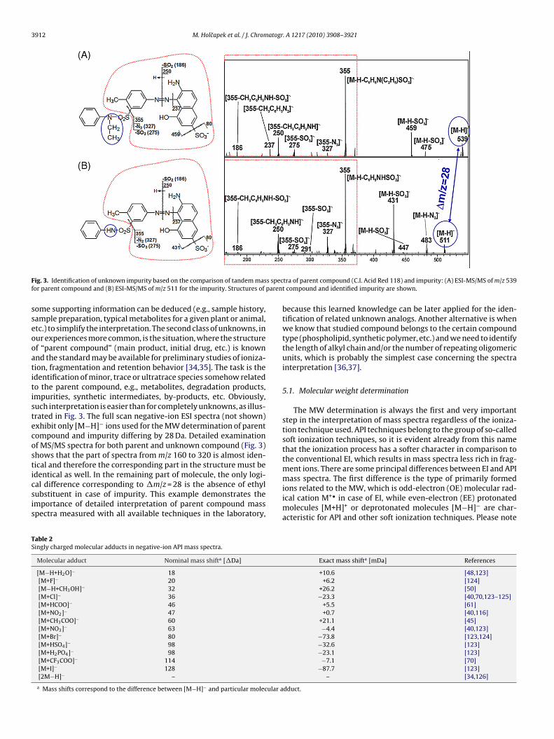
3912 M. Holcapek et al. / J. Chromatogr. A 1217 (2010) 3908–3921
F s specf paren
sseooatitistecosticsis
TS
ig. 3. Identification of unknown impurity based on the comparison of tandem masor parent compound and (B) ESI-MS/MS of m/z 511 for the impurity. Structures of
ome supporting information can be deduced (e.g., sample history,ample preparation, typical metabolites for a given plant or animal,tc.) to simplify the interpretation. The second class of unknowns, inur experiences more common, is the situation, where the structuref “parent compound” (main product, initial drug, etc.) is knownnd the standard may be available for preliminary studies of ioniza-ion, fragmentation and retention behavior [34,35]. The task is thedentification of minor, trace or ultratrace species somehow relatedo the parent compound, e.g., metabolites, degradation products,mpurities, synthetic intermediates, by-products, etc. Obviously,uch interpretation is easier than for completely unknowns, as illus-rated in Fig. 3. The full scan negative-ion ESI spectra (not shown)xhibit only [M−H]− ions used for the MW determination of parentompound and impurity differing by 28 Da. Detailed examinationf MS/MS spectra for both parent and unknown compound (Fig. 3)hows that the part of spectra from m/z 160 to 320 is almost iden-ical and therefore the corresponding part in the structure must be
dentical as well. In the remaining part of molecule, the only logi-al difference corresponding to �m/z = 28 is the absence of ethylubstituent in case of impurity. This example demonstrates themportance of detailed interpretation of parent compound masspectra measured with all available techniques in the laboratory,able 2ingly charged molecular adducts in negative-ion API mass spectra.
Molecular adduct Nominal mass shifta [�Da]
[M−H+H2O]− 18[M+F]− 20[M−H+CH3OH]− 32[M+Cl]− 36[M+HCOO]− 46[M+NO2]− 47[M+CH3COO]− 60[M+NO3]− 63[M+Br]− 80[M+HSO4]− 98[M+H2PO4]− 98[M+CF3COO]− 114[M+I]− 128[2M−H]− –
a Mass shifts correspond to the difference between [M−H]− and particular molecular a
tra of parent compound (C.I. Acid Red 118) and impurity: (A) ESI-MS/MS of m/z 539t compound and identified impurity are shown.
because this learned knowledge can be later applied for the iden-tification of related unknown analogs. Another alternative is whenwe know that studied compound belongs to the certain compoundtype (phospholipid, synthetic polymer, etc.) and we need to identifythe length of alkyl chain and/or the number of repeating oligomericunits, which is probably the simplest case concerning the spectrainterpretation [36,37].
5.1. Molecular weight determination
The MW determination is always the first and very importantstep in the interpretation of mass spectra regardless of the ioniza-tion technique used. API techniques belong to the group of so-calledsoft ionization techniques, so it is evident already from this namethat the ionization process has a softer character in comparison tothe conventional EI, which results in mass spectra less rich in frag-ment ions. There are some principal differences between EI and API
mass spectra. The first difference is the type of primarily formedions related to the MW, which is odd-electron (OE) molecular rad-ical cation M+• in case of EI, while even-electron (EE) protonatedmolecules [M+H]+ or deprotonated molecules [M−H]− are char-acteristic for API and other soft ionization techniques. Please noteExact mass shifta [mDa] References
+10.6 [48,123]+6.2 [124]
+26.2 [50]−23.3 [40,70,123–125]
+5.5 [61]+0.7 [40,116]
+21.1 [45]−4.4 [40,123]
−73.8 [123,124]−32.6 [123]−23.1 [123]−7.1 [70]
−87.7 [123]– [34,126]
dduct.

M. Holcapek et al. / J. Chromatogr. A 1217 (2010) 3908–3921 3913
Table 3Typical fragmentation behavior observed in MS and MS/MS spectra for individual functional groups.
Functional groupa Nominal mass shiftb [�Da] Exact mass shiftb [mDa] Fragment/product ions References
Phosphate(RPO4H2)
+96 −38.8 [M−H–HPO3]− [60][M−H–H3PO4]− [60][H2PO4]− [59–61,64][PO3]− [59–61,64]
Phosphonate(RPO3H2)
+80 −33.7 [H2PO3]− [63][PO3]− [62,63][PO2]− [62,63]
Sulfate(RSO4H)
+96 −48.3 [M−H–SO3]− [33,43][M−H–H2SO4]− [33,43][HSO4]− [33,43]
Sulfonic acid (RSO3H) +80 −43.2 [M−H–SO2]− [31,33,43,67,74,101][M−H–SO3]− [31,33,43,67,74,101][SO3]−• [33,43][SO2]−• [43]
Sulfoxide (R1SO2R2) +64 −38.1c [M+H–OH]+• [68][M+H–Ri]+• [68]
Nitrate(RNO3)
+61 −20.0 [M−H–NO]−• [116][M−NO2]− [40][M−H–NO2]−• [40,116][M−H–ONO2]−• [116]
Nitro(RNO2)
+45 −14.9 [M+H–OH]+• [67,104,127][M–OH]− [116][M+H–H2O]+ [72][M+H–NO]+• [67,127][M+H–NO2]+• [67,101,104,127][M–NO]− [71,116][M−H–NO]−• [43,72,74][M−H–OH]−• [67][M−H–HNO]− [67][M–NO2]− [71,116][M−H–NO2]−• [67,72,74][M−H–HNO2]− [67,73,128][NO2]− [72]
Nitroso (RNO) +29 −9.8 [M+H–NO]+ [46,75,129]
N-oxide (RN+O−) +29 −9.8 [M+H–O]+ [77–83][M+H–OH]+• [34,79,81,99,101][M+H–H2O]+ [79,80]
Azo(R1N = NR2)
+28 +6.1c [M+H–N2]+ [43,86,103][M+H–Ri]+
[M+H–RiNH]+
[M+H–RiN2]+
Amide, alkylamide (R1CONH2, R1CONHR2,R1CONR2R3)
+43 +5.8 [M+H–NH3]+ [M+H–R2NH2]+ [67,85][M+H–R2R3NH]+
Amine (R1NH2,R1NHR2, R1NR2R3)
+15 +10.9 [M+H–NH3]+ [67,86,88,99,101,130–133][M+H–R2NH2]+
[M+H–R2R3NH]+
[M+H–HCN]+ [67,86]
Nitrile (RCN) +25 −4.8 [M+H–HCN]+ [67,86]
Hydroperoxide(ROOH)
+32 −10.2 [M+H–H2O]+ [105,106][M+H–H2O2]+ [105,106]
Epoxides (RO) +16 −5.1 [M+H–H2O]+ [105]
Carboxylicacid(RCOOH)
+44 −10.2 [M+H–H2O]+ [67][M+H–CO2]+ [67,96][M+H–H2O–CO]+ [41,67][M−H–CO2]− [35,41,43,67,74,92,93,96,101]
Alcohol (ROH) +16 −5.1 [M+H–H2O]+ [67,101]
Phenol(ROH)
+16 −5.1 [M+H–H2O]+ [90,96,97][M+H–CO]+ [103][M−H–H2O]− [33,43,134][M−H–CO]− [43,103]
Methoxy(ROCH3)
+30 +10.6 [M+H–CH3]+• [86,97–100][M+H–CH3O]+• [100,101][M+H–CH3OH]+ [86,97,98][M+H–HCOH]+ [97,102]

3914 M. Holcapek et al. / J. Chromatogr. A 1217 (2010) 3908–3921
Table 3 (Continued )
Functional groupa Nominal mass shiftb [�Da] Exact mass shiftb [mDa] Fragment/product ions References
Ester(R1COOR2)
+44 −10.2c [M+H–R2OH]+ [67,99,101,131,135][M+H–R2OH–CO]+ [67,135]
Ketone(R1COR2)
+28 −5.1c [M+H–H2O]+ [104][M+H–CO]+ [67,97,104][RiCO]+ [67]
Aldehyde(RCHO)
+28 −5.1 [M+H–CO]+ [67][M−H–CO]− [67]
Halides(RX)
+18 (F), +34 (Cl),+78 (Br), +126 (I)
−9.4 [M+H–X]+• Cl—[67,101,104]−39.0 Br—[67,101]−89.5 [M+H–HX]+ F—[67,99,101]
−103.4 Cl—[67,84,102,104]Br—[43]
[M−H–X]−• Br—[43]I—[45]
[M−H–HX]− Cl—[43,67,101,128,136]Br—[43,136]
[I]− [45,102][Br]− [43,45,102][Cl]− [43,102]
a i
functf = NO2
and k
ti(a
spw(hwmthptai
[idasa(nbs
Dwadpareamc
R or R means alkyl/aryl.b Mass shifts correspond to the difference between the molecule with a particular
or sulfonic acid = RSO3H–RH = SO3 = 80 or the mass shift for nitro group = RNO2–RHc For R1XR2 functional groups (sulfoxide R1SO2R2, azo R1N = NR2, ester R1COOR2
hat terms “protonated molecular ions” and “deprotonated molecularons” are incorrect and their use is discouraged [38]. In addition tode)protonated molecules, there are some characteristic moleculardducts useful for the verification of MW determination.
In the positive-ion mode (Table 1), the most typical adduct isodiated molecule [M+Na]+ often accompanied by less abundantotassiated molecule [M+K]+. The relative abundance of the adductith ammonium ion [M+NH4]+ strongly depends on the presence
or absence) of ammonium additives in the mobile phase and theistory of ammonium use on the particular instrument. Adductsith other inorganic cations (e.g., Li, Ag and less frequently otheretal ions) can be generated, when these ions are added to the solu-
ion to support the formation of desired ions. Some types of adductsave special applications, such as the determination of double bondositions in long fatty acid chains from the characteristic fragmen-ation of lithium adducts [39]. Other special application of metal iondducts is the fragmentation of ternary complexes with the copperon used for the chiral recognition [25].
In the negative-ion mode (Table 2), the typical base peakM−H]− may be in certain cases accompanied or replaced by adductons with small inorganic ions. The presence and relative abun-ances of these adducts usually strongly depend on the presencend concentration of these anionic species in the solution, so theelective adduct formation can be supported by adding the selectednion to the solution. The special application is the use of chlorideor other small inorganic anions) to determine MWs of aliphaticitrates used as explosives [40], where no MW information cane obtained without this interesting hint. In case of amines andubstituted amines, the formation of chloride adduct is common.
Other less common ions can occur in both polarity modes.imeric or in general polymeric ions are common especially in casehere the analyte concentration is too high or the compound has an
ffinity to form dimeric or polymeric ions [41]. The most commonimeric ions are [2M+H]+ or [2M−H]− depending on the measuredolarity, but other adducts analogous to monomeric ions can belso expected, e.g., with sodium ion (Table 1). Other possible MW
elated ions are doubly and multiply charged ions, which can beasily determined by the fact that distances between isotopic peaksre equal to one half for doubly charged ions or in general 1/n forultiply charged ions with n charges. Moreover, peaks of multiplyharged ions occur at m/z corresponding to (MW + nH)/n. Except
ional group and the same molecule but without any substituent, e.g., the mass shift–H = 45.
etone R1COR2), mass shifts are compared to the parent molecule R1R2.
for biomacromolecules not covered in this review, typical exam-ples for small molecules are polyaromatic compounds providingdoubly charged ions in the positive-ion mode and polysulfonatedcompounds providing multiply charged ions in the negative-ion ESI[32,42,43]. Non-covalent adducts with solvents coming from themobile phase usually have low to negligible relative abundanceswith some exceptions. The most common adducts are with ace-tonitrile [31,44–46], methanol [45,47–50] or water [45,48]. Theimportance of these adducts should not be underestimated regard-less of low relative abundances, because if they are positivelydetected and correctly interpreted, then they serve as a valuableconfirmation of MW determination.
The last example of unusual MW related ions is the formation ofOE molecular ions, which is known for example from the negative-ion APCI of compounds containing some functional groups, such asnitro group(s) on the polyaromatic system (preferably heterocyclescontaining nitrogen or sulfur). It is supposed that the electron cap-ture mechanism is responsible for the formation of M−• ions, butthe situation can be rather complex and more structural subunitsmay contribute to this mechanism [51,52]. For positive-ion APCIand ESI modes, the formation of molecular radicals M+• is knownfor highly conjugated systems found in polyaromatic compounds[17,43,52–55].
5.2. Typical fragmentation behavior for individual functionalgroups
Table 3 lists common functional groups sorted approximatelyin the order of their influence on the fragmentation and clusteredin groups according to the type of heteroatom: (1) phosphorouscontaining functional groups (phosphate, phosphonate), (2) sulfur-containing functional groups (sulfate, sulfonic acid, sulfoxide),(3) nitrogen containing functional groups (nitrate, nitro, nitroso,N-oxide, azo, amide, alkylamide, amine, nitrile), (4) oxygen con-taining functional groups (hydroperoxide, epoxide, carboxylic acid,alcohol, phenol, methoxy, ester, ketone, aldehyde), (5) halogen sub-
stituents, and (6) other structural types (alkyl/aryl substitutionon the aromatic ring, polycyclic aromatic hydrocarbons, alkene,alkyne). In general, the highest impact on the fragmentation hasfunctional groups formally derived from strong inorganic acids, e.g.,phosphate can be considered as a monoester of phosphoric acid,
M. Holcapek et al. / J. Chromatogr. A 1217 (2010) 3908–3921 3915
F e funp id, an
softbbtosttis
ig. 4. Characteristic fragmentation behavior for a hypothetical molecule with onhosphate, (B) sulfate, (C) sulfonic acid, (D) azo, (E) nitro, (F) nitro, (G) carboxylic ac
ulfate as the monoester of sulfuric acid and nitrate as the esterf nitric acid to list three functional groups with the most distinctragmentation. We have tried to exclude cases, where the fragmen-ation behavior of one functional group is substantially influencedy other functional group in the molecule to present a clear linketween the functional group and its fragmentation. In practice,he presence of more functional groups and their mutual effectsn the fragmentation are very common and highly complex, butuch discussion is out of the scope of this introductory review on
he interpretation of API mass spectra. In accordance with Ref. [56],he term “fragment ion” is used here to describe all ions originat-ng from the fragmentation in the full scan mode and tandem masspectra to simplify the discussion. Fig. 4 illustrates the characteristicctional group for negative-ion (A, B, C, F, G) or positive-ion (D, E, H) modes: (A)d (H) methoxy group.
mass spectra for hypothetical molecules containing single func-tional group for a better visualization of fragmentation behaviordescribed in Table 3. In practice, the relative abundances of indi-vidual ions may differ significantly and some fragment ions may beabsent depending on the structure of whole molecule, especiallyother functional groups. Fig. 5 shows proposals for mechanismsof common NLs and the formation of fragment ions described inthe following chapters and Table 3. These mechanisms are pro-posed based on the knowledge of starting point (i.e., [M+H]+ or
[M−H]− ions depending on the polarity mode) and final productions (Table 3) taking into account basic fragmentation mechanismsdescribed for EI [1]. Some mechanisms are not listed, because anal-ogous mechanisms can be found, e.g., the NL of ROH from ester has
3916 M. Holcapek et al. / J. Chromatogr. A 1217 (2010) 3908–3921
Fig. 5. Proposed mechanisms of characteristic NLs and the formation of fragment ions for selected functional groups in the negative-ion mode (A–F and L) from [M−H]− iona ) alkyl( sulfoN peroxo H from
to
5
cpocbtnrTca[(
5
fo
nd in the positive-ion mode (G–K and M–P) from [M+H]+ ion, R means (substitutedC) NL of SO3 from sulfate, (D) formation of [HSO4]− from sulfate, (E) NL of SO3 fromL of NH3 from amine, (I) NL of H2O from hydroperoxide, (J) NL of H2O2 from hydrof H2O from phenol or alcohol, (N) NL of CH3
• from methoxy group, (O) NL of CH3O
he identical mechanism as the NL of H2O from alcohol (Fig. 5M),nly hydrogen is replaced by R.
.2.1. Phosphorous containing functional groupsOrganophosphorous compounds are common especially in the
hemistry of phosphorylated proteins (out of scope of this review),hospholipids [57] and pesticides [58]. In this review, we focusnly on phosphate and phosphonate monoesters. Diesters are veryommon for phospholipids, but their fragmentation is governedy the type of other substituent of the phosphate group [56]. Dueo the presence of two acidic protons in phosphate and phospho-ate monoesters and quite labile character of these groups, the onlyecommended ionization mode is the negative-ion ESI [59–64].he characteristic ions are small negatively charged phosphorousontaining fragment ions: [PO3]− for both phosphates [59–61,64]nd phosphonates [62,63], [H2PO4]− for phosphates [59–61,64],H2PO3]− and [PO2]− for phosphonates [62,63]. The neutral lossNL) of HPO3 [60] and H3PO4 [60] from [M−H]− can occur as well.
.2.2. Sulfur-containing functional groupsSulfur-containing functional groups (especially sulfates and sul-
onic acids) have a very high impact on the fragmentation behaviorf compounds carrying these functional groups. It is typical even
or aryl: (A) NL of HPO3 from phosphate, (B) formation of [H2PO4]− from phosphate,nic acid, (F) formation of [SO3]−• from sulfonic acid, (G) NL of NH3 from amide, (H)ide, (K) NL of H2O from carboxylic acid, (L) NL of CO2 from carboxylic acid, (M) NLmethoxy group, and (P) NL of HX from halides.
for compounds with multiple different functional groups that thefragmentation process starts from the NLs associated with thesetwo groups, i.e., the NL of H2SO4 for sulfates [33,43], SO3 for bothsulfates and sulfonic acids [33,43,65] and SO2 for sulfonic acids [43].The complementary ions [HSO4]− and [SO3]−• are often observedin the spectra as well, rarely also low abundant [SO2]−• for sulfonicacid. The NL of SO2 has been also reported for arylsulfonyl esters[66]. It is interesting to note that the radical ion [SO3]−• is highlydiagnostic, while its EE analog [HSO3]− is not common in the spec-tra. The negative charge of both sulfate and sulfonic groups logicallydetermines the use of negative-ion ESI mode, while negative-ionAPCI (or APPI) can be applied with a reduced sensitivity only forcompounds with up to two sulfonic acid groups in the molecule[42]. The positive-ion mode is not convenient and only weak signalsfor monosulfonic acids containing other proton-acceptor groupscan be expected, e.g., dyes containing hydroxyl, amino group oranthraquinone skeleton [42,67]. The important group of water sol-uble organic dyes carries multiple anionic groups (sulfonic acid,
carboxylic acid and sulfate). Their negative-ion ESI spectra showthe series of multiply charged negative-ions [M−xH]x− and theirsodiated adducts [M−(x + y)H + yNa]x− [31–33,42,43,65]. The totalnumber of acid groups is equal to the highest observed charge orthe highest number of acid protons replaceable by sodium ions in
atogr. A 1217 (2010) 3908–3921 3917
[bH
5
gEtgznnc
Etcsmhalaa(it
iocdwcfcbocTvfqN
ntmtooIsvtTNhotTbgd
M. Holcapek et al. / J. Chrom
M−(x + y)H + yNa]x− series. The radical losses of OH• and R• haveeen published for sulfoxides [68]. For thiols, the only known NL is2S similarly as for EI spectra [1].
.2.3. Nitrogen containing functional groupsThe first thing related to the nitrogen containing functional
roups must be the knowledge of so-called nitrogen rule. If theE rule [69] is valid without any exception for API mass spectra,hen all ions with even m/z values have an odd number of nitro-en atoms, while ions with odd m/z values have even number orero nitrogen atoms. Unfortunately, exceptions from EE rule can-ot be neglected, because OE ions are relatively common for someitrogen containing groups, e.g., nitro, nitroso, N-oxide or nitrogenontaining heterocyclic compounds.
The nitrate is rather labile group typical for explosives.xtremely strong tendency of this functional group for the spon-aneous fragmentation even at the softest possible ESI conditionsan result in the absence of [M−H]− ions in the full scan spectrahowing only fragment ions [NO3]− and [NO2]− in the negative-ionode without any information on the MW. There is one interesting
int applied in the negative-ion APCI that (poly)nitrated aliphaticnd aromatic explosives can be detected in the form of molecu-ar adducts with small inorganic anions, such as chloride, bromide,cetate, formate [40,70]. The source of halide ions can be either theddition of small amount of inorganic salt or chlorinated solventfor example CH2Cl2 or CHCl3). The trace concentration of theseons even in chromatographic solvents is often sufficient to formhese stable adducts without the deliberate addition.
Nitro compounds have a quite complex fragmentation and ion-zation behavior starting from the initial MW related ions. It is quiteften that negative-ion API spectra of nitro and especially polynitroompounds show the radical ion M−• [70,71], but the formation ofeprotonated molecule [M−H]− is also known depending on thehole structure of particular nitro compound [72–74]. The typi-
al NLs are radicals NO• and NO2• often occurring as the starting
ragmentation process on condition that no phosphorous or sulfur-ontaining functional groups are present. These NLs can occur foroth M−• and [M−H]− initial ions, which brings a complex surveyf possible fragment ions for nitro compounds (see Table 3). Theomplementary radical ion [NO2]−• can be observed as well [72].he third NL known for the nitro group is the loss of oxygen itself (atery low abundance, if at all), which is something rather unusualor all other functional groups except for N-oxides. Nitroso group isuite rare except for certain applications and the only predictableL is NO [46,75].
N-oxide is a common metabolic oxidation product for someitrogen containing drugs [34,76–78]. The characteristic fragmen-ation of N-oxides is deoxygenation occurring only in the full scan
ass spectra [77–83] and it is explained by the thermal degrada-ion in the APCI source [77]. Some authors reported that the NL ofxygen in APCI is replaced by NLs of OH• and H2O in ESI [77], whilethers [81] observed the loss of oxygen also in ESI mass spectra.n all cases, the [M+H–O]+• is reported only for the full scan masspectra but not for tandem mass spectra. It is likely that the pre-ailing fragmentation pathway strongly depends on the setting ofuning parameters and API technique used for the measurement.he second unusual fragmentation pathway known for tertiary-oxides is the elimination of aldehyde or ketone after Meisen-eimer N–R to N–O rearrangement [78,79]. Another fragmentationf N-oxides (i.e., N,N-dimethylamino-N-oxide) is the cleavage of
he nitrogen–carbon bond resulting in the NL of (CH3)2NOH [34].wo isobaric oxidation metabolites, hydroxylated vs. N-oxide, cane distinguished based on the characteristic fragmentation of bothroups, where the dehydration is typical for hydroxyl, while theeoxygenation is a clear marker of N-oxide [77,79].Fig. 6. Correlation between the fragmentation behavior and multiple functionalgroups (1 sulfo, 1 nitro, 2 hydroxyl and 1 azo groups) for dye C.I. Mordant Black15 in the negative-ion MS/MS spectrum of [M−H]− .
The azo group is the characteristic functional group for a widerange of organic dyes, often accompanied by other functionalgroups including sulfonic acid(s) because of a better solubility inwater. There are several possibilities of cleavages of azo grouptogether or without the transfer of hydrogen atom(s), i.e., cleavagesbetween nitrogen and neighboring carbon atoms and between twonitrogen atoms in the azo group [43], as illustrated on the examplein Fig. 6. The rearrangement loss of N2 is known for both EI and APIspectra [43]. Azo compounds without any other functional groupprovide signal only in the positive-ion mode, but in practice organicazo dyes carrying anionic functional groups are measured in thenegative-ion ESI mode with the observation of the rearrangementloss of N2 as well [31,33,43,84].
Amide compounds without other electronegative functionalgroups are suitable for the measurement in the positive-ion mode,where the characteristic NL of NH3 (RNH2 for alkylamides andR1NHR2 for dialkylamides) for amides results in the formation ofstable carbonyl ions [85]. This is an analogy of peptide cleavageleading to the formation of y and b ions. The same NL of NH3 can beexpected for amines, but in fact this loss is typical only for aliphaticamines, but not so common for aromatic amines, where it occursonly in case of absence of other functional groups [67,86]. TheNL of HCN is characteristic for both nitriles [67,87] and aromaticamines without other functional groups [67,86]. It is interesting tonote that very similar behavior is known for aliphatic alcohols vs.phenols, where H2O loss is common for alcohols but rare for phe-nols. Substituted amines provide losses of alkyl- and dialkylamines,such as methyl amine [34,88], CH3N CH2 [34,89], dimethylamine[34,88–90]. Nitrile compounds containing other electronegativesubstituents can provide the signal in the negative-ion mode withcommon NL of HCN or even the nitrile ion [CN]− can be detected[91]. For compounds with multiple functional groups, the fragmen-tation pathways of amides, amines and nitriles can be suppressedby other competitive fragmentation reactions.
5.2.4. Oxygen containing functional groupsThe fragmentation behavior of oxygen containing functional
groups has many common features with EI. Regardless the fact+ −
that the initial ion in API mass spectra is EE [M+H] or [M−H]unlike to OE ion M+• in EI spectra, the resulting fragment ions areoften the same or at least part of them. The strongest effect onthe fragmentation among common oxygen containing functionalgroups has carboxylic acids and alcohols, if we neglect relatively

3 atogr
utgtiikmt
ap[innuaifIoIsen
msgafir
wmgwti[om
ngaottfio
itithtMi(aE
918 M. Holcapek et al. / J. Chrom
nusual hydroperoxides and epoxides. Oxygen containing func-ional groups are often present together with other functionalroups, so it is not easy to recommend the most suitable ioniza-ion mode for individual functional groups. The negative-ion modes more sensitive for carboxylic acids, while the positive-ion modes generally more convenient for other functional groups. Esters,etones and ethers may not provide any signal in the negative-ionode, unless other suitable functional group improves the ioniza-
ion efficiency.Carboxylic acids have quite specific behavior with the char-
cteristic NL of CO2 observed in all types of spectra, i.e., bothositive and negative modes, full scan and tandem mass spectra35,43,92,93]. We know only one common isobaric NL correspond-ng to �m/z = 44, which is the radical NH2CO• occurring for someitrogen containing heterocycles with the carbonyl group next toitrogen [94,95], but this loss never occurs in both polarity modesnlike to the carboxylic acid. In principle, the carboxylic acid canlso provide [HCOO]− ion at m/z 45 in the negative-ion mode, butt is rarely reported due to the fact that the mass range is mostly setrom m/z 50 and for some manufacturers it is the instrument limit.n the positive-ion mode, the typical non-specific NL is water [67],ften followed by the loss of carbon monoxide (H2O + CO) [31,67].t is interesting to note that Levsen et al. [67] excludes the pos-ibility of CO2 NL in the positive-ion mode, while we have somexamples on this fact [92,93,96], but it is less frequent than for theegative-ion mode.
Aliphatic alcohols provides the abundant NL of water in anyeasurement mode as expected, but unfortunately this NL is not
pecific and can occur almost for any oxygen containing functionalroup, but the relative abundance of water loss is the highest forlcohols and always present already in the full scan mode. It isrequently the base peak in tandem mass spectra. The situations rather different for phenols, where the most typical is the rear-angement loss of CO and the loss of water is less common.
Concerning the ether functional group, the fragmentation path-ays should be similar as with EI, however most examples of APIass spectra in the literature deal only with the aromatic methoxy
roup, so we have focused only on this group. Ether compoundsithout other functional group usually do not provide a signal in
he negative-ion mode. There are four possible NLs in the positive-on mode, radicals CH3
• [35,86,97–100] and less frequently CH3O•
100,101] and the neutral molecule of methanol [86,97,98,102]r methanal [97,102]. The presence of OE ions is typical for theethoxy group [58,67].Ester functional group has a relatively low polarity and therefore
ot so high importance on the fragmentation if other more polarroups are present. The characteristic fragmentation is the NL oflcohol (methanol for methylesters, ethanol for ethylesters, etc.)ften followed by the NL of CO. If ester is the only functional group,hen the positive-ion APCI (or APPI) is the most convenient andhe negative-ion mode does not provide any signal. Products of theragmentation in API mass spectra of ketones are identical as for EI,.e., [R1CO]+ and [R2CO]+ [67,103]. Low abundant NL of water mayccur as well [104]. For aldehydes, the only reported NL is CO [67].
Epoxy and especially hydroperoxy groups are not often detectedn organic molecules, because they are known as unstable oxida-ion intermediates. Epoxides are more stable, so their detections more likely. Both groups induce an extensive fragmenta-ion with the typical NL of water for both groups or H2O2 forydroperoxides [105,106]. The extensive fragmentation and alsohe adduct formation may complicate the right determination of
Ws [49]. Their MW can be also determined with the positive-on APCI mode at the special soft setting of tuning parameterslow temperatures and low flow rates) based on [M+H + methanol]+
dducts [49] and by silver-ion adduct formation in the positive-ionSI [107].
. A 1217 (2010) 3908–3921
5.2.5. Halogen substituentsPossible fragmentation pathways for halides are relatively easy
with NLs of HX or radical X•. The probability of radical ionappearance increases in the order F < Cl < Br < I (Table 3). For poly-halogenated compounds, repetitive NLs are observed and the rel-ative abundance of (de)protonated molecules can be significantlydecreased. Halides can be, in principal, measured in both polar-ity modes with all API techniques depending on other functionalgroups present in the molecule. Chlorine (35Cl:37Cl = approximately3:1) and bromine (79Br:81Br = approximately 1:1) atoms havehighly characteristic isotopic doublets enabling their easy identi-fication in individual ions in the spectra, while fluorine (19F) andiodine (127I) are monoisotopic. If more Cl/Br atoms are present inthe molecule, then the ratio of isotopic peaks differing by two unitsis calculated according to the binomial equation. For example fortwo chlorine and one bromine atoms, the equation is (3a + b)2(a + b)and the calculated coefficients are equal to relative abundances ofM: M + 2: M + 4, etc. In the negative-ion mode, anionic halides canbe observed, especially with the quadrupole analyzer [43,45,102].The formation of radical molecular anions M−• has been reportedfor polybrominated diphenyl ethers followed by NLs of Br• and Br2[108] and for brominated polyaromatic hydrocarbons [43]. If thefluorine atom is present next to other functional groups, then theNL of HF is often suppressed.
5.2.6. Other structural typesIt is worthy to comment in this chapter also the fragmentation
behavior of some structural subunits without heteroatoms, such asalkyl/aryl substitution, polycyclic aromatic hydrocarbons and thepresence of double and triple bonds. Very common structural fea-ture is the alkyl or aryl substitution on the aromatic and other cyclicand also acyclic structures. The typical NL for the alkyl substituentis alkene [95,102,109], for example the NL of butene for the butylsubstitution. The NL of alkane is also known [94], if hydrogen is ster-ically available, but based on the literature and own experiences,the NL of alkene is generally preferred. The radical loss of alkyl isreported mainly in the negative-ion APCI [94,95]. For the phenyl (ingeneral aryl) substitution, the logical NL is C6H6 [104] or C6H4 [95],less frequently radical C6H5
• [95]. For organometallic compoundscontaining a carbon–metal bond, the cleavage of this bond result-ing in NLs of alkene and alkane for the alkyl substitution [48,84,92]and C6H6 for the phenyl substitution [48,89,93,102] is typical.
Aromatic compounds with highly conjugated structures (prefer-ably nitrogen [17,52,54] or sulfur [55] containing heterocyclicsystems) have an ability to stabilize an unpaired electron resultingin the molecular radical cation M+• formation [17,35,43,52,54,55],which is typical for polycyclic aromatic hydrocarbons, porphyrins,etc. The ability to delocalize the unpaired electron can be retainedalso for some fragment ions. In practice, there is often a competi-tion between the formation of radical cation M+• and protonatedmolecule [M+H]+ depending on various parameters, such as otherfunctional groups, the alkyl chain length, used solvents, flow rates,the type of instrument, API ionization technique [43,54].
The presence of double or triple carbon–carbon bonds resultsin corresponding shifts of MWs, which may help to determinethe number of unsaturations, but there is no as significant con-sequences for the fragmentation of alkenes and alkynes as forheteroatom containing functional groups. If the position of unsat-uration has to be determined (e.g., for unsaturated fatty acids),then there are several derivation procedures (additions on the dou-ble bond) resulting in the subsequent fragmentation used for the
localization of the double bond due to the characteristic fragmen-tation behavior. These approaches are known for both EI [110] andAPI techniques [111]. The difference between isobaric monoun-saturated and cyclic saturated compounds is not so difficult torecognize, because monounsaturated compounds have very sim-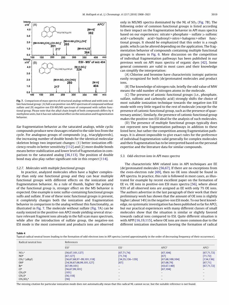
M. Holcapek et al. / J. Chromatogr.
Fig. 7. Comparison of mass spectra of structural analogs without and with ionic sul-fate functional group: (A) full scan positive-ion APCI spectrum of compound withoutstmb
icctscmpb
5
iffoesibietwE
TT
T
ulfate and (B) negative-ion ESI-MS/MS spectrum of compound with sulfate func-ional group. Please note that the alkyl chain length of both compounds differs by 4
ethylene units, but it has not substantial effect on the ionization and fragmentationehavior.
lar fragmentation behavior as the saturated analogs, while cyclicompounds produce new cleavages related to the side loss from theycle. For analogous groups of compounds (e.g., triacylglycerols),he increasing number of double bonds for the identical molecularkeleton brings two important changes: (1) better ionization effi-iency results in better sensitivity [112] and (2) more double bondseans better stabilization and lower level of fragmentation in com-
arison to the saturated analog [36,113]. The position of doubleond may also play rather significant role in this respect [114].
.2.7. Molecules with multiple functional groupsIn practice, analyzed molecules often have a higher complex-
ty than only one functional group and they can bear multipleunctional groups with different effects on the ionization andragmentation behavior. As a rule of thumb, higher the polarityf the functional group is, stronger effect on the MS behavior isxpected. One example is ionic sulfur-containing functional groupsulfo and sulfate. If one of these ionic functional groups is present,t completely changes both the ionization and fragmentationehavior in comparison to the analog without this functionality, as
llustrated in Fig. 7. The molecule without sulfate (Fig. 7A) can beasily ionized in the positive-ion APCI mode yielding several struc-ure relevant fragment ions already in the full scan mass spectrum,hile after the introduction of sulfate group, the negative-ion
SI mode is the most convenient and products ions are observed
able 4ypical radical neutral losses leading to the formation of odd-electron ions in API spectra
Radical neutral loss References
ESI+ ES
NO2• [56,67,101,127] [6
NO• [67,127] [7CH3
• (alkyl) [56,67,68,97–99,101,118] [3OH• [34,56,67,68,99,101,127] [6Br• [67,99,101] –Cl• [56,67,99,101] –CH3O• [101] –CH3S• [56,118] –CH3SO2
• [56] –
he missing citation for particular ionization mode does not automatically mean that this
A 1217 (2010) 3908–3921 3919
only in MS/MS spectra dominated by the NL of SO3 (Fig. 7B). Thefollowing order of common functional groups is listed accordingto their impact on the fragmentation behavior in API mass spectrabased on our experiences: nitrate > phosphate ∼ sulfate � sulfonicacid > carboxylic acid > hydroxyl > nitro > halogens > other func-tional groups. It should be emphasized that this order is a roughguide, which can be altered depending on the application. The frag-mentation behavior of compounds containing multiple functionalgroups is shown in Fig. 6. More discussion on the competitionof individual fragmentation pathways has been published in ourprevious work on API mass spectra of organic dyes [42]. Somegeneral comments are valid in most cases and their knowledgecan simplify the interpretation:
(A) Chlorine and bromine have characteristic isotopic patternseasily recognized for both (de)protonated molecules and productions.
(B) The knowledge of nitrogen rule, briefly the odd value of MWmeans the odd number of nitrogen atoms in the molecule.
(C) The presence of anionic functional groups (i.e., phosphate,sulfate, sulfonic and carboxylic acid) strongly shifts the choice ofmost suitable ionization technique towards the negative-ion ESImode with very little regard to the rest of molecule (except for thepresence of cationic functional group, such as the presence of quar-ternary amine). Similarly, the presence of cationic functional groupmakes the positive-ion ESI ideal for the analysis of such molecules.
(D) The presence of multiple functional groups typically doesnot represent new fragmentation pathways in addition to theselisted here, but rather the competition among fragmentation path-ways. It is almost impossible to give exact rules for the preferenceof individual fragmentation valid generally for complex moleculesand their fragmentation has to be interpreted based on the personalexpertise and the literature data for similar compounds.
5.3. Odd-electron ions in API mass spectra
The characteristic MW related ions in API techniques are EE(de)protonated molecules [56,67]. If there are no exceptions fromthe even-electron rule [69], then no OE ions should be found inAPI spectra. In practice, this rule is followed in most cases, as illus-trated for example by recent excellent paper on the formation ofEE vs. OE ions in positive-ion ESI mass spectra [56], where about93% of all observed ions are assigned as EE with only 7% OE ions.The authors advertise in the last paragraph of their work that theirpreliminary work has shown that the amount of OE ions is slightlyhigher (about 14%) in the negative-ion ESI mode. To our best knowl-edge, no systematic investigation has been published so far for APCI,
but our practical experiences with many different classes of smallmolecules show that the situation is similar or slightly favoredtowards radical ions compared to ESI. Quite different situation iswith APPI [16,19,115], where OE ions are more common due to thedifferent ionization mechanism favoring the formation of radical(sorted approximately in the order of decreasing frequency of their occurrence).
I− APCI+ APCI−
7,71,74] [67,104] [67,71,72]1,74] [67] [71,72]4,35,136–139] [67,68,100,104] [134,136]7] [67,68,79] [67,71]
[43,67,140] [67,71,72][67,104] –– –– –– –
radical NL cannot occur, but the suitable reference is not found.

3 atogr
itbim
tiotsti(TtfnTa[macamicpirera(f
6
nbobTmfgifsbdtopiaaoasmLit
920 M. Holcapek et al. / J. Chrom
ons. Concerning OE ions, APPI is somewhere in between soft ioniza-ion techniques and EI. In certain cases, molecular radical ions cane observed due to the charge exchange mechanism in both polar-
ty modes and the electron capture mechanism in the negative-ionode [16,19,52].Table 4 summarized the most common radical NLs leading
o the formation of OE ions in API spectra sorted approximatelyn the order of their importance based on our experiences andther references as well. In our opinion, the most typical func-ional group for the formation of OE ions is the nitro grouptarting from the formation of the radical molecular ion M−• byhe electron capture mechanism [52,70] and three common rad-cal NLs, NO2
•, NO• and OH• occurring in both polarity modesTable 4). Similar losses are known also for nitrates [70,116].ypically, nitro group characteristic NLs are observed as the ini-ial fragmentation step even for compounds containing multipleunctional groups [43]. The same NL (NO) is known for theitroso group, but this functional group is relatively rare [75].he radical NL is common for the methyl substitution on theromatic ring or other cyclic structures, N-methyl substitution56,67], (poly)methoxylated flavonoids [97,100], drugs containing
ethoxy group [34,35], etc. For alkyl/aryl substitution, NLs of EElkene/arene or alkane are characteristic, but the radical alkyl lossan be also observed, especially in the negative-ion APCI modend for heterocyclic compounds [94,95,109,117]. The presence ofethoxy group sometimes exhibits the second radical CH3O•, but
t is not as common as the methyl radical loss. The analogous radi-al loss is known for CH3S• [118]. The radical loss can also occur foroly- and perhalogenated compounds (Table 4), while the NL of HX
s usually preferred, if sterically available hydrogen is present. Theadical NL of OH• has been reported for certain functional groups,.g., nitro group, N-oxide, sulfoxide, while we have not found anyeport for this radical loss from alcohol, where the NL of water hasstrong preference. If CH3SO2 group is present in the molecule
e.g., pesticides), then the corresponding radical NL leads to theormation of OE ion with relative abundances up to 100% [56].
. Concluding remarks
Nowadays, HPLC–MS and other liquid-phase separation tech-iques coupled to MS are routinely used in analytical laboratories,ut the potential of MS is often not fully explored due to the absencef basic knowledge of mass spectra interpretation partially causedy the lack of appropriate literature for soft ionization techniques.his review describes the basic rules for the interpretation of APIass spectra of small molecules, especially their ionization and
ragmentation behavior related to the type of particular functionalroups. On the other hand, the broad range of functional groupsncluded in our review may cause that the information on particularunctional groups may not be enough detailed. It should be empha-ized that the expertise in the mass spectra interpretation cannote obtained by studying the literature only, but the practical every-ay interpretation of mass spectra is essential. Experiences withhe similar or identical class of compounds either from previouswn measurements or from the literature are invaluable in solvingractical problems. The most sophisticated Fourier transformation
nstruments Orbitrap and ICR with the ultrahigh resolving powernd excellent mass accuracy below 1 ppm provides the most reli-ble data for the structural elucidation of complex structures, butn the other hand even the simplest low resolution quadrupolenalyzer can – in certain cases – be applied for the identification of
impler compounds derived from the parent structure, e.g., drugetabolites, degradation products and technological impurities.imitations of MS in the structure elucidation should be also keptn mind (e.g., positional isomers, enantiomers) and – if possible –he complementary data from other spectral techniques and the
. A 1217 (2010) 3908–3921
knowledge of retention behavior should be used together with thebasic chemical sense. The final step in the unambiguous structuralelucidation should be the comparison with the identical standardeither commercial available or synthesized.
Acknowledgments
This work was supported by the grant project no.MSM0021627502 sponsored by the Ministry of Education, Youthand Sports of the Czech Republic and project no. 203/08/1536sponsored by the Czech Science Foundation. We are obligedto anonymous reviewers for their valuable comments how toimprove this manuscript.
References
[1] F.W. McLafferty, F. Turecek, Interpretation of Mass Spectra, University ScienceBooks, Mill Valley, CA, USA, 1993.
[2] F.G. Kitson, B.S. Larsen, C.N. McEwen, Gas Chromatography and Mass Spec-trometry: A Practical Guide, Academic Press, Inc., San Diego, CA, USA, 1996.
[3] R.M. Smith, Understanding Mass Spectra: A Basic Approach, John Wiley &Sons, Inc., Hoboken, NJ, USA, 2005.
[4] R. Jirásko, M. Holcapek, Mass Spectrom. Rev., submitted for publication.[5] J.C. Holmes, F.A. Morrell, Appl. Spectrosc. 11 (1957) 86.[6] V.L. Talroze, G.V. Karpov, I.G. Gorodetskii, V.E. Skurat, Russ. J. Phys. Chem.
USSR 42 (1968) 1658.[7] C.R. Blakley, M.J. McAdams, M.L. Vestal, J. Chromatogr. 158 (1978) 261.[8] M.L. Alexandrov, L.N. Gall, N.V. Krasnov, V.I. Nikolaev, V.A. Pavlenko, V.A.
Shkurov, Dokl. Akad. Nauk. SSSR 277 (1984) 379.[9] M. Yamashita, J.B. Fenn, J. Phys. Chem. 88 (1984) 4451.
[10] J.B. Fenn, M. Mann, C.K. Meng, S.F. Wong, C.M. Whitehouse, Science 246 (1989)64.
[11] http://nobelprize.org/ (downloaded 27.10.09).[12] F. Hillenkamp, M. Karas, R.C. Beavis, B.T. Chait, Anal. Chem. 63 (1991) A1193.[13] A. Makarov, Anal. Chem. 72 (2000) 1156.[14] A. Makarov, M. Scigelová, J. Chromatogr. A, this issue.[15] R. Elviri, F. Speroni, M. Careri, A. Mangia, L. Sanità Di Toppi, M. Zottini, J.
Chromatogr. A, this issue.[16] D.B. Robb, M.W. Blades, Anal. Chim. Acta 627 (2008) 34.[17] J.M. Purcell, R.P. Rodgers, C.L. Hendrickson, A.G. Marshall, J. Am. Soc. Mass
Spectrom. 18 (2007) 1265.[18] L.C. Short, K.A. Hanold, S.S. Cai, J.A. Syage, Rapid Commun. Mass Spectrom. 21
(2007) 1561.[19] D.R. Smith, D.B. Robb, M.W. Blades, J. Am. Soc. Mass Spectrom. 20 (2009) 73.[20] E. de Hoffmann, V. Stroobant, Mass Spectrometry: Principles and Applications,
John Wiley & Sons Ltd., Chichester, England, 2007.[21] R. Jirásko, M. Holcapek, L. Kolárová, M. Nádvorník, A. Popkov, J. Mass Spec-
trom. 43 (2008) 1274.[22] R. Jirásko, M. Holcapek, E. Rosenberg, Int. J. Mass Spectrom. 280 (2009) 198.[23] R.E. Ardey, Liquid Chromatography–Mass Spectrometry: An Introduction,
John Wiley & Sons Ltd., Chichester, England, 2003.[24] W.M.A. Niessen, Liquid Chromatography–Mass Spectrometry, Taylor & Fran-
cis Inc., Boca Raton, FL, USA, 2006.[25] W.A. Tao, D.X. Zhang, E.N. Nikolaev, R.G. Cooks, J. Am. Chem. Soc. 122 (2000)
10598.[26] K.A. Schug, W. Lindner, J. Sep. Sci. 28 (2005) 1932.[27] M. Holcapek, H. Velínská, M. Lísa, P. Cesla, J. Sep. Sci. 23 (2009) 3672.[28] M. Holcapek, K. Volná, P. Jandera, L. Kolárová, K. Lemr, M. Exner, A. Církva, J.
Mass Spectrom. 39 (2004) 43.[29] B.K. Matuszewski, M.L. Constanzer, C.M. Chavez-Eng, Anal. Chem. 75 (2003)
3019.[30] I. Marchi, V. Viette, F. Badoud, M. Fathi, M. Saugy, S. Rudaz, J.L. Veuthey, J.
Chromatogr. A, this issue.[31] D. Ansorgová, M. Holcapek, P. Jandera, J. Sep. Sci. 26 (2003) 1017.[32] M. Holcapek, P. Jandera, P. Zderadicka, J. Chromatogr. A 926 (2001) 175.[33] D. Vanerková, A. Sakalis, M. Holcapek, P. Jandera, A. Voulgaropoulos, Rapid
Commun. Mass Spectrom. 20 (2006) 2807.[34] P. Císar, M. Nobilis, Z. Vybíralová, M. Holcapek, L. Kolárová, M. Pour, J. Kunes,
J. Klimes, J. Pharm. Biomed. Anal. 37 (2005) 1059.[35] M. Nobilis, J. Kopecky, J. Kvetina, Z. Svoboda, M. Pour, J. Kunes, M. Holcapek,
L. Kolárová, J. Pharm. Biomed. Anal. 32 (2003) 641.[36] M. Holcapek, P. Jandera, P. Zderadicka, L. Hrubá, J. Chromatogr. A 1010 (2003)
195.[37] P. Jandera, M. Holcapek, G. Theodoridis, J. Chromatogr. A 813 (1998) 299.[38] K.K. Murray, J. Chromatogr. A, this issue.
[39] F.F. Hsu, J. Turk, J. Am. Soc. Mass Spectrom. 19 (2008) 1673.[40] X.M. Zhao, J. Yinon, J. Chromatogr. A 977 (2002) 59.[41] M. Nobilis, M. Holcapek, L. Kolárová, J. Kopecky, M. Kunes, Z. Svoboda, J.Kvetina, J. Chromatogr. A 1031 (2004) 229.[42] M. Holcapek, P. Jandera, J. Prikryl, Dyes Pigm. 43 (1999) 127.[43] M. Holcapek, K. Volná, D. Vanerková, Dyes Pigm. 75 (2007) 156.

atogr.
[138] R. Vessecchi, C.A. Carollo, J.N.C. Lopes, A.E.M. Crotti, N.P. Lopes, S.E. Galembeck,J. Mass Spectrom. 44 (2009) 1224.
M. Holcapek et al. / J. Chrom
[44] M. Holcapek, H. Virelizier, J. Chamot-Rooke, P. Jandera, C. Moulin, Anal. Chem.71 (1999) 2288.
[45] D. Baglio, D. Kotzias, B.R. Larsen, J. Chromatogr. A 854 (1999) 207.[46] D. Tsikas, M. Raida, J. Sandmann, S. Rossa, W.G. Forssmann, J.C. Frolich, J.
Chromatogr. B 742 (2000) 99.[47] G.C. Kite, C.A. Stoneham, N.C. Veitch, B.K. Stein, K.E. Whitwell, J. Chromatogr.
B 838 (2006) 63.[48] R. Jirásko, M. Holcapek, L. Kolárová, T.S.B. Baul, J. Mass Spectrom. 42 (2007)
918.[49] D. Rondeau, R. Vogel, J.C. Tabet, J. Mass Spectrom. 38 (2003) 931.[50] S. Grujic, T. Vasiljevic, M. Lausevic, T. Ast, Rapid Commun. Mass Spectrom. 22
(2008) 67.[51] R. Rodriguez-Acuna, E. Brenne, F. Lacoste, J. Agric. Food Chem. 56 (2008) 6241.[52] M. Holcapek, M. Lísa, K. Volná, N. Almonasy, J. Prikryl, J. Mass Spectrom. 42
(2007) 1645.[53] S.M. van Leeuwen, H. Hayen, U. Karst, Anal. Bioanal. Chem. 378 (2004) 917.[54] M. Schafer, M. Drayss, A. Springer, P. Zacharias, K. Meerholz, Eur. J. Org. Chem.
(2007) 5162.[55] L.C. Herrera, L. Ramaley, J.S. Grossert, Rapid Commun. Mass Spectrom. 23
(2009) 571.[56] E.M. Thurman, I. Ferrer, O.J. Pozo, J.V. Sancho, F. Hernandez, Rapid Commun.
Mass Spectrom. 21 (2007) 3855.[57] M. Pulfer, R.C. Murphy, Mass Spectrom. Rev. 22 (2003) 332.[58] W.M.A. Niessen, J. Chromatogr. A, this issue.[59] H. De Brabandere, N. Forsgard, L. Israelsson, J. Petterson, E. Rydin, M. Walde-
back, P.J.R. Sjoberg, Anal. Chem. 80 (2008) 6689.[60] F.F. Hsu, J. Turk, J. Am. Soc. Mass Spectrom. 11 (2000) 986.[61] C. Antonio, T. Larson, A. Gilday, I. Graham, E. Bergstroem, J. Thomas-Oates, J.
Chromatogr. A 1172 (2007) 170.[62] K. Huikko, T. Kotiaho, J. Yli-Kauhaluoma, R. Kostiainen, J. Mass Spectrom. 37
(2002) 197.[63] L. Goodwin, J.R. Startin, D.M. Goodall, B.J. Keely, Rapid Commun. Mass Spec-
trom. 17 (2003) 963.[64] H. Hernandez, Rapid Commun. Mass Spectrom. 10 (1996) 1543.[65] K. Volná, M. Holcapek, L. Kolárová, K. Lemr, J. Cáslavsky, P. Kacer, J. Poustka,
M. Hubálek, Rapid Commun. Mass Spectrom. 22 (2008) 101.[66] L. Xu, D.C. Spink, Anal. Biochem. 375 (2008) 105.[67] K. Levsen, H.M. Schiebel, J.K. Terlouw, K.J. Jobst, M. Elend, A. Preib, H. Thiele,
A. Ingendoh, J. Mass Spectrom. 42 (2007) 1024.[68] P. Wright, A. Alex, D. Gibson, R. Jones, P. Macrae, Rapid Commun. Mass Spec-
trom. 19 (2005) 2005.[69] M. Karni, A. Mandelbaum, Org. Mass Spectrom. 15 (1980) 53.[70] I. Cotte-Rodriguez, Z. Takats, N. Talaty, H.W. Chen, R.G. Cooks, Anal. Chem. 77
(2005) 6755.[71] X.F. Fu, Y. Zhang, S.H. Shi, F. Gao, D.W. Wen, W. Li, Y.P. Liao, H.W. Liu, Rapid
Commun. Mass Spectrom. 20 (2006) 2906.[72] J. Dron, E. Abidi, I. El Haddad, N. Marchand, H. Wortham, Anal. Chim. Acta 618
(2008) 184.[73] H. Kangas, R. Franzen, J. Tois, J. Taskinen, R. Kostiainen, Rapid Commun. Mass
Spectrom. 13 (1999) 1680.[74] A.C. Schmidt, R. Herzschuh, F.M. Matysik, W. Engewald, Rapid Commun. Mass
Spectrom. 20 (2006) 2293.[75] S. Diem, B. Gutsche, M. Herderich, J. Agric. Food Chem. 49 (2001) 5993.[76] J. Maláková, M. Nobilis, Z. Svoboda, M. Lísa, M. Holcapek, J. Kvetina, J. Klimes,
V. Palicka, J. Chromatogr. B 853 (2007) 265.[77] I.M. Peiris, W. Lam, S. Michael, R. Ramanathan, J. Mass Spectrom. 39 (2004)
600.[78] S.G. Ma, Y. Xu, M.G. Shou, Rapid Commun. Mass Spectrom. 23 (2009) 1446.[79] S.G. Ma, S.K. Chowdhury, K.B. Alton, Anal. Chem. 77 (2005) 3676.[80] R. Ramanathan, A.D. Su, N. Alvarez, N. Blumenkrantz, S.K. Chowdhury, K.
Alton, J. Patrick, Anal. Chem. 72 (2000) 1352.[81] X.S. Miao, R.E. March, C.D. Metcalfe, Int. J. Mass Spectrom. 230 (2003) 123.[82] X.Y. Qi, B. Wu, Y.Y. Cheng, H.B. Qu, Rapid Commun. Mass Spectrom. 23 (2009)
291.[83] Y.Y. Wang, X.Y. Chen, Q. Li, D.F. Zhong, Rapid Commun. Mass Spectrom. 22
(2008) 1843.[84] T.S.B. Baul, A. Mizar, A.K. Chandra, X.Q. Song, G. Eng, R. Jirásko, M. Holcapek,
D. de Vos, A. Linden, J. Inorg. Biochem. 102 (2008) 1719.[85] J. Canezin, A. Cailleux, A. Turcant, A. Le Bouil, P. Harry, P. Allain, J. Chromatogr.
B 765 (2001) 15.[86] S.P. Yang, J. Han, Y.F. Huan, Y.J. Cui, X. Zhang, H.W. Chen, H.W. Gu, Anal. Chem.
81 (2009) 6070.[87] A. Kamel, P. Jeanville, K. Colizza, L.E. J-Rivera, J. Am. Soc. Mass Spectrom. 19
(2008) 1579.[88] C. Joyce, W.F. Smyth, V.N. Ramachandran, E. O’Kane, D.J. Coulter, J. Pharm.
Biomed. Anal. 36 (2004) 465.[89] M. Holcapek, L. Kolárova, A. Ruzicka, R. Jambor, P. Jandera, Anal. Chem. 78
(2006) 4210.[90] Y.Y. Wang, X.Y. Chen, Q. Li, D.F. Zhong, J. Mass Spectrom. 43 (2008) 1099.[91] F. Tellier, R. Fritz, L. Kerhoas, P.H. Ducrot, J. Einhorn, A. Carlin-Sinclair, P.
Leroux, J. Agric. Food Chem. 56 (2008) 8050.[92] T.S.B. Baul, K.S. Singh, M. Holcapek, R. Jirásko, A. Linden, X.Q. Song, A. Zapata,
G. Eng, Appl. Organomet. Chem. 19 (2005) 935.
A 1217 (2010) 3908–3921 3921
[93] T.S.B. Baul, K.S. Singh, M. Holcapek, R. Jirásko, E. Rivarola, A. Linden, J.Organomet. Chem. 690 (2005) 4232.
[94] A. Klásek, A. Lycka, M. Holcapek, Tetrahedron 63 (2007) 7059.[95] A. Klásek, A. Lycka, M. Holcapek, I. Hoza, Helv. Chim. Acta 91 (2008) 354.[96] M. Nobilis, Z. Vybíralová, K. Sládková, M. Lísa, M. Holcapek, J. Kvetina, J. Chro-
matogr. A 1119 (2006) 299.[97] D.O. Otieno, H. Rose, N.P. Shah, Food Chem. 105 (2007) 1642.[98] K. Chen, N.S. Rannulu, Y. Cai, P. Lane, A.L. Liebl, B.B. Rees, C. Corre, G.L. Challis,
R.B. Cole, J. Am. Soc. Mass Spectrom. 19 (2008) 1856.[99] W.F. Smyth, J. Chromatogr. B 824 (2005) 1.
[100] D.Y. Zhou, D.L. Chen, Q. Xu, X.Y. Xue, F.F. Zhang, X.M. Liang, J. Pharm. Biomed.Anal. 43 (2007) 1692.
[101] W.F. Smyth, V.N. Ramachandran, E. O’Kane, D. Coulter, Anal. Bioanal. Chem.378 (2004) 1305.
[102] L. Kolárová, M. Holcapek, R. Jambor, L. Dostál, M. Nádvorník, A. Ruzicka, J.Mass Spectrom. 39 (2004) 621.
[103] M. Holcapek et al., unpublished results.[104] Y.P. Tai, S.F. Pei, J.P. Wan, X.J. Cao, Y.J. Pan, Rapid Commun. Mass Spectrom.
20 (2006) 994.[105] W.C. Byrdwell, W.E. Neff, J. Chromatogr. A 905 (2001) 85.[106] M.C. Reinnig, J. Warnke, T. Hoffmann, Rapid Commun. Mass Spectrom. 23
(2009) 1735.[107] H.Y. Yin, N.A. Porter, Lipidomics and Bioactive Lipids, 2007, p. 193.[108] A. Riu, D. Zalko, L. Debrauwer, Rapid Commun. Mass Spectrom. 20 (2006)
2133.[109] A. Klásek, K. Korístek, A. Lycka, M. Holcapek, Tetrahedron 59 (2003) 1283.[110] J. Cvacka, E. Kofronová, S. Vasícková, K. Stránsky, P. Jiros, O. Hovorka, J. Kindl,
I. Valterová, Lipids 43 (2008) 441.[111] W.C. Yang, J. Adamec, F.E. Regnier, Anal. Chem. 79 (2007) 5150.[112] M. Holcapek, P. Jandera, J. Fischer, B. Prokes, J. Chromatogr. A 858 (1999)
13.[113] M. Holcapek, M. Lísa, P. Jandera, N. Kabatová, J. Sep. Sci. 28 (2005)
1315.[114] M. Lísa, M. Holcapek, T. Rezanka, N. Kabátová, J. Chromatogr. A 1146 (2007)
67.[115] H. Kersten, V. Funcke, M. Lorenz, K.J. Brockmann, T. Benter, R. O’Brien, J. Am.
Soc. Mass Spectrom. 20 (2009) 1868.[116] J. Yinon, J.E. McClellan, R.A. Yost, Rapid Commun. Mass Spectrom. 11 (1997)
1961.[117] A. Klásek, A. Lycka, M. Holcapek, I. Hoza, Tetrahedron 60 (2004) 9953.[118] Z.J. Wu, G.Y. Li, D.M. Fang, H.Y. Qi, W.J. Ren, G.L. Zhang, J. Mass Spectrom. 42
(2007) 749.[119] B. Bennion, S. Dasgupta, E.L. Hogan, S.B. Levery, J. Mass Spectrom. 42 (2007)
598.[120] S.G. Roussis, R. Proulx, Anal. Chem. 74 (2002) 1408.[121] L. Boutreau, E. Leon, J.Y. Salpin, B. Amekraz, C. Moulin, J. Tortajada, Eur. J. Mass
Spectrom. 9 (2003) 377.[122] B.A. Perera, M.P. Ince, E.R. Talaty, M.J. Van Stipdonk, Rapid Commun. Mass
Spectrom. 15 (2001) 615.[123] D.J. Harvey, J. Am. Soc. Mass Spectrom. 16 (2005) 622.[124] J.F. Sheen, G.R. Her, Rapid Commun. Mass Spectrom. 18 (2004) 1911.[125] W.M. Draper, D. Wijekoon, M. McKinney, P. Behniwal, S.K. Perera, C.P. Flessel,
J. Agric. Food Chem. 50 (2002) 1852.[126] T.R.I. Cataldi, A. Rubino, F. Lelario, S.A. Bufo, Rapid Commun. Mass Spectrom.
21 (2007) 2374.[127] M. Thevis, M. Kohler, A. Thomas, N. Schlorer, W. Schanzer, Rapid Commun.
Mass Spectrom. 22 (2008) 2471.[128] J.K. Prasain, R. Patel, M. Kirk, L. Wilson, N. Botting, V.M. Darley-Usmar, S.
Barnes, J. Mass Spectrom. 38 (2003) 764.[129] L. Debrauwer, D. Zalko, G. Bories, J. Tulliez, Rapid Commun. Mass Spectrom.
11 (1997) 1089.[130] F. Becher, E. Duriez, H. Volland, J.C. Tabet, E. Ezan, Anal. Chem. 79 (2007) 659.[131] M. Nobilis, T. Jira, M. Lísa, M. Holcapek, B. Szotaková, J. Lamka, L. Skálová, J.
Chromatogr. A 1149 (2007) 112.[132] S.S. Jensen, X. Ariza, P. Nielsen, J. Vilarrasa, F. Kirpekar, J. Mass Spectrom. 42
(2007) 49.[133] S. Bourcier, Y. Hoppilliard, Rapid Commun. Mass Spectrom. 23 (2009) 93.[134] B. Portet, N. Fabre, R. Rozenberg, J.L. Habib-Jiwan, C. Moulis, J. Quetin-Leclercq,
J. Chromatogr. A 1210 (2008) 45.[135] J. Vynuchal, S. Lunák, A. Hatlapatková, R. Hrdina, A. Lycka, L. Havel, K.
Vynuchalová, R. Jirásko, Dyes Pigm. 77 (2008) 266.[136] H. Gallart-Ayala, E. Moyano, M.T. Galceran, Rapid Commun. Mass Spectrom.
21 (2007) 4039.[137] J. Timonen, P. Aulaskari, P. Hirva, P. Vainiotalo, Eur. J. Mass Spectrom. 15 (2009)
595.
[139] J.G. Kang, L.A. Hick, W.E. Price, Rapid Commun. Mass Spectrom. 21 (2007)857.
[140] M. Nobilis, M. Pour, P. Senel, J. Pavlík, J. Kunes, M. Voprsalová, L. Kolárová, M.Holcapek, J. Chromatogr. B 853 (2007) 10.