Embed Size (px)
Citation preview
67
6 Fluid Management
Basics of Blood Management, Second Edition. Petra Seeber and Aryeh Shander.© 2013 John Wiley & Sons, Ltd. Published 2013 by John Wiley & Sons, Ltd.
Circulating intravascular volume is essential for survival. Therefore, fl uid therapy is one of the fi rst measures taken to resuscitate a patient. In the context of blood manage-ment, fl uids serve several purposes as they expand the plasma volume. They not only guarantee cardiac output by means of the Frank – Starling mechanism, but also carry red cells, oxygen, nutrients, metabolic byproducts, and drugs. Besides, they cause changes in the microvas-culature, coagulation profi le, and rheology. All intravas-cular fl uids have a profound impact on the water and electrolyte balance of the body. Effects may be benefi cial or detrimental. An in - depth understanding of fl uids is needed to convey the maximum benefi t to the patient from fl uid management.
exceed 30 kDa. By defi nition, crystalloids have zero colloid osmotic pressure.
Colloids: Solutions that contain substances with a molecular weight exceeding 30 kDa. The dispersed parti-cles are between 1 and 10 nm in diameter and cannot be separated by fi ltration or gravity. Colloids exert a colloid - osmotic pressure.
Volume therapy: The replacement or expansion of plasma volume to achieve an optimal intravascular fl uid level, to restore osmotic pressure, to infl uence rheology, and to ensure microvascular perfusion.
A brief history
The history of intravenously administered fl uids dates back to Christopher Wren in the middle of the 17th century when he described the fi rst vascular access by quill and bladder. He and his colleagues at the Royal Society, England, experimented with different fl uids as “ blood substitutes, ” including wine, beer, and milk.
A more scientifi c approach to fl uid management was taken about 200 years later. O ’ Shaughnessy and Brooke observed pathophysiological changes in cholera patients. They wrote: “ The blood drawn in the worst of cases of the cholera has lost a large proportion of its water. It has lost also a great proportion of its neutral saline ingredi-ents ” [1] . Based on this observation, Thomas Latta administered saline solution intravenously. One source wrote: “ The great desideratum of restoring the natural current in the veins and arteries, of improving the color of the blood, and recovering the functions of the lungs, in Cholera Asphyxia, may be accomplished by injecting a weak saline solution into the veins of the patient. To Dr
Objectives
1. To explain the importance of fl uid therapy in blood management. 2. To learn about the role of blood products in volume therapy. 3. To explain different models of the human fl uid balance and their implications for fl uid management. 4. To describe how the choice and timing of fl uid therapy infl uences coagulation parameters and blood loss.
Defi nitions
Plasma substitutes: Any liquids that are used to replace blood plasma. Sometimes, the term plasma substituterefers only to colloid solutions.
Crystalloids: Solutions that contain electrolytes or other small solutes. Their molecular weight does not
68 Chapter 6
system. Attention moved to other colloids. Dextran solutions appeared to be an alternative. Sponsored by the sugar industry, Swedish chemists performed research on sugar beets and identifi ed dextran. In order to detect even small amounts of this sugar, they tried to produce antibodies by injecting dextran into rabbits. However, no matter how hard they tried, the rabbits did not develop antibodies. At the same time and in the same place, other researchers tried to dry blood plasma and ship it to regions where the First World War was raging. From conversations among both groups of researchers, the idea was born to use dextran as a plasma expander, since it seemed to be non - antigenic. The urgent need for such a product accelerated the research and, very soon, the fi rst clinical investigations started [5] . Raw dextran solutions were fi rst used in 1943 in animals. The dextran in these solutions had a very high molecular weight and was highly antigenic. Reducing the size of the dextran molecule by hydrolysis made it fi t for human use. In 1947, “ Macrodex, ” a dextran with an average molecular weight of about 75 000 Da, was introduced into clinical practice.
Finally, starch solutions were modifi ed and proved to be a usable colloid. Hydroxyethyl starch (HES) was intro-duced as plasma replacement in 1957 by Wiedersheim. Its career in clinical use began in the late 1960s and contin-ues to this day.
Why do we need fl uid therapy?
Judicious fl uid management is thought to correlate with a favorable outcome [6] . Restoration of blood volume, blood pressure, and cardiac output is what classically is aimed at and indicates macrovascular resuscitation. It is hoped that macrovascular resuscitation translates into optimal tissue perfusion, tissue oxygenation, and removal of toxic metabolic byproducts. The ultimate goal of volume therapy, though, is to prevent patients from undue morbidity and mortality.
When considering fl uid management, it is prudent to distinguish between the settings that call for fl uid therapy. Absolute (physiological) fl uid losses, whether in normal or increased quantity, include fl uid losses via urine and perspiration. These losses are not accom-panied by losses of oncotically active substances. Relative fl uid losses, where fl uids are not lost to the body, but have left their compartment by fl uid shifts across membranes, may occur with or without oncotically active substances, the latter occurring especially when
Thomas Latta . . . is due the merit of fi rst having recourse to this practice. . . . To produce the effect referred to, a large quantity must be injected, from fi ve to ten pounds in an adult ” [2] .
Some years later, in 1860, normal saline was used by Barnes and Little to treat hemorrhage. Bull and col-leagues recommended the use of saline as a transfusion substitute for blood [3] .
In 1880, Sydney Ringer, who experimented with frog hearts, used normal saline as well. Not being content with the results, he added other electrolytes to normal saline, developing a more physiological electrolyte solution. Consequently, he became the father of Ringer ’ s solution. In the 1930s, Alexis Hartmann added a lactate buffer to an electrolyte solution to treat metabolic acidosis. This addition produced what we now call Hartmann ’ s solution.
Finally, as the results of Penfi eld ’ s work, the fi rst suc-cessful use of hypertonic saline solutions for medical purposes was reported in 1919. But it was not until 1980 that DeFelippe and colleagues undertook systematic investigations on the clinical use of hypertonic sodium chloride solutions. Today ’ s sophisticated solutions are the result of innumerable refi nements in the type and amount of ions added to resuscitation fl uids.
The history of the colloids began in 1861 when Thomas Graham experimented with different solutions. He passed them through a membrane. Some fl uids were unable to pass through the membrane. Graham called these fl uids colloids, coming from the Latin word “ collo , ” meaning glue.
The medical use of colloids began in the early part of the 20th century. During the First World War, Arabian gum solutions were advocated for the treatment of severe hemorrhage. However, their toxicity led them to fall into disrepute by the late 1930s. A more successful approach to volume substitution with a colloid was the use of gelatin. It has been in clinical use since 1915 [4] . In the beginning, gelatin was produced by boiling the connec-tive tissues of animals, which developed a jelly - like fl uid. These solutions had the advantage of having a signifi cant oncotic effect. Unfortunately, they tended to gel on cooling — which makes infusion diffi cult. To overcome this unwanted effect, modifi ed gelatin solutions were introduced. These became known as new - generation gelatins.
During the Second World War, polyvinylpyrrolidone (PVP), a synthetic polymer of vinyl pyrrolidone, was used as a colloid. PVP was soon abandoned as it was found to be stored permanently in the reticuloendothelial
Fluid Management 69
taking the osmotic and hydrostatic pressures on both sides of a membrane into consideration, was thought to govern fl uid movements across membranes. These assumptions were used to explain the behavior of the various intravenous fl uids after being infused. However, the issue is not as simplistic. Many regulatory mecha-nisms alter the amount of fl uids found in the body ’ s compartments. When changing from the lying to the sitting position, plasma volume is shifted from the intra-vascular to the interstitial space. Infl ammatory reactions (e.g., due to infection, surgical manipulation or trauma) increase vascular permeability, resulting in a shift of fl uids from the intravascular to the interstitial space. Atrial natriuretic peptide, which is excreted in conditions of hypervolemia, also increases vascular permeability, causing similar fl uid shifts. Such hypervolemia may be caused by an actual increase in intravascular fl uids, e.g., by intravenous fl uid therapy, or by vasoconstrictory infl uences. All these fi ndings may be important in guiding effective fl uid therapy [8] .
Before starting the review of the various fl uids, check Table A.6 . It contains some defi nitions of terms that are used in the following paragraphs.
Intravenous fl uids
There are two basic groups of intravenous fl uids availa-ble: crystalloids and colloids. Crystalloids are solutions that contain electrolytes or other small solutes, their molecular weight not exceeding 30 kDa. By defi nition, crystalloids have zero colloid osmotic pressure. In con-trast, colloids contain substances with a molecular weight exceeding 30 kDa. The dispersed particles are between 1 and 1000 nm in diameter and cannot be separated by fi ltration or gravity. Colloids exert a colloid - osmotic pressure.
Crystalloid solutions Attempts have been made to classify crystalloids. They can be classifi ed by their clinical use as replacement fl uids, maintenance fl uids, and fl uids for special purposes.
Another way to classify crystalloids is to differentiate between balanced and unbalanced solutions. Balanced solutions are those with electrolyte concentrations similar to normal human plasma. Logically, balanced fl uids are recommended for plasma (volume) replacement. Bal-anced solutions may have fewer side effects than unbal-anced solutions.
membranes leak. The second form of absolute fl uid losses is pathological fl uid losses, the most common form being blood loss. Thereby, oncotically active substances are also lost [7] .
When clinically relevant, all the above mentioned fl uid losses may call for fl uid therapy. In the realm of blood management, fl uid therapy is needed to replace lost blood. Besides, when fl uids are given for reasons other than blood loss, prudent choice of the best possible fl uid management may prevent coagulopathy and kidney failure, both of which may counteract optimal blood management.
At fi rst glance, treating like with like, namely treating blood loss and anemia with blood transfusions, seems reasonable. But if increasing the cardiac output is the goal, transfusing red cells and whole blood is not the treatment of choice. Cellular fl uids increase the viscosity of blood. Whole blood and erythrocyte transfusions sometimes even reduce blood fl ow and increase oxygen defi ciency in tissue.
Basics of volume balance and intravenous fl uids
The human body has an intricate system of volume control. To elucidate the mechanisms controlling the human fl uid balance, physiology research has proposed a simplifi ed model of this. It tries to explain what may happen when a particular fl uid is infused. The model estimates that the total body water makes up 50 – 60% of the fat - free body mass. The total body water is divided in compartments or spaces. About 15% of the total body water is found in connective tissue and bones. Water exchange with this compartment takes a long time and in an acute situation, this compartment can be neglected in calculations. There are three further compartments or spaces relevant for clinical considerations regarding fl uid management: • Intracellular fl uids (ICF) — 55% of total body fl uid • Interstitial fl uids (ISF) — 20% of total body fl uid • Intravascular fl uids (IVF) — about 7.5% of total body fl uid.
Based on these estimates, it is assumed that the follow-ing volume ratios are true: ICF/ECF about 2:1 ISF/IVF about 3:1 (approx 75% of the extracellular fl uid
is in the interstitium and 25% is in the vessels). It was thought that the fl uids in the respective com-
partments are kept relatively constant. Starling ’ s law,
70 Chapter 6
solutions ( > 10%) are used to provide metabolic substrate or to treat hyperkalemia (together with insulin).
Glucose solutions are not suitable for volume substitu-tion. Why? According to the fl uid compartment model, 1 L of 5% glucose solution given intravenously is rapidly metabolized, leaving free water behind. This free water distributes throughout the whole body water space. Each space receives its share in proportion to its contribution to total body water. We know that only 7.5% of the total body water constitutes the intravascular volume. Conse-quently, only 7.5% of the liter of glucose solution remains in the blood stream, i.e., only 75 mL. This is less than ideal when it comes to plasma volume expansion. There-fore, glucose solutions are not a good choice for volume substitution.
Other points argue against the use of glucose as mere volume replacement as well. Severely sick patients may not be able to metabolize glucose properly. It is thus metabolized to lactate, which is thought to be toxic. Accu-mulation of glucose itself exerts osmotic pressure and leads to cell dehydration. If excess glucose is excreted by the kidney, it takes with it water and thereby promotes dehydration. Additionally, carbon dioxide production during metabolism may be problematic to patients on ventilation.
Hypertonic fl uids Hypertonic fl uids contain an unphysiologically high per-centage of electrolytes and come in varying osmolarities (500 – 2400 mOsm/kg). Among the most commonly used is hypertonic saline, e.g., with a 1.8% or 7.5% saline content. Rapid volume expansion is expected after infu-sion of small amounts of such hypertonic fl uids (e.g., 250 mL). When iso - osmotic solutions are used, this effect, however, is only transient. The addition of colloids such as dextran or HES to increase the colloid osmotic pres-sure of the solution is thought to prolong their duration of action in keeping fl uid in the intravascular system.
Hypertonic solutions are relatively cheap. It is even possible to prepare hypertonic solutions on the spot.
Sodium chloride 0.9% ( normal saline) Normal saline is the prototype of all crystalloid solutions. It contains 154 mmol sodium and 154 mmol chloride. Although normal saline is referred to as “ physiological ” sodium chloride solution, its electrolyte levels are not physiological. The sodium level is a little higher than in normal plasma and the chloride level is much higher. Thus, normal saline is an example of an unbalanced crystalloid.
What happens to 1 L of normal saline after rapid intra-venous infusion? As already mentioned, the sodium content of normal saline is similar to that found in the extracellular fl uid compartment. This limits its distribu-tion to the extracellular space. In theory, the infused solution disperses according to the distribution of the extracellular fl uid — namely, 25% remains intravascularly and 75% is shifted interstitially. Theoretically, 25% of the infused liter remains in the vascular system and expands it by about 250 mL. This is the reason behind the historical recommendation to substitute 1 L of blood loss with 3 – 4 L of normal saline. This recommendation was drawn from the test results of laboratory physiology, but seems to contradict clinical fi ndings [9] . Clinically, when measured in postoperative patients, the plasma volume expansion from infusion of 1 L normal saline amounted to 180 mL only. As will be outlined later, the plasma volume expansion with intravenous fl uids in context - sensitive.
Ringer’s, Hartmann ’s, etc. Besides sodium and chloride, other ions may be added to a crystalloid solution — constituting a distinct solution or brand, e.g., Ringer ’ s solution, Hartmann ’ s solution, E153, and Plasmalyte. Among those added ions are potassium, calcium, and magnesium. Sometimes bicarbonate is added to these solutions. Lactate (or its racemate), gluco-nate, or acetate may be added as well. They are bicarbo-nate precursors that are metabolized to bicarbonate in the liver.
The distribution of all pure electrolyte solutions with a near - physiological sodium content in the body ’ s fl uid compartment follows the same principles as those that govern the distribution of normal saline, as outlined above.
Glucose solutions Glucose is a small organic molecule. If dissolved in water, it also constitutes a crystalloid solution. Isotonic glucose solutions (about 5% glucose in water) are used to treat dehydration by providing free water. Hypertonic glucose
Practice tip Preparing a hypertonicsolution
You can prepare a 1.8% NaCl solution by adding 150 mEq NaCl to 1 L of normal saline (NaCl 0.9%).
The way hypertonic fl uids work is very simple. The sodium content of the solution limits its distribution to
Fluid Management 71
possible, but unlikely because the solution is given only in increments of 250 mL. There is also the potential for excessive blood loss when the hypertonic solution is given before active hemorrhage is stopped. Increased intravas-cular volume increases vascular wall tension and inter-feres with clotting.
Hypertonic solutions are used for so - called small - volume resuscitation. When compared with isotonic crystalloids, a smaller infusion volume is required to provide equal volume expansion. Less edema formation was observed. The volume - sparing effect of hypertonic solutions is especially desirable in cardiac patients and in patients undergoing extensive procedures, which nor-mally require large amounts of fl uids.
Despite more than 20 years of research yielding encouraging results, small - volume resuscitation has not changed clinical practice. Over the last few years, trials have been conducted in prehospital trauma patients, after cardiac bypass surgery, burns, endotoxic shock, brain edema, and other conditions. A recent trial with hyper-tonic saline in the trauma setting yielded discouraging results and the trial was stopped prematurely [10] .
Colloid solutions
Plasma proteins such as albumin are the major colloids of human plasma. As such, they are important since they exert osmotic forces across the vessel wall. Due to their size, usually only small amounts of colloids leave the intact vessel. If the vessel wall is damaged, capillary leakage may result, and relatively large molecules may leave the blood vessel and travel to the interstitial space. By mimicking natural plasma colloids, synthetic colloids, called “ plasma substitutes, ” may augment or replace plasma volume and act as a substitute for the osmotic effects of lost endogenous colloids.
The origin of therapeutically used colloids varies. There are naturally occurring colloids, such as albumin, and synthetically produced colloids, such as gelatin, dextran, and starch.
There is a particular terminology that is used in con-junction with colloid solutions: • Dispersion divides colloids in monodisperse and polydisperse solutions. Albumin is a molecule that has a relatively constant molecular weight, being a monodis-perse solution. In contrast, the molecular weight of synthetic colloid molecules varies, since their size follows the Gaussian distribution. These solutions are called polydisperse.
the extracellular space, since sodium cannot cross cell membranes easily. Water, in contrast, crosses cell mem-branes with ease and follows the osmotic gradient. The intravenous injection of hypertonic sodium chloride results in an increase of plasma sodium concentration and creates an osmotic gradient across the cell mem-brane. Like a sponge in a bucket of water, hypertonic sodium chloride draws water out of the tissue into the vessels. Endogenous intracellular fl uids are mobilized and the intravascular volume increases. This is a kind of autotransfusion, tapping on the total body water, which is a huge reservoir amounting to more than 30 L in the adult.
Volume expansion is not the only effect hypertonic solutions exert. Due to the resulting volume expansion, cardiac output increases. Possible vasodilatatory effects in combination with venoconstriction lead to a drop in the afterload and overall changes in vascular tone. Taken together, oxygen delivery may improve. Addition-ally, myocardial performance improves since an increased serum osmolarity acts as an inotrope. Other reported benefi cial effects are an enhanced renal perfusion, induction of diuresis, improvement of blood fl uidity, re - establishment of spontaneous arteriolar vasomotion, and activation of the sympathetic nervous system. Benefi cial effects of hypertonic saline and even more so of hypertonic saline with dextran have also been reported for the microcirculation. Since hypertonic saline draws water out of cells into vessels, swelling of the endothelial cell lining in the vasculature is reduced. Small vessels that have been blocked by this swelling may become patent again. This potentially improves tissue perfusion. Adhesion and activation of neutrophils is reduced by hypertonic solutions as well. Fewer sub-stances that damage the endothelium are released by neutrophils. However, this positive effect was observed only if hypertonic solutions were given early after a physi-ological insult, such as a trauma, before the activation of neutrophils.
Unfortunately, hypertonic fl uids also cause some unwanted effects. Infusion leads to electrolyte imbalance, resulting in hypernatremia, hyperchloremia, hyperosmo-larity, and hypokalemia. Rapid infusion is to be avoided due to the danger of causing cardiac arrhythmias and cardiac failure. Severe hypernatremia and other electro-lyte imbalances have the potential to cause neurological sequelae.
Hypertonic fl uids occasionally irritate the vessel wall, resulting in pain at the injection site and thrombophle-bitis. Coagulation disorders due to added dextran are
72 Chapter 6
Another characteristic of HES solutions is the pattern of substitution. This is determined by the position of the glucose groups on the entire HES molecule. It is some-times expressed as the C2/C6 ratio. A high ratio means that there are many glucose molecules on the second carbon atom in the HES molecules. The more glucose substitution on a C2 molecule in an HES molecule, the longer the starch remains in the circulation. The reason for this is that glucose in the C2 position hinders the breakdown of the HES molecule by amylases. The less hydroxylation, the shorter is the stay in the circulation.
A further characteristic of a starch solution is the carrier solution. Initially, the starches were dissolved in normal saline. More recently, a balanced electrolyte solu-tion has been used.
HES is excreted by the kidney. If this is hindered by the size and substitution pattern of the molecule, accumulation of HES in the reticuloendothelial system occurs. Accumulation is responsible for many of the side effects of HES: renal and immunological impairment, pruritus (notably in long - term administration), changes in the coagulation system, and an increase in blood vis-cosity. Pruritus is especially troubling for patients. It commences several weeks after HES administration and the effects can last as long as 2 years, with no therapy being effective [11] .
HES solutions exert effects on the microvasculature. Some experimental evidence exists for the benefi cial
• The molecular weight indicates the weight of one mol-ecule of the colloid. The actual molecular weight of colloid molecules in the solution follows the Gaussian distribution. The molecular weight indicated on the bottle of such solutions denotes the average weight of all molecules in that bottle. What is clinically important, though, is the so - called intravascular molecular weight. In vivo , the average molecular weight may change. This may be due to changes from enzymatic breakdown of the colloid molecule, e.g., starch. Besides, small molecules of a polydisperse colloid, especially if they are below the renal threshold of 50 – 60 kDa, are rapidly excreted. Other molecules may easily be broken down by blood enzymes. Such molecules do not contribute to the volume effect. What counts are the molecules that actually remain in the circulation for a given time. It is the number of molecules present, and not their size, that is responsible for the osmotic, water - binding effect.
Hydroxyethyl starch solutions HES is a synthetic colloid made from corn or potato starch. This starch is the basis for the production of amy-lopectin, a glucose chain. For the production process, corn starch is preferred over potato starch because it already consists of more than 95% amylopectin, whereas potato starch consists of only 80% amylopectin. Hydrox-yethyl groups are added to amylopectin (a process called hydroxylation) to synthesize the fi nal product. Hydroxy-lation protects the HES molecules from degradation by serum amylases.
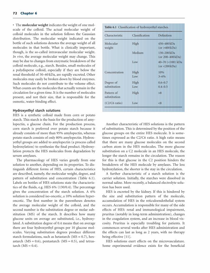
The pharmacology of HES varies greatly from one solution to another, depending on its properties. To dis-tinguish different forms of HES, certain characteristics are described, namely, the molecular weight, degree, and pattern of substitution and concentration (Table 6.1 ). Labels on bottles of HES solutions state the characteris-tics of the fl uids, e.g. HES 6% (130/0.4). The percentage gives the concentration of the starch solution. A 6% solution is considered iso - oncotic, a 10% solution hyper-oncotic. The fi rst number in the parentheses denotes the average molecular weight of the colloid, and the second number is the substitution degree or molar sub-stitution (MS) of the starch. It describes how many glucose units on average are substituted, i.e., hydroxy-lated. A substitution degree of 0.4 means that on average there are four hydroxyethyl groups per 10 glucose mol-ecules. Varying substitution degrees produce different starch formulations, such as hetastarch (MS = 0.7), hex-astarch (MS = 0.6), pentastarch (MS = 0.5), and tetras-tarch (MS = 0.4).
Table 6.1 Classifi cation of hydroxyethyl starches.
Characteristic Classifi cation Defi nition
Molecular weight
High 450 – 480 kDa (or > 400 kDa)
Medium 130 – 200 kDa (or 200 – 400 kDa)
Low 40 – 70 ( < 100) kDa (or < 200 kDa)
Concentration High 10% Low 3 – 6%
Degree of substitution
High 0.6 – 0.7 Low 0.4 – 0.5
Pattern of substitution
High > 8
(C2/C6 ratio) Low < 8

Fluid Management 73
60 kDa. Gelatin is therefore rapidly excreted. The plasma half - life is only short, namely about 2 hours. Repeated doses are needed to maintain an appropriate intravascu-lar volume.
High-viscosity fl uids: Alginate solutions Alginate solutions are not new. Studies with these agents were launched in the 1950s [15] . However, alginate solutions were not widely used clinically. It was only a few years ago that the interest in alginate was rekindled when searching for an agent to increase the viscosity of blood. In animal experiments, high - viscosity alginates are currently under investigation for use as a plasma expander [16] .
Last but not least: The albumin story Albumin is a naturally occurring colloid with an average molecular weight of about 66 kDa. It account for about 55% of the total blood protein pool of healthy persons. It is responsible for approximately 75% of the blood ’ s colloid - osmotic pressure. Albumin solutions for clinical use are prepared from pooled human plasma. Commer-cially available solutions are either iso - oncotic (4/5%) or hyperoncotic (20/25%) [17] .
Some think of albumin as the gold standard of colloid therapy. Others are absolutely against its use. Why? The proponents of albumin therapy cite albumin ’ s ability to maintain the colloid osmotic pressure, its anti - infl ammatory and antioxidant properties, its protective effects on the endothelium, and its buffering properties. The opponents not only cite its high costs, but also its pro - oxidant effects, the potential for pathogen transmis-sion, and its lack of additional benefi t or even survival over other fl uid resuscitation regimens.
One rationale for the use of albumin may be to correct hypoalbuminemia. It is true that the serum albumin level of critically ill patients correlates with their outcome. A low albumin level predicts an unfavo-rable outcome. Nevertheless, the artifi cial correction of the albumin level by albumin supplementation does not improve the outcome and may even be associated with a worse outcome [18] . Hypoalbuminemia is best cor-rected with suffi cient nutrition and treatment of the underlying condition.
Another reason that has been given for using albumin is to treat edema since albumin is a colloid that binds water. However, this is not the case. The reason for this is easy to understand when we consider that albumin is also present in the interstitial space. Also, when intrave-nously administered, albumin disperses freely in the
effect of HES. It was claimed for some HES that the extravascular leakage of colloids caused by microvascular hyperpermeability may be reduced, but this effect is not universally demonstrated [12] . Postulated underlying mechanisms may include the HES ’ sealing effect on the capillaries or the inhibition of endothelial activation and damage. HES may also modify severe infl ammatory responses and reduce endothelial dysfunction. It may therefore be organ protective in sepsis and surgeries that elicit severe systemic infl ammatory responses.
Dextran solutions Dextran is a glucose polymer. It is synthesized by enzymes of Leuconostoc mesenteroides . This bacterium produces dextran of very high molecular weight. Hydrolytic break-down and fractionation fi nally lead to different dextran preparations. Dextran solutions are available as dextran 40 (average molecular weight of 40 kDa) and dextran 70 (average molecular weight of 70 kDa). They are available in concentrations of 3 – 10% [13] .
Initially, dextran has an excellent volume - increasing effect but the effect is only short - lived. Because of the small molecule size, dextran is excreted rapidly. Dextran 40 increases plasma volume more and acts for shorter duration than dextran 70.
Although initial experiments with dextran failed to demonstrate the antigenicity of dextran, antibodies are developed occasionally. To prevent anaphylactic reac-tions, a monovalent hapten, dextran 1, is usually given prior to the infusion of dextran 40 or 70 to block possible antibody development.
Nowadays, the use of dextran is limited and it has largely been replaced with HES. Dextran still has some use in combination with hypertonic saline.
Gelatin solutions The gelatins available today are the so - called new - generation gelatins. They are modifi ed polypeptides from bovine collagen. Three different modifi cations are used: cross - linked gelatin (e.g., Gelifundol), urea - linked gelatin (e.g., Polygelin), and succinylated gelatin (e.g., Gelofu-sine). Gelatin solutions differ with regard to their electro-lyte concentrations. This is of clinical importance. Urea - linked gelatin has a high calcium and potassium concentration, while the succinylated preparations are low in calcium and potassium. Gelatin is available in solutions of concentrations of 4%, 6%, and 10%, and is dissolved in sodium chloride solution [13, 14] .
The average molecular weight of gelatin is 30 – 35 kDa. This is far below the renal threshold of approximately
74 Chapter 6
on the body ’ s ability to clot is not as pronounced as with dextran or some HES solutions [23] .
Dextran is known for its inhibition of thrombocyte aggregation by coating platelets (inhibition of factor VIII) and its ability to increase fi brinolysis. Further, it reduces blood viscosity. Such properties may not be favo-rable in patients who are hemorrhaging, but they can be used to prevent thrombosis and to improve the blood ’ s rheology.
The effect of HES on coagulation varies with the HES formulation. Some HES formulations cause an acquired von Willebrand syndrome. These may also alter the platelet ’ s ability to participate effectively in the clotting process, either by coating the platelet or by interfering with its receptors. Some HES solutiomns decrease the expression of platelet receptors, while others increase it. DDAVP (1 - deamino - 8 - d - arginine vasopressin) can offset some of the side effects of HES on coagulation [24] . There is a historical restriction of the amount of HES given. In an attempt to prevent coagulopathy, HES infu-sions were restricted to 20 mL/kg body weight/day. This recommendation was given in the early days of HES therapy, when only HES with a high molecular weight and a high substitution ratio was available. Although not universally agreed upon, medium molecular weight HES with a low substitution ratio and HES with a low molecu-lar weight seem to alter coagulation much less than high molecular weight HES [23, 25] . Thus, it has been said that their maximum daily dose can be increased to 50 mL/kg body weight/day. The magnitude of their effects on the clotting process is comparable with those of gelatin solution [26] .
Apart from the colloid itself, its carrier solution may infl uence the colloid ’ s effects on the coagulation profi le. A lack of calcium in the unbalanced solution was pro-posed to impair clotting. Colloids in calcium - containing carrier solutions may impair clotting less than the same colloid formulated in normal saline [27] .
Kidney function Virtually every colloid may impair kidney function and is associated with a higher rate of renal replacement therapy. For most colloids, this effect is dose dependent. The effect of albumin on kidney function is ambiguous, with some studies demonstrating a renoprotective, others a neutral, and others a detrimental effect. Gelatins are also thought to impair kidney function minimally. HES, and probably especially the high molecular weight formulations, are nephrotoxic and seem to cause higher rates of acute kidney failure and the need for dialysis.
interstitium, an effect that is even more pronounced in critically ill patients with capillary leakage. That is why albumin may even cause edema.
A third reason given for the use albumin is to admin-ister the “ ideal ” resuscitation fl uid. As the protein designed to maintain the colloid osmotic pressure of blood, it is thought to naturally have the lowest rate of side effects, even when solutions produced from donated blood are used. Nevertheless, when albumin solutions were tested in clinical routine for fl uid resuscitation, they had little proven advantages over HES or saline [19] . In some groups of patients, albumin seems to be outrightly detrimental, such as in patients with traumatic brain injury. In some other groups of patients, albumin dem-onstrated some benefi t [20] . Based on such contradictory fi ndings, some authors claim that albumin reduces mor-bidity and mortality, while others refute this claim. To date, it has not been clarifi ed which patients, if any, may benefi t and which patients may be harmed by albumin resuscitation.
Side effects of intravenous fl uids
Despite decades of research on fl uid therapy, the choice of resuscitation fl uid is often nothing more than a creed. Taking into consideration what side effects the fl uids may elicit may make it easier to decide on the therapy for an individual patient.
Coagulation Every intravenous fl uid has the potential to impair hemostasis. They may dilute clotting factors, fl ush away newly made clots, and have specifi c effects on coagulation factors or platelets. Nevertheless, crystalloids may also enhance hemostasis. They were shown to cause a mild hypercoagulopathy, lasting several hours. The clinical impact of intravenous fl uids on coagulation is not only fl uid - specifi c but also depends on the amount and manner of fl uid administration.
Albumin was thought of as the gold standard of fl uid therapy, since it was postulated that it does not cause coagulopathy. Nevertheless, it was shown that albumin exerts an intrinsic anticoagulatory effect and may impair coagulation, especially in higher grades of hemodilution [21] . The same is true of gelatins. They were also thought not to infl uence coagulation, even in higher doses. However, specialized clotting tests (e.g., ristocetin time) were able to show some infl uence of gelatin on coagula-tion [22] . Even so, the infl uence of albumins and gelatins
Fluid Management 75
trauma, depending on the velocity of blood loss [29] . When fl uids are administered during this time, i.e., the time the body needs to produce stable clots, intrinsic hemostasis is impaired and only weak clots are produced [30] . Fluid resuscitation increases the blood pressure and fl uid volume may wash away the developing clots. Early fl uid administration during the initiation of hemostasis may prolong the time to achieve hemostasis, and with it blood loss increases. Fluid administration after the for-mation of the initial clot triggers rebleeding. Total blood loss increases as well [31. The remaining clotting factors are diluted. Hypothermic coagulopathy may be worsened if the resuscitation fl uids are not warmed adequately. Thus, artifi cially, the patient is rendered coagulopathic and hypothermic. To avoid this, alternative resuscitation strategies for the hypovolemic patient with active or threatened rebleeding have been proposed. These strate-gies include hypotensive resuscitation and delayed resuscitation.
The goal of hypotensive resuscitation is to maintain a low blood pressure by volume resuscitation. It is only after defi nite surgical hemostasis that hypotensive resuscitation switches to normotensive resuscitation. In humans, the goal of hypotensive resuscitation is mean arterial pressure of about 40 – 60 mmHg or mean systolic blood pressure of 80 – 90 mmHg. When blood pressure measurement is not available, infusions at a fi xed rate (one that empirically does not raise the systolic blood pressure above 90 mmHg) are administered. This consti-tutes so - called controlled resuscitation.
Hypotensive resuscitation was shown in animal models and in some human studies to improve survival of hem-orrhagic shock [32] . However, the benefi ts of hypotensive resuscitation are time - dependent. Evidence suggests that the benefi t of low blood pressure is offset by the detri-ments of this resuscitation strategy after about 1 – 8 hours (depending on the study) have elapsed.
Another form or resuscitation is delayed resuscitation. In comparison with traditional resuscitation, it reduces blood loss and mortality while maintaining tissue oxy-genation. However, mortality increases when resuscita-tion is delayed for hours. Current evidence suggests delayed resuscitation may be benefi cial only when the patient is awake and has a palpable radial pulse, and hemostasis is achievable within about 15 minutes. For all other cases, hypotensive resuscitation may be more appropriate [32] .
The timing of fl uid therapy may also be important when it comes to surgical procedures. When fl uid infu-sion is delayed until after the phase with potential or
In patients with renal insuffi ciency, HES should only be given in low concentration and with suffi cient co - administration of crystalloids to prevent kidney damage [25] .
Allergic reactions According to a study by Laxenaire et al. [28] , the follow-ing incidences of anaphylactoid reactions after colloid infusion occurred: 0.345% for gelatin, 0.273% for dextran, 0.099% for albumin, and 0.058% for HES. In 20% of the cases, these reactions were serious. Patients with a history of drug allergies were especially at risk of developing an anaphylactoid reaction. Gelatins and dex-trans therefore should be avoided in patients with a known history of drug allergies.
Hyperchloremic metabolic acidosis Hyperchloremic metabolic acidosis is a non - respiratory acidosis with an increase of chloride in plasma. The most common avoidable cause of this acidosis is an intrave-nous fl uid with a high chloride content, as is found in unbalanced fl uids and especially in normal saline. Bal-anced fl uids do not cause this derangement. Hyperchlo-remic metabolic acidosis was reported to have many effects on red cells, coagulation, the gastrointestinal tract, and renal function. Although the clinical relevance of those effects is controversial, it was stated that balanced solutions could be superior to unbalanced solutions in preventing the development of this acidosis.
Fluids at work
When should fl uid be given? Does the timing of fl uid resuscitation play a role? In general, the earlier the patient has an adequate fl uid status, the better. In surgical and trauma patients, early fl uid optimization is more effi cacious than delayed resus-citation in maintaining or restoring normal tissue per-fusion and oxygenation. The shorter the time a patient is in shock, the lower their risk of developing postinjury coagulopathy. However, early fl uid resuscitation to achieve normovolemia and to restore a normal blood pressure in the actively bleeding patient seems to be det-rimental. It favors bleeding or rebleeding when adminis-tered before defi nitive surgical hemostasis is achieved. The human body usually initiates clot formation within 10 minutes when the mean arterial blood pressure drops to 60 mmHg. In actively bleeding patients, such low blood pressures may be reached some minutes after the
76 Chapter 6
patient is already normovolemic. With the limited evi-dence we have on this subject, bolus infusions should be discouraged.
When intravenous access is not available or feasible, there are other routes of fl uid administration: intraos-seous, subcutaneous, oral, rectal, and intraperitoneal. There is not much scientifi c evidence regarding effi cacy, benefi ts, and risks of the alternative routes of fl uid administration. However, when intravenous access is not achievable or in austere environments, administering fl uids via alternative routes may be effective in reversing hemorrhagic or hypovolemic shock [36, 37] .
How much fl uid should be given? Hypovolemia is detrimental since it precipitates tissue hypoxia. Hypervolemia may be just as detrimental. Hyp-ervolemia fi nally leads to tissue edema and tissue hypoxia results as well. Besides, hypervolemia may lead to pulmo-nary edema and paralytic ileus. That is why it is benefi cial to know the optimum fl uid level of any individual.
Monitoring the volume status of individual patients is an art. There is no number or symptom which tells whether a patient is normovolemic or will benefi t from further volume. At the level of the macrocirculation, hypovolemia is usually assumed when there is orthostatic hypotension (which may indicate an estimated volume loss of at least 20%) or when there is supine hypotension (which may indicate an estimated volume loss of at least 30%). Other clinical signs, such as the heart rate, skin temperature, urine output or a dry oral mucosa, may or may not be associated with hypovolemia. Whether or not clinical signs indicative of hypovolemia are present, one cannot tell whether there already is a volume need at the level of the microcirculation in general or in one organ system in particular.
A variety of measurable goals have been defi ned as surrogate markers for tissue perfusion. Traditionally, volume therapy was targeted at static intravascular pressures, such as the arterial blood pressure, central venous pressure, and pulmonary capillary wedge pres-sure. Low pressures may indicate hypovolemia, but may also be caused by other factors. Normal pressure, however, does not rule out hypovolemia and tissue hypoxia. Intravascular pressures may be useful if their trends are considered, but a single pressure reading does not indicate how to proceed with the volume therapy. Measures of the global blood fl ow, such as cardiac output, stroke volume, stroke volume variation or its delta - down component, oxygen delivery, and oxygen consumption, have been assumed to indicate
actual blood loss, overall surgical blood loss may be reduced without detriment to the patient [33] .
What fl uid should be given? There is an ongoing debate about the ideal agent for fl uid management. The heated discussions among the propo-nents and opponents of the different fl uids can be fol-lowed in the current medical literature. Since no one resuscitation fl uid has clear - cut survival benefi ts over another [11, 34] , there is no point in becoming dogmatic about the choice of fl uids. Nevertheless, some differences of the available fl uids can be kept in mind when the deci-sion about a fl uid regimen is to be made for an individual patient.
Benefi ts may be conveyed when the fl uid ’ s effects on the coagulation system, immune system, kidney, and overall fl uid volume are taken into account. Also, cost considerations may play a role.
How should fl uid be given? Nowadays, fl uids are usually administered intravenously. Questions remain regarding the velocity of infusion. Some fl uid management strategies recommend bolus infusions, others continuous infusions. In a recent trial multicenter trial (FEAST) [35] , bolus infusions for patients in hypotensive septic shock increased mortality compared with patients treated with continuous fl uid resuscitation, and the trial was halted prematurely. The authors of the study speculated that the vasoconstriction in shock may be a protective measure that should not be disturbed by bolus infusion. Besides, reperfusion injury and subclinical effects on the lung, heart, and brain may play a role in the detrimental effect of bolus fl uid infusion.
Bolus infusions may also be detrimental in hemor-rhagic patients. They may increase bleeding and disturb endogenous clotting, as outlined above [29, 31] .
Besides, the infusion rate affects the amount of fl uid that is kept in the vasculature. When fl uid is administered slowly, more will remain in the vascular bed compared with bolus administration. For instance, more than 90% of a medium molecular weight starch may remain intra-vascularly when given slowly, while only 60% of the same fl uid will remain there when given as a bolus. This may be due to the sudden increase in blood pressure that is elicited by the rapid infusion. Other factors may play a role as well. The rate of intravascular fl uid retention depends also on the condition of the patient at com-mencement of fl uid infusion. If the patient is hypovo-lemic, more fl uid remains intravascularly than if the
Fluid Management 77
Few high - quality data are available that guide the choice of fl uids regarding prevention of coagulopathy. The little information that can be gleaned from the medical literature is summarized below.
In general, crystalloids exert the least infl uence on coagulation and blood loss. When it comes to colloids, all of them seem to impair one or another in vitro marker of coagulation disturbance. Some studies report that this translates into increased blood loss and transfusions [39] . Physiologically balanced fl uids or colloids (HES) dis-solved in them seem to lessen blood loss and trigger fewer transfusions than unbalanced fl uids. Low molecular weight HES in saline impairs coagulation less than high molecular weight HES in a balanced solution [40] . Gelatin, albumin, and low - to - medium molecular weight HES, preferably in a balanced salt solution, seem to cause the least coagulopathy and increase in blood loss of all colloids [11, 41] . However, few trials comparing different HES solutions head - to - head regarding their effect on blood loss have been published so far [25] .
Avoiding or reducing postinjurycoagulopathy Resuscitation fl uids are not the only culprit for develop-ing coagulopathy. Trauma patients may develop a special kind of coagulopathy, the so - called postinjury coagulopa-thy, a condition without an agreed defi nition. Its patho-physiology is complex. Factors such as tissue injury itself, hemorrhage with concomitant circulatory impairment and immunomodulation, hypothermia, hypocalcemia, clotting factor dilution, acidosis, and pre - existing condi-tions may all contribute to its development. In addition, resuscitation injury may contribute to the development of postinjury coagulopathy.
Presumably, the development of postinjury coagulopa-thy can be modifi ed by fl uid management. Logically, fl uids known for their marked coagulopathic effect should be avoided. All fl uids given should be warmed to body temperature in order to avoid hypothermia. Rapid and full volume resuscitation may initially prevent or treat shock effectively, and thus reduce the ischemic and reperfusion component of postinjury coag-ulopathy. On the other hand, normotensive resuscitation may encourage further bleeding with its concomitant loss of clotting factors, reduced clot strength, and disturbance of intrinsic hemostatic measures [30] . Hypotensive resuscitation of trauma patients reduces this risk. Coagu-lopathy and coagulopathic bleeding with its resulting mortality is reduced when hypotensive resuscitation is employed [42] .
hypovolemia. Volume therapy to achieve goals (goal - directed therapy), such as an appropriate cardiac output, has been shown to improve survival in some patient groups. However, neither intravascular pressures nor variables of the global blood fl ow answer the important question: Do I optimize tissue perfusion and oxygenation with my current fl uid resuscitation strategy? Since tissue oxygenation and perfusion is currently considered the major goal in volume therapy, monitoring of them seems promising. Direct measurements of tissue oxygenation are not available. Some surrogate markers gained by gastric tonometry (an indirect estimate of mucosa per-fusion) and near infrared spectroscopy may help estimate tissue oxygenation in selected patients. For the great majority of patients, however, good clinical judgment combined with some clinical and paraclinical parameters should be used to guide fl uid therapy.
Infl uence of fl uid therapy on bloodmanagement
The last question in this chapter is: Does the choice of fl uid affect blood management - related outcome varia-bles, such as blood loss, anemia, coagulation and mortal-ity? Well, it does in so far that blood is not a volume replacement. For volume resuscitation, crystalloids or colloids, but not transfusions, is indicated. In the great majority of settings, acellular fl uids are actually superior to blood as far as their ability to optimize cardiac output is concerned.
Avoiding or reducingpharmacologically-induced coagulopathy Resuscitation fl uids themselves may lead to coagulopathy. The right choice of resuscitation fl uid does prevent this, at least partially. When a patient undergoes surgery with high anticipated blood loss, when even minor bleeding may be detrimental, or when the patient is coagulopathic to begin with, avoidance of fl uid - induced coagulopathy is essential.
Impairment of coagulation by resuscitation fl uids has been tested in vitro (e.g., by thromboelastography, factor levels, platelet function assays). The results, however, often do not correlate with clinical endpoints such as blood loss and postoperative anemia [38] . Therefore, it is diffi cult to extrapolate information gained from in vitrotesting to clinical medicine. It is not known which labora-tory parameter of coagulation correlates with blood loss and postoperative anemia.
78 Chapter 6
colloids are equally effective in optimizing the cardiac output, if the correct dose is given at the right time and in the right manner. • The choice of fl uid therapy may infl uence the total blood loss. Though not tested in large trials, some fl uids may be more effective in preventing coagulopathy and other blood loss than others. The timing and manner of fl uid resuscitation may be equally important to reduce coagulopathy and bleeding. • High - viscosity fl uids may improve the microcirculation. Normalizing blood viscosity in states of severe hemodilu-tion maintains tissue perfusion and oxygenation.
Questions for review
1. What is the difference between crystalloids and colloids? 2. What crystalloids are there and how do they differ from each other? 3. What colloids are there? 4. What do the following terms mean: polydisperse, sub-stitution degree, hypertonic, and replacement fl uid? 5. What four terms are typically used to describe HES? 6. How do timing and manner of fl uid resuscitation infl uence blood management - related outcomes?
Exercises and practice cases
Reducing blood loss Overall blood loss can be reduced with judicious fl uid resuscitation. Hypotensive resuscitation to a goal of a mean arterial pressure of 50 mmHg in trauma patients with hemorrhagic shock reduces transfusions and postoperative mortality [42] . Giving only restricted amounts of fl uid during major surgery and postponing full fl uid replacement to the end of surgery is reported to reduce intraoperative blood loss and red cell transfusions [33, 43] .
Preservation of microcirculation in severehemodilution/anemia Another important factor for blood management is the resuscitation fl uid ’ s ability to impair or to preserve microcirculation during bleeding and in anemic states. In states of shock, microcirculatory impairment with a reduced functional capillary density results [44] . Simi-larly, patients resuscitated with large volumes of low - viscosity fl uids, such as crystalloids, show signs of impaired microcirculation. Reduced blood viscosity, and not anemia, is the limiting factor in such settings. Fluid management, thus, should aim at restoring or preserving microvascular perfusion.
Among other factors, resuscitation fl uid viscosity infl uences the microcirculation. Viscosity increases the shear stress on the microvasculature and nitric oxide is produced. Vessels dilate and the functional capillary density increases. Once microcirculatory blood fl ow is restored, signs typically associated with severe anemia may disappear. It is believed that using viscous fl uids in severe hemodilution or in hemorrhagic shock may reduce transfusions and improve survival.
Knowing this, preclinical data point to advantages of increasing the viscosity of the resuscitation fl uid once a critically low value of blood viscosity has been reached. Different viscosity modifi ers have been proposed: sodium alginate, high molecular weight starches, dextrans, PVP, keratin and polyethylene glycol - conjugated albumin [45] . It has been postulated that polyethylene glycol - conjugated albumin yields the best results [46] . Such new viscogenic colloids are in preclinical trials and may provide future resuscitation fl uids.
Key points
• Restoring blood volume in hypovolemic patients is more important than correcting anemia. Crystalloids and
How much of the following solutions are needed to restore his blood volume? — normal saline, lactated Ringer ’ s, HES 6% (450/0.7); 10% dextran 40, gelatin 3%, and NaCl 7.5%. Explain your answers.
Suggestions for further research
1. What solutions are suitable plasma substitutes for therapeutic plasma exchange, e.g., in myasthenic crisis? 2. What specifi c considerations are needed for fl uid therapy in babies? 3. What fl uids are acceptable to strict vegetarians?
A boxer weighing 100 kg experiences severe epistaxis. He loses 1.5 L of blood.
Fluid Management 79
16. Cabrales P , Tsai AG , Intaglietta M . Alginate plasma expander maintains perfusion and plasma viscosity during extreme hemodilution . Am J Physiol Heart Circ Physiol 2005 ; 288 : H1708 – H1716 .
17. Rena NM , Wibawa ID . Albumin infusion in liver cirrhotic patients . Acta Med Indones 2010 ; 42 : 162 – 168 .
18. Myburgh JA , Finfer S . Albumin is a blood product too — Is it safe for all patients? Crit Care Resusc 2009 ; 11 : 67 – 70 .
19. Liberati A , Moja L , Moschetti I , Gensini GF , Gusinu R . Human albumin solution for the resuscitation and volume expansion in critically ill patients . Intern Emerg Med 2006 ; 1 : 243 – 245 .
20. SAFE Study Investigators . Impact of albumin compared to saline on organ function and mortality of patients with severe sepsis . Intensive Care Med 2011 ; 37 : 86 – 96 .
21. Roche AM , James MF , Bennett - Guerrero E , Mythen MG. A head - to - head comparison of the in vitro coagulation effects of saline - based and balanced electrolyte crystalloid and colloid intravenous fl uids . Anesth Analg 2006 ; 102 : 1274 – 1279 .
22. Thaler U , Deusch E , Kozek - Langenecker SA. In vitro effects of gelatin solutions on platelet function: a comparison with hydroxyethyl starch solutions . Anaesthesia 2005 ; 60 : 554 – 559 .
23. Van der Linden P , Ickx BE. The effects of colloid solutions on hemostasis . Can J Anaesth 2006 ; 53 ( Suppl 6 ): S30 – 39 .
24. Conroy JM , Fishman RL , Reeves ST , Pinosky ML , Lazarchick J . The effects of desmopressin and 6% hydroxyethyl starch on factor VIII:C . Anesth Analg 1996 ; 83 : 804 – 807 .
25. Groeneveld AB , Navickis RJ , Wilkes MM . Update on the comparative safety of colloids: A Systematic review of clini-cal studies . Ann Surg 2011 ; 253 : 470 – 483 .
26. Vanhoonacker J , Ongenae M , Vanoverschelde H , Donadoni R . Hydroxyethyl starch 130/0.4 versus modifi ed fl uid gelatin for cardiopulmonary bypass priming: The effects on post-operative bleeding and volume expansion needs after elec-tive CABG . Acta Anesth Belg 2009 ; 60 : 91 – 97 .
27. Boldt J , Mengistu A . A new plasma - adapted hydroxyethyl starch preparation: In vitro coagulation studies . J Cardiot-horac Vasc Anesth 2010 ; 24 : 394 – 398 .
28. Laxenaire MC , Charpentier C , Feldman L . Anaphylactoid reactions to colloid plasma substitutes: incidence, risk factors, mechanisms. A French multicenter prospective study . Ann Fr Anesth Reanim 1994 ; 13 : 301 – 310 .
29. Fouche Y , Sikorski R , Dutton RP . Changing paradigms in surgical resuscitation . Crit Care Med 2010 ; 38 ( Suppl ): S411 – 420 .
30. Rezende - Neto JB , Rizoli SB , Andrade MV , et al . Permissive hypotension and desmopressin enhance clot formation . J Trauma 2010 ; 68 : 42 – 50 ; discussion 50 – 51 .
31. Hirshberg A , Hoyt DB , Mattox KL . Timing of fl uid resusci-tation shapes the hemodynamic response to uncontrolled hemorrhage: analysis using dynamic modeling . J Trauma 2006 ; 60 : 1221 – 1227 .
Homework
List all available fl uids in your hospital, classify them as crystalloid or colloid, and make a table containing all fl uids and their content of electrolytes, molecular weights, substitution degree, etc.
Find out where you can get the best available colloids and crystalloids. Record the contact details for the sources (e.g., a pharmacy or a pharmaceutical company).
References
1. O ’ Shaughnessy D , Brooke W . Experiments on the blood in cholera [letter] . Lancet 1831 ; 1 : 490 .
2. Lewins R , Latta T . Injection of saline solutions in extraordi-nary quantities into the veins in cases of malignant cholera . Lancet 1831 ; 32 : 243 – 244 .
3. Bull WT. On the intra - venous injection of saline solutions as a substitute for transfusion of blood . Med Rec 1884 : 6 – 8 .
4. Gruber UF. Blutersatz . Fortschr Med 1969 ; 87 : 631 – 634 . 5. Gronwall A , Ingelman B . The introduction of dextran as a
plasma substitute . Vox Sang 1984 ; 47 : 96 – 99 . 6. Myburgh JA. Fluid resuscitation in acute illness — time to
reappraise the basics . N Engl J Med 2011 ; 364 : 2543 – 2544 . 7. Chappell D , Jacob M , Hofmann - Kiefer K , Conzen P , Rehm
M. A rational approach to perioperative fl uid management . Anesthesiology 2008 ; 109 : 723 – 740 .
8. Iijima T. Complexity of blood volume control system and its implications in perioperative fl uid management . J Anesth 2009 ; 23 : 534 – 542 .
9. Hartok CS , et al . A systematic review of third - generation hydroxyethyl starch (HES 130/0.4) in resuscitation: Safety not adequately addressed . Anesth Analg 2011 ; 112 : 635 – 645 .
10. Bulger EM , May S , Kerby JD , et al . Out - of - hospital hyper-tonic resuscitation after traumatic hypovolemic shock. A randomized, placebo controlled trial . Ann Surg 2011 ; 253 : 431 – 441 .
11. Murphy GS , Greenberg SB . The new generation hydroxyethyl starch solutions: The holy grail of fl uid therapy of just another starch? J Cardiothorac Vasc Anesth 2010 ; 24 : 389 – 393 .
12. Ando Y , Terao Y , Fukusaki M , et al. Infl uence of low molecu-lar weight hydroxyethyl starch on microvascular permeabil-ity in patients undergoing abdominal surgery: comparison with crystalloids . J Anesth 2008 ; 22 : 391 – 396 .
13. Niemi TT , Miyashita R , Yamakage M . Colloid solutions: a clinical update . J Anesth 2010 ; 24 : 913 – 925 .
14. Mitra S , Khandelwal P . Are all colloids same? How to select the right colloid? Indian J Anaesthes 2009 ; 53 : 592 – 607 .
15. Tomoda M , Inokuchi K . Sodium alginate of lowered polym-erization (alginon). A new plasma expander . J Int Coll Surg 1959 ; 32 : 621 – 635 .
80 Chapter 6
terior lumbar interbody fusion using pedicle screws and cages . Spine (Phila Pa 1976) 2010 ; 35 : 829 – 834 .
41. Choi YS , Shim JK , Hong SW , Kim JC , Kwak YL . Comparing the effects of 5% albumin and 6% hydroxyethyl starch 130/0.4 on coagulation and infl ammatory response when used as priming solutions for cardiopulmonary bypass . Minerva Anesthesiol 2010 ; 76 : 584 – 591 .
42. Morrison CA , Carrick MM , Norman MA , et al . Hypotensive resuscitation strategy reduces transfusion requirements and severe postoperative coagulopathy in trauma patients with hemorrhagic shock: preliminary results of a randomized controlled trial . J Trauma 2011 ; 70 : 652 – 663 .
43. Vretzakis G , Kleitsaki A , Stamoulis K , et al . The impact of fl uid restriction policy in reducing the use of red blood cells in cardiac surgery . Acta Anaesth Belg 2009 ; 60 : 221 – 228 .
44. Komori M , Takada K , Tomizawa Y , Uezono S , Nishiyama K , Ozaki M . Effects of colloid resuscitation on peripheral microcirculation, hemodynamics, and colloidal osmotic pressure during acute severe hemorrhage in rabbits . Shock 2005 ; 23 : 377 – 382 .
45. Zhao L , You G , Liao F , et al . Sodium alginate as viscosity modifi er may induce aggregation of red blood cells . Artif Cells Blood Substit Immobil Biotechnol 2010 ; 38 : 267 – 276 .
46. Villela N , Salazar V á zquez BY , Intaglietta M. Microcircula-tory effects of intravenous fl uids in critical illness: plasma expansion beyond crystalloids and colloids . Curr Opin Anaesthesiol 2009 ; 22 : 163 – 167 .
32. Santry HP , Alam HB . Fluid resuscitation: Past, present and the future . Shock 2010 ; 33 : 229 – 241 .
33. Fujita Y , Takeuchi A , Sugiura T . Before - after study of a restricted fl uid infusion strategy for management of donor hepatectomy for living - donor liver transplantation . J Anesth 2009 ; 23 : 67 – 74 .
34. Bunn F , et al . Colloid solutions for fl uid resuscitation . Cochrane Database Syst Rev 2011 ;( 3 ): CD001319 .
35. Maitland K , Kiguli S , Opoka RO , et al ; FEAST Trial Group. Mortality of fl uid bolus in children with severe infection . NEngl J Med 2011 ; 364 : 2483 – 2495 .
36. Grocott MPW. Resuscitation from hemorrhagic shock using rectally administered fl uids in a wilderness environment . Wilderness Environ Med 2005 ; 16 : 209 – 211 .
37. Cancio LC , Kramer GC , Hoskins SL . Gastrointestinal fl uid resuscitation of thermally injured patients . J Burn Care Res 2006 ; 27 : 561 – 569 .
38. Schramko A , Suojaranta - Ylinen R , Kuitunen A , Raivio P , Kukkonen S , Niemi T. Hydroxyethylstarch and gelatin solu-tions impair blood coagulation after cardiac surgery: a pro-spective randomized trial . Br J Anaesth 2010 ; 104 : 691 – 697 .
39. Hecht - Dolnik M , Barkan H , Taharka A , Loftus J. Hetastarch increases the risk of bleeding complications in patients after off - pump coronary bypass surgery: A randomized clinical trial . J Thorac Cardiovasc Surg 2009 ; 138 : 703 – 711 .
40. Choi SJ , Ahn HJ , Chung SS , et al . Hemostatic and electrolyte effects of hydroxyethyl starches in patients undergoing pos-