Embed Size (px)
Citation preview

BasicsBasics of of RealReal Time 3D Time 3D EchocardiographyEchocardiography
J.S. McGhieJ.S. McGhieW.B. VletterW.B. VletterF. F. MeijboomMeijboomR. R. FrowijnFrowijnH. Van de ZwaanH. Van de Zwaan
No No disclosuresdisclosures
Image aquisition and manipulation
Outline
Introduction
3D imaging
Data set manipulation

Image aquisition and manipulation
Outline
Introduction
3D imaging3D imaging3D imaging
Data set manipulationData set manipulationData set manipulation

Ultrasound imaging modalities in Cardiology

xMatrix X3-1 transducer
xMatrix array technologyutilizes 2400 fully-sampledelements for 360-degree focusing and steering

24 x 15 24 x 2024 x 15
Purewave technology – wider bandwith higher sensitivity
X3-1 X7-2
Footprint15 x 24mm
X7-2t
TTEpediatric
<20 kg
TEEadult
Footprint15 x 20mm
TTEadult>20 kg
RT3DE transducers iE33

Outline
IntroductionIntroductionIntroduction
3D imaging
Data set manipulationData set manipulationData set manipulation
Image aquisition and manipulation

3D Echo Protocol – Imaging modes
LIVE 3D
small sector - thin slice acquisition
zoom mode (enlargement of a subsegment of this sector)
xPlane (lateral tilt image)
3D Echo Protocol – Imaging modes
LIVE 3D
small sector - thin slice acquisition
zoom mode (enlargement of a sub segment of this sector)zoom mode (enlargement of a sub segment of this sector)zoom mode (enlargement of a sub segment of this sector)
xPlanexPlanexPlane (lateral tilt image)(lateral tilt image)(lateral tilt image)

The current display format suffers from attempting to show3D images on a 2D display
Small sector – Thin slice mode

Full-volume dataset not real-time(sector 101° x 104°)
Real-time datase (sector 30° x 60°)
Small sector – Thin slice mode

3D Echo Protocol – Imaging modes
LIVE 3D
smallsmallsmall sectorsectorsector--- thinthinthin slice acquisitionslice acquisitionslice acquisition
zoom mode (enlargement of a subsegment of this sector)
xPlanexPlanexPlane (lateral tilt image)(lateral tilt image)(lateral tilt image)

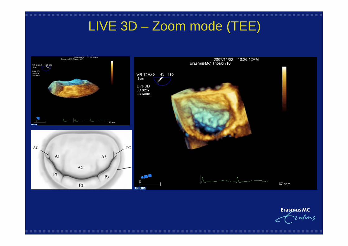
LIVE 3D – Zoom mode (TEE)
LIVE 3D – Zoom mode (TEE)

3D Echo Protocol – Imaging modes
LIVE 3D
smallsmallsmall sectorsectorsector--- thinthinthin slice acquisitionslice acquisitionslice acquisition
zoom mode (enlargement of a zoom mode (enlargement of a zoom mode (enlargement of a subsegmentsubsegmentsubsegment of this sector)of this sector)of this sector)
xPlane (lateral tilt 2D image)
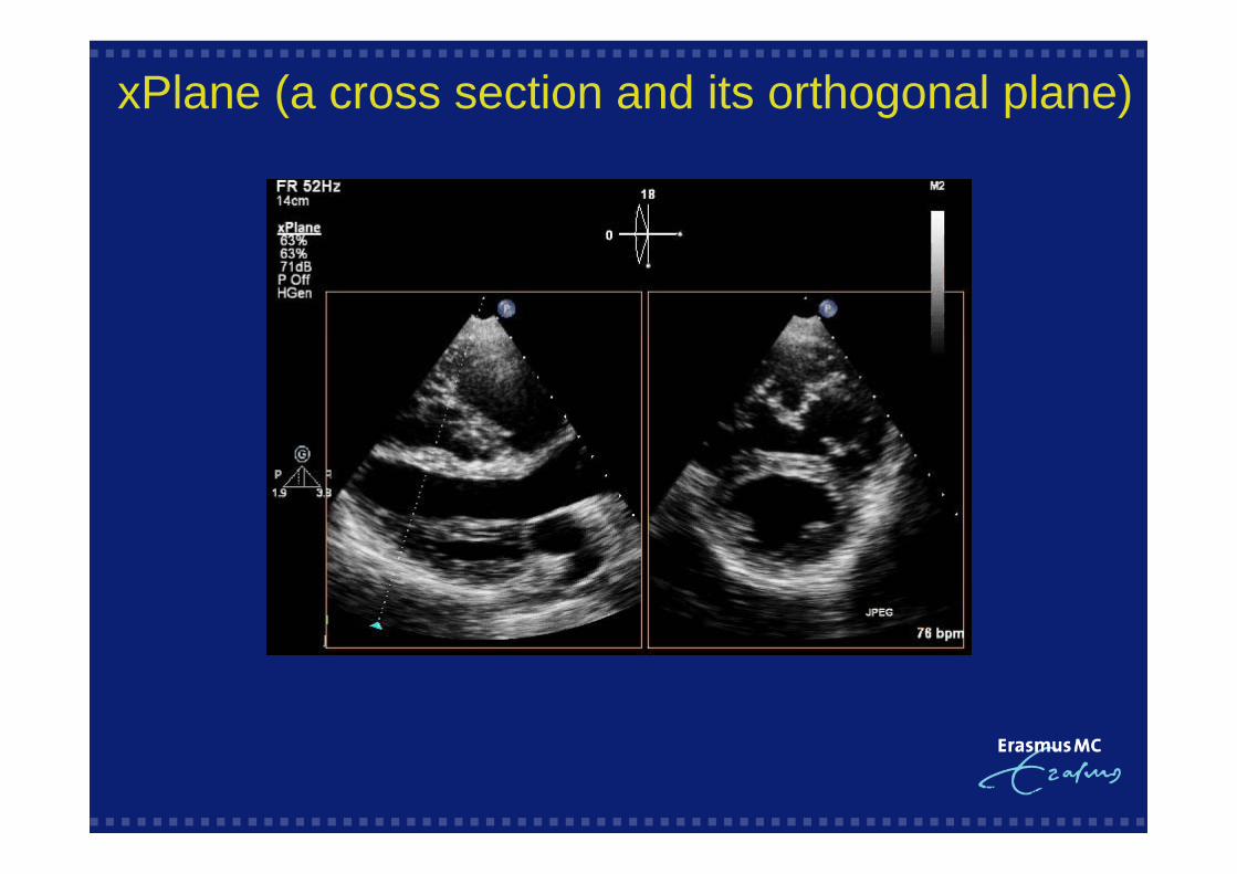
xPlane (a cross section and its orthogonal plane)

Lateral tilting
xPlane (a cross section and its orthogonal plane)

2D to 3D full volume
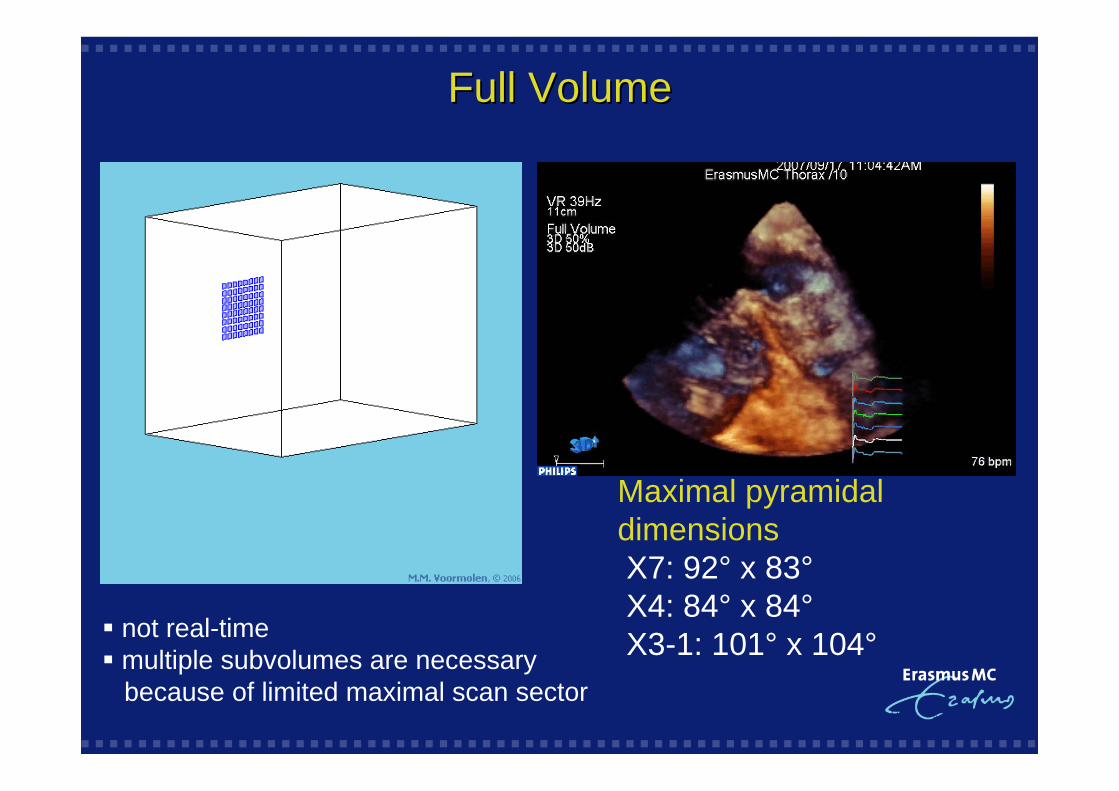
Full VolumeFull Volume
not real-time multiple subvolumes are necessary
because of limited maximal scan sector
Maximal pyramidaldimensionsX7: 92° x 83°X4: 84° x 84°X3-1: 101° x 104°

3D Echo Protocol: Full Volume
Patient and machine preparation
1. Good ECG signal with clear R-wave(3D Full volume triggering)
2. Adjust machine settings (follow the rules as for 2D) for the best 3D resolution: Harmonics – fundamental, adjust gain setting
(clear blood-tissue border - minimize noise) Region of interest between the dotted lines Minimize sector ( angle, depth, density) Maximise number of subvolumes Increase overall gain before recording

3D aquisition work flow

3D aquisition work flow: Gainsetting
Overall gain TGC

3D Analysis
• Cardiac Function
••• CardiacCardiacCardiac morpholgymorpholgymorpholgy

Image optimisation for the LV and RV
Priority: Good quality 2D image
Frame rate as high as possible - acquisition beats: 7
- optimise depth setting
Complete ventricle in dataset - optimise density setting
Extreem dilatation - acquisition beats: 5 (wide angle)
- notice: drop in frame rate

Frame Rate 3D Volume Data Set
Ultrasound speed : 1500 m/sec
Depth of 16cm : transmitting + receiving time : 220 micro sec
2D ( 90 scanlines ) requires 19,8 msec → FR : 50 Hz
3D ( 2400 scanlines ) requires 528 msec → FR : 1,9 Hz
How to improve the frame rate
■ Receive multiple beams for each transmit event(parallel processing)
■ Decrease image size either laterally or in depth
■ Acquire data over multiple cardiac cycles ( 4 – 7 ) and build
a composite volume data set

Depth13 cm
Depth16 cm
35 Hz35 Hz
30 Hz30 Hz
Depth: frame rate

Number of subvolumes – frame rate
20 Hz20 Hz
35 Hz35 Hz

Wideangle
35 Hz35 Hz
18 Hz18 Hz
Wide angle – frame rate

Subvolumes
Reasons to use less subvolumes
Arrhythmias / severe bradycardia
Breathhold problems
Big heart: use wide angle / 5 subvolumes

Low
High
35 Hz35 Hz
35 Hz35 Hz
Line density – frame rate
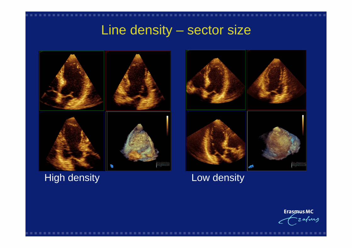
Line density – sector size
High density Low density

2D images within the 3D data set
Multiplane reconstruction

What do I trace?
Endocardial boarder detection using contrast

LV volume and ejection fraction

Echo drop out in full volume data set
Sometimes unavoidable!

Apical four-chamber view Laterally modified apicalfour-chamber view
2D: RV view before RT3DE acquisition

Full volume RT3DE RV: acquisition dataset
RV volumes and ejection fraction.
For the first time possible with echo!

What do I trace?
RV endocardial boarder detection using contrast
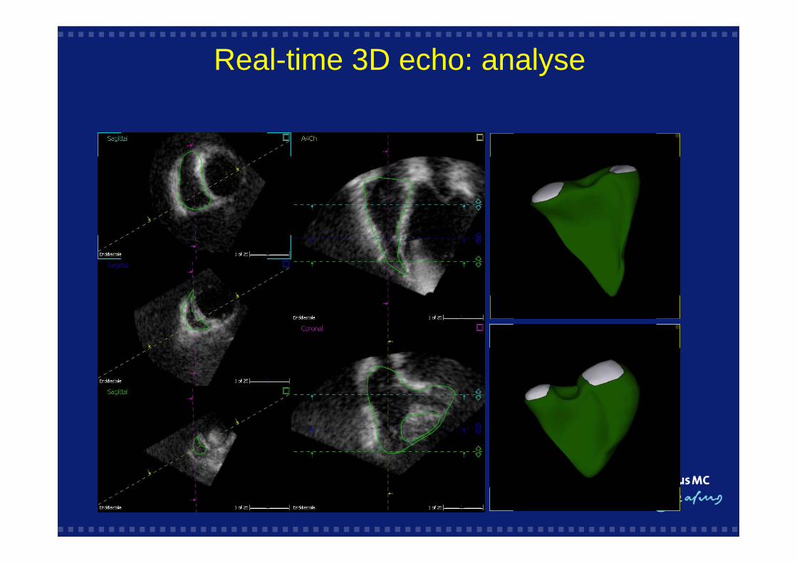
Real-time 3D echo: analyse

3D Analysis
••• CardiacCardiacCardiac FunctionFunctionFunction
• Cardiac morpholgy

90º Bi-plane view mitral valve
Dotted lines: ROI = mitral valve

Normal Mitral Valve

Dimension ResolutionAxial BestLateral Medium Elevation Least
~0.5 mm ~3.0 mm
~2.5 mm
Resolution: the best plane?

Ultrasound beam perpendicular to ROI

Parasternal LAX Parasternal SAXMV seen from the LA
Full Volume – Colour

3D Echo pitfalls: stitching artifact

3D Full volume data set
Multiple subvolumesIncreased chance of artefact
One - shot
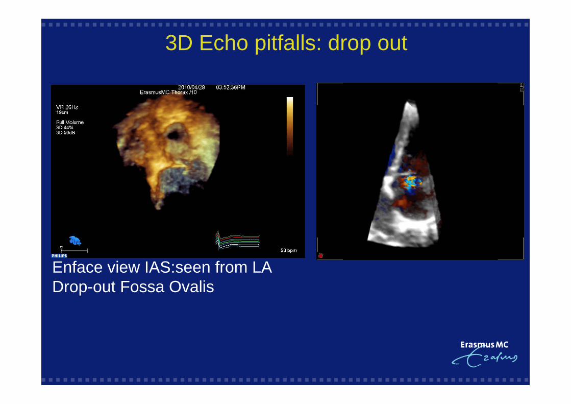
3D Echo pitfalls: drop out
Enface view IAS:seen from LADrop-out Fossa Ovalis
Image aquisition and manipulation
Outline
IntroductionIntroductionIntroduction
Tips and tricks for an optimal 3D data setTips and tricks for an optimal 3D data setTips and tricks for an optimal 3D data set
Data set manipulation

3D Multiplane reconstruction: 4Fallot
• The ability to move through a 3D dataset in any 2D image plane
• Interogate the data set on or of-line
• Allows precise identification and localization of abnormalities
• Allows quantification.

3D analysis – the crop-box

3D analysis – the crop-box

Crop till you Drop!!!

X5-1: the all in one transducer
2D imaging2D colour DopplerPW and CW Doppler
3D imagingLive 3D imaging and colourLive 3D zoomFull volume imaging and colour
+

2D imaging
S5-1 X3-1
X5-1

3D imaging
X3-1 X5-1
Conclusions: Image acquisition and manipulation
In 3D the cardiac structures are shown in relationship to each otherin all three spacial dimensions
The cardiac structures can be rotated or viewed from different orientations
The ability to “move through” a 3D data set in any 2D image planeallows better appreciation of cardiac anatomy in complex structural heartdisease.
3D images are more intuitive than 2D images allowing quicker appreciationof cardiac anatomy by other health care workers.
The clinical role of 3d echocardiography will continue to evolveas technology advances
Conclusions: Image acquisition and manipulation
The limitations of 2D are also true for 3D echo
3D echo is not a stand-alone feature, but should be used next to / on top of other echo
modalities.
But remember :

Overall gain

Overall gain

Source: SA of the Heart: Wilcock/Cook/Anderson
Left A-V Valve

Bicuspid Ao Valve
Aortic valve en-face view from Aorta

From RV
Normal Tricupid Valve

• Only the closure line of PV is visible!• Dynamic RVOT during the
cardiac cycle
Normal Pulmonary Valve
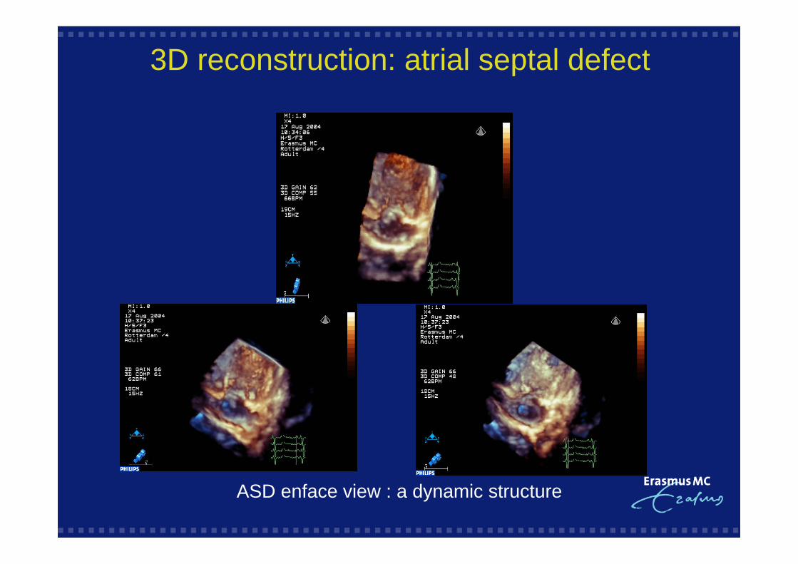
3D reconstruction: atrial septal defect
ASD enface view : a dynamic structure

3D reconstruction: VSD by 4Fallot(baby)
En face VSD from LV En face VSD from RV

TEE: Enface view of the IAS
ASD II-SVC-Aorta En face view LA: Device insitu