Embed Size (px)
Citation preview
Contents lists available at ScienceDirect
BBA - General Subjects
journal homepage: www.elsevier.com/locate/bbagen
Quantification of kinetic rate constants for transcytosis of polymericnanoparticle through blood-brain barrier
Aminul Islam Khan, Qian Lu, Dan Du, Yuehe Lin, Prashanta Dutta⁎
School of Mechanical and Materials Engineering, Washington State University, Pullman, WA 99164-2920, United States
A R T I C L E I N F O
Keywords:Blood-brain barrierPolymeric nanoparticleDrug deliveryArtificial neural networkParameter estimation
A B S T R A C T
Background: Polymeric nanoparticles (PNP) have received significant amount of interests for targeted drugdelivery across the blood-brain barrier (BBB). Experimental studies have revealed that PNP can transport drugmolecules from microvascular blood vessels to brain parenchyma in an efficient and non-invasive way. Despitethat, very little attention has been paid to theoretically quantify the transport of such nanoparticles across BBB.Methods: In this study, for the first time, we developed a mathematical model for PNP transport through BBBendothelial cells. The mathematical model is developed based on mass-action laws, where kinetic rate para-meters are determined by an artificial neural network (ANN) model using experimental data from in-vitro BBBexperiments.Results: The presented ANN model provides a much simpler way to solve the parameter estimation problem byavoiding integration scheme for ordinary differential equations associated with the mass-action laws.Furthermore, this method can efficiently deal with both small and large data set and can approximate highlynonlinear functions. Our results show that the mass-action model, constructed with ANN based rate parameters,can successfully predict the characteristics of the polymeric nanoparticle transport across the BBB.Conclusions: Our model results indicate that exocytosis of nanoparticles is seven fold slower to endocytosissuggesting that future studies should focus on enhancing the exocytosis process.General significance: This mathematical study will assist in designing new drug carriers to overcome the drugdelivery problems in brain. Furthermore, we anticipate that this model will form the basis of future compre-hensive models for drug transport across BBB.
1. Introduction
The blood-brain barrier (BBB) is a distinct and highly selectivebarricade formed by the microvascular endothelial cells together withthe other neurovascular elements such as astrocytes, pericytes, andmicroglial cells [1,2] to protect the brain against harmful agents. Theendothelial cells in the BBB are connected to each other through tightjunctions. These tight junctions preclude paracellular transport of so-lutes across BBB except some water-soluble substances. Several trans-cellular routes, such as transporter protein mediated, receptor-medi-ated, adsorptive mediated, cell-mediated transcytosis etc. are availablefor transportation of metabolites and other essential components forproper functioning of brains [3]. However, these transport routescannot be used for delivering large-molecule drugs and> 98% of small-molecule drugs across the BBB [4]. To enhance the efficacy of ther-apeutic delivery through the BBB, several techniques have been testedwith varying degrees of success. Among various techniques, tight
junction disruption, drug molecules modification and carrier-mediatedtransportation have dominated the drug delivery research.
Tight junction opening is reported using biological, chemical, andphysical stimuli [3]. Biologics such as virus, macrophage, cereport,zonula occludens toxin etc. can increase paracellular transport by eitheropening tight junctions or using Trojan horse effect [3]. Selected che-micals such as cyclodextrin and poloxamers can extract water and othersubstances (e.g. cholesterol) from endothelial cells, which lead toopening of gaps between cells for paracellular transport. Physical me-chanisms such as ultrasound, microwave, and electromagnetic field canalso open tight junctions by protein translocation, which enhance theBBB permeability. Although various stimuli can potentially increase thepenetration of drugs to the brain, high concentration of these stimulicompounds can compromise the BBB.
Drug delivery across BBB through modification of drugs is at-tempted either by direct conjugation of drugs to a BBB transporter (suchas glucose transporter) or by targeting lipid-mediated transport of
https://doi.org/10.1016/j.bbagen.2018.08.020Received 24 June 2018; Received in revised form 29 August 2018; Accepted 30 August 2018
⁎ Corresponding author.E-mail address: [email protected] (P. Dutta).
BBA - General Subjects 1862 (2018) 2779–2787
Available online 31 August 20180304-4165/ © 2018 Elsevier B.V. All rights reserved.
T
drugs. However, because of highly selective nature of transporters,drugs transport by direct conjugation to a transporter is only possible ifdrug molecules meet all criteria of endogenous ligands [5]. Lipidizationis done by attaching lipid-like molecules on the drug structure bymodifying hydrophilic moieties. Although high lipophilicity favoredhigher permeability of drugs, this lipidization does not ensure targeteddrug delivery because the permeability of lipidized-drugs increasesacross all biological membranes in the body [6]. Moreover, this ap-proach is only suitable for drugs having molecular weight< 500 Dasince higher molecular weight compounds cannot cross BBB throughpassive lipid soluble mechanism.
Another promising avenue for drug transport across BBB is throughcarriers such as liposomes, nanoparticles, nanospheres, nanosuspen-sions, polymer micelles, and nanogels [7]. Among them, nanoparticle-based therapeutic delivery is extensively studied because of their non-invasiveness and targeted drug delivery capability [8]. As a results,several nanoparticle-based system such as mesoporous silica [9,10],silver [11], superparamagnetic iron oxide [12], gold [13], polymeric[14] nanoparticle have been developed and tested as a drug deliverymechanism through BBB with different levels of success. Polymericnanoparticles offer several advantages over non-polymeric ones be-cause of their similarity with natural carriers such as serum lipoproteinsand viruses. Moreover, they can be targeted not only to the particularorgan/tissue, but also to a particular cell or even an intracellularcompartment [7]. In addition, polymeric nanoparticles increase drugsolubility, improve bio-distribution of drugs, and can potentially de-crease side effects [14].
Although significant efforts have been made to study the nano-particle transport through BBB experimentally, not much attention hasbeen paid to theoretically quantify the transport mechanism.Mathematical modeling is a powerful tool to understand the biologicalprocesses such as transport across BBB, where experiments are intricate,costly and sometimes ethically wrong. Hinow et al. [15] developed amathematical model to describe the transport of free drugs from bloodto brain once drugs get released from liposomes under the applicationof focused ultrasound. However, their model neglects drug accumula-tion by the endothelial cell and active efflux of drugs from brain toblood. The quantification of accumulated drugs is very important be-cause accrual of particles can increase the toxicity in endothelial cellswhich may alter the BBB integrity. Loeser [16] modeled the drug re-lease from a temperature-sensitive liposome and the diffusion of drugsin a brain cancer cell, but their model does not include the transport ofliposomes from blood to targeted brain cancer cells. Recently, Khanet al. [17] developed a mathematical model for receptor-mediatedtranscytosis of iron across BBB. Although our previous model addressesthe transcytosis, it can't be used to predict the nano-carrier based drugtransport across the BBB because of difference in mechanisms andpathways between iron molecule and nanoparticles.
The goal of the current research is to develop a mathematical modelfor nanoparticle transport across the BBB using the laws of mass-action.Mass action laws can generally be formed as a set of ordinary or partialdifferential equations. The estimation of parameters is the crucial stepin the development of these type of models. Several methods are de-veloped and used for the parameter estimation such as least square[18], Bayesian approach [19], incremental approach [20], artificialneural network approach [21], preprocessing method [22] etc. Amongthese methods, least square is the oldest, simplest and widely used forparameter estimation. However, this method suffers from convergenceproblems. In addition, this approach may get trapped into the localoptimal solution instead of the global one [21,23]. In this work, anartificial neural network (ANN) based model is presented for parameterestimation. Our ANN based parameter estimation method is muchsimpler to solve since it does not require an integration scheme fordifferential Eqs. [21]. In addition, ANN can deal with large data sets,can efficiently approximate highly nonlinear functions, and can be usedfor multi input-output variables [24]. To train our ANN model, we have
considered the transcytosis of poly[Triphenylamine-4-vinyl-(P-methoxy-benzene)] (TEB)-based nanoparticles through BBB. We speci-fically selected TEB-polymeric nanoparticles because TEB nanoparticlesexhibit excellent fluorescence properties which eliminates the necessityof tagging with additional fluorescence markers. Thus, this type ofnanoparticles can potentially be used in both imaging and drug de-livery. Moreover, TEB nanoparticles can be synthesized as small as20 nm. It has been reported that smaller size (~20 nm) nanoparticlesyield higher transcytosis across BBB [13,25,26]. In addition, the TEBnanoparticles are highly biocompatible. Controlled transcytosis ex-periments of TEB nanoparticles across BBB are performed on an in vitroBBB model which is constructed based on mouse cerebral endothelialcells (bEnd.3).
Rest of the paper is organized as follows. In the following section,mathematical model for nanoparticle transport through BBB en-dothelial cells is presented. Next, an ANN model is presented for esti-mation of kinetic parameters. In section 4, we describe the in-vitro ex-periments of both endocytosis and exocytosis to determine thenanoparticle transport rate across the BBB. In section 5, we presentimportant results such as estimated rate parameters for nanoparticletranscytosis and time required for nano-carrier penetration. Finally, wepresent our conclusions and future research outlook.
2. Mathematical model
A mass-action based mathematical model is presented to study thetranscytosis of nanoparticle from the upper (luminal) chamber to lower(abluminal) chamber as shown in Fig. 1. Endocytosis of nanoparticlescan occur by various active transport mechanisms. At the same time,endocytosed nanoparticles may recycle back from cell to upper com-partment due to the presence of active efflux pumps in BBB en-dothelium. The endocytosis and recycling of nanoparticles can be ex-pressed with the following reversible kinetic equation.
←→−
N Nupk
kcl
1
1
(1)
where Nup and Ncl represent the number of TEB nanoparticles in uppercompartment and cell, respectively, k1 and k−1 are the overall rate ofendocytosis and recycling of nanoparticles, respectively. The fusionmachinery of BBB endothelium transports the nanoparticles from cell tolower compartment of transwell through basolateral membrane by anexocytosis mechanism [27,28]. Like recycling process through theapical membrane in the luminal side, transported nanoparticles can beinternalized from abluminal side (lower compartment) to endothelialcell through basolateral membrane. This process is termed as re-endocytosis in the literature [29]. The exocytosis and reendocytosis ofnanoparticles can be expressed by the following reversible first orderkinetic equation.
←→−
N Nclk
klo
2
2
(2)
Fig. 1. Schematic of nanoparticle (filled circle) transcytosis through a transwellBBB model. At time t=0, nanoparticles are introduced on the upper chamberfor transcytosis.
A.I. Khan et al. BBA - General Subjects 1862 (2018) 2779–2787
2780
where Nlo represents the number of TEB nanoparticles in lower com-partment, k2 and k−2 are the overall rate of exocytosis and re-endocytosis of nanoparticles, respectively.
By applying the mass-action laws, the first order reversible reactions(Eq. 1–2) can be converted into the following first order ODEs:
= − + −d n
dtk n k nup
up cl1 1 (3)
= − + +− −d ndt
k n k k n k n( )clup cl lo1 1 2 2 (4)
= − −d n
dtk n k nlo
cl lo2 2 (5)
where nup, ncl and nlo represents concentration of nanoparticles in uppercompartment, cells and lower compartment, respectively at any time t.Eqs. (3–5) can be solved if the initial concentrations (nup,0, ncl,0 andnlo,0) and various rate constants (k1, k−1, k2 and k−2) are known.Nanoparticle concentrations in the upper compartment, cell, and lowercompartment at the start of experiment (incubation) can be taken asinitial conditions. Obviously, at the start of transport study, nano-particle concentration in the upper compartment, nup,0, can be found bydividing the amount of nanoparticle placed in upper compartment withthe volume of medium placed in the upper compartment. On the otherhand, nanoparticle concentrations both inside the cell, ncl,0, and in thelower compartment, nlo,0, are zero at the start of transport assay. For thesake of simplicity, all variables are normalized as.
= = = =nn
nn n
nn n
nτ t
T, , andup
up
upcl
cl
uplo
lo
up,0 ,0 ,0 (6)
where T can be any appropriate time scale for normalization purpose.Using these non-dimensional variables, Eqs. (3–5) can be reduced to
= − + −d n
dτk n k nup
up cl1 1 (7)
= − + +− −d ndτ
k n k k n k n( )clup cl lo1 1 2 2 (8)
= − −d ndτ
k n k nlocl lo2 2 (9)
where =k k T1 1 , =− −k k T1 1 , =k k T2 2 and =− −k k T2 2 . Although nano-particle concentration at the beginning of the experiments can be foundreadily ( =n 1.0up,0 ; =n 0.0cl,0 and =n 0.0lo,0 ), determination of appro-priate rate constant parameters (k1, −k 1, k2 and −k 2) is extremely im-portant for completeness of this type of model. In next section, wedescribe our method for finding the rate constant parameters, which isthe primary objective of this work.
3. Parameter estimation
Traditionally, the rate constant parameters are determined by sol-ving the following optimization problem [21]:
∑ ∑= −εmin
k n τ n τ n τ, ( ) ( ( ) ( ))pϵP jϵJ
j p j p2
(10)
where n is a J dimensional vector of state variables, k is the vector ofparameters, n τ( )j p are the experimentally observed values of the statevariables at time τp, and n τ( )j p are the corresponding model prediction.In our model, J=3 for variables nup, ncl and nlo and = − −k k k k k[ , , , ]1 1 2 2
as presented in Eqs. (7–9). The k must be estimated such that the ob-jective function (ε) is minimized.
Although traditional method described above is simple to imple-ment, it suffers from convergence problems. More importantly, thetraditional method is get trapped into the local optimal solution insteadof the global one [23]. To circumvent these concerns, we present anartificial neural network (ANN) based model to estimate parameters k( )
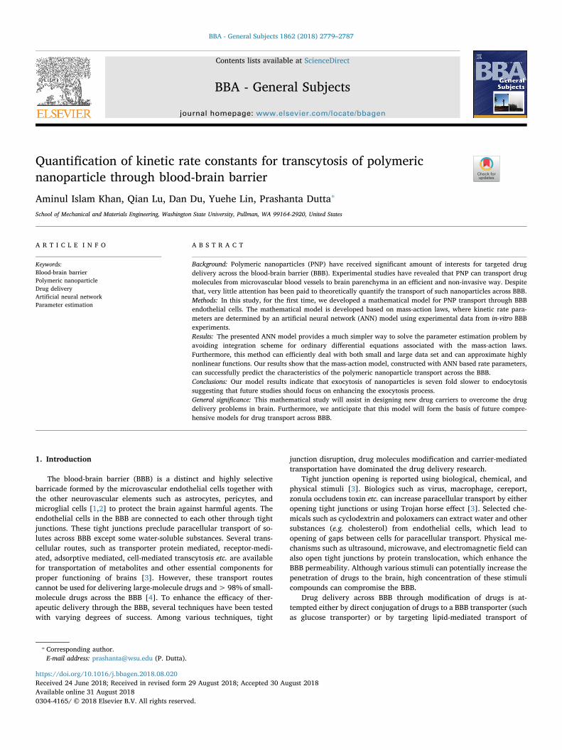
by following the work of Dua [21].The general architecture of the ANN used in this work is shown in
Fig. 2, where only fundamental steps are presented for parameter es-timation related to a system of ordinary differential equations (ODEs).The ANN model shown in Fig. 2 has only one node in the input layerbecause in our ODE-based model time is the only independent variable.The number of nodes in the output layer must be equal to the number ofoutputs (J). The number of hidden layers and the number of nodes ineach hidden layer can be arbitrary. In our model, we have considered asingle hidden layer with I number of nodes.
For a linear transfer function and no threshold in the input layer, theoutput of the input layer is exactly equal to the input (τ). Thus, the totalactivation for the hidden layer, xi can be given as
= +x v τ qi i i (11)
where qi is the threshold in the ith node of hidden layer and vi is theweight factor between the node of input layer and the ith node of hiddenlayer. Considering a sigmoid transfer function in the hidden layer,output of hidden layer, yi can be given as
=+ −y
e1
1i xi (12)
Here, we choose a sigmoid transfer function because, in general, thisfunction yields well-behaved networks [30]. Thus, the total activationfor the output layer, (zj) can be expressed as
∑= +=
z w y rji
I
ij i j1 (13)
where rj is the threshold in the jth node of output layer and wij is theweight factor between the ithnode of hidden layer and the jth node ofoutput layer for the input yi. Similar to hidden layer, considering asigmoid transfer function in the output layer, output of this layer can bewritten as
=+ −n
e1
1jANN
zj (14)
Thus, for our ANN model, unknowns are the threshold vectors (qiand rj) and the weight factor matrices (vi and wij). Values of thethreshold vectors and the weight factor matrices can be determined bysolving the following optimization problem:
Fig. 2. Schematic of an artificial neural network (ANN), consisting of one input,one hidden and one output layer, for a system of ODEs presented in Eqs. (7–9).Input layer has only one node, while the hidden layer and the output layer aremade of I and J nodes, respectively. The cap in the node sums up all theweighted inputs and feeds as total activation. For details about the ANN,readers are referred to [24,30].
A.I. Khan et al. BBA - General Subjects 1862 (2018) 2779–2787
2781
∑ ∑= −δmin
n q r v w n τ n τ, , , , ( ( ) ( ))ANNpϵP jϵJ
j p jANN
p2
(15)
A number of efficient algorithms, such as backpropagation algo-rithm [31], generalized delta rule, radial bias algorithm [30], etc. areavailable in the literatures for solving the aforementioned optimizationproblem (Eq. 15). Once the threshold vectors and weight factor ma-trices are found, the derivative of the state variables can be determinedby differentiating Eq. (14) with respect to nondimensional time, τ as
∑=+ +
−
−=
−
−
dndτ
ee
w ee
v(1 ) (1 )
jANN z
zi
I
ijx
x i21
2
j
j
i
i (16)
Note that this will provide the rate of change of state variables (left-hand side of Eqs. 7–9). Thus, one can estimate the required parametersfor the model by solving the following optimization problem [21]:
∑ ∑= ⎛
⎝⎜ − ⎞
⎠⎟ε min
kdn τ
dτf n τ k τ
( )( ( ), , )
pϵP jϵJ
jANN
pj
ANNp p
2
(17)
subject to: >k 0.Here f n τ k τ( ( ), , )j
ANNp p represents the right-hand side of Eqs. (7–9).
This optimization problem is much simpler to solve because this doesnot require an integration scheme. The first term on the right-hand sideof Eq. (17) can be easily obtained from Eq. (16), while the second termcan be calculated from the right-hand side terms of Eqs. (7–9) using theartificial neural network (nj
ANN ) solution of state variables (Eq. 14).Here, it is noted that traditional method (Eq. 10) minimizes error be-tween observed and model predicted values of state variables, whereas,our model (Eq. 17) minimizes error between the derivatives of the statevariables. This derivative based optimization method is used in otherwork [22], however, in their work, derivatives are determined using apreprocessing method.
4. Experimental section
4.1. Materials and reagents
Transwells (Polycarbonate membrane 0.4 μm) were purchased fromFisher Scientific (Waltham, MA, USA). Biological reagents, such as borncalf serum, gentamycin, penicillin, streptomycin, fetal bovine serum(FBS), and ATCC® 30–2002™ Dulbecco's Modified Eagle's Medium(DMEM) were obtained from ATCC (Manassas, VA, USA). All otherchemicals and solvents were obtained from Sigma-Aldrich (St. Louis,MO, USA).
4.2. Preparation of poly-[Triphenylamine-4-vinyl-(P-methoxy-benzene)](TEB)-based nanoparticles
In order to obtain TEB-based nanoparticles, the TEB polymer wasfirst synthesized from the reaction of two intermediates: intermediate1[4,4′-diformyl-triphenylamine], and intermediate2 [2,5-di- (ethox-yphosphorylene) -1,4-dimethoxybenzene (phospholipid)]. The inter-mediate1 was prepared through a series of chemical reactions of N, N-dimethylformamide, phosphorus oxychloride and triphenylamine. Theobtained final product was purified for further use. The intermediate2was obtained from the final product of a series of chemical reactions of1,4-dioxane, 1,4-dimethoxybenzene, hydrochloric acid, formaldehyde,and triethyl phosphite. The obtained product was re-crystallized withacetone to gain intermediate2. Both intermediate1 and intermediate2were solid powders. Finally, the TEB polymer was synthesized throughWittig-Horner reaction of intermediate1 and intermediate2. All che-mical products during the synthesis process were characterized andconfirmed using the nuclear magnetic resonance (NMR). The obtainedTEB polymer was dissolved in the solution of tetrahydrofuran, poly(styrene-co-maleic anhydride) and water. Nitrogen gas was then bub-bled through the solution to evaporate the solvent. As the solution was
concentrated, the fluorescent TEB-based nanoparticles were obtainedby co-precipitation from the solution.
4.3. Physical and chemical characterization of TEB-based nanoparticles
Previous studies suggest that the nanoparticle transport efficiencyacross the BBB decreases with the increase of particle size. For instance,several studies reported that the maximum transport efficiency isachieved for particle size ranging between 20–30 nm [10,13,25]. Thus,the synthesis steps are controlled to form nanoparticles in that ballpark.The size and morphology of TEB-based nanoparticles are characterizedby transmission electron microscopy (TEM, Philips CM200 UT, FieldEmission Instruments, USA).
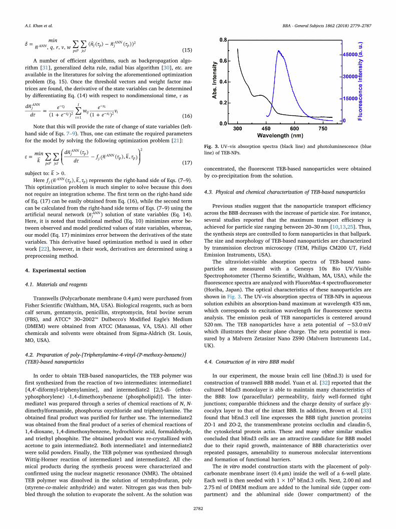
The ultraviolet-visible absorption spectra of TEB-based nano-particles are measured with a Genesys 10s Bio UV/VisibleSpectrophotometer (Thermo Scientific, Waltham, MA, USA), while thefluorescence spectra are analyzed with FluoroMax-4 spectrofluorometer(Horiba, Japan). The optical characteristics of these nanoparticles areshown in Fig. 3. The UV–vis absorption spectra of TEB-NPs in aqueoussolution exhibits an absorption-band maximum at wavelength 435 nm,which corresponds to excitation wavelength for fluorescence spectraanalysis. The emission peak of TEB nanoparticles is centered around520 nm. The TEB nanoparticles have a zeta potential of −53.0mVwhich illustrates their shear plane charge. The zeta potential is mea-sured by a Malvern Zetasizer Nano ZS90 (Malvern Instruments Ltd.,UK).
4.4. Construction of in vitro BBB model
In our experiment, the mouse brain cell line (bEnd.3) is used forconstruction of transwell BBB model. Yuan et al. [32] reported that thecultured bEnd3 monolayer is able to maintain many characteristics ofthe BBB: low (paracellular) permeability, fairly well-formed tightjunctions; comparable thickness and the charge density of surface gly-cocalyx layer to that of the intact BBB. In addition, Brown et al. [33]found that bEnd.3 cell line expresses the BBB tight junction proteinsZO-1 and ZO-2, the transmembrane proteins occludin and claudin-5,the cytoskeletal protein actin. These and many other similar studiesconcluded that bEnd3 cells are an attractive candidate for BBB modeldue to their rapid growth, maintenance of BBB characteristics overrepeated passages, amenability to numerous molecular interventionsand formation of functional barriers.
The in vitro model construction starts with the placement of poly-carbonate membrane insert (0.4 μm) inside the well of a 6-well plate.Each well is then seeded with 1× 10⁵ bEnd.3 cells. Next, 2.00ml and2.75ml of DMEM medium are added to the luminal side (upper com-partment) and the abluminal side (lower compartment) of the
Fig. 3. UV–vis absorption spectra (black line) and photoluminescence (blueline) of TEB-NPs.
A.I. Khan et al. BBA - General Subjects 1862 (2018) 2779–2787
2782
transwell, respectively. Then, the cells are cultured inside the transwellin a humidified incubator at 37 °C with 5% CO2. The culture medium ischanged every day for cell growth.
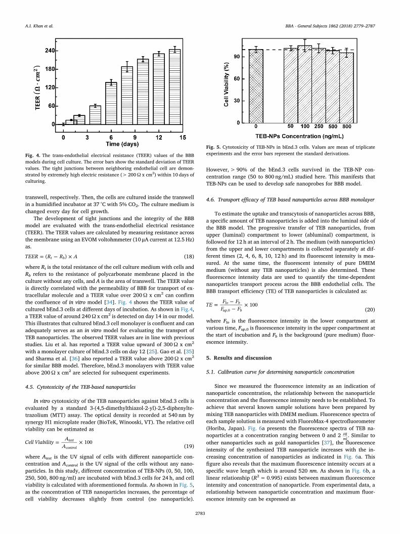
The development of tight junctions and the integrity of the BBBmodel are evaluated with the trans-endothelial electrical resistance(TEER). The TEER values are calculated by measuring resistance acrossthe membrane using an EVOM voltohmmeter (10 μA current at 12.5 Hz)as.
= − ×TEER R R A( )t b (18)
where Rt is the total resistance of the cell culture medium with cells andRb refers to the resistance of polycarbonate membrane placed in theculture without any cells, and A is the area of transwell. The TEER valueis directly correlated with the permeability of BBB for transport of ex-tracellular molecule and a TEER value over 200Ω x cm2 can confirmthe confluence of in vitro model [34]. Fig. 4 shows the TEER value ofcultured bEnd.3 cells at different days of incubation. As shown in Fig.4,a TEER value of around 240Ω x cm2 is detected on day 14 in our model.This illustrates that cultured bEnd.3 cell monolayer is confluent and canadequately serves as an in vitro model for evaluating the transport ofTEB nanoparticles. The observed TEER values are in line with previousstudies. Liu et al. has reported a TEER value upward of 300Ω x cm2
with a monolayer culture of bEnd.3 cells on day 12 [25]. Gao et al. [35]and Sharma et al. [36] also reported a TEER value above 200Ω x cm2
for similar BBB model. Therefore, bEnd.3 monolayers with TEER valueabove 200Ω x cm2 are selected for subsequent experiments.
4.5. Cytotoxicity of the TEB-based nanoparticles
In vitro cytotoxicity of the TEB nanoparticles against bEnd.3 cells isevaluated by a standard 3-(4,5-dimethylthiazol-2-yl)-2,5-diphenylte-trazolium (MTT) assay. The optical density is recorded at 540 nm bysynergy H1 microplate reader (BioTeK, Winooski, VT). The relative cellviability can be estimated as
= ×Cell Viability AA
100test
control (19)
where Atest is the UV signal of cells with different nanoparticle con-centration and Acontrol is the UV signal of the cells without any nano-particles. In this study, different concentration of TEB-NPs (0, 50, 100,250, 500, 800 ng/ml) are incubated with bEnd.3 cells for 24 h, and cellviability is calculated with aforementioned formula. As shown in Fig. 5,as the concentration of TEB nanoparticles increases, the percentage ofcell viability decreases slightly from control (no nanoparticle).
However,> 90% of the bEnd.3 cells survived in the TEB-NP con-centration range (50 to 800 ng/mL) studied here. This manifests thatTEB-NPs can be used to develop safe nanoprobes for BBB model.
4.6. Transport efficacy of TEB based nanoparticles across BBB monolayer
To estimate the uptake and transcytosis of nanoparticles across BBB,a specific amount of TEB nanoparticles is added into the luminal side ofthe BBB model. The progressive transfer of TEB nanoparticles, fromupper (luminal) compartment to lower (abluminal) compartment, isfollowed for 12 h at an interval of 2 h. The medium (with nanoparticles)from the upper and lower compartments is collected separately at dif-ferent times (2, 4, 6, 8, 10, 12 h) and its fluorescent intensity is mea-sured. At the same time, the fluorescent intensity of pure DMEMmedium (without any TEB nanoparticles) is also determined. Thesefluorescence intensity data are used to quantify the time-dependentnanoparticles transport process across the BBB endothelial cells. TheBBB transport efficiency (TE) of TEB nanoparticles is calculated as:
= −−
×TE F FF F
100lo b
up b,0 (20)
where Flo is the fluorescence intensity in the lower compartment atvarious time, Fup,0 is fluorescence intensity in the upper compartment atthe start of incubation and Fb is the background (pure medium) fluor-escence intensity.
5. Results and discussion
5.1. Calibration curve for determining nanoparticle concentration
Since we measured the fluorescence intensity as an indication ofnanoparticle concentration, the relationship between the nanoparticleconcentration and the fluorescence intensity needs to be established. Toachieve that several known sample solutions have been prepared bymixing TEB nanoparticles with DMEM medium. Fluorescence spectra ofeach sample solution is measured with FluoroMax-4 spectrofluorometer(Horiba, Japan). Fig. 6a presents the fluorescence spectra of TEB na-noparticles at a concentration ranging between 0 and 2 μg
ml. Similar to
other nanoparticles such as gold nanoparticles [37], the fluorescenceintensity of the synthesized TEB nanoparticle increases with the in-creasing concentration of nanoparticles as indicated in Fig. 6a. Thisfigure also reveals that the maximum fluorescence intensity occurs at aspecific wave length which is around 520 nm. As shown in Fig. 6b, alinear relationship (R2= 0.995) exists between maximum fluorescenceintensity and concentration of nanoparticle. From experimental data, arelationship between nanoparticle concentration and maximum fluor-escence intensity can be expressed as
Fig. 4. The trans-endothelial electrical resistance (TEER) values of the BBBmodels during cell culture. The error bars show the standard deviation of TEERvalues. The tight junctions between neighboring endothelial cell are demon-strated by extremely high electric resistance (> 200Ω x cm2) within 10 days ofculturing.
Fig. 5. Cytotoxicity of TEB-NPs in bEnd.3 cells. Values are mean of triplicateexperiments and the error bars represent the standard derivations.
A.I. Khan et al. BBA - General Subjects 1862 (2018) 2779–2787
2783
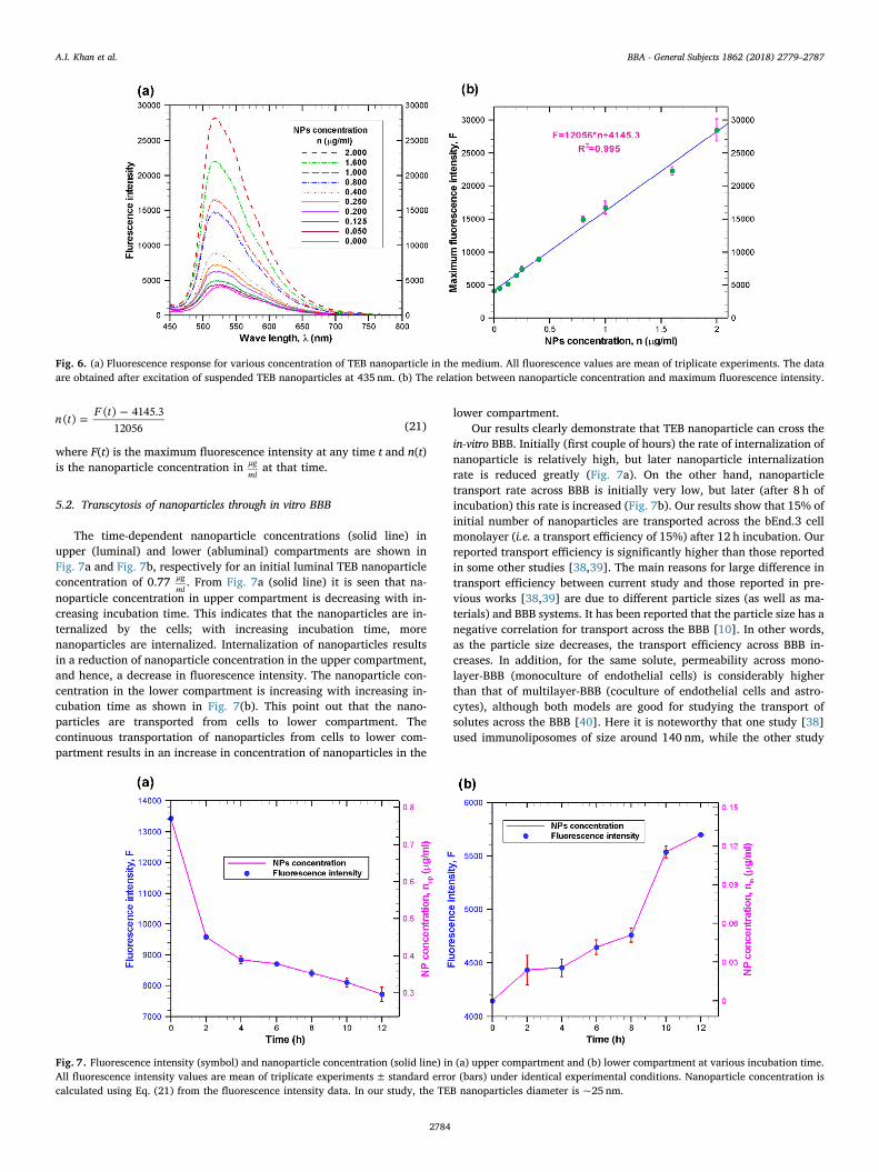
= −n t F t( ) ( ) 4145.312056 (21)
where F(t) is the maximum fluorescence intensity at any time t and n(t)is the nanoparticle concentration in μg
mlat that time.
5.2. Transcytosis of nanoparticles through in vitro BBB
The time-dependent nanoparticle concentrations (solid line) inupper (luminal) and lower (abluminal) compartments are shown inFig. 7a and Fig. 7b, respectively for an initial luminal TEB nanoparticleconcentration of 0.77 μg
ml. From Fig. 7a (solid line) it is seen that na-
noparticle concentration in upper compartment is decreasing with in-creasing incubation time. This indicates that the nanoparticles are in-ternalized by the cells; with increasing incubation time, morenanoparticles are internalized. Internalization of nanoparticles resultsin a reduction of nanoparticle concentration in the upper compartment,and hence, a decrease in fluorescence intensity. The nanoparticle con-centration in the lower compartment is increasing with increasing in-cubation time as shown in Fig. 7(b). This point out that the nano-particles are transported from cells to lower compartment. Thecontinuous transportation of nanoparticles from cells to lower com-partment results in an increase in concentration of nanoparticles in the
lower compartment.Our results clearly demonstrate that TEB nanoparticle can cross the
in-vitro BBB. Initially (first couple of hours) the rate of internalization ofnanoparticle is relatively high, but later nanoparticle internalizationrate is reduced greatly (Fig. 7a). On the other hand, nanoparticletransport rate across BBB is initially very low, but later (after 8 h ofincubation) this rate is increased (Fig. 7b). Our results show that 15% ofinitial number of nanoparticles are transported across the bEnd.3 cellmonolayer (i.e. a transport efficiency of 15%) after 12 h incubation. Ourreported transport efficiency is significantly higher than those reportedin some other studies [38,39]. The main reasons for large difference intransport efficiency between current study and those reported in pre-vious works [38,39] are due to different particle sizes (as well as ma-terials) and BBB systems. It has been reported that the particle size has anegative correlation for transport across the BBB [10]. In other words,as the particle size decreases, the transport efficiency across BBB in-creases. In addition, for the same solute, permeability across mono-layer-BBB (monoculture of endothelial cells) is considerably higherthan that of multilayer-BBB (coculture of endothelial cells and astro-cytes), although both models are good for studying the transport ofsolutes across the BBB [40]. Here it is noteworthy that one study [38]used immunoliposomes of size around 140 nm, while the other study
Fig. 6. (a) Fluorescence response for various concentration of TEB nanoparticle in the medium. All fluorescence values are mean of triplicate experiments. The dataare obtained after excitation of suspended TEB nanoparticles at 435 nm. (b) The relation between nanoparticle concentration and maximum fluorescence intensity.
Fig. 7. Fluorescence intensity (symbol) and nanoparticle concentration (solid line) in (a) upper compartment and (b) lower compartment at various incubation time.All fluorescence intensity values are mean of triplicate experiments± standard error (bars) under identical experimental conditions. Nanoparticle concentration iscalculated using Eq. (21) from the fluorescence intensity data. In our study, the TEB nanoparticles diameter is ~25 nm.
A.I. Khan et al. BBA - General Subjects 1862 (2018) 2779–2787
2784
[39] is based on gold nanoparticles of overall size between 70 and100 nm. Moreover, in both studies, BBB is established by co-culture ofbrain endothelial cells and astrocytes. On the other hand, in our study,monolayer BBB is formed with bEnd.3 cell and transport assay is carriedout with TEB nanoparticles of size ~ 25 nm. Since, the particle size isvery low and BBB is formed with bEnd.3 monolayer, current studyyields higher transport efficiency. This observed transport efficiency isin line with previous works having similar BBB system and comparableparticle size. For instance, Liu et al. [25] reported a transport efficiencyof 26.1 ± 8.9% across the BBB (formed by bEND.3 cell monolayer)after 12 h incubation with poly(ethylene glycol) (PEG) coated silicananoparticles (25 nm diameter). Qiao et al. [41] also showed thattransport efficiency of PEG-coated Fe3O4 nanoparticles across mono-layer BBB (formed by porcine brain capillary endothelial cells) can beas high as 22.5 ± 1.4%. Here it is noteworthy to mention that evenwith three layer BBB system (co culture of bEND.3 cell line, pericytesand astrocytes), 25 nm silica nanoparticle yields a much higher trans-port efficiency [10].
Next, we determined the nanoparticle accumulation inside the cell.Considering negligible degradation of TEB nanoparticles inside the cell,the mass balance equation can be given as follows:
+ + =dn
dtdndt
dndt
0up cl lo(22)
Integrating this equation from starting time (t=0) to any time twith initial conditions, nup,0= 0.77, ncl,0 = 0 and nlo,0= 0, we get fol-lowing mass balance equation for nanoparticle accumulation inside theendothelial cells:
= − −n n n0.77cl up lo (23)
The progressive accumulation of TEB nanoparticles inside the en-dothelial cell is shown in Fig. 8. During the first 4 h, there is a rapidaccumulation of nanoparticles inside the cell because of high en-docytosis and low exocytosis. Fig. 8 indicates that TEB nanoparticleaccumulation gets saturated after 4 h. This kind of saturation char-acteristics is quite common for other particles such as iron transportacross BBB [42]. The saturation in nanoparticle accumulation is due tothe fact that cells have a limiting capacity to hold nanoparticles. Thissaturation in nanoparticle accumulation also indicates that endocytosisis in equilibrium with exocytosis [42].
5.3. Estimation of kinetic rate constant
The primary focus of this study is to find the rate constants formodel equations using experimental data. For simplicity, the analysiswas performed for normalized governing equations presented in Eqs.(7–9). Thus, the experimental data of nanoparticle concentrations arenormalized with nup,0 = 0.77 μg
ml, while the time is normalized with =10
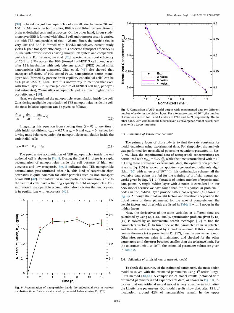
h. Using these normalized experimental data, the optimization problemgiven in Eq. (15) is solved by applying a generalized delta rule algo-rithm [30] with an error of 10−3. In this optimization scheme, all theavailable data points are fed for the training of artificial neural net-works given by Eqs. (11–14) because of limited number of experimentaldata points. A single hidden layer with 3 nodes is considered in ourANN model because we have found that, for this particular problem, 3nodes in the hidden layer provide faster convergence (as shown inFig. 9). Although the final weight factors and thresholds depend on theinitial guess of these parameter, for the sake of completeness, theweight factors and thresholds are listed in Table 1 with 3 nodes in thehidden layers.
Next, the derivatives of the state variables at different time arecalculated by using Eq. (16). Finally, optimization problem given by Eq.(17) is solved by an incremental search technique [17] to find theparameters vector, k . In brief, one of the parameter value is selectedand then its value is changed by a random amount. If this change de-creases the error (ε) as presented in Eq. (17), then the new value is kept.Otherwise, previous value is maintained and checked for the otherparameters until the error becomes smaller than the tolerance limit. Forthe tolerance limit 1× 10⁻³, the estimated parameter values are givenin Table 2.
5.4. Validation of artificial neural network model
To check the accuracy of the estimated parameters, the mass actionmodel is solved with the estimated parameters using 4th order Runge-Kutta method [43,44]. A comparison of model results (obtained withestimated parameters) and experimental data, as shown in Fig. 10, in-dicates that our artificial neural model is very effective in estimatingthe kinetic rate parameters. Our model results show that, after 12 h ofincubation, around 42% of nanoparticles remain in the upper
Fig. 8. Accumulation of nanoparticles inside the endothelial cells at variousincubation time. Data are calculated by material balance using Eq. (23).
Fig. 9. Comparison of ANN model output with experimental data for differentnumber of nodes in the hidden layer. For a tolerance limit of 10−3,the numberof iterations needed for 3 and 4 nodes are 1203 and 1409, respectively. On theother hand, with 2 nodes in the hidden layer, a convergence cannot be achievedeven with 12,000 iterations.
A.I. Khan et al. BBA - General Subjects 1862 (2018) 2779–2787
2785
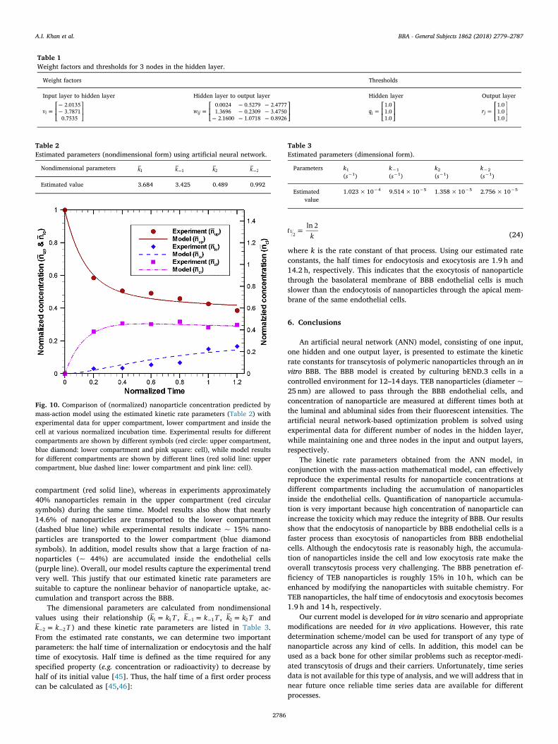
compartment (red solid line), whereas in experiments approximately40% nanoparticles remain in the upper compartment (red circularsymbols) during the same time. Model results also show that nearly14.6% of nanoparticles are transported to the lower compartment(dashed blue line) while experimental results indicate ~ 15% nano-particles are transported to the lower compartment (blue diamondsymbols). In addition, model results show that a large fraction of na-noparticles (~ 44%) are accumulated inside the endothelial cells(purple line). Overall, our model results capture the experimental trendvery well. This justify that our estimated kinetic rate parameters aresuitable to capture the nonlinear behavior of nanoparticle uptake, ac-cumulation and transport across the BBB.
The dimensional parameters are calculated from nondimensionalvalues using their relationship ( =k k T1 1 , =− −k k T1 1 , =k k T2 2 and
=− −k k T2 2 ) and these kinetic rate parameters are listed in Table 3.From the estimated rate constants, we can determine two importantparameters: the half time of internalization or endocytosis and the halftime of exocytosis. Half time is defined as the time required for anyspecified property (e.g. concentration or radioactivity) to decrease byhalf of its initial value [45]. Thus, the half time of a first order processcan be calculated as [45,46]:
=tk
ln 21
2 (24)
where k is the rate constant of that process. Using our estimated rateconstants, the half times for endocytosis and exocytosis are 1.9 h and14.2 h, respectively. This indicates that the exocytosis of nanoparticlethrough the basolateral membrane of BBB endothelial cells is muchslower than the endocytosis of nanoparticles through the apical mem-brane of the same endothelial cells.
6. Conclusions
An artificial neural network (ANN) model, consisting of one input,one hidden and one output layer, is presented to estimate the kineticrate constants for transcytosis of polymeric nanoparticles through an invitro BBB. The BBB model is created by culturing bEND.3 cells in acontrolled environment for 12–14 days. TEB nanoparticles (diameter ~25 nm) are allowed to pass through the BBB endothelial cells, andconcentration of nanoparticle are measured at different times both atthe luminal and abluminal sides from their fluorescent intensities. Theartificial neural network-based optimization problem is solved usingexperimental data for different number of nodes in the hidden layer,while maintaining one and three nodes in the input and output layers,respectively.
The kinetic rate parameters obtained from the ANN model, inconjunction with the mass-action mathematical model, can effectivelyreproduce the experimental results for nanoparticle concentrations atdifferent compartments including the accumulation of nanoparticlesinside the endothelial cells. Quantification of nanoparticle accumula-tion is very important because high concentration of nanoparticle canincrease the toxicity which may reduce the integrity of BBB. Our resultsshow that the endocytosis of nanoparticle by BBB endothelial cells is afaster process than exocytosis of nanoparticles from BBB endothelialcells. Although the endocytosis rate is reasonably high, the accumula-tion of nanoparticles inside the cell and low exocytosis rate make theoverall transcytosis process very challenging. The BBB penetration ef-ficiency of TEB nanoparticles is roughly 15% in 10 h, which can beenhanced by modifying the nanoparticles with suitable chemistry. ForTEB nanoparticles, the half time of endocytosis and exocytosis becomes1.9 h and 14 h, respectively.
Our current model is developed for in vitro scenario and appropriatemodifications are needed for in vivo applications. However, this ratedetermination scheme/model can be used for transport of any type ofnanoparticle across any kind of cells. In addition, this model can beused as a back bone for other similar problems such as receptor-medi-ated transcytosis of drugs and their carriers. Unfortunately, time seriesdata is not available for this type of analysis, and we will address that innear future once reliable time series data are available for differentprocesses.
Table 1Weight factors and thresholds for 3 nodes in the hidden layer.
Weight factors Thresholds
Input layer to hidden layer Hidden layer to output layer Hidden layer Output layer
= ⎡
⎣⎢
−−
⎤
⎦⎥v
2.01353.7871
0.7535i = ⎡
⎣⎢
− −− −
− − −
⎤
⎦⎥w
0.0024 0.5279 2.47771.3696 0.2309 3.4750
2.1600 1.0718 0.8926ij = ⎡
⎣⎢
⎤
⎦⎥q
1.01.01.0
i = ⎡
⎣⎢
⎤
⎦⎥r
1.01.01.0
j
Table 2Estimated parameters (nondimensional form) using artificial neural network.
Nondimensional parameters k1 −k 1 k2 −k 2
Estimated value 3.684 3.425 0.489 0.992
Fig. 10. Comparison of (normalized) nanoparticle concentration predicted bymass-action model using the estimated kinetic rate parameters (Table 2) withexperimental data for upper compartment, lower compartment and inside thecell at various normalized incubation time. Experimental results for differentcompartments are shown by different symbols (red circle: upper compartment,blue diamond: lower compartment and pink square: cell), while model resultsfor different compartments are shown by different lines (red solid line: uppercompartment, blue dashed line: lower compartment and pink line: cell).
Table 3Estimated parameters (dimensional form).
Parameters k1(s−1)
k−1
(s−1)k2(s−1)
k−2
(s−1)
Estimatedvalue
1.023× 10⁻⁴ 9.514× 10⁻⁵ 1.358×10⁻⁵ 2.756× 10⁻⁵
A.I. Khan et al. BBA - General Subjects 1862 (2018) 2779–2787
2786
Acknowledgment
This work was supported by the National Institute of GeneralMedical Sciences of the US National Institutes of Health under AwardNumber R01GM122081. The content is solely the responsibility of theauthors and does not necessarily represent the official views of theNational Institutes of Health.
References
[1] R. Cecchelli, V. Berezowski, S. Lundquist, M. Culot, M. Renftel, M.P. Dehouck,L. Fenart, Modelling of the blood-brain barrier in drug discovery and development,Nat. Rev. Drug Discov. 6 (2007) 650–661.
[2] N.J. Abbott, A.A. Patabendige, D.E. Dolman, S.R. Yusof, D.J. Begley, Structure andfunction of the blood–brain barrier, Neurobiol. Dis. 37 (2010) 13–25.
[3] Y. Chen, L.H. Liu, Modern methods for delivery of drugs across the blood-brainbarrier, Adv. Drug Deliv. Rev. 64 (2012) 640–665.
[4] W.M. Pardridge, The blood-brain barrier: bottleneck in brain drug development,NeuroRx 2 (2005) 3–14.
[5] L. Juillerat-Jeanneret, The targeted delivery of cancer drugs across the blood-brainbarrier: chemical modifications of drugs or drug-nanoparticles? Drug Discov. Today13 (2008) 1099–1106.
[6] M.M. Patel, B.R. Goyal, S.V. Bhadada, J.S. Bhatt, A.F. Amin, Getting into the brain,CNS Drugs 23 (2009) 35–58.
[7] A.V. Kabanov, E.V. Batrakova, Polymer nanomaterials for drug delivery across theblood brain barrier, Neuroimmune Pharmacology, Springer, 2017, pp. 847–868.
[8] P.R. Lockman, R.J. Mumper, M.A. Khan, D.D. Allen, Nanoparticle technology fordrug delivery across the blood-brain barrier, Drug Dev. Ind. Pharm. 28 (2002) 1–13.
[9] Y. Shen, B. Cao, N.R. Snyder, K.M. Woeppel, J.R. Eles, X.T. Cui, ROS responsiveresveratrol delivery from LDLR peptide conjugated PLA-coated mesoporous silicananoparticles across the blood-brain barrier, Journal of Nanobiotechnology 16(2018) 13.
[10] Y. Song, D. Du, L. Li, J. Xu, P. Dutta, Y.H. Lin, In vitro study of receptor-mediatedsilica nanoparticles delivery across blood-brain barrier, ACS Appl. Mater. Interfaces9 (2017) 20410–20416.
[11] H.C. Lin, M.Y. Ho, C.M. Tsen, C.C. Huang, C.C. Wu, Y.J. Huang, I.L. Hsiao,C.Y. Chuang, From the cover: comparative proteomics reveals silver nanoparticlesalter fatty acid metabolism and amyloid beta clearance for neuronal apoptosis in atriple cell coculture model of the blood-brain barrier, Toxicol. Sci. 158 (2017)151–163.
[12] A. Ivask, E.H. Pilkington, T. Blin, A. Kakinen, H. Vija, M. Visnapuu, J.F. Quinn,M.R. Whittaker, R.R. Qiao, T.P. Davis, P.C. Ke, N.H. Voelcker, Uptake and trans-cytosis of functionalized superparamagnetic iron oxide nanoparticles in an in vitroblood brain barrier model, Biomaterials Science 6 (2018) 314–323.
[13] O. Betzer, M. Shilo, R. Opochinsky, E. Barnoy, M. Motiei, E. Okun, G. Yadid,R. Popovtzer, The effect of nanoparticle size on the ability to cross the blood-brainbarrier: an in vivo study, Nanomedicine 12 (2017) 1533–1546.
[14] J.M. Chan, P.M. Valencia, L. Zhang, R. Langer, O.C. Farokhzad, Polymeric nano-particles for drug delivery, Cancer Nanotechnology, Springer (2010) 163–175.
[15] P. Hinow, A. Radunskaya, S.M. MacKay, J.N.J. Reynolds, M. Schroeder, E.W. Tan,I.G. Tucker, Signaled drug delivery and transport across the blood-brain barrier,Journal of Liposome Research 26 (2016) 233–245.
[16] V.F. Loeser, Modeling of Anticancer Drug Delivery by Temperature-SensitiveLiposomes, the University of Wisconsin-Milwaukee, 2017 PhD Dissertation.
[17] A.I. Khan, J. Liu, P. Dutta, Iron transport kinetics through blood-brain barrier en-dothelial cells, Biochimica et Biophysica Acta (BBA)-General Subjects 1862 (2018)1168–1179.
[18] R.J. Ritchie, T. Prvan, Current statistical methods for estimating the Km and Vmaxof Michaelis-Menten kinetics, Biochem. Educ. 24 (1996) 196–206.
[19] H. Putter, S.H. Heisterkamp, J.M.A. Lange, F. de Wolf, A Bayesian approach toparameter estimation in HIV dynamical models, Stat. Med. 21 (2002) 2199–2214.
[20] C. Michalik, B. Chachuat, W. Marquardt, Incremental global parameter estimationin dynamical systems, Ind. Eng. Chem. Res. 48 (2009) 5489–5497.
[21] V. Dua, An Artificial Neural Network approximation based decomposition approachfor parameter estimation of system of ordinary differential equations, Comput.Chem. Eng. 35 (2011) 545–553.
[22] O. Strebel, A preprocessing method for parameter estimation in ordinary differ-ential equations, Chaos, Solitons Fractals 57 (2013) 93–104.
[23] B.P. Bezruchko, D.A. Smirnov, I.V. Sysoev, Identification of chaotic systems withhidden variables (modified Bock's algorithm), Chaos, Solitons Fractals 29 (2006)82–90.
[24] V. Dua, A mixed-integer programming approach for optimal configuration of arti-ficial neural networks, Chemical Engineering Research & Design 88 (2010) 55–60.
[25] D. Liu, B.Q. Lin, W. Shao, Z. Zhu, T.H. Ji, C.Y. Yang, In vitro and in vivo studies onthe transport of PEGylated silica nanoparticles across the blood-brain barrier, ACSAppl. Mater. Interfaces 6 (2014) 2131–2136.
[26] G. Sonavane, K. Tomoda, K. Makino, Biodistribution of colloidal gold nanoparticlesafter intravenous administration: effect of particle size, Colloids and Surfaces B-Biointerfaces 66 (2008) 274–280.
[27] S.A. Predescu, D.N. Predescu, K. Shimizu, I.K. Klein, A.B. Malik, Cholesterol-de-pendent syntaxin-4 and SNAP-23 clustering regulates caveolar fusion with the en-dothelial plasma membrane, J. Biol. Chem. 280 (2005) 37130–37138.
[28] Q.Y. Zhu, M. Yamakuchi, C.J. Lowenstein, SNAP23 regulates endothelial exocytosisof von Willebrand factor, PLoS One 10 (2015) e0118737.
[29] L. Descamps, M.-P. Dehouck, G. Torpier, R. Cecchelli, Receptor-mediated transcy-tosis of transferrin through blood-brain barrier endothelial cells, Am. J. Phys. HeartCirc. Phys. 270 (1996) H1149–H1158.
[30] D.R. Baughman, Y.A. Liu, Neural Networks in Bioprocessing and ChemicalEngineering, Academic Press, San Diego, CA, 2014.
[31] R. Vaidyanathan, Process fault Detection and diagnosis Using Neural Netw., PhDDissertation, Purdue University (1991).
[32] W. Yuan, G.L. Li, B.M. Fu, Effect of surface charge of immortalized mouse cerebralendothelial cell monolayer on transport of charged solutes, Ann. Biomed. Eng. 38(2010) 1463–1472.
[33] R.C. Brown, A.P. Morris, R.G. O'Neil, Tight junction protein expression and barrierproperties of immortalized mouse brain microvessel endothelial cells, Brain Res.1130 (2007) 17–30.
[34] A. Wong, M. Ye, A. Levy, J. Rothstein, D. Bergles, P.C. Searson, The blood-brainbarrier: an engineering perspective, Frontiers in Neuroengineering 6 (2013) 7.
[35] H.L. Gao, J. Qian, S.J. Cao, Z. Yang, Z.Q. Pang, S.Q. Pan, L. Fan, Z.J. Xi, X.G. Jiang,Q.Z. Zhang, Precise glioma targeting of and penetration by aptamer and peptidedual-functioned nanoparticles, Biomaterials 33 (2012) 5115–5123.
[36] G. Sharma, A. Modgil, T.C. Zhong, C.W. Sun, J. Singh, Influence of short-chain cell-penetrating peptides on transport of doxorubicin encapsulating receptor-targetedliposomes across brain endothelial barrier, Pharm. Res. 31 (2014) 1194–1209.
[37] C.C. Huang, Z. Yang, K.H. Lee, H.T. Chang, Synthesis of highly fluorescent goldnanoparticles for sensing mercury (II), Angew. Chem. 119 (2007) 6948–6952.
[38] K.B. Johnsen, A. Burkhart, F. Melander, P.J. Kempen, J.B. Vejlebo, P. Siupka,M.S. Nielsen, T.L. Andresen, T. Moos, Targeting transferrin receptors at the blood-brain barrier improves the uptake of immunoliposomes and subsequent cargotransport into the brain parenchyma, Sci. Rep. 7 (2017) 10396.
[39] K.B. Johnsen, M. Bak, P.J. Kempen, F. Melander, A. Burkhart, M.S. Thomsen,M.S. Nielsen, T. Moos, T.L. Andresen, Antibody affinity and valency impact brainuptake of transferrin receptor-targeted gold nanoparticles, Theranostics 8 (2018)3416–3436.
[40] G.L. Li, M.J. Simon, L.M. Cancel, Z.D. Shi, X.Y. Ji, J.M. Tarbell, B. Morrison, M. Fu,Permeability of Endothelial and Astrocyte Cocultures: in vitro blood-brain barriermodels for drug delivery studies, Ann. Biomed. Eng. 38 (2010) 2499–2511.
[41] R.R. Qiao, Q.J. Jia, S. Huwel, R. Xia, T. Liu, F.B. Gao, H.J. Galla, M.Y. Gao,Receptor-mediated delivery of magnetic nanoparticles across the blood-brain bar-rier, ACS Nano 6 (2012) 3304–3310.
[42] T.J. Raub, C.R. Newton, Recycling kinetics and transcytosis of transferrin in pri-mary cultures of bovine brain microvessel endothelial cells, J. Cell. Physiol. 149(1991) 141–151.
[43] S.C. Chapra, R.P. Canale, Numerical Methods for Engineers, McGraw Hill, NewYork, 1998.
[44] R.L. Burden, J.D. Faires, Numerical Analysis. 2001, Brooks/Cole, Boston, USA,2001.
[45] K.M. Mayle, A.M. Le, D.T. Kamei, The intracellular trafficking pathway of trans-ferrin, Biochimica et Biophysica Acta (BBA)-General Subjects 1820 (2012)264–281.
[46] A. Ciechanover, A. Schwartz, A. Dautry-Varsat, H. Lodish, Kinetics of internaliza-tion and recycling of transferrin and the transferrin receptor in a human hepatomacell line. Effect of lysosomotropic agents, J. Biol. Chem. 258 (1983) 9681–9689.
A.I. Khan et al. BBA - General Subjects 1862 (2018) 2779–2787
2787