Embed Size (px)
Citation preview
BCH 313 Metabolism of Amino acids and Proteins (3 UNITS)
Proteins are polymers of amino acids, with each amino acid residue joined to its neighbour
by a specific type of covalent bond. (The term ―residue‖ reflects the loss of the elements of
water when one amino acid is joined to another.) Proteins can be broken down (hydrolysed)
to their constituent amino acids by a variety of methods, and the earliest studies of proteins
naturally focused on the free amino acids derived from them. Twenty different amino acids
are commonly found in proteins. The first to be discovered was asparagine, in 1806. The last
of the 20 to be found, threonine, was not identified until 1938. All the amino acids have
trivial or common names, in some cases derived from the source from which they were first
isolated. Asparagine was first found in asparagus, and glutamate in wheat gluten; tyrosine
was first isolated from cheese (its name is derived from the Greek tyros, ―cheese‖); and
glycine (Greek glykos,―sweet‖) was so named because of its sweet taste.
Amino Acids Share Common Structural Features
All 20 of the common amino acids are α-amino acids. They have a carboxyl group and an
amino group bonded to the same carbon atom (the α-carbon). They differ from each other in
their side chains, or R groups, which vary in structure, size, and electric charge, and which
influence the solubility of the amino acids in water. In addition to these 20 amino acids there
are many less common ones. Some are residues modified after a protein has been
synthesized; others are amino acids present in living organisms but not as constituents of
proteins. The common amino acids of proteins have been assigned three-letter abbreviations
and one-letter symbols, which are used as shorthand to indicate the composition and sequence
of amino acids polymerized in proteins.
Two conventions are used to identify the carbons in an amino acid—a practice that can be
confusing. The additional carbons in an R group are commonly designated β, γ, δ, ε, and so
forth, proceeding out from the carbon. For most other organic molecules, carbon atoms are
simply numbered from one end, giving highest priority (C-1) to the carbon with the
substituent containing the atom of highest atomic number. Within this latter convention, the
carboxyl carbon of an amino acid would be C-1 and the α-carbon would be C-2. In some
cases, such as amino acids with heterocyclic R groups, the Greek lettering system is
ambiguous and the numbering convention is therefore used. For all the common amino acids
except glycine, the carbon is bonded to four different groups: a carboxyl group, an amino
group, an R group, and a hydrogen atom in glycine, the R group is another hydrogen atom).
The α-carbon atom is thus a chiral centre. Because of the tetrahedral arrangement of the
bonding orbitals around the α-carbon atom, the four different groups can occupy two unique
spatial arrangements, and thus amino acids have two possible stereoisomers. Since they are
no superimposable mirror images of each other, the two forms represent a class of
stereoisomers called enantiomers. All molecules with a chiral centre are also optically
active—that is, they rotate plane-polarized light.
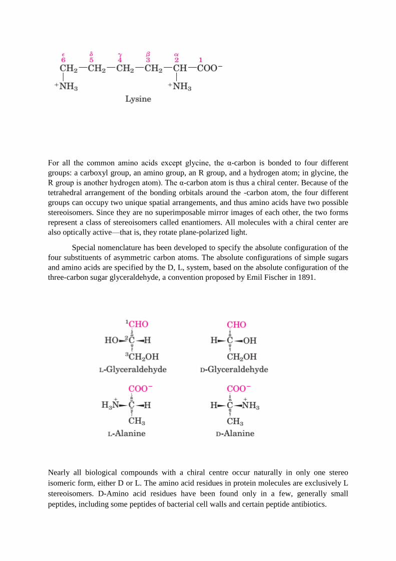
For all the common amino acids except glycine, the α-carbon is bonded to four different
groups: a carboxyl group, an amino group, an R group, and a hydrogen atom; in glycine, the
R group is another hydrogen atom). The α-carbon atom is thus a chiral center. Because of the
tetrahedral arrangement of the bonding orbitals around the -carbon atom, the four different
groups can occupy two unique spatial arrangements, and thus amino acids have two possible
stereoisomers. Since they are no superimposable mirror images of each other, the two forms
represent a class of stereoisomers called enantiomers. All molecules with a chiral center are
also optically active—that is, they rotate plane-polarized light.
Special nomenclature has been developed to specify the absolute configuration of the
four substituents of asymmetric carbon atoms. The absolute configurations of simple sugars
and amino acids are specified by the D, L, system, based on the absolute configuration of the
three-carbon sugar glyceraldehyde, a convention proposed by Emil Fischer in 1891.
Nearly all biological compounds with a chiral centre occur naturally in only one stereo
isomeric form, either D or L. The amino acid residues in protein molecules are exclusively L
stereoisomers. D-Amino acid residues have been found only in a few, generally small
peptides, including some peptides of bacterial cell walls and certain peptide antibiotics.

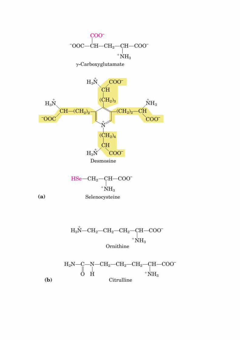
Uncommon Amino Acids Also Have Important Functions
In addition to the 20 common amino acids, proteins may contain residues created by
modification of common residues already incorporated into a polypeptide. Among these
uncommon amino acids are 4-hydroxyproline, a derivative of proline, and 5-hydroxylysine,
derived from lysine. The former is found in plant cell wall proteins, and both are found in
collagen, a fibrous protein of connective tissues. 6-NMethyllysineis a constituent of myosin,
a contractile protein of muscle. Another important uncommon amino acid is -
carboxyglutamate, found in the blood clotting protein prothrombin and in certain other
proteins that bind Ca2+
as part of their biological function.
More complex is desmosine,a derivative of four Lys residues, which is found in the fibrous
protein elastin. Selenocysteine is a special case. This rare amino acid residue is introduced
during protein synthesis rather than created through a post synthetic modification. It contains
selenium rather than the sulfur of cysteine. Actually derived from serine, selenocysteine is a
constituent of just a few known proteins. Some 300 additional amino acids have been found
in cells. They have a variety of functions but are not constituents of proteins. Ornithine and
citrulline deserve special note because they are key intermediates (metabolites) in the
biosynthesis of arginine and in the urea cycle.
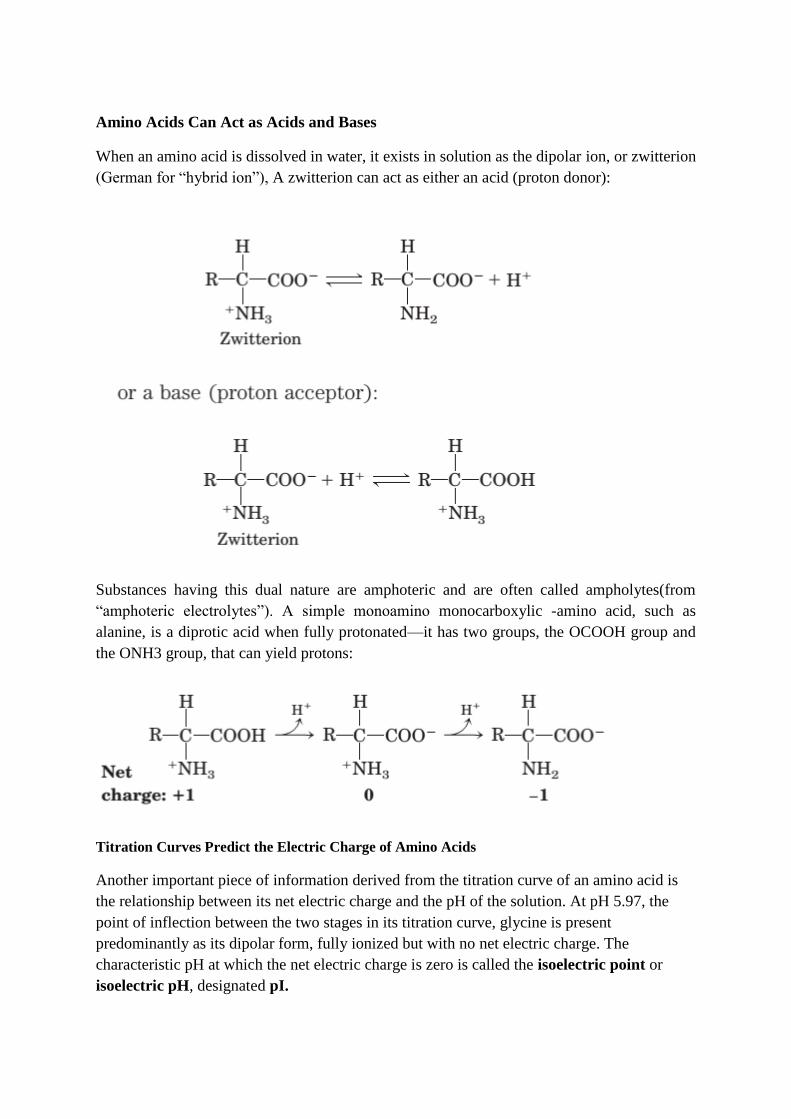
Amino Acids Can Act as Acids and Bases
When an amino acid is dissolved in water, it exists in solution as the dipolar ion, or zwitterion
(German for ―hybrid ion‖), A zwitterion can act as either an acid (proton donor):
Substances having this dual nature are amphoteric and are often called ampholytes(from
―amphoteric electrolytes‖). A simple monoamino monocarboxylic -amino acid, such as
alanine, is a diprotic acid when fully protonated—it has two groups, the OCOOH group and
the ONH3 group, that can yield protons:
Titration Curves Predict the Electric Charge of Amino Acids
Another important piece of information derived from the titration curve of an amino acid is
the relationship between its net electric charge and the pH of the solution. At pH 5.97, the
point of inflection between the two stages in its titration curve, glycine is present
predominantly as its dipolar form, fully ionized but with no net electric charge. The
characteristic pH at which the net electric charge is zero is called the isoelectric point or
isoelectric pH, designated pI.
Amino Acids Can Be Classified by R Group
Knowledge of the chemical properties of the common amino acids is central to an
understanding of biochemistry. The topic can be simplified by grouping the amino acids into
five main classes based on the properties of their R groups (Table 3–1), in particular, their
polarity, or tendency to interact with water at biological pH (near pH 7.0). The polarity of the
R groups varies widely, from nonpolar and hydrophobic (water-insoluble) to highly polar and
hydrophilic (water-soluble) within each class there are gradations of polarity, size, and shape
of the R groups. Nonpolar, Aliphatic R Groups the R groups in this class of amino acids are
nonpolar and hydrophobic. The side chains of alanine, valine, leucine, and isoleucine tend to
cluster together within proteins, stabilizing protein structure by means of hydrophobic
interactions. Glycine has the simplest structure. Although it is formally nonpolar, it’s very
small side chain makes no real contribution to hydrophobic interactions. Methionine, one of
the two sulfur-containing amino acids, has a nonpolar thioether group in its side chain.
Proline has an aliphatic side chain with a distinctive cyclic structure. The secondary amino
(imino) group of proline residues is held in a rigid conformation that reduces the structural
flexibility of polypeptide regions containing proline.
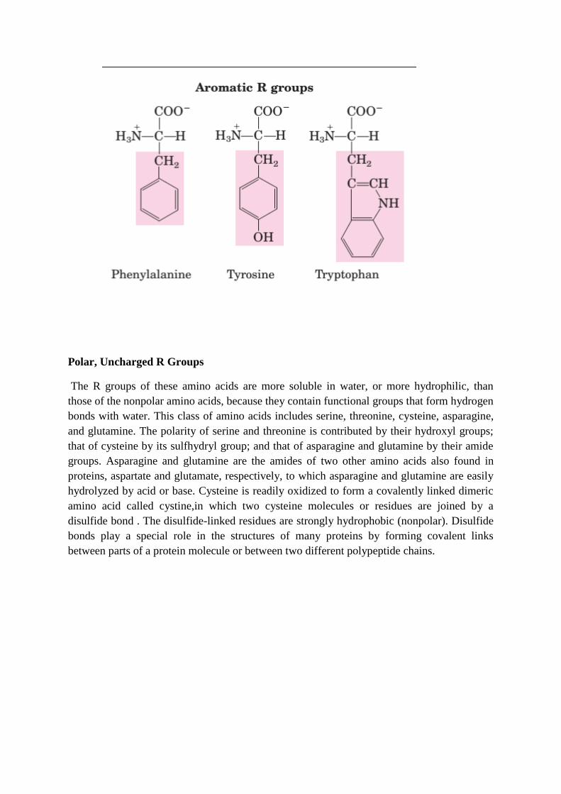
Aromatic R Groups
Phenylalanine, tyrosine, and tryptophan, with their aromatic side chains, are relatively
nonpolar (hydrophobic). All can participate in hydrophobic interactions. The hydroxyl group
of tyrosine can form hydrogen bonds, and it is an important functional group in some
enzymes. Tyrosine and tryptophan are significantly more polar than phenylalanine, because
of the tyrosine hydroxyl group and the nitrogen of the tryptophan indole ring.
Tryptophan and tyrosine, and to a much lesser extent phenylalanine, absorb ultraviolet light.
This accounts for the characteristic strong absorbance of light by most proteins at a
wavelength of 280 nm, a property exploited by researchers in the characterization of proteins.
Polar, Uncharged R Groups
The R groups of these amino acids are more soluble in water, or more hydrophilic, than
those of the nonpolar amino acids, because they contain functional groups that form hydrogen
bonds with water. This class of amino acids includes serine, threonine, cysteine, asparagine,
and glutamine. The polarity of serine and threonine is contributed by their hydroxyl groups;
that of cysteine by its sulfhydryl group; and that of asparagine and glutamine by their amide
groups. Asparagine and glutamine are the amides of two other amino acids also found in
proteins, aspartate and glutamate, respectively, to which asparagine and glutamine are easily
hydrolyzed by acid or base. Cysteine is readily oxidized to form a covalently linked dimeric
amino acid called cystine,in which two cysteine molecules or residues are joined by a
disulfide bond . The disulfide-linked residues are strongly hydrophobic (nonpolar). Disulfide
bonds play a special role in the structures of many proteins by forming covalent links
between parts of a protein molecule or between two different polypeptide chains.
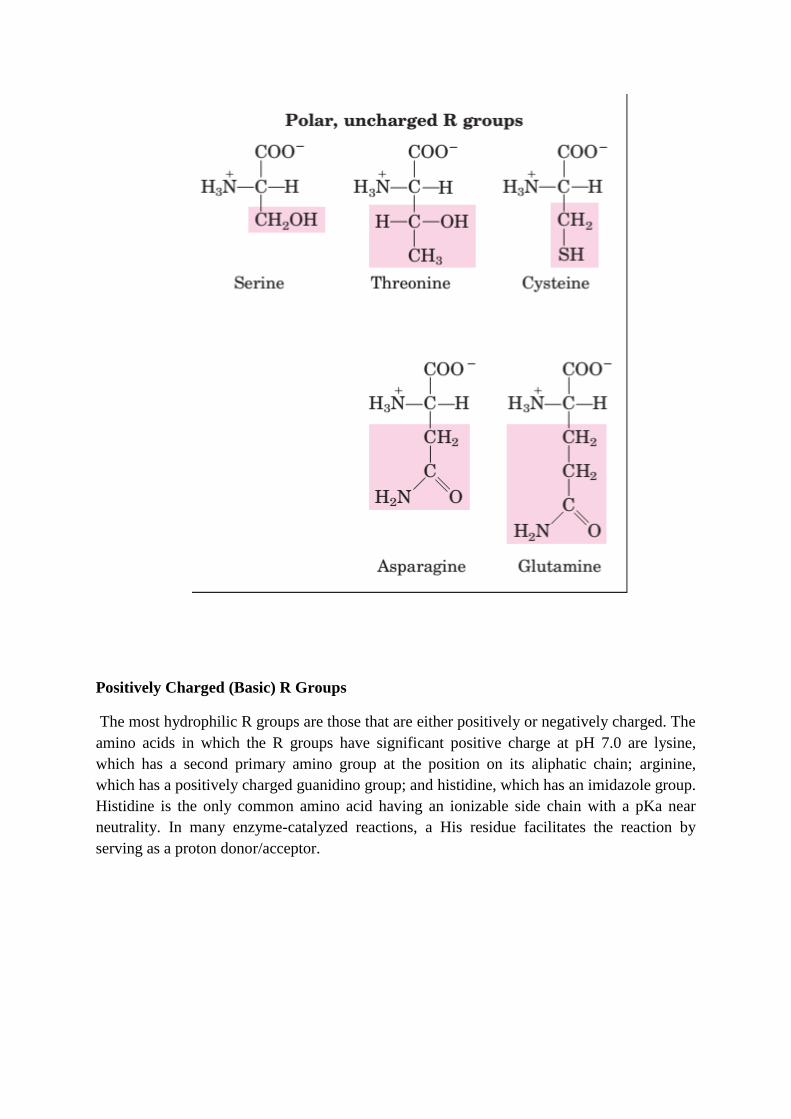
Positively Charged (Basic) R Groups
The most hydrophilic R groups are those that are either positively or negatively charged. The
amino acids in which the R groups have significant positive charge at pH 7.0 are lysine,
which has a second primary amino group at the position on its aliphatic chain; arginine,
which has a positively charged guanidino group; and histidine, which has an imidazole group.
Histidine is the only common amino acid having an ionizable side chain with a pKa near
neutrality. In many enzyme-catalyzed reactions, a His residue facilitates the reaction by
serving as a proton donor/acceptor.
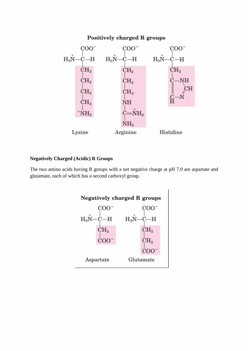
Negatively Charged (Acidic) R Groups
The two amino acids having R groups with a net negative charge at pH 7.0 are aspartate and
glutamate, each of which has a second carboxyl group.
Nutritional classification:
1- Essential amino acids: These amino acids can’t be formed in the body and so, it is
essential to be taken in diet. Their deficiency affects growth, health and protein synthesis.
Ten amino acids present in proteins (arginine, histidine, isoleucine, leucine, threonine, lysine,
methionine, phenylalanine, tryptophan, valine) are required in the diet of a growing human.
Arginine and histidine, although not required in the diets of adults, are required for growth
(children and adolescents), because the amounts that can be synthesized are not sufficient to
maintain normal growth rates. Larger amounts of phenylalanine are required if the diet is low
in tyrosine because tyrosine is synthesized from phenylalanine. Larger amounts of
methionine are required if the diet is low in cysteine because the sulfur of methionine is
donated for the synthesis of cysteine.
2- Non-essential amino acids: These are formed in the body but not in sufficient amount for
body requirements especially in children. Twelve amino acids present in proteins are
synthesized in the body - eleven (serine, glycine, cysteine, alanine, aspartate, asparagine,
glutamate, glutamine, proline, arginine, histidine) are produced from glucose, one (tyrosine)
is produced from phenylalanine.
Metabolic classification: according to metabolic or degradation products of amino acids
they may be:
1- Ketogenic amino acids: which give ketone bodies. Lysine and Leucine are the only pure
ketogenic amino acids.
2- Mixed ketogenic and glucogenic amino acids: which give both ketone bodies and glucose.
These are: isoleucine, phenylalanine, tyrosine and tryptophan.
3- Glucogenic amino acids: Which give glucose. They include the rest of amino acids. These
amino acids by catabolism yields products that enter in glycogen and glucose formation.
Classification of Amino Acids
An alternative classification scheme.
1. Acidic amino acids and their amides: aspartic acid, asparagine, glutamic acid,
glutamine.
2. Basic amino acids: histidine, lysine, arginine.
3. Aromatic amino acids: phenylalanine, tyrosine, tryptophan.
4. Sulphur containing amino acids: cysteine, methionine.
5. Imido acid: proline.
6. Hydrophobic side chains: glycine, alanine, valine, leucine, isoleucine.
7. Hydroxylic amino acids: serine, threonine, (tyrosine).
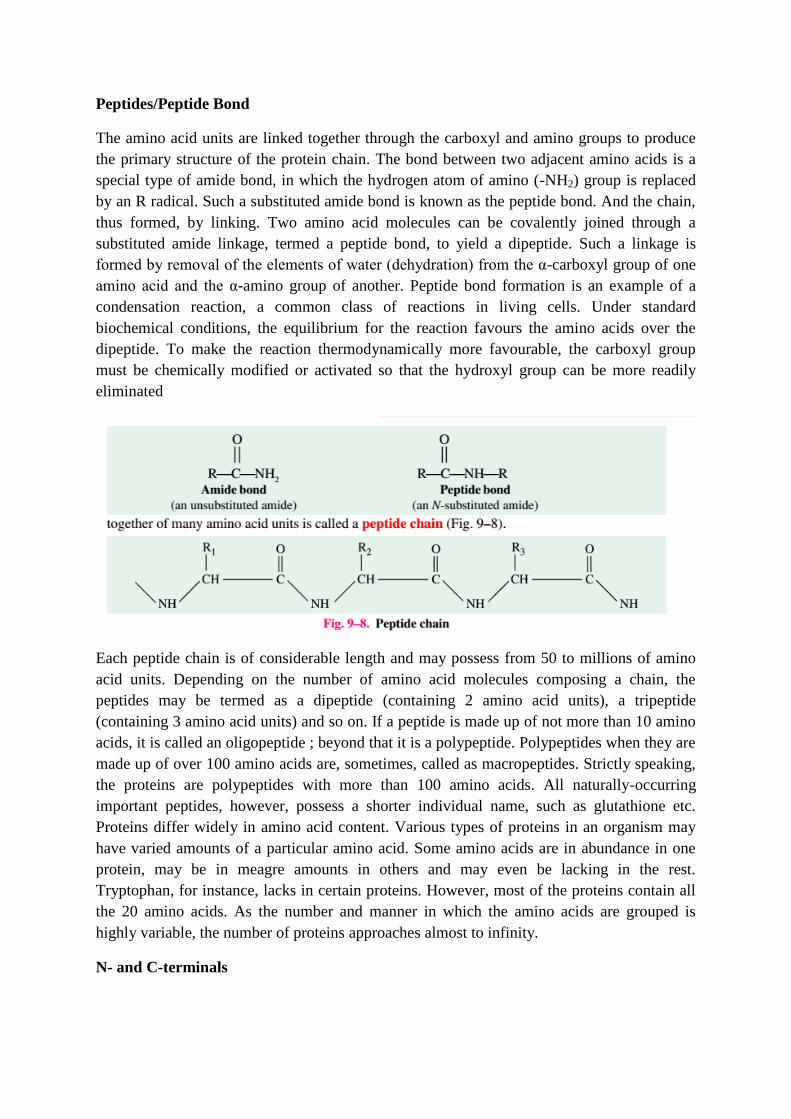
Peptides/Peptide Bond
The amino acid units are linked together through the carboxyl and amino groups to produce
the primary structure of the protein chain. The bond between two adjacent amino acids is a
special type of amide bond, in which the hydrogen atom of amino (-NH2) group is replaced
by an R radical. Such a substituted amide bond is known as the peptide bond. And the chain,
thus formed, by linking. Two amino acid molecules can be covalently joined through a
substituted amide linkage, termed a peptide bond, to yield a dipeptide. Such a linkage is
formed by removal of the elements of water (dehydration) from the α-carboxyl group of one
amino acid and the α-amino group of another. Peptide bond formation is an example of a
condensation reaction, a common class of reactions in living cells. Under standard
biochemical conditions, the equilibrium for the reaction favours the amino acids over the
dipeptide. To make the reaction thermodynamically more favourable, the carboxyl group
must be chemically modified or activated so that the hydroxyl group can be more readily
eliminated
Each peptide chain is of considerable length and may possess from 50 to millions of amino
acid units. Depending on the number of amino acid molecules composing a chain, the
peptides may be termed as a dipeptide (containing 2 amino acid units), a tripeptide
(containing 3 amino acid units) and so on. If a peptide is made up of not more than 10 amino
acids, it is called an oligopeptide ; beyond that it is a polypeptide. Polypeptides when they are
made up of over 100 amino acids are, sometimes, called as macropeptides. Strictly speaking,
the proteins are polypeptides with more than 100 amino acids. All naturally-occurring
important peptides, however, possess a shorter individual name, such as glutathione etc.
Proteins differ widely in amino acid content. Various types of proteins in an organism may
have varied amounts of a particular amino acid. Some amino acids are in abundance in one
protein, may be in meagre amounts in others and may even be lacking in the rest.
Tryptophan, for instance, lacks in certain proteins. However, most of the proteins contain all
the 20 amino acids. As the number and manner in which the amino acids are grouped is
highly variable, the number of proteins approaches almost to infinity.
N- and C-terminals
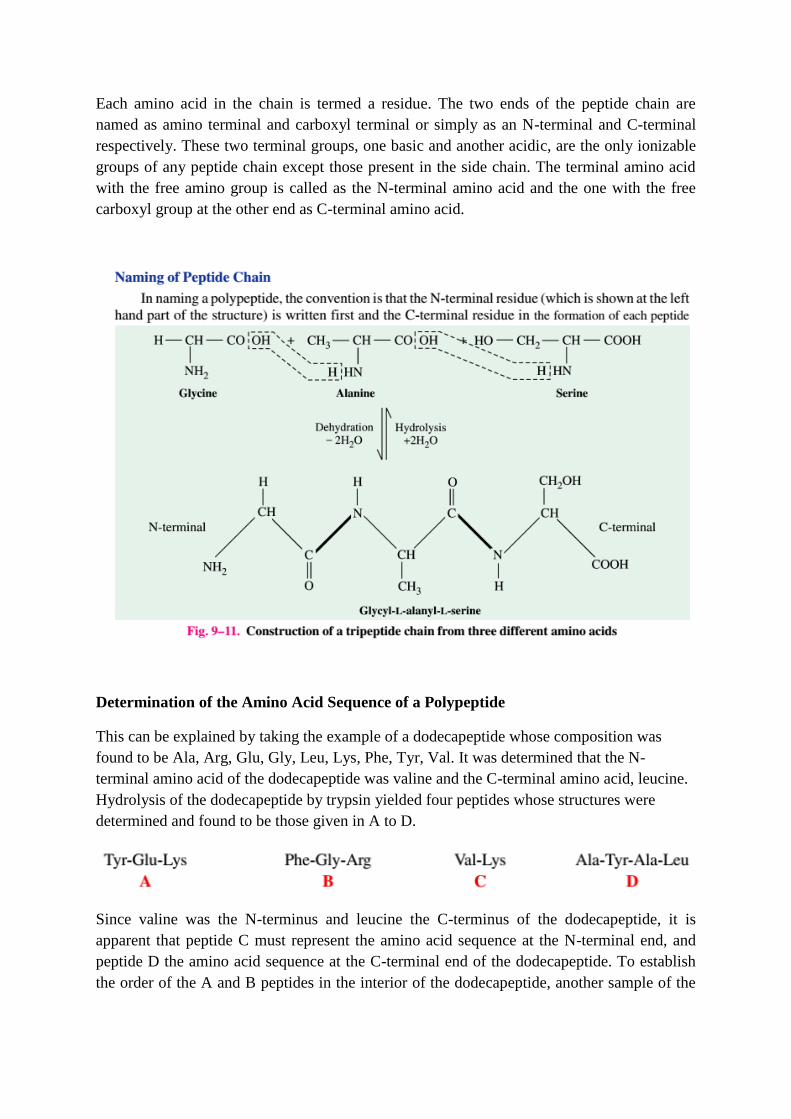
Each amino acid in the chain is termed a residue. The two ends of the peptide chain are
named as amino terminal and carboxyl terminal or simply as an N-terminal and C-terminal
respectively. These two terminal groups, one basic and another acidic, are the only ionizable
groups of any peptide chain except those present in the side chain. The terminal amino acid
with the free amino group is called as the N-terminal amino acid and the one with the free
carboxyl group at the other end as C-terminal amino acid.
Determination of the Amino Acid Sequence of a Polypeptide
This can be explained by taking the example of a dodecapeptide whose composition was
found to be Ala, Arg, Glu, Gly, Leu, Lys, Phe, Tyr, Val. It was determined that the N-
terminal amino acid of the dodecapeptide was valine and the C-terminal amino acid, leucine.
Hydrolysis of the dodecapeptide by trypsin yielded four peptides whose structures were
determined and found to be those given in A to D.
Since valine was the N-terminus and leucine the C-terminus of the dodecapeptide, it is
apparent that peptide C must represent the amino acid sequence at the N-terminal end, and
peptide D the amino acid sequence at the C-terminal end of the dodecapeptide. To establish
the order of the A and B peptides in the interior of the dodecapeptide, another sample of the
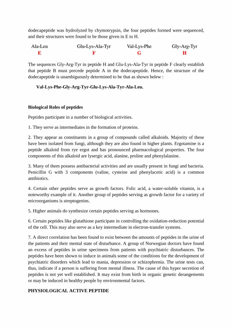
dodecapeptide was hydrolyzed by chymotrypsin, the four peptides formed were sequenced,
and their structures were found to be those given in E to H.
The sequences Gly-Arg-Tyr in peptide H and Glu-Lys-Ala-Tyr in peptide F clearly establish
that peptide B must precede peptide A in the dodecapeptide. Hence, the structure of the
dodecapeptide is unambiguously determined to be that as shown belew :
Val-Lys-Phe-Gly-Arg-Tyr-Glu-Lys-Ala-Tyr-Ala-Leu.
Biological Roles of peptides
Peptides participate in a number of biological activities.
1. They serve as intermediates in the formation of proteins.
2. They appear as constituents in a group of compounds called alkaloids. Majority of these
have been isolated from fungi, although they are also found in higher plants. Ergotamine is a
peptide alkaloid from rye ergot and has pronounced pharmacological properties. The four
components of this alkaloid are lysergic acid, alanine, proline and phenylalanine.
3. Many of them possess antibacterial activities and are usually present in fungi and bacteria.
Penicillin G with 3 components (valine, cysteine and phenylacetic acid) is a common
antibiotics.
4. Certain other peptides serve as growth factors. Folic acid, a water-soluble vitamin, is a
noteworthy example of it. Another group of peptides serving as growth factor for a variety of
microorganisms is streptogenins.
5. Higher animals do synthesize certain peptides serving as hormones.
6. Certain peptides like glutathione participate in controlling the oxidation-reduction potential
of the cell. This may also serve as a key intermediate in electron-transfer systems.
7. A direct correlation has been found to exist between the amounts of peptides in the urine of
the patients and their mental state of disturbance. A group of Norwegian doctors have found
an excess of peptides in urine specimens from patients with psychiatric disturbances. The
peptides have been shown to induce in animals some of the conditions for the development of
psychiatric disorders which lead to mania, depression or schizophrenia. The urine tests can,
thus, indicate if a person is suffering from mental illness. The cause of this hyper secretion of
peptides is not yet well established. It may exist from birth in organic genetic derangements
or may be induced in healthy people by environmental factors.
PHYSIOLOGICAL ACTIVE PEPTIDE
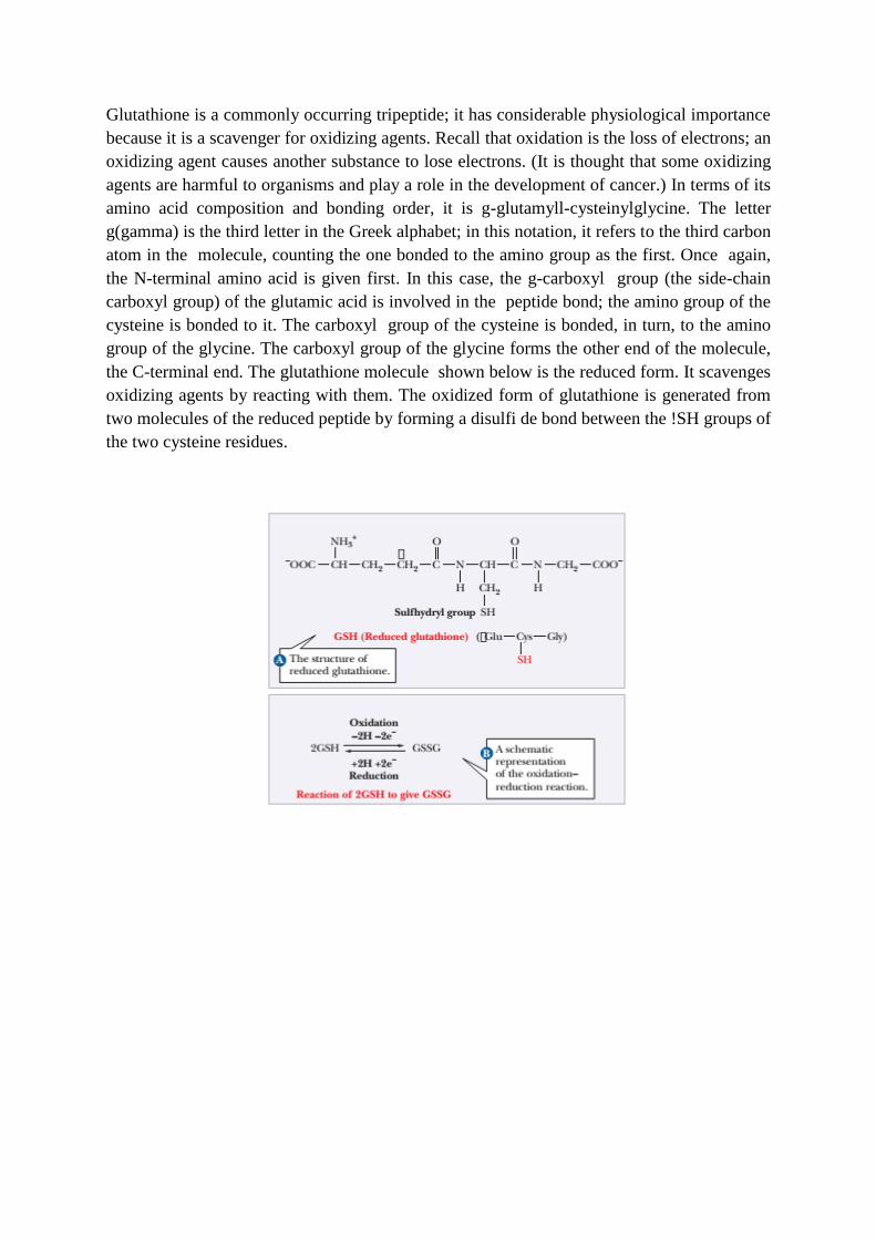
Glutathione is a commonly occurring tripeptide; it has considerable physiological importance
because it is a scavenger for oxidizing agents. Recall that oxidation is the loss of electrons; an
oxidizing agent causes another substance to lose electrons. (It is thought that some oxidizing
agents are harmful to organisms and play a role in the development of cancer.) In terms of its
amino acid composition and bonding order, it is g-glutamyll-cysteinylglycine. The letter
g(gamma) is the third letter in the Greek alphabet; in this notation, it refers to the third carbon
atom in the molecule, counting the one bonded to the amino group as the first. Once again,
the N-terminal amino acid is given first. In this case, the g-carboxyl group (the side-chain
carboxyl group) of the glutamic acid is involved in the peptide bond; the amino group of the
cysteine is bonded to it. The carboxyl group of the cysteine is bonded, in turn, to the amino
group of the glycine. The carboxyl group of the glycine forms the other end of the molecule,
the C-terminal end. The glutathione molecule shown below is the reduced form. It scavenges
oxidizing agents by reacting with them. The oxidized form of glutathione is generated from
two molecules of the reduced peptide by forming a disulfi de bond between the !SH groups of
the two cysteine residues.
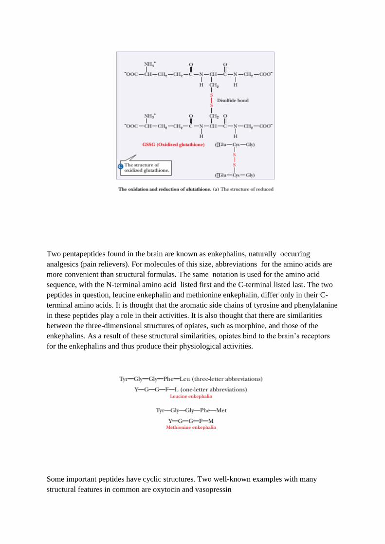
Two pentapeptides found in the brain are known as enkephalins, naturally occurring
analgesics (pain relievers). For molecules of this size, abbreviations for the amino acids are
more convenient than structural formulas. The same notation is used for the amino acid
sequence, with the N-terminal amino acid listed first and the C-terminal listed last. The two
peptides in question, leucine enkephalin and methionine enkephalin, differ only in their C-
terminal amino acids. It is thought that the aromatic side chains of tyrosine and phenylalanine
in these peptides play a role in their activities. It is also thought that there are similarities
between the three-dimensional structures of opiates, such as morphine, and those of the
enkephalins. As a result of these structural similarities, opiates bind to the brain’s receptors
for the enkephalins and thus produce their physiological activities.
Some important peptides have cyclic structures. Two well-known examples with many
structural features in common are oxytocin and vasopressin
PROTEIN STRUCTURE
The covalent backbone of a typical protein contains hundreds of individual bonds. Because
free rotation is possible around many of these bonds, the protein can assume a very
large number of conformations. However, each protein has a specific chemical or
structural function, strongly suggesting that each has a unique three-dimensional
structure. Given that, generally, the ordered array of molecules in a crystal can form
only if the molecular units are identical, the finding that many proteins could be
crystallized was evidence that even very large proteins are discrete chemical entities
with unique structures. This conclusion revolutionized thinking about proteins and their
functions This section of the note will examine how sequence of amino acids in a
polypeptide chain is translated into a discrete, three-dimensional protein structure. This
note will emphasize five themes. First, the three-dimensional structure of a protein is
determined by its amino acid sequence. Second, the function of a protein depends on its
structure. Third, an isolated protein usually exists in one or a small number of stable
structural forms. Fourth, the most important forces stabilizing the specific structures
maintained by a given protein are non-covalent interactions. Finally, amid the huge
number of unique protein structures, we can recognize some common structural
patterns that help to organize our understanding of protein architecture.
The spatial arrangement of atoms in a protein is called its conformation. The possible
conformations of a protein include any structural state it can achieve without breaking
covalent bonds. A change in conformation could occur, for example, by rotation about
single bonds. Of the many conformations that are theoretically possible in a protein
containing hundreds of single bonds, one or (more commonly) a few generally
predominate under biological conditions. The need for multiple stable conformations
reflects the changes that must take place in most proteins as they bind to other
molecules or catalyze reactions. The conformations existing under a given set of
conditions are usually the ones that are thermodynamically the most stable-that is,
having the lowest Gibbs free energy (G). Proteins in any of their functional, folded
conformations are called native proteins.
Hydrophobic interactions are clearly important in stabilizing conformation; the interior
of a protein is generally a densely packed core of hydrophobic amino acid side chains.
It is also important that any polar or charged groups in the protein interior have
suitable partners for hydrogen bonding or ionic interactions. One hydrogen bond seems
to contribute little to the stability of a native structure, but the presence of hydrogen
bonding groups without partners in the hydrophobic core of a protein can be so
destabilizing that conformations containing these groups are often thermodynamically
untenable. The favorable free-energy change resulting from the combination of several
such groups with partners in the surrounding solution can be greater than the free-energy
difference between the folded and unfolded states. In addition, hydrogen bonds between
groups in a protein form cooperatively (formation of one makes the next one more
likely) in repeating secondary structures that optimize hydrogen bonding. Hydrogen
bonds often have an important role in guiding the protein-folding process. The interaction of
oppositely charged groups that form an ion pair, or salt bridge, can have either a
stabilizing or destabilizing effect on protein structure. As in the case of hydrogen bonds,
charged amino acid side chains interact with water and salts when the protein is
unfolded, and the loss of those interactions must be considered when evaluating the
effect of a salt bridge on the overall stability of a folded protein. However, the strength
of a salt bridge increases as it moves to an environment of lower dielectric constant.
Primary structure is the order in which the amino acids are covalently linked together. The
peptide Leu-Gly-Thr-Val-Arg-Asp-His (recall that the N-terminal amino acid is listed first)
has a different primary structure from the peptide Val-His-Asp-Leu-Gly-Arg-Thr, even
though both have the same number and kinds of amino acids. Note that the order of amino
acids can be written on one line. The primary structure is the one-dimensional first step in
specifying the three-dimensional structure of a protein. Some biochemists define primary
structure to include all covalent interactions, including the disulfide bonds that can be formed
by cysteines; however, we shall consider the disulfide bonds to be part of the tertiary
structure. One of the most striking demonstrations of the importance of primary structure is
found in the hemoglobin associated with sickle-cell anemia. In this genetic disease, red blood
cells cannot bind oxygen efficiently. The red blood cells also assume a characteristic sickle
shape, giving the disease its name. The sickled cells tend to become trapped in small blood
vessels, cutting off circulation and thereby causing organ damage. These drastic
consequences stem from a change in one amino acid residue in the sequence of the primary
structure.
Secondary Structure of Proteins
The secondary structure of proteins is the hydrogen-bonded arrangement of the backbone of the
protein, the polypeptide chain. The nature of the bonds in the peptide backbone plays an important
role here. Within each amino acid residue are two bonds with reasonably free rotation: (1) the bond
between the α-carbon and the amino nitrogen of that residue and (2) the bond between the α-
carbon and the carboxyl carbon of that residue. The combination of the planar peptide group and
the two freely rotating bonds has important implications for the three-dimensional conformations of
peptides and proteins. A peptide-chain backbone can be visualized as a series of playing cards, each
card representing a planar peptide group. The cards are linked at opposite corners by swivels,
representing the bonds about which there is considerable freedom of rotation (Figure 4.1). The side
chains also play a vital role in determining the three-dimensional shape of a protein, but only the
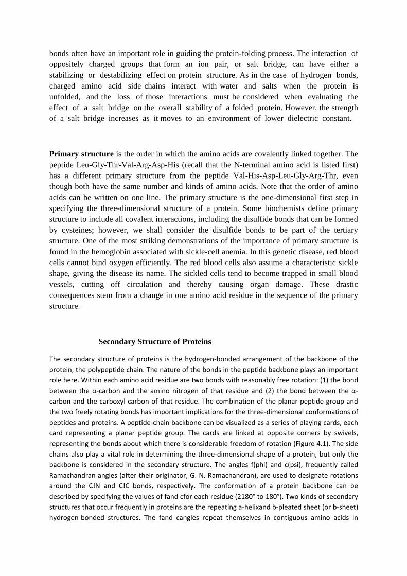
backbone is considered in the secondary structure. The angles f(phi) and c(psi), frequently called
Ramachandran angles (after their originator, G. N. Ramachandran), are used to designate rotations
around the C!N and C!C bonds, respectively. The conformation of a protein backbone can be
described by specifying the values of fand cfor each residue (2180° to 180°). Two kinds of secondary
structures that occur frequently in proteins are the repeating a-helixand b-pleated sheet (or b-sheet)
hydrogen-bonded structures. The fand cangles repeat themselves in contiguous amino acids in
regular secondary structures. The a-helix and b-pleated sheet are not the only possible secondary
structures, but they are by far the most important.
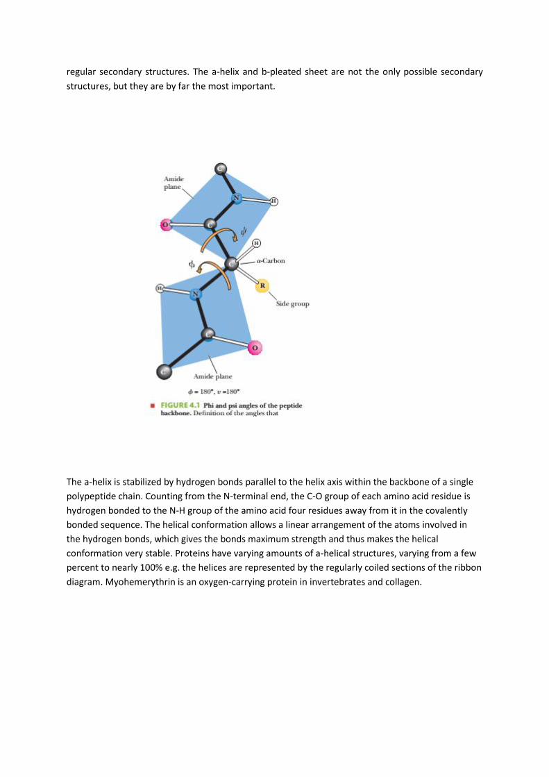
The a-helix is stabilized by hydrogen bonds parallel to the helix axis within the backbone of a single
polypeptide chain. Counting from the N-terminal end, the C-O group of each amino acid residue is
hydrogen bonded to the N-H group of the amino acid four residues away from it in the covalently
bonded sequence. The helical conformation allows a linear arrangement of the atoms involved in
the hydrogen bonds, which gives the bonds maximum strength and thus makes the helical
conformation very stable. Proteins have varying amounts of a-helical structures, varying from a few
percent to nearly 100% e.g. the helices are represented by the regularly coiled sections of the ribbon
diagram. Myohemerythrin is an oxygen-carrying protein in invertebrates and collagen.
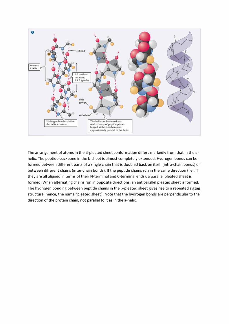
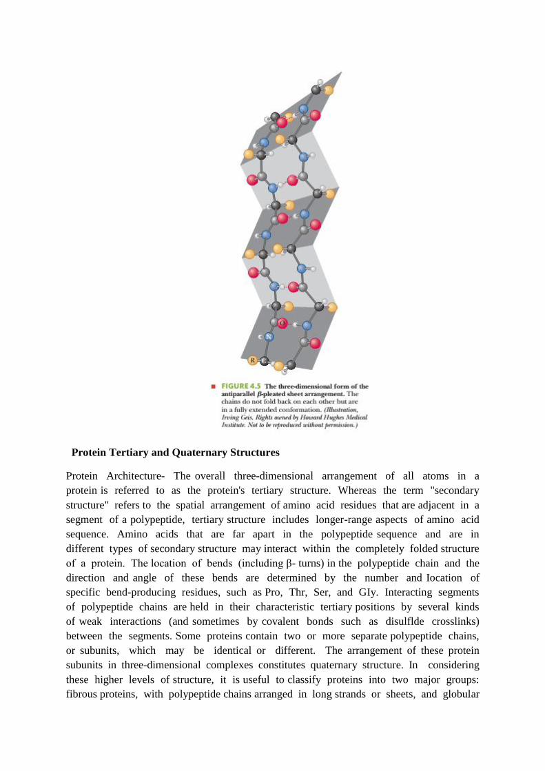
The arrangement of atoms in the β-pleated sheet conformation differs markedly from that in the a-
helix. The peptide backbone in the b-sheet is almost completely extended. Hydrogen bonds can be
formed between different parts of a single chain that is doubled back on itself (intra-chain bonds) or
between different chains (inter-chain bonds). If the peptide chains run in the same direction (i.e., if
they are all aligned in terms of their N-terminal and C-terminal ends), a parallel pleated sheet is
formed. When alternating chains run in opposite directions, an antiparallel pleated sheet is formed.
The hydrogen bonding between peptide chains in the b-pleated sheet gives rise to a repeated zigzag
structure; hence, the name “pleated sheet”. Note that the hydrogen bonds are perpendicular to the
direction of the protein chain, not parallel to it as in the a-helix.
Protein Tertiary and Quaternary Structures
Protein Architecture- The overall three-dimensional arrangement of all atoms in a
protein is referred to as the protein's tertiary structure. Whereas the term "secondary
structure" refers to the spatial arrangement of amino acid residues that are adjacent in a
segment of a polypeptide, tertiary structure includes longer-range aspects of amino acid
sequence. Amino acids that are far apart in the polypeptide sequence and are in
different types of secondary structure may interact within the completely folded structure
of a protein. The location of bends (including β- turns) in the polypeptide chain and the
direction and angle of these bends are determined by the number and Iocation of
specific bend-producing residues, such as Pro, Thr, Ser, and GIy. Interacting segments
of polypeptide chains are held in their characteristic tertiary positions by several kinds
of weak interactions (and sometimes by covalent bonds such as disulflde crosslinks)
between the segments. Some proteins contain two or more separate polypeptide chains,
or subunits, which may be identical or different. The arrangement of these protein
subunits in three-dimensional complexes constitutes quaternary structure. In considering
these higher levels of structure, it is useful to classify proteins into two major groups:
fibrous proteins, with polypeptide chains arranged in long strands or sheets, and globular
proteins, with polypeptide chains folded into a spherical or globular shape. The two
groups are structurally distinct. Fibrous proteins usually consist largely of a single type
of secondary structure, and their tertiary structure is relatively simple. Globular proteins
often contain several types of secondary structure. The two groups also differ
functionally: the structures that provide support, shape, and external protection to
vertebrates are made of flbrous proteins, whereas most enzymes and regulatory proteins
are globular proteins.
In a globular protein, different segments of the polypeptide chain (or multiple
polypeptide chains) fold back on each other, generating a more compact shape than is
seen in the fibrous proteins (Fig. 4-14). The folding also provides the structural
diversity necessary for proteins to carry out a wide array of biological functions. Globular
proteins include enzymes, transport proteins, motor proteins, regulatory proteins,
immunoglobulins, and proteins with many other functions. The first breakthrough in
understanding the three-dimensional structure of a globular protein came from x-ray
diffraction studies of myoglobin carried out by John Kendrew and his colleagues in
the1950s. Myoglobin is a relatively small (M, 16,700),oxygen-binding protein of
muscle cells. It functions both to store oxygen and to facilitate oxygen diffusion in
rapidly contracting muscle tissue. Myoglobin contains a single polypeptide chain of
153 amino acid residues of known sequence and a single iron protoporphyrin, or heme,
group. The same heme group that is found in myoglobin is found in hemoglobin, the
oxygen-binding protein of erythrocytes, and is responsible for the deep red-brown color
of both myoglobin and hemoglobin.
α-Keratin, collagen, and silk flbroin nicely illustrate the relationship between protein
structure and biological function (Table 4-2). Fibrous proteins share properties that give
strength and./or flexibility to the structures in which they occur. In each case, the
fundamental structural unit is a simple repeating element of secondary structure. All
fibrous proteins are insoluble in water, a property conferred by a high concentration of
hydrophobic amino acid residues both in the interior of the protein and on its surface.
These hydrophobic surfaces are largely buried as many similar polypeptide chains are
packed together to form elaborate supramolecular complexes. α-Keratin The α-keratins
have evolved for strength. Found only in mammals, these proteins constitute almost the
entire dry weight of hair, wool, nails, claws, quills, horns, hooves, and much of the
outer layer of skin. The a-keratins are part of a broader family of proteins called
intermediate f,lament (IF) proteins. Other IF proteins are found in the cytoskeletons of
animal cells. All IF proteins have a structural function and share the structural features
exemplified by the a-keratins.