Embed Size (px)
Citation preview

Available online at www.sciencedirect.com
www.elsevier.com/locate/brainres
b r a i n r e s e a r c h 1 4 7 3 ( 2 0 1 2 ) 1 6 1 – 1 7 2
0006-8993/$ - see frohttp://dx.doi.org/10
nCorresponding autE-mail address:
Research Report
BDNF may play a differential role in the protective effect ofthe mGluR2/3 agonist LY379268 on striatal projectionneurons in R6/2 Huntington’s disease mice
A. Reinera,n, H.B. Wanga, N. Del Mara, K. Sakatab, W. Yooc, Y.P. Denga
aDepartment of Anatomy and Neurobiology, The University of Tennessee Health Science Center, 855 Monroe Ave., Memphis, TN 38163, USAbDepartment of Pharmacology, The University of Tennessee Health Science Center, Memphis, TN 38163, USAcBiostatistics and Epidemiology Unit of Department of Preventive Medicine, The University of Tennessee Health Science Center, Memphis,
TN 38163, USA
a r t i c l e i n f o
Article history:
Accepted 12 July 2012
We have found that daily subcutaneous injection with a maximum tolerated dose (MTD) of
the mGluR2/3 agonist LY379268 (20 mg/kg) beginning at 4 weeks dramatically improves the
Available online 20 July 2012
Keywords:
Huntington’s disease
Therapy
mGluR2/3
Striatum
BDNF
nt matter & 2012 Elsevie.1016/j.brainres.2012.07.0
hor. Fax: þ1 [email protected] (A. R
a b s t r a c t
phenotype in R6/2 mice. For example, we observed normalization of motor function in
distance traveled, speed, the infrequency of pauses, and the ability to locomote in a
straight line, and a rescue of a 15–20% striatal neuron loss at 10 weeks. As acute LY379268
treatment is known to increase cortical BDNF production, and BDNF is known to be
beneficial for striatal neurons, we investigated if the benefit of daily LY379268 in R6/2 mice
for striatal projection neurons was associated with increases in corticostriatal BDNF, with
assessments done at 10 weeks of age after daily MTD treatment since the fourth week of
life. We found that LY379268 increased BDNF expression in layer 5 neurons in motor cortex,
which project to striatum, partly rescued a preferential loss of enkephalinergic striatal
neurons, and enhanced substance P (SP) expression by SP striatal projection neurons. The
enhanced survival of enkephalinergic striatal neurons was correlated with the cortical
BDNF increase, but the enhanced SP expression by SP striatal neurons was not. Thus,
LY379268 may protect the two main striatal projection neuron types by different mechan-
isms, enkephalinergic neurons by the trophic benefit of BDNF, and SP neurons by a
mechanism not involving BDNF. The SP neuron benefit may perhaps instead involve the
anti-excitotoxic action of mGluR2/3 receptor agonists.
& 2012 Elsevier B.V. All rights reserved.
1. Introduction
We have found that daily subcutaneous injection with a
maximum tolerated dose (MTD) of the mGluR2/3 agonist
LY379268 (20 mg/kg) beginning at 4 weeks dramatically
r B.V. All rights reserved.26
.einer).
improves the phenotype in R6/2 mice (Reiner et al., 2012).
For example, this regimen of LY379268 administration pre-
vents a 15–20% striatal neuron loss seen at 10 weeks in R6/2
mice. We also observed normalization of motor function at
10 weeks in R6/2 mice treated with daily LY379268 in such

b r a i n r e s e a r c h 1 4 7 3 ( 2 0 1 2 ) 1 6 1 – 1 7 2162
parameters as distance traveled, speed, the frequency of
pauses, and the ability to locomote in a straight line. The
basis of this benefit is uncertain, but the finding that acute
LY379268 treatment increases cortical brain-derived neuro-
trophic factor (BDNF) production (Di Liberto et al., 2010) raises
the possibility that the benefit is, at least in part, mediated by
BDNF. Four lines of evidence add weight to this possibility.
First, pyramidal neurons of cerebral cortex synthesize
BDNF (Altar et al., 1997; Altar and DiStefano, 1998; Cattaneo
et al., 2001, 2005; Baquet et al., 2004), and those in layer 5 in
particular transport it to striatum, where it is released from
terminals and binds to TrkB receptors on striatal neurons
(Ivkovic and Ehrlich, 1999; Mizuno et al., 1994; Weiss et al.,
1986). Striatal projection neurons are the main targets of this
input (Reiner et al., 2003b), and BDNF is a potent trophic and
survival factor for these neurons (Mizuno et al., 1994; Widmer
and Hefti, 1994; Nakao et al., 1995; Martınez-Serrano and
Bjorklund, 1996; Alcantara et al., 1997; Ivkovic and Ehrlich,
1999; Aggerman and Ernfors, 2003; Grosse et al., 2005;
Ventimiglia et al., 1995). Second, wild-type huntingtin
promotes BDNF production by cortical neurons but polyglu-
tamine-expanded mutant huntingtin promotes BDNF production
less effectively, and accordingly cortical BDNF production is
reduced in human Huntington’s disease (HD) and in trans-
genic HD mice (Cattaneo et al., 2001, 2005; Zuccato et al.,
2001, 2003, 2005, 2008; Zuccato and Cattaneo, 2007). Depletion
of WT huntingtin in cortical neurons by sequestration in
mutant protein aggregates and impaired transport of BDNF
by cortical neuron axons further exacerbate the striatal and
cortical BDNF insufficiency caused by the HD mutation
(Gauthier et al., 2004; Cattaneo et al., 2005). Third, studies in
various mutant mice indicate that diminished cortical pro-
duction of BDNF harms striatum (Gorski et al., 2003; Baquet
et al., 2004; Canals et al., 2004; Saylor et al., 2006; Strand et al.,
2007). Finally, intrastriatal BDNF delivery and selective fore-
brain over expression of BDNF reverse cortical and striatal
injury, and improve motor performance in transgenic HD
mice (Canals et al., 2004; Gharami et al., 2008; Xie et al., 2010;
Giralt et al., 2011).
Thus, BDNF deficiency has been implicated in HD patho-
genesis, and BDNF over expression in successful HD treat-
ment. As LY379268 given acutely boosts cortical BDNF
production, we investigated if the benefit of daily LY379268
in R6/2 mice was associated with increases in cortical BDNF,
and if any BDNF boost was correlated with the improved
survival and neurochemistry of either of the two main
striatal projection neuron types. Our results show that the
LY379268 benefit for ENK but not SP neurons seems linked to
a BDNF enhancement.
2. Results
2.1. Behavior
The total distance traveled in a 30-min open field session in
vehicle-treated R6/2 (R62V) mice at ten weeks of age was
significantly less than in vehicle-treated WT mice (WTV)
(57.5% of vehicle-treated WT) (Table 1). By contrast, distance
traveled in the LY379268-treated R6/2 mice (R62LY) at ten
weeks of age was not significantly different from that in WTV
mice (101.3% of WTV), and was significantly more than in
R62V mice. Similarly, R62V mice were significantly slower in
open field than WTV mice at ten weeks of age (64.8% of
vehicle-treated WT), and LY379268 rendered R6/2 speed at ten
weeks of age significantly more than in R62V but not
significantly different from that in WTV (93.8% of vehicle-
treated WT) (Table 1). Thus, daily MTD LY379268 normalized
R6/2 mice at 10 weeks on these two motor parameters, which
are representative of the motor benefit seen in open field at
this age with LY379268 treatment (Reiner et al., 2012).
2.2. Quantitative PCR (qPCR)
Our qPCR analysis (Fig. 1) showed that daily LY379268 treat-
ment in WT mice (WTLY) had no significant effect (p¼0.8345)
on overall BDNF expression in the frontal pole of cerebral
cortex (WTLY was 105.8% of WTV). BDNF expression in the
frontal pole of cerebral cortex of R62V mice was, however,
significantly reduced to 43.3% of that in WTV mice (p¼0.0429).
Daily LY379268 boosted cortical BDNF message in R6/2 mice
to 83.4% of that in WTV mice, which was not significantly
different than in vehicle-treated WT (p¼0.5176). Thus, qPCR
shows that the R6/2 mutation causes more than a 50%
reduction in overall BDNF expression in frontal cortex at 10
weeks, which was restored to indistinguishable from normal
WT levels by daily LY379268.
2.3. In situ hybridization histochemistry (ISHH)—Neuronabundance
Blinded counts of labeled neurons (Fig. 2, Table 2) revealed
significantly fewer BDNF neurons in layer 5 of M1 in R62V
mice compared to WTV mice (80.6% of WTV) (p¼0.0197). The
abundance of BDNF neurons in layer 5 of M1 in R62LY mice
was, however, not significantly different than in WTV mice
(96.8% of WTV), and was significantly more than in R62V mice
(p¼0.0406). Similarly, relative BDNF message in layer 5 of M1
in R62V mice was also significantly less (po0.0001) than in
WTV mice (47.1% of WTV), and was elevated by LY379268
treatment to 78.9% of WTV mice (Table 2). The layer 5 BDNF
message level in R6/2 after daily LY379268 was significantly
greater than in R62V mice (p¼0.0093), and no longer signifi-
cantly different from WTV mice (p¼0.0782). Thus, LY379268
treatment normalized layer 5 BDNF neuron abundance and
boosted BDNF message in layer 5. Note that although our
measurements were confined to layer 5 of M1, ISHH sup-
ported our qPCR findings that LY379268 had a beneficial effect
for all cortical layers and regions (Fig. 2).
The abundance of ENK neurons in striatum in R62V mice
was also significantly less than in WTV mice (71.2% of WTV)
(po0.0001) (Fig. 3; Table 2). While the abundance of ENK
neurons in striatum in LY379268-treated R6/2 mice was
significantly more than in R62V mice (p¼0.0240), it remained
significantly less (83.1% of WTV) than in the WTV mice
(p¼0.0017). Thus, LY379268 treatment increased striatal ENK
neuron abundance in R6/2 mice, but did not normalize it. No
shortfall was seen in the abundance of SP striatal neurons in
R62V mice, and LY379268 treatment did not alter their
abundance in R6/2 mice (Fig. 4; Table 2). Notably, however,

Table 1 – Effect of LY379268 on activity and speed in open field in WT and R6/2 mice, compared to vehicle treatment, at 10weeks of age. Numbers of animals per group, and repeat length for the R6/2 groups are shown. R6/2 mice receiving vehicleare significantly impaired in activity and speed, but daily LY379268 treatment normalized them.
Groups Distance traveled in cm Maximum speed in cm/s Number of animals CAG repeat length
WT–Vehicle 32,026.271829.5 40.671.9 15
WT–LY379268 31,110.471120.1 37.071.5 17
R62–Vehicle 18,427.271920.8n# 26.372.6n# 12 124.7
R62–LY379268 32,431.373431.2 38.174.0 15 124.5n Significant vs. WTV.# Significant vs. R62LY.
Fig. 1 – Graph showing the effect of LY379268 on BDNF message in frontal cortex in WT and R6/2 mice, compared to vehicle
treatment, at 10 weeks of age, as assessed by qPCR. R6/2 mice receiving vehicle have significantly decreased BDNF
expression compared to WT mice, but daily LY379268 treatment nearly normalized R6/2 mice. Error bars show SEMs.
b r a i n r e s e a r c h 1 4 7 3 ( 2 0 1 2 ) 1 6 1 – 1 7 2 163
LY379268 reduced SP striatal neuron abundance in WT mice
to 90.9% of that in WTV mice, and this reduction was
significant (p¼0.0496).
2.4. ISHH—Message per neuron
In a prior study, we reported that relative striatal signal for
ENK and SP message was significantly reduced in R62V mice,
with the ENK signal more than halved and the SP signal
reduced by about 25% (Reiner et al., 2012). We further found
in that study that LY379268 treatment normalized the striatal
SP message but not the striatal ENK message in R6/2 mice. In
the present study, we report on the effect of the R6/2
mutation and daily LY379268 on ENK and SP message per
striatal neuron in these same animals (Table 3). We found
that ENK message per ENK neuron was significantly
increased (p¼0.0268) in WTLY mice (109.2% of vehicle-treated
WT), and significantly reduced (po0.0001) in vehicle-treated
R6/2 mice (61.3% of vehicle-treated WT). Despite its beneficial
effect on ENK neuron abundance, LY379268 slightly but
significantly (p¼0.0225) reduced ENK message per neuron
below that seen in R62V mice to 50.4% of WTV. By contrast,
for SP message per SP neuron, no difference was seen
between vehicle-treated and LY37928-treated WT mice. SP
message per neuron was, however, significantly reduced
(p¼0.0010) in R62V mice (77.3% of WTV), and daily LY379268
treatment normalized relative SP message per SP neuron in
R6/2 mice so that it was no longer significantly different
(p¼0.4355) than WTV (94.3% of WTV).
2.5. ISHH—Correlation between cortical BDNF and ENKstriatal neuron abundance
Since cortical neurons provide striatum with trophic support
via BDNF, and LY379268 treatment boosted cortical BDNF and
rescued striatal ENK neurons in R6/2 mice, we next sought to
assess the relationship between cortical layer-5 BDNF neuron
abundance and striatal ENK neuron abundance, to determine
if variation in striatal ENK neuron abundance was related to
variation in cortical BDNF in those same mice. The trends
were similar for vehicle-treated and LY379268-treated mice,
and thus we combined results per genotype (Fig. 5). For WT
mice, we observed no significant correlation of ENK striatal
neuron abundance (r¼0.2089) with BDNF cortical neuron
abundance (p¼0.3272). By contrast, although the range of
variation in cortical BDNF neuron abundance was similar for
WT and R6/2 mice, ENK striatal neuron abundance was
significantly correlated (r¼0.4299) with cortical layer-5 BDNF
neuron abundance in R6/2 mice (p¼0.0309). Thus, while ENK
striatal neuron abundance does not significantly depend on
cortical BDNF neuron abundance in WT mice, such depen-
dence seemingly develops with the mutation in R6/2 mice
(Fig. 5). ANCOVA showed that although ENK neuron survival
was BDNF-dependent in R6/2 but not WT mice, ENK neuron

Fig. 2 – Images of right medial cerebral cortex at the level of M1, at low power (A)–(D) and high power (E)–(H), showing ISHH
labeling for BDNF message, from 10-week old WT mice treated daily with either vehicle (WTV) or LY379268 (WTLY) compared
to 10-week old R6/2 mice treated daily with either vehicle (R62V) or LY379268 (R62LY). Note that cortical BDNF message is
somewhat reduced in the R6/2 mice, and LY379268 increases it in R6/2 mice. Images (A)–(D) are to the same scale, and
images (E)–(H) are to the same scale.
Table 2 – Effect of LY379268 on cortical BDNF neuron abundance and message in layer 5 of M1, and on striatal SP and ENKneuron abundance in WT and R6/2 mice, at 10 weeks of age. Numbers of animals per group, and repeat length for the R6/2groups are shown. R6/2 mice receiving vehicle (R62V) show significantly lower BDNF neuron abundance and message inlayer 5 of M1, and significantly fewer ENK neurons in striatum than do WT mice receiving either vehicle (WTV) orLY379268 (WTLY). Daily LY379268 treatment normalized cortical BDNF neuron abundance in R6/2 mice (R62LY), andsignificantly increased cortical BDNF message and striatal ENK neuron abundance.
BDNF neurons in M1
layer 5 (%)
BDNF message in M1
layer 5 (%)
ENK neurons in
striatum (%)
SP neurons in
striatum (%)
Number of
animals
CAG repeat
length
WTV 100.076.2 100.0711.5 100.073.6 100.073.4 11, 11, 12, 12
WTLY 107.575.7 110.577.9 105.774.6 90.973.1n 13, 13, 13, 13
R62V 80.673.3n,# 47.174.7n,# 71.273.3n,# 101.973.7 11, 11, 12, 13 125.7
R62LY 96.876.4 78.978.9 83.173.2n 101.372.9 13, 13, 13, 13 126.5n Significant vs. WTV.# Significant vs. R62LY.
b r a i n r e s e a r c h 1 4 7 3 ( 2 0 1 2 ) 1 6 1 – 1 7 2164
loss remained significantly greater in R6/2 than WT mice for
any given BDNF neuron abundance. A similar effect was
evident for layer 5 BDNF message. Thus, as shown by the
ANCOVA, the BDNF boost by LY379268 could improve but not
entirely normalize ENK neuron survival.
2.6. ISHH—Correlation between cortical BDNF andstriatal neuron message
Because the graphed trends appeared to differ for vehicle versus
LY379268 treatment in R6/2 mice, we present the correlations of
cortical BDNF neuron abundance in M1 with striatal SP message
per neuron separately for each group. The striatal SP signal per
neuron showed no significant correlation with M1 layer 5 BDNF
neuron abundance in WT mice (Fig. 6) and the regression lines
were essentially identical for vehicle and LY379268 treated mice
(r¼�0.3383 for WTV; �0.3467 for WTLY) mice. The results for
R6/2 mice were more complex (Fig. 6). R62V mice showed a
suggestive but not significant trend toward a correlation
(r¼0.4503) between cortical BDNF and striatal SP message per
neuron (p¼0.1646). Although the mean striatal SP signal per
neuron was rendered normal by the drug treatment in R62LY
mice, the striatal SP signal showed no correlation with M1 layer 5
BDNF neuron abundance (r¼0.1137). Thus, the SP message
deficit in R62V mice was not demonstrably attributable to the
reduced cortical BDNF, and the LY379268 benefit for striatal
neuron SP message in R6/2 mice was unambiguously not
attributable to the boosting effect of the drug on cortical BDNF.

Fig. 4 – Images of right half of telencephalon anterior to
anterior commissure, showing ISHH labeling for SP
message, from 10-week old WT mice treated daily with
either vehicle (WTV) or LY379268 (WTLY) compared to
10-week old R6/2 mice treated daily with either vehicle
(R62V) or LY379268 (R62LY). Note that SP message is slightly
reduced in the R6/2 mice, and LY379268 appears to increase
message so that it is similar to that in WTV mice. All
images are to the same scale.
Fig. 3 – Images of right half of telencephalon anterior to
anterior commissure, showing ISHH labeling for ENK
message, from 10-week old WT mice treated daily with
either vehicle (WTV) or LY379268 (WTLY) compared to
10-week old R6/2 mice treated daily with either vehicle
(R62V) or LY379268 (R62LY). Note that ENK message is
clearly reduced in the R6/2 mice, and LY379268 does not
have a benefit that is evident in either WT or R6/2 mice.
All images are to the same scale.
b r a i n r e s e a r c h 1 4 7 3 ( 2 0 1 2 ) 1 6 1 – 1 7 2 165
3. Discussion
This study has three major findings. First, our neuron counts
indicate that the striatal neurons reported to be lost in
symptomatic R6/2 mice in previous studies (Stack et al.,
2005, 2007; Reiner et al., 2012) represent ENK neurons but
not SP neurons at 10 weeks of age. Second, our results show
that daily LY379268 treatment boosts BDNF message in layer
5 neurons of cortex in R6/2 mice, which is known to be the
main source of the cortical input to the striatum (Reiner et al.,
2003b, 2010). A recent study demonstrated that WT mice
acutely treated with LY379268 show up regulation of BDNF in
cortex and hippocampus (Di Liberto et al., 2010). Our results
thus extend this observed BDNF boosting effect of LY379268
administration to chronic treatment in R6/2 HD mutant mice.
Our results also suggested a possible slight increase in
cortical BDNF expression in WT mice treated daily with
MTD LY379268. Finally, our results suggest that LY379268
may yield its neuroprotective effect on striatal ENK neurons
in R6/2 mice by its enhancement of the production of BDNF
by cortical neurons and its transport to striatum. Consistent
with this, our ELISA data indicate that BDNF protein is
increased greatly in R6/2 striatum by daily LY379268 admin-
istration at an MTD (Reiner et al., 2011).
BDNF is produced by cortical neurons and transported
axonally to their cortical and striatal projection targets
(Altar et al., 1997; Zuccato et al., 2001; Gauthier et al., 2004;
Zuccato and Cattaneo, 2007). BDNF promotes development,
differentiation, plasticity and survival of neurons in cortex
and striatum (Lessmann et al., 2003; Poo, 2001; Zuccato and
Cattaneo, 2007), and mutant huntingtin reduces cortical
BDNF expression and protein transport in HD mice (Zuccato
et al., 2001). Placing the R6/1 HD transgene on a hemizygous
BDNF knock-out background exacerbates striatal ENK but not
SP loss (Canals et al., 2004). Moreover, cortex-specific BDNF
knock-out results in cortical and striatal pathology that
mimics human HD (Gorski et al., 2003; Baquet et al., 2004;
Strand et al., 2007), and embryonic deletion of the BDNF
receptor (TrkB) from striatal neurons results in their profound
loss, especially ENK neurons (Baydyuk et al., 2011). Conver-
sely, behavioral performance is improved and disease pro-
gression slowed in transgenic HD mice overexpressing BDNF
in telencephalon (Gharami et al., 2008; Xie et al., 2010; Arregui
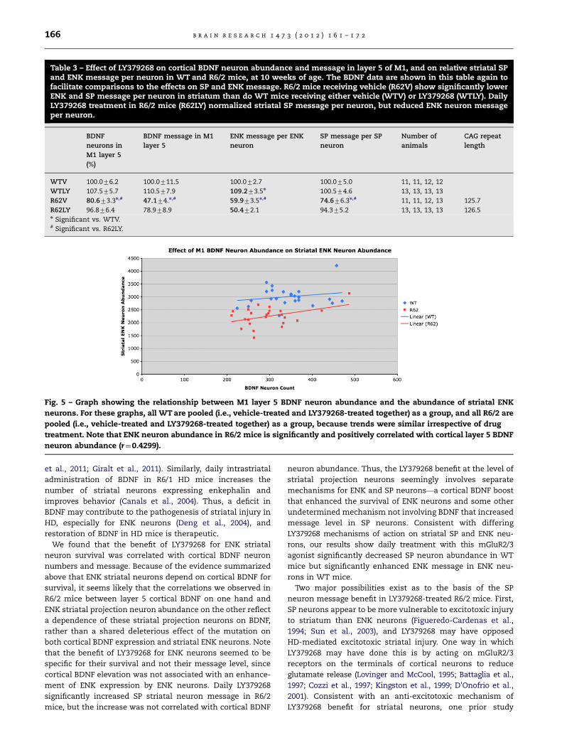
Table 3 – Effect of LY379268 on cortical BDNF neuron abundance and message in layer 5 of M1, and on relative striatal SPand ENK message per neuron in WT and R6/2 mice, at 10 weeks of age. The BDNF data are shown in this table again tofacilitate comparisons to the effects on SP and ENK message. R6/2 mice receiving vehicle (R62V) show significantly lowerENK and SP message per neuron in striatum than do WT mice receiving either vehicle (WTV) or LY379268 (WTLY). DailyLY379268 treatment in R6/2 mice (R62LY) normalized striatal SP message per neuron, but reduced ENK neuron messageper neuron.
BDNF
neurons in
M1 layer 5
(%)
BDNF message in M1
layer 5
ENK message per ENK
neuron
SP message per SP
neuron
Number of
animals
CAG repeat
length
WTV 100.076.2 100.0711.5 100.072.7 100.075.0 11, 11, 12, 12
WTLY 107.575.7 110.577.9 109.273.5n 100.574.6 13, 13, 13, 13
R62V 80.673.3n,# 47.174.n,# 59.973.5n,# 74.676.3n,# 11, 11, 12, 13 125.7
R62LY 96.876.4 78.978.9 50.472.1 94.375.2 13, 13, 13, 13 126.5n Significant vs. WTV.# Significant vs. R62LY.
Fig. 5 – Graph showing the relationship between M1 layer 5 BDNF neuron abundance and the abundance of striatal ENK
neurons. For these graphs, all WT are pooled (i.e., vehicle-treated and LY379268-treated together) as a group, and all R6/2 are
pooled (i.e., vehicle-treated and LY379268-treated together) as a group, because trends were similar irrespective of drug
treatment. Note that ENK neuron abundance in R6/2 mice is significantly and positively correlated with cortical layer 5 BDNF
neuron abundance (r¼0.4299).
b r a i n r e s e a r c h 1 4 7 3 ( 2 0 1 2 ) 1 6 1 – 1 7 2166
et al., 2011; Giralt et al., 2011). Similarly, daily intrastriatal
administration of BDNF in R6/1 HD mice increases the
number of striatal neurons expressing enkephalin and
improves behavior (Canals et al., 2004). Thus, a deficit in
BDNF may contribute to the pathogenesis of striatal injury in
HD, especially for ENK neurons (Deng et al., 2004), and
restoration of BDNF in HD mice is therapeutic.
We found that the benefit of LY379268 for ENK striatal
neuron survival was correlated with cortical BDNF neuron
numbers and message. Because of the evidence summarized
above that ENK striatal neurons depend on cortical BDNF for
survival, it seems likely that the correlations we observed in
R6/2 mice between layer 5 cortical BDNF on one hand and
ENK striatal projection neuron abundance on the other reflect
a dependence of these striatal projection neurons on BDNF,
rather than a shared deleterious effect of the mutation on
both cortical BDNF expression and striatal ENK neurons. Note
that the benefit of LY379268 for ENK neurons seemed to be
specific for their survival and not their message level, since
cortical BDNF elevation was not associated with an enhance-
ment of ENK expression by ENK neurons. Daily LY379268
significantly increased SP striatal neuron message in R6/2
mice, but the increase was not correlated with cortical BDNF
neuron abundance. Thus, the LY379268 benefit at the level of
striatal projection neurons seemingly involves separate
mechanisms for ENK and SP neurons—a cortical BDNF boost
that enhanced the survival of ENK neurons and some other
undetermined mechanism not involving BDNF that increased
message level in SP neurons. Consistent with differing
LY379268 mechanisms of action on striatal SP and ENK neu-
rons, our results show daily treatment with this mGluR2/3
agonist significantly decreased SP neuron abundance in WT
mice but significantly enhanced ENK message in ENK neu-
rons in WT mice.
Two major possibilities exist as to the basis of the SP
neuron message benefit in LY379268-treated R6/2 mice. First,
SP neurons appear to be more vulnerable to excitotoxic injury
to striatum than ENK neurons (Figueredo-Cardenas et al.,
1994; Sun et al., 2003), and LY379268 may have opposed
HD-mediated excitotoxic striatal injury. One way in which
LY379268 may have done this is by acting on mGluR2/3
receptors on the terminals of cortical neurons to reduce
glutamate release (Lovinger and McCool, 1995; Battaglia et al.,
1997; Cozzi et al., 1997; Kingston et al., 1999; D’Onofrio et al.,
2001). Consistent with an anti-excitotoxic mechanism of
LY379268 benefit for striatal neurons, one prior study

Fig. 6 – Graphs showing the relationship between M1 layer 5 BDNF neuron abundance and striatal SP message per SP neuron
in vehicle-treated (A) and LY379268-treated mice (B). For these graphs, vehicle-treated and LY379268-treated mice are
presented separately, because R6/2 showed what might be different trends. Although SP message per neuron appeared
possibly correlated with M1 layer 5 BDNF neuron abundance (r¼0.4503) in vehicle-treated but not LY379268-treated R6/2
mice (B), this proved not significant. In WT mice with either treatment (A), SP message per neuron was uncorrelated with M1
layer 5 BDNF neuron abundance. Thus, SP message per neuron was independent of cortical BDNF in WT and R6/2 mice, and
the benefit of daily LY379268 treatment for SP neuron message was not mediated by BDNF.
b r a i n r e s e a r c h 1 4 7 3 ( 2 0 1 2 ) 1 6 1 – 1 7 2 167
prevented corticostriatal glutamate release in R6/2 mice by
ablating cortex, and thereby reversed striatal neuron shrink-
age (Stack et al., 2007). Note that LY379268 may have also
opposed excitotoxic striatal injury by acting directly on
striatal projection neurons that possess mGluR3 receptors.
Approximately half of striatal projection neurons express
mGluR3 (but none GluR2) (Testa et al., 1994; Allen Brain
Atlas), and it is possible this half is predominantly the SP
type. Agonist binding to mGluR2/3 receptors, which are
negatively coupled to adenylyl cyclase (Nakanishi, 1992;
Schoepp et al., 1999), reduces Ca2þ currents via N, L-type
Ca2þ channels and increases Kþ currents via IRKC channels,
and thereby decreases neuronal excitability (Davies et al.,
1995; King and Liu, 1996; McCool et al., 1996; Colwell and
Levine, 1999). Finally, striatal astrocytes express mGluR3
(Ohishi et al., 1993; Testa et al., 1994; Liu et al., 1998), and
LY379268 increases striatal glial production of TGF-b (D’Onofrio
et al., 2001). Agonists for mGluR3 counteract neuronal NMDA
toxicity in vitro and in vivo, and this protection is mediated
by astrocytic release of TGF-b1 (Bruno et al., 1997, 1998), and
is absent in mGluR3 knock-out mice and in co-cultures of
mGluR3�/� astrocytes and mGluR3þ/þ neurons (Corti et al.,
2007). Note, however, that TGF-b1 levels are reduced in the
brains of human HD victims and in HD mice (R6/2 and
YAC128), and acute administration of LY379268 does not
elevate cortical or striatal TGF-b1 in R6/2 or YAC128 mice
(Battaglia et al., 2011). It is uncertain thus if the beneficial
effects of daily LY379268 could have involved boosting cor-
tical and striatal TGF-b1 in our R6/2 mice.
Our results have relevance to the processes mediating
striatal projection neuron death in HD. Despite the great
variety of processes perturbed by the HD mutation and thus
the seeming great variety of possible mechanisms mediating
cell death (Reiner et al., 2003a; Zeron et al., 2002; Maglione
et al., 2010), several authors have posited on theoretical
grounds that a so-called one-hit cell death mechanism is
responsible for striatal neuron death in HD (Clarke et al.,
2000, 2001; Clarke and Lumsden, 2005; Miller et al., 2010). In a
genetic disease such as HD, the mutant protein is conceived
as causing a homeostatic defect that renders the cells
vulnerable to single fatal hits by some other stressor(s) that
do not cause death in normal cells. Both differences in the

b r a i n r e s e a r c h 1 4 7 3 ( 2 0 1 2 ) 1 6 1 – 1 7 2168
fatal stressor and the extent of the mutation-engendered
homeostatic defect between cell types can result in differ-
ences between cell types in disease vulnerability (Clarke and
Lumsden, 2005). Our present evidence suggests that the HD
mutation alters SP and ENK striatal neuron homeostasis so as
to render ENK neurons much more dependent on BDNF than
normal for their survival. Moreover, our results suggest, for
two reasons, that the hit that kills ENK neurons in R6/2
striatum (and perhaps in HD itself) might be the single hit of
a drop in corticostriatal BDNF below some critical level, while
the hit that kills SP neurons must involve some other
mechanism, such as excitotoxic injury. First, a drop in
corticostriatal BDNF that kills ENK neurons is not adequate
to kill SP neurons. Second, ENK neurons can be rescued by
the boost in BDNF achieved by LY379268, but the rescue of SP
message level by LY379268 is independent of its BDNF boost.
This interpretation implies that the two cell types require
separate drug treatment strategies, or a single strategy (such
as mGluR2/3 agonist therapy) that remedies the differing
effects of the HD mutation on them.
4. Experimental procedure
R6/2 mice were maintained from founders obtained from JAX
(Bar Harbor, ME). The repeat length in the mutant transgene
had shortened during progressive breeding cycles at JAX from
its original 150 CAG to about 120 CAG, unbeknown to JAX at
the time we obtained our founders. Our colony was main-
tained by breeding R6/2 mice with CBA�C57BL/6 F1 (B6CBAF1)
mice. Genotype and CAG repeat length were determined by
PCR-based amplification using genomic DNA extracted from
tail biopsies (Dragatsis et al., 2009). Genotype analysis was
carried out by the Laragen Corporation (Culver City, CA). Two
sets of mice (all including both males and females) were
studied: (1) mutant and wild-type (WT) mice treated with
vehicle or LY379268 for behavioral assessment of LY379268
efficacy (open field) (n¼59); (2) mutant and WT mice treated
with vehicle or LY379268 for qPCR assessment of BDNF
expression in frontal cortex (n¼24); and (3) mutant and WT
mice treated with vehicle or LY379268 for in situ hybridiza-
tion histochemical (ISHH) assessment of LY379268 efficacy
(cortical BDNF, and striatal SP and ENK expression) (n¼51).
Details on animal numbers per treatment group in the
behavioral and histological studies are provided in the
Results, but typically about 15 mice were studied per group
in the behavioral studies, 6 per group in the qPCR studies, and
about 12 per group in the histological studies. Repeat length
was about 125 CAG, and mean repeat length per R6/2 study
group is also provided in Section 2.
4.1. Behavioral studies
Male and female mice were run in behavioral studies, until
death in the case of R6/2 mice or the 18th week in the case of
WT mice (by which time all R6/2 mice had died). Note that for
concordance to the time point analyzed histologically, only
behavioral data at 10 weeks is presented here. All mice were
weighed daily, and maintained according to best practice stan-
dards for mouse care, which involve feed and environmental
enrichment, and group housing (Carter et al., 2000; Hockly
et al., 2003). Mutant mice were fed Purina Lab Diet 5001 (as a
wet a mash) placed on the cage bottom as symptoms
developed. An environmentally enriched cage contains a
transparent Mouse Tunnel, (BioServ Product K3323) and a
Petite Gumabone (BioServ Product K3214), and shredded
paper. Behavioral data for open-field testing at the ten-week
time point from 52 mice reported in Reiner et al. (2012) are
presented here, as well as from 7 additional mice.
4.1.1. Open fieldWe conducted weekly automated 30-min assessment of open
field behavior, using a Noldus EthoVision video tracking
system (Noldus Information Technology, The Netherlands),
and the SEE software of Drai and Golani (2001) to analyze the
mouse motor behavior. The SEE software algorithms dichot-
omize mouse movements into lingering episodes and pro-
gression segments, and allow rapid characterization of about
30 endpoints related to locomotion, motivation, navigation,
spatial memory and learning (Drai et al., 2000; Drai and
Golani, 2001). Each animal was brought from its housing
room, introduced into the arena and returned after the
30 min session. The arena was 200 cm in diameter with a
non-porous gray floor and a 50 cm high gray wall, which
provided a high-contrast background for video tracking of the
mice. The arena was illuminated with two ceiling-mounted
40 W neon bulbs. We report here on two open field para-
meters showing a clear R6/2–WT difference, total distance
and maximum speed, to evidence the behavioral benefit of
LY37968 at the 10-week time point assessed in the qPCR and
ISHH studies.
4.2. qPCR studies
4.2.1. RNA extraction and reverse transcriptase polymerasechain reactionTotal RNA extraction from frontal cortex, and conversion to
cDNA were performed as described previously (Jha et al.,
2011). Twenty-five nanograms of cDNA was preamplified in a
reaction mix with primers (final concentration, 200 nM) and
PreAmp Master Mix (Applied Biosystems, Carlsbad, CA) fol-
lowing manufacture’s instructions. The qPCR for BDNF
expression was performed using the BioMark 96�96 real-
time PCR system (Fluidigm, South San Francisco, CA) follow-
ing the fabricant instructions. Briefly, for each individual
assay, 5 mL of an assay mix that contains 2 mM forward primer,
2 mM reverse primer, and 1 mM UPL probe, and 1�assay
reagent (Fluidigm, PN85000736) was loaded into one of the
Assay Inlets of the M96 Dynamic Array. In the sample inlets,
5 mL of a sample mix that contains 2.5 mL of preamplified
cDNA, 3.25 mL of 2�Universal TaqMan Master Mix (Applied
Biosystems), and 0.32 mL of 20�Sample Loading Solution
(Fluidigm, PN85000746). The cycling program used consisted
of 2 min at 50 1C, 10 min at 95 1C, followed by 40 cycles of
95 1C for 15 s and 1 min at 60 1C. The CT values were obtained
by using the BioMark Gene Expression Data Analysis after
automatic inspection for quality. CTs higher than 27 and low-
quality reactions were excluded and considered as not avail-
able. Relative gene expression values were determined
by using the 2�DDCT method of Livak and Schmittgen (2001).

b r a i n r e s e a r c h 1 4 7 3 ( 2 0 1 2 ) 1 6 1 – 1 7 2 169
Six house keeping genes (HGPRT, cyclophilin D, TBP, b-actin,
b-tublin, S19) were tested in a pre-assay, and HGPRT and
Cyclophilin D were used as reference genes because they
showed the minimum deviation among samples. Expression
for all individual BDNF exons was determined, summed, and
averaged between the two reference genes. Primer design and
probes have been described previously (Jha et al., 2011).
4.3. Histological studies
4.3.1. ISHH methodsWe analyzed tissue from vehicle-treated and MTD LY379268-
treated 10-week old R6/2 and WT mice (treated daily since the
fourth week of life) that had been fresh-frozen processed for
ENK, SP and BDNF mRNA detection by ISSH, using previously
described methods (Sun et al., 2002; Wang et al., 2006; Reiner
et al., 2012). ISHH was performed on 20 mm thick fresh frozen
cryostat sections through the striatum. The sections were
collected onto pre-cleaned Superfrosts/Plus microscope slides,
dried on a slide warmer, and stored at �80 1C until used for
ISHH. To process sections for ISHH, the slides were removed
from �80 1C, quickly thawed and dried using a hair dryer. After
fixation with 2% paraformaldehyde in saline sodium citrate
(2�SSC) for 5 min, the sections were acetylated with 0.25%
acetic anhydride/0.1 M triethanolamine hydrochloride (pH 8.0)
for 10 min, dehydrated through a graded ethanol series, and air-
dried. Digoxigenin-UTP labeled cRNA probes (i.e., riboprobes)
for preproenkephalin (PPE) and preprotachykinin (PPT), respec-
tively, were transcribed from plasmids with PPE cDNA or PPT
cDNA inserts (817 bp and 900 bp in size, respectively), generated
by us using RT-PCR. Primers for PPE PCR were: Sense:
50-TTCCTGAGGCTTTGCACC-30, and Antisense: 50-TCACTGCTG-
GAAAAGGGC-30. Primers for PPT PCR were: Sense: 50-TCGAA-
CATGAAAATCCTCGTGGCC-30, and Antisense: 50-CACATCATA-
CAATGACTGAAGACC-30. Primers for BDNF PCR were: Sense: 50-
GGCGCCCATGAAAGAAGTAAAC-30, and Antisense: 50-CGGCAA-
CAAACCACAACATTAT-30. The PPE riboprobe was directed
against nucleotides 312–1128 (GenBank accession number
NM_001002927), while the PPT riboprobe was directed against
nucleotides 95–994 (GenBank accession number D17584). The
digoxigenin-labeled BDNF riboprobe was directed against nucleo-
tides 715–1634 (GenBank accession number MN_007540), which
includes the protein coding region of BDNF and part of the
adjacent 3-prime untranslated sequence. Note that all BDNF
transcripts share this sequence, found within exon IX of the
BDNF gene, and thus our probe detected all BDNF transcripts
(Aid et al., 2007). The sections were incubated with digoxigenin
(DIG)-labeled probe in hybridization buffer containing 50% for-
mamide, 4�SSC, 1�Denhardt’s solution, 200 mg/mL denatured
salmon sperm DNA, 250 mg/mL yeast tRNA, 10% dextran sulfate,
and 20 mM dithiothreitol (DTT) at 63 1C overnight. After hybridi-
zation, the slices were washed at 58 1C consecutively in 4�SSC,
50% formamide with 4�SSC, 50% formamide with 2�SSC, and
then 2�SSC, followed by treatment with RNase A (20 mg/mL) for
30 min at 37 1C. Finally, sections were washed at 55 1C in 1�SSC,
0.5�SSC, 0.25�SSC, dehydrated through a graded ethanol
series, and air-dried. Digoxigenin labeling was detected using
anti-digoxigenin Fab fragments conjugated to alkaline phospha-
tase (AP), as visualized with nitroblue tetrazolium (NBT)
histochemistry (Roche, Indianapolis, IN). Sections were cover-
slipped with a 1% gelatin-based aqueous solution.
4.4. Analysis
4.4.1. Open fieldThe open field data were analyzed using SAS software and a
mixed-model ANOVA, considering genotype, drug, and their
interactions in the analysis. Fisher PLSD was used for indivi-
dual group comparisons.
4.4.2. qPCRThe BDNF expression data from the qPCR were analyzed
using SAS software and a mixed-model ANOVA, considering
genotype, drug, and their interactions in the analysis. Fisher
PLSD was used for individual group comparisons.
4.4.3. ISHHBlinded neuron counts were performed bilaterally on cap-
tured images of cortex and striatum from one section from
each animal. Cortical counts were performed for layer 5 of
M1, while striatal counts were performed for dorsolateral and
central striatum at a level rostral to globus pallidus externus
(GPe), which represents a striatal territory to which motor
cortex heavily projects (Reiner et al., 2003b, 2010). Neuron
abundance reported here for BDNF represents the labeled
neuron number per unit area (mm2) of layer 5 of M1. Relative
BDNF message was calculated as the product of the area of
layer 5 containing BDNF labeled neurons and their mean
signal intensity (as measured by ImageJ). Due to striatal
shrinkage in R6/2 mice (with compression of surviving
neurons), striatal neuron abundance was calculated as the
striatal area multiplied by the labeled striatal neuron count
per unit area. Thus, the ENK and SP neuron abundance
reflected abundance for the entire striatum in the sections
analyzed. Densitometric analysis of striatal signal for PPT and
PPE mRNA was carried out on the ISHH tissue from vehicle-
treated and MTD LY379268-treated WT and R6/2 mice using
ImageJ. For the SP and ENK signal, an image of a section from
each mouse just anterior to the level of GPe was captured at
4800 dpi using an Epson scanner. Using ImageJ, the area and
signal intensity in striatum were measured, and their product
taken to reflect striatal message. The striatal message data
are presented in Reiner et al. (2012). Striatal message for each
animal was then divided by the neuron count to determine
message per ENK and per SP neuron, as presented here. Both
sides of the brain were analyzed in the case of neuron counts
and striatal message per neuron type, and averaged. Note
that the optical density measurements reflect relative mes-
sage signal, and may not be linear with message abundance.
Group results were analyzed using SAS software and a
mixed-model ANOVA, considering genotype, drug, and their
interactions in the analysis. Fisher PLSD was used for indivi-
dual group comparisons. Linear regression modeling was
used to assess the relationship between M1 BDNF neuron
abundance and striatal ENK neuron abundance, and between
M1 BDNF neuron abundance and SP message per striatal
neuron. BDNF neuron abundance and BDNF message were
highly correlated, so we only present correlations for BDNF
neuron number. Analysis of covariance (ANCOVA) was used

b r a i n r e s e a r c h 1 4 7 3 ( 2 0 1 2 ) 1 6 1 – 1 7 2170
to compare fitted regression lines between groups since fitted
lines were of similar slope.
Acknowledgments
We thank Aminah Henderson, Marion Joni, and Ting Wong
for histological assistance, and Michael Piantedosi and
Trevon Clark for assistance with behavioral studies and
mouse colony maintenance. Supported by the CHDIF (AR),
and NIH NS28721 (AR). The authors have no financial interest
in the research reported here.
r e f e r e n c e s
Aggerman, K., Ernfors, P., 2003. Differential influence of BDNF andNT3 on the expression of calcium binding proteins andneuropeptide Y in vivo. NeuroReport 14, 2183–2187.
Aid, T., Kazantseva, A., Piirsoo, M., Palm, K., Timmusk, T., 2007.Mouse and rat BDNF gene structure and expression revisited.J. Neurosci. Res. 85, 525–535.
Alcantara, S., Frisen, J., del Rio, J.A., Soriano, E., Barbacid, M.,Silos-Santiago, I., 1997. TrkB signaling is required for postnatalsurvival of CNS neurons and protects hippocampal and motorneurons from axotomy-induced cell death. J. Neurosci. 17,3623–3633.
Allen Brain Atlas. /http://mouse.brain-map.org/welcome.doS.Altar, C.A., Ning, C.A., Bliven, T., Juhasz, M., Conner, M., Acheson,
A.L., Lindsay, R.M., Wiegand, S.J., 1997. Anterograde transportof brain-derived neurotrophic factor and its role in the brain.Nature 389, 856–860.
Altar, C.A., DiStefano, P.S., 1998. Neurotrophin trafficking byanterograde transport. Trends Neurosci. 21, 433–437.
Arregui, L., Benıtez, J.A., Razgado, L.F., Vergara, P., Segovia, J., 2011.Adenoviral astrocyte-specific expression of BDNF in thestriata of mice transgenic for Huntington’s disease delaysthe onset of the motor phenotype. Cell. Mol. Neurobiol. 31,1229–1243.
Baquet, Z.C., Gorski, J.A., Jones, K.R., 2004. Early striatal dendritedeficits followed by neuron loss with advanced age in theabsence of anterograde cortical brain-derived neurotrophicfactor. J. Neurosci. 24, 4250–4258.
Battaglia, G., Monn, J.A., Schoepp, D.D., 1997. In vivo inhibition ofveratridine-evoked release of striatal excitatory amino acidsby the group II metabotropic glutamate receptor agonistLY354740. Neurosci. Lett. 229, 161–164.
Battaglia, G., Cannella, M., Riozzi, B., Orobello., S., Maat-Schieman,M.L., Aronica, E., Busceti, C.L., Ciarmiello, A., Alberti, S.,Amico, E., Sassone, J., Sipione, S., Bruno, V., Frati, L., Nicoletti,F., Squitieri, F., 2011. Early defect of transforming growthfactor b1 formation in Huntington’s disease. J. Cell. Mol.Med. 15, 555–571.
Baydyuk, M., Russell, T., Liao, G.Y., Zang, K., An, J.J., Reichardt, L.F.,Xu, B., 2011. TrkB receptor controls striatal formation byregulating the number of newborn striatal neurons. Proc.Nat. Acad. Sci. U.S.A. 108, 1669–1674.
Bruno, V., Battaglia, G., Casabona, G., Copani, A., Caciagli, F.,Nicoletti, F., 1998. Neuroprotection by glial metabotropicglutamate receptors is mediated by transforming growthfactor-beta. J. Neurosci. 18, 9594–9600.
Bruno, V., Sureda, F.X., Storto, M., Casabona, G., Caruso, A.,Knopfel, T., Kuhn, R., Nicoletti, F., 1997. The neuroprotectiveactivity of group-II metabotropic glutamate receptors requiresnew protein synthesis and involves a glial-neuronal signaling.J. Neurosci. 17, 1891–1897.
Canals, J.M., Pineda, J.R., Torres-Peraza, J.F., Bosch, M., Martın-Ibanez, R., Munoz, M.T., Mengod, G., Ernfors, P., Alberch, J.,2004. Brain-derived neurotrophic factor regulates the onsetand severity of motor dysfunction associated with enkepha-linergic neuronal degeneration in Huntington’s disease.J. Neurosci. 24, 7727–7739.
Carter, R.J., Hunt, M.J., Morton, A.J., 2000. Environmental stimula-tion increases survival in mice transgenic for exon 1 of theHuntington’s disease gene. Mov. Disord. 15, 925–937.
Cattaneo, E., Rigamonti, D., Goffredo, D., Zuccato, C., Squitieri, F.,Sipione, S., 2001. Loss of normal huntingtin function:new developments in Huntington’s disease research. TrendsNeurosci. 24, 182–188.
Cattaneo, E., Zuccato, C., Tartari, M., 2005. Normal Huntingtinfunction: an alternative approach to Huntington’s disease.Nat. Rev. Neurosci. 6, 919–930.
Clarke, G., Collins, R.A., Leavitt, B.R., Andrews, D.F., Hayden, M.R.,Lumsden, C.J., McInnes, R.R., 2000. A one-hit model of cell deathin inherited neuronal degenerations. Nature 406, 195–199.
Clarke, G., Lumsden, C.J., McInnes, R.R., 2001. Inherited neurode-generative diseases: the one-hit model of neurodegeneration.Hum. Mol. Genet. 10, 2269–2275.
Clarke, G., Lumsden, C.J., 2005. Heterogeneous cellular environ-ments modulate one-hit neuronal death kinetics. Brain Res.Bull. 65, 59–67.
Colwell, C.S., Levine, M.S., 1999. Metabotropic glutamate receptormodulation of excitotoxicity in the neostriatum: role ofcalcium channels. Brain Res. 833, 234–241.
Corti, C., Battaglia, G., Molinaro, G., Riozzi, B., Pittaluga, A., Corsi,M., Mugnaini, M., Nicoletti, F., Bruno, V., 2007. The use ofknock-out mice unravels distinct roles for mGlu2 and mGlu3metabotropic glutamate receptors in mechanisms of neuro-degeneration/neuroprotection. J. Neurosci. 27, 8297–8308.
Cozzi, A., Attucci, S., Peruginelli, F., Marinozzi, M., Luneia, R.,Pellicciari, R., Moroni, F., 1997. Type 2 metabotropic glutamate(mGlu) receptors tonically inhibit transmitter release in ratcaudate nucleus: in vivo studies with (2S,10S,20S,30R)-2-(20-carboxy-30-phenylcyclopropyl) glycine, a new potent andselective antagonist. Eur. J. Neurosci. 9, 1350–1355.
Davies, C.H., Clarke, V.R., Jane, D.E., Collingridge, G.L., 1995.Pharmacology of postsynaptic metabotropic glutamate recep-tors in rat hippocampal CA1 pyramidal neurones. Br. J.Pharmacol. 116, 1859–1869.
Deng, Y.P., Penney, J.B., Young, A.B., Albin, R.L., Anderson, K.D.,Reiner, A., 2004. Differential loss of striatal projection neuronsin Huntington’s disease: a quantitative immunohistochemicalstudy. J. Chem. Neuroanat. 27, 143–164.
Di Liberto, V., Bonomo, A., Frinchi, M., Belluardo, N., Mudo, G.,2010. Group II metabotropic glutamate receptor activation byagonist LY379268 treatment increases the expression of brainderived neurotrophic factor in the mouse brain. Neuroscience165, 863–873.
D’Onofrio, M., Cuomo, L., Battaglia, G., Ngomba, R.T., Storto, M.,Kingston, A.E., Orzi, F., De Blasi, A., Di Iorio, P., Nicoletti, F.,Bruno, V., 2001. Neuroprotection mediated by glial group-IImetabotropic glutamate receptors requires the activation ofthe MAP kinase and the phosphatidylinositol-3-kinase path-ways. J. Neurochem. 78, 435–445.
Dragatsis, I., Goldowitz, D., Del Mar, N., Deng, Y.P., Meade, C.A.,Liu, L., Sun, Z., Dietrich, P., Yue, J., Reiner, A., 2009. CAG repeatlengths Z335 attenuate the phenotype in the R6/2 Hunting-ton’s disease transgenic mouse. Neurobiol. Dis. 33, 315–330.
Drai, D., Golani, I., 2001. SEE: a tool for the visualization andanalysis of rodent exploratory behavior. Neurosci. Biobehav.Rev. 25, 409–426.
Drai, D., Benjamini, Y., Golani, I., 2000. Statistical discriminationof natural modes of motion in rat exploratory behavior.J. Neurosci. Methods 96, 119–131.
b r a i n r e s e a r c h 1 4 7 3 ( 2 0 1 2 ) 1 6 1 – 1 7 2 171
Figueredo-Cardenas, G., Anderson, K.D., Chen, Q., Veenman, C.L.,Reiner, A., 1994. Relative survival of striatal projection neu-rons and interneurons after intrastriatal injection of quino-linic acid in rats. Exp. Neurol. 129, 37–56.
Gauthier, L.R., Charrin, B., Borrell-Pages, M., Dompierre, J., Ran-gone, H., Cordelieres, F., De Mey, J., MacDonald, M., Lessmann,H.S., 2004. Huntingtin controls neurotrophic support andsurvival of neurons by enhancing BDNF vesicular transportalong microtubules. Cell 118, 127–138.
Gharami, K., Xie, Y., An, J.J., Tonegawa, S., Xu, B., 2008. BDNF overexpression in the forebrain ameliorates Huntington’s diseasephenotypes in mice. J. Neurochem. 105, 369–379.
Giralt, A., Carreton, O., Lao-Peregrin, C., Martın, E.D., Alberch, J.,2011. Conditional BDNF release under pathological conditionsimproves Huntington’s disease pathology by delaying neuro-nal dysfunction. Mol. Neurodegen 6, 71–86.
Gorski, J.A, Zeiler, S.R., Tamowski, S., Jones, K.R., 2003. Brain-derived neurotrophic factor is required for the maintenance ofcortical dendrites. J. Neurosci. 23, 6856–6865.
Grosse, G., Djalali, S., Deng, D.R., Holtje, M., Hinz, B., Schwartzkopff,K., Cygon, M., Rothe, T., Stroh, T., Hellweg, R., Ahnert-Hilger,G., Hortnagl, H., 2005. Area-specific effects of brain-derivedneurotrophic factor (BDNF) genetic ablation on variousneuronal subtypes of the mouse brain. Dev. Brain Res. 156,111–126.
Hockly, E., Woodman, B., Mahal, A., Lewis, C.M., Bates, G., 2003.Standardization and statistical approaches to therapeutictrials in the R6/2 mouse. Brain Res. Bull. 61, 469–479.
Ivkovic, S., Ehrlich, M.E., 1999. Expression of the striatal DARPP-32/ARPP-21 phenotype in GABAergic neurons requires neuro-trophins in vivo and in vitro. J. Neurosci. 19, 5409–5419.
Jha, S., Dong, B., Sakata, K., 2011. Enriched environment treat-ment reverses depression-like behavior and restores reducedhippocampal neurogenesis and protein levels of brain-derivedneurotrophic factor in mice lacking its expression throughpromoter IV. Trans. Psychiatry 1, e40, http://dx.doi.org/10.1038/tp.2011.33.
King, A.E., Liu, X.H., 1996. Dual action of metabotropic glutamatereceptor agonists on neuronal excitability and synaptic trans-mission in spinal ventral horn neurons in vitro. Neurophar-macol 35, 1673–1680.
Kingston, A.E., O’Neill, M.J., Lam, A., Bales, K.R., Monn, J.A.,Schoepp, D.D., 1999. Neuroprotection by metabotropic gluta-mate receptor agonists: LY2354740, LY379268 and LY389795.Eur. J. Pharmacol. 377, 155–165.
Lessmann, V., Gottmann, K., Malcangio, M., 2003. Neurotrophinsecretion: current facts and future prospects. Prog. Neurobiol.69, 341–374.
Liu, X.B., Munoz, A., Jones, E.G., 1998. Changes in subcellularlocalization of metabotropic glutamate receptor subtypesduring postnatal development of mouse thalamus. J. Comp.Neurol. 395, 450–465.
Livak, K.J., Schmittgen, T.D., 2001. Analysis of relative geneexpression data using real-time quantitative PCR and the2(-Delta Delta C(T)) method. Methods 4, 402–408.
Lovinger, D.M., McCool, B.A., 1995. Metabotropic glutamate recep-tor-mediated presynaptic depression at corticostriatalsynapses involves mGLuR2 or 3. J. Neurophys. 73, 1076–1083.
Maglione, V., Marchi, P., Di Pardo, A., Lingrell, S., Horkey, M.,Tidmarsh, E., Sipione, S., 2010. Impaired ganglioside metabo-lism in Huntington’s disease and neuroprotective role of GM1.J. Neurosci. 30, 4072–4080.
Martınez-Serrano, A., Bjorklund, A., 1996. Protection of the neos-triatum against excitotoxic damage by neurotrophin-produ-cing, genetically modified neural stem cells. J. Neurosci. 16,4604–4616.
McCool, B.A., Pin, J.P., Brust, P.F., Harpold, M.M., Lovinger, D.M.,1996. Functional coupling of rat group II metabotropic
glutamate receptors to an omega-conotoxin GVIA-sensitivecalcium channel in human embryonic kidney 293 cells. Mol.Pharmacol. 50, 912–922.
Miller, J., Arrasate, M., Shaby, B.A., Mitra, S., Masliah, E., Finkbei-ner, S., 2010. Quantitative relationships between huntingtinlevels, polyglutamine length, inclusion body formation, andneuronal death provide novel insight into Huntington’s dis-ease molecular pathogenesis. J. Neurosci. 30, 10541–10550.
Mizuno, K., Carnahan, J., Nawa, H., 1994. Brain-derived neuro-trophic factor promotes differentiation of striatal GABAergicneurons. Dev. Biol. 165, 243–256.
Nakanishi, S., 1992. Molecular diversity of glutamate receptorsand implications for brain function. Science 258, 597–603.
Nakao, N., Brundin, P., Funa, K., Lindvall, O., Odin, P., 1995.Trophic and protective actions of brain-derived neurotrophicfactor on striatal DARPP-32-containing neurons in vitro. Dev.Brain Res. 90, 92–101.
Ohishi, H., Shigemoto, R., Nakanishi, S., Mizuno, N., 1993. Dis-tribution of the mRNA for a metabotropic glutamate receptor(mGluR3) in the rat brain: an in situ hybridization study. J.Comp. Neurol. 335, 252–266.
Poo, M.M., 2001. Neurotrophins as synaptic modulators. Nat. Rev.Neurosci. 2, 24–32.
Reiner, A., Dragatsis, I., Zeitlin, S.O., Goldowitz, D., 2003a. Wild-type huntingtin plays a role in brain development and neu-ronal survival. Mol. Neurobiol. 28, 259–275.
Reiner, A., Jiao, Y., Del Mar, N., Laverghetta, A.V., Lei, W.L., 2003b.Differential morphology of pyramidal-tract type and intrate-lencephalically-projecting type corticostriatal neurons andtheir intrastriatal terminals in rats. J. Comp. Neurol. 457,420–440.
Reiner, A., Hart, N.M., Lei, W.L., Deng, Y.P., 2010. Corticostriatalprojection neurons—dichotomous types and dichotomousfunctions. Front. Neuroanat. 4http://dxdoi.org/10.3389/fnana.2010.00142 (Article 142).
Reiner, A., Deng, Y.P., Wang, H.B., Lafferty, D.C., Del Mar, N.,Sakata, K., Wang, B., Liao, F., 2011. Striatal neuroprotection inR6/2 mice by the group 2 metabotropic glutamate receptoragonist LY379268 may be mediated by the BDNF-Akt pathway.Soc. Neurosci. Abst. 557.15.
Reiner, A., Lafferty, D.C., Wang, H.B., Del Mar, N., Deng, Y.P., 2012.The group 2 metabotropic glutamate receptor agonistLY379268 rescues neuronal, neurochemical and motorabnormalities in R6/2 Huntington’s disease mice. Neurobiol.Dis. 47, 75–91.
Saylor, A.J., Meredith, G.E., Vercillo, M.S., Zahm, D.S., McGinty, J.F.,2006. BDNF heterozygous mice demonstrate age-relatedchanges in striatal and nigral gene expression. Exp. Neurol.199, 362–372.
Schoepp, D.D., Jane, D.E., Monn, J.A., 1999. Pharmacologicalagents acting at subtypes of metabotropic glutamate recep-tors. Neuropharmacol 38, 1431–1476.
Stack, E.C., Kubilus, J.K., Smith, K., Cormier, K., Del Signore, S.J.,Guelin, E., Ryu, H., Hersch, S.M., Ferrante, R.J., 2005. Chronol-ogy of behavioral symptoms and neuropathological sequela inR6/2 Huntington’s disease transgenic mice. J. Comp. Neurol.490, 354–370.
Stack, E.C., Dedeoglu, A., Smith, K.M., Cormier, K., Kubilus, J.K.,Bogdanov, M., Matson, W.R., Yang, L., Jenkins, B.G., Luthi-Carter, R., Kowall, N.W., Hersch, S.M., Beal, M.F., Ferrante, R.J.,2007. Neuroprotective effects of synaptic modulation in Hun-tington’s disease R6/2 mice. J. Neurosci. 27, 12908–12915.
Strand, A.D., Baquet, Z.C., Aragaki, A.K., Holmans, P., Yang, L.,Cleren, C., Beal, M.F., Jones, L., Kooperberg, C., Olson, J.M.,Jones, K.R., 2007. Expression profiling of Huntington’s diseasemodels suggests that brain-derived neurotrophic factor deple-tion plays a major role in striatal degeneration. J. Neurosci. 27,11758–11768.

b r a i n r e s e a r c h 1 4 7 3 ( 2 0 1 2 ) 1 6 1 – 1 7 2172
Sun, Z., Del Mar, N., Meade, C., Goldowitz, D., Reiner, A., 2002.Differential changes in striatal projection neurons in R6/2mice transgenic for Huntington’s disease. Neurobiol. Dis. 11,369–385.
Sun, Z., Chen, Q., Reiner, A., 2003. Enkephalinergic but notsubstance P-containing striatal projection neurons lose vul-nerability to quinolinic acid with age in rats. Exp. Neurol. 184,1034–1042.
Testa, C.M., Standaert, D.G., Young, A.B., Penney Jr., J.B., 1994.Metabotropic glutamate receptor mRNA expression in thebasal ganglia of the rat. J. Neurosci. 14, 3005–3008.
Ventimiglia, R., Mather, P.E., Jones, B.E., Lindsay, R.M., 1995. Theneurotrophins BDNF, NT-3 and NT-4/5 promote survival andmorphological and biochemical differentiation of striatalneurons in vitro. Eur. J. Neurosci. 7, 213–222.
Wang, H.B., Laverghetta, A.V., Foehring, R.F., Deng, Y.P., Sun, Z.,Yamamoto, K., Lei, W.L., Jiao, Y., Reiner, A., 2006. Single-cellRT-PCR, in situ hybridization histochemical, and immunohis-tochemical studies of substance P and enkephalin co-occur-rence in striatal projection neurons in rats. J. Chem. Neurol.31, 178–199.
Weiss, S., Pin, J.P., Sebben, M., Kemp, D.E., Sladeczek, F., Gabrion,J., Bockaert, J., 1986. Synatogenesis of cultured striatal neuronsin serum-free medium: a morphological and biochemicalstudy. Proc. Nat. Acad. Sci. U.S.A. 83, 2238–2243.
Widmer, H.R., Hefti, F., 1994. Neurotrophin-4/5 promotes survivaland differentiation of rat striatal neurons developing inculture. Eur. J. Neurosci. 6, 1669–1679.
Xie, Y., Hayden, M.R., Xu, B., 2010. BDNF over expression in theforebrain rescues Huntington’s disease phenotypes in YAC128mice. J. Neurosci. 30, 14708–14718.
Zeron, M.M., Hansson, O., Chen, N., Wellington, C.L., Leavitt, B.R.,Brundin, P., Hayden, M.R., Raymond, L.A., 2002. Increasedsensitivity to N-methyl-D-aspartate receptor-mediated excito-toxicity in a mouse model of Huntington’s disease. Neuron 33,849–860.
Zuccato, C., Ciammola, A., Rigamonti, D., Leavitt, B.R., Goffredo,D., Conti, L., MacDonald, M.E., Friedlander, R.M., Silani, V.,Hayden, M.R., Timmusk, T., Sipione, S., Cattaneo, E., 2001. Lossof huntingtin-mediated BDNF gene transcription in Hunting-ton’s disease. Science 293, 493–498.
Zuccato, C., Tartari, M., Crotti, A., Goffredo, D., Valenza, M., Conti,L., Cataudella, T., Leavitt, B.R., Hayden, M.R., Timmusk, T.,Rigamonti, D., Cattaneo, E., 2003. Huntingtin interacts withREST/NRSF to modulate the transcription of NRSE-controlledneuronal genes. Nat. Gen. 35, 76–83.
Zuccato, C., Liber, D., Ramos, C., Tarditi, A., Rigamonti, D., Tartari,M., Valenza, M., Cattaneo, E., 2005. Progressive loss of BDNF ina mouse model of Huntington’s disease and rescue by BDNFdelivery. Pharmacol. Res. 52, 133–139.
Zuccato, C., Marullo, M., Conforti, P., McDonald, M.E., Tartari, M.,Cattaneo, E., 2008. Systematic assessment of BDNF and itsreceptor levels in human cortices affected by Huntington’sdisease. Brain Path 18, 225–238.
Zuccato, C., Cattaneo, E., 2007. Role of brain-derived neurotrophicfactor in Huntington’s disease. Prog. Neurobiol. 81, 294–330.