Embed Size (px)
Citation preview
Behavioral sensitization to amphetamine resultsfrom an uncoupling between noradrenergicand serotonergic neuronsLucas Salomon*, Christophe Lanteri*, Jacques Glowinski, and Jean-Pol Tassin†
Institut National de la Sante et de la Recherche Medicale Unite 114, College de France, 11, Place Marcelin Berthelot, 75231 Paris Cedex 05, France
Edited by Richard D. Palmiter, University of Washington School of Medicine, Seattle, WA, and approved March 23, 2006 (received for reviewFebruary 1, 2006)
In rodents, drugs of abuse induce locomotor hyperactivity, andrepeating injections enhances this response. This effect, calledbehavioral sensitization, persists many months after the last ad-ministration, thus mimicking long-term sensitivity to drugs ob-served in human addicts. We show here that, in naıve animals,noradrenergic and serotonergic systems, besides their behavioralactivating effects, inhibit each other by means of the stimulationof �1b-adrenergic and 5-HT2A receptors and that this mutualinhibition vanishes with repeated injections of d-amphetamine;this uncoupling may be responsible for behavioral sensitizationand for an increased reactivity of dopaminergic neurons. First, afterrepeated d-amphetamine injections, a d-amphetamine challengeinduces a dramatic increase in cortical extracellular norepinephrine(NE) levels. This increased cortical NE release still occurs after 1month of withdrawal but is diminished or blocked if sensitizationis performed in the presence of prazosin, SR46349B, or both�1-adrenergic and 5-HT2A receptor antagonists, respectively. Astrong correlation between increases in cortical extracellular NElevels and the expression of behavioral sensitization was found.Second, repeated d-amphetamine injections induce an increasedreactivity of serotonergic neurons measured by cortical extracel-lular serotonin (5-HT) levels after the administration of a 5-HTreleaser, p-chloroamphetamine. Third, knockout mice for �1b-adrenergic (�1b-AR KO) or 5-HT2A (5-HT2A-R KO) receptor, respec-tively, exhibit a behavioral and biochemical hyperreactivity to theacute injection of p-chloroamphetamine (�1b-AR KO; 5-HT levels)and d-amphetamine (5-HT2A-R KO; NE levels). Uncoupling betweennoradrenergic and serotonergic neurons may occur not only inaddiction but also during chronic stressful situations, thus facili-tating the onset of mental illness.
d-amphetamine � microdialysis � norepinephrine � serotonin � behavioralsensitization
Psychostimulants and opiates, two major groups of drugs ofabuse, produce locomotor stimulant effects that become
enhanced with repeated intermittent injections. This enhancedbehavioral response, named behavioral sensitization, is enduringand can last up to 1 year after drug exposure (1). Studies of theneurobiological basis of behavioral sensitization have focused,despite conflicting data (2–6), on the midbrain dopamine (DA)system because of evidence suggesting that this system mediateslocomotor stimulation as well as the ability of drugs to elicitcraving and to lead to abuse (7). Indeed, it was established thatmost drugs abused by humans increase DA release in the nucleusaccumbens, a structure innervated by midbrain DA neurons (8).Moreover, animals readily self-administer agents that increaseDA transmission, such as amphetamine and cocaine (9). Fur-thermore, it has been proposed that the rewarding properties ofopiates, such as morphine or heroin, are produced by thedisinhibition of midbrain DA cells firing via the stimulation of�-opiate receptors located on GABAergic midbrain interneu-rons that negatively regulate DA cells firing (10). Recently,however, we have shown that psychostimulant�opiate-induced
locomotor stimulation and behavioral sensitization are entirelydependent on the stimulation of two nondopaminergic mono-aminergic receptors, �1b-adrenergic and 5-HT2A (11). Knockoutmice for the �1b-adrenergic receptor (�1b-AR KO) (12) and theantagonists of �1-adrenergic and 5-HT2A receptors (prazosinand SR46349B, respectively) were used to define �1b-adrenergicand 5-HT2A components in drug-induced locomotor activity. Wehave shown that prazosin blocks most of the morphine-evokedlocomotor response in WT mice and that, as expected, mor-phine-evoked locomotor response in �1b-AR KO mice was notaffected by prazosin (11). Surprisingly, morphine-evoked loco-motor response was 3-fold higher in �1b-AR KO mice than inWT mice when both species were treated with prazosin (11).Because SR46349B entirely inhibited morphine-induced loco-motor response in �1b-AR KO mice, it was suggested that5-HT2A receptors could compensate for the genetic deletion of�1b-adrenergic receptors (11). However, when both species wererepeatedly treated with morphine, morphine-evoked locomotorresponse in presence of prazosin increased in WT mice andbecame similar to that observed in �1b-AR KO mice (11). Thisfinding suggests that prazosin limits the 5-HT2A component ofmorphine-evoked locomotor activity in naıve WT mice and thatthis limitation disappears when animals are sensitized. A possi-bility could be that, in addition to their behavioral activatingeffects, noradrenergic and serotonergic systems are coupled (i.e.,limit or stimulate each other) in naıve animals and becomeindependent after repeated injections of psychostimulants oropiates, explaining accordingly the development of behavioralsensitization.
To test the first hypothesis, i.e., whether 5-HT2A receptorscompensate for the genetic deletion of �1b-adrenergic receptorsand also whether �1b-adrenergic receptors compensate in 5-HT2Areceptor knockout mice (5-HT2A-R KO) (13), densities of �1b-adrenergic and 5-HT2A binding sites were measured by autoradiog-raphy in 5-HT2A-R KO and �1b-AR KO mice and compared withthose of WT mice. Binding sites were studied in the prefrontalcortex (PFC) because of the role of cortical �1b-adrenergic recep-tors in stimulant-induced locomotor activity (14, 15) and becauseboth receptors are colocalized in layers III–V in this structure (14,16). The absence of obvious interactions between �1b-adrenergicand 5-HT2A receptors led us to study, as mentioned above, thepossibility of a modification of a mutual relationship betweennoradrenergic and serotonergic neurons after repeated treatments
Conflict of interest statement: No conflicts declared.
This paper was submitted directly (Track II) to the PNAS office.
Abbreviations: DA, dopamine; NE, norepinephrine; 5-HT, serotonin; PCA, p-chloroamphet-amine; VTA, ventral tegmental area; PFC, prefrontal cortex; �1b-AR KO, knockout for the�1b-adrenergic receptor; 5-HT2A-R KO, knockout for the 5-HT2A receptor.
*L.S. and C.L. contributed equally to this work.
†To whom correspondence should be addressed. E-mail: [email protected].
© 2006 by The National Academy of Sciences of the USA
7476–7481 � PNAS � May 9, 2006 � vol. 103 � no. 19 www.pnas.org�cgi�doi�10.1073�pnas.0600839103
with psychostimulants or opiates. We present here data obtainedwith repeated d-amphetamine treatments.
First, mice locomotor responses and prefrontocortical extracel-lular norepinephrine (NE) levels were determined in differentconditions, i.e., after acute and repeated treatments with d-amphetamine, after 4 days or 1 month of withdrawal and whenrepeated d-amphetamine treatments were done in presence ofprazosin, SR46349B, or a mixture of both antagonists. Then,extracellular prefrontocortical serotonin (5-HT) levels were deter-mined in naıve animals and in those having received repeatedd-amphetamine treatments. However, because d-amphetamine didnot modify cortical 5-HT extracellular levels in our experimentalconditions, reactivity of serotonergic neurons was estimated afterthe injection of a 5-HT releaser, p-chloroamphetamine (PCA), acompound analogous to ecstasy and known to induce locomotorhyperactivity and behavioral sensitization in mice (17).
Finally, locomotor and biochemical responses to d-amphetamineor PCA were measured in 5-HT2A-R KO or �1b-AR KO mice,respectively.
ResultsAbsence of Direct Interactions Between �1b-Adrenergic and 5-HT2A
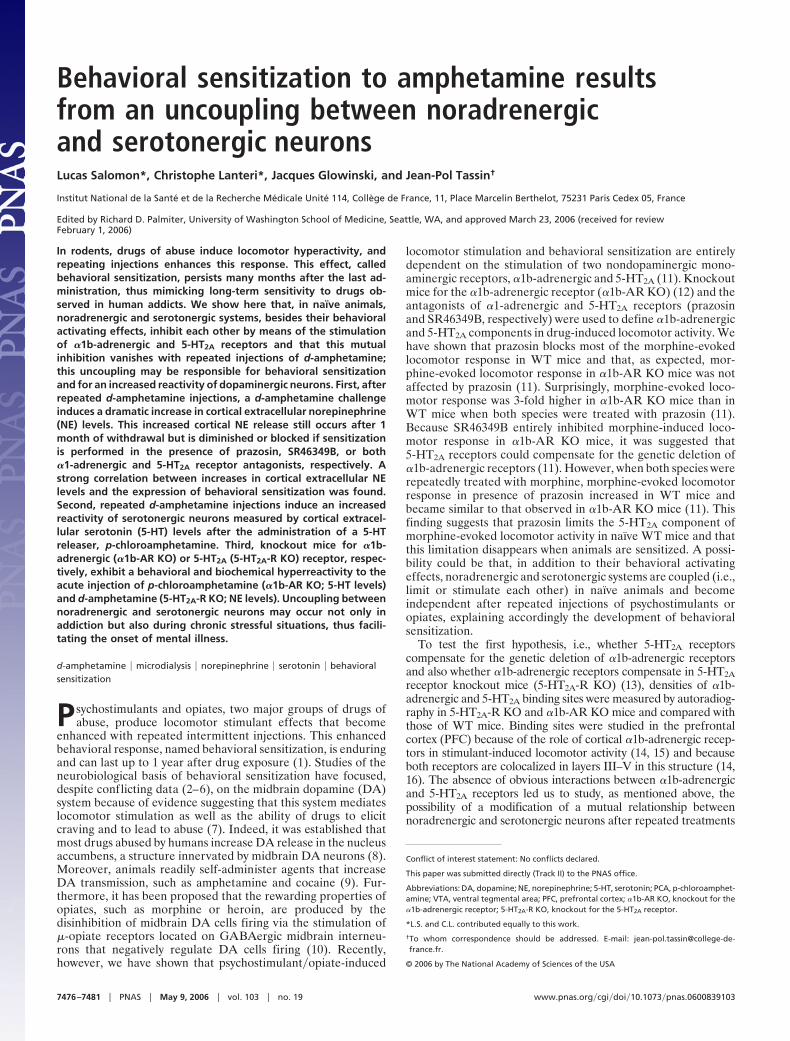
Receptors. Fig. 1 shows that, whereas binding of tritiated prazosinis almost completely abolished (P � 0.001) in �1b-AR KO mice,there is no change in tritiated ketanserin binding when comparedwith WT mice. Similarly, in 5-HT2A-R KO mice, despite theimportant decrease in tritiated ketanserin binding (P � 0.001),no change occurs in tritiated prazosin binding when comparedwith WT mice. This result clearly indicates that �1b-adrenergicand 5-HT2A receptors do not compensate for each other andsuggests that the dependency between both systems observed innaıve animals is due to interactions that are presynaptic to
�1b-adrenergic and 5-HT2A receptors, i.e., between noradren-ergic and serotonergic neurons.
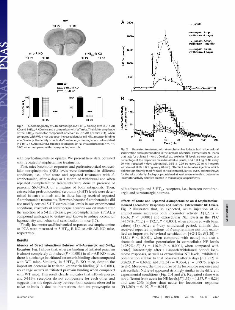
Effects of Acute and Repeated d-Amphetamine on d-Amphetamine-Induced Locomotor Responses and Cortical Extracellular NE Levels.Fig. 2 illustrates that, as expected, acute injection of d-amphetamine increases both locomotor activity [F(1,273) �166.6; P � 0.0001] and extracellular NE levels in the PFC[�167%; F(1,5) � 172.7; P � 0.0001, when compared with basalvalues] (18). After a 4-day withdrawal period, animals thatreceived repeated injections of d-amphetamine not only exhib-ited an important behavioral sensitization [�241%; F(1,20) �315.1; P � 0.0001, when compared with acute] but also adramatic and similar potentiation in extracellular NE levels[�259%; F(1,5) � 116.9; P � 0.0001, when compared withacute]. Interestingly, after a 1-month withdrawal period, loco-motor responses, as well as extracellular NE levels, exhibited apotentiation similar to that observed after 4 days [F(1,252) �0.2620, P � 0.6092; and F(1,54) � 0.0664, P � 0.7976, respec-tively). Moreover, the time course of the locomotor response andextracellular NE level appeared strikingly similar in the differentexperimental conditions (Fig. 2 A and B). Repeated saline wasnot different from acute for NE levels [F(1,37) � 1.157, P � 0.29]and was 20% higher than acute for locomotor response[F(1,269) � 6.107, P � 0.014].
Fig. 1. Autoradiography of �1b-adrenergic and 5-HT2A binding sites in �1b-ARKO and 5-HT2A-R KO mice and a comparison with WT mice. The higher amplitudeof the 5-HT2A locomotor component observed in �1b-AR KO mice (11), whencompared with WT, is not due to an increased density in 5-HT2A receptor bindingsites. Similarly, the density of cortical �1b-adrenergic binding sites is not modifiedin 5-HT2A-R KO mice. 3H Kt, tritiated ketanserin; 3H Pz, tritiated prazosin. ***, P �0.001 when compared with corresponding controls.
Fig. 2. Repeated treatment with d-amphetamine induces both a behavioralsensitization and a potentiation in the increase of cortical extracellular NE levelsthat lasts for at least 1 month. Cortical extracellular NE levels are expressed as apercentage of the respective mean basal value (acute, 0.64 � 0.1 pg of NE every20 min; repeated 4-days withdrawal, 0.55 � 0.09 pg every 20 min; 1-monthwithdrawal, 0.56 � 0.1 pg every 20 min). Effects of acute saline injection, whichdid not significantly modify basal cortical extracellular NE levels, are not shownfor the sake of clarity. Each group contained at least seven animals to determinelocomotor activity and five animals in microdialysis experiments.
Salomon et al. PNAS � May 9, 2006 � vol. 103 � no. 19 � 7477
NEU
ROSC
IEN
CE
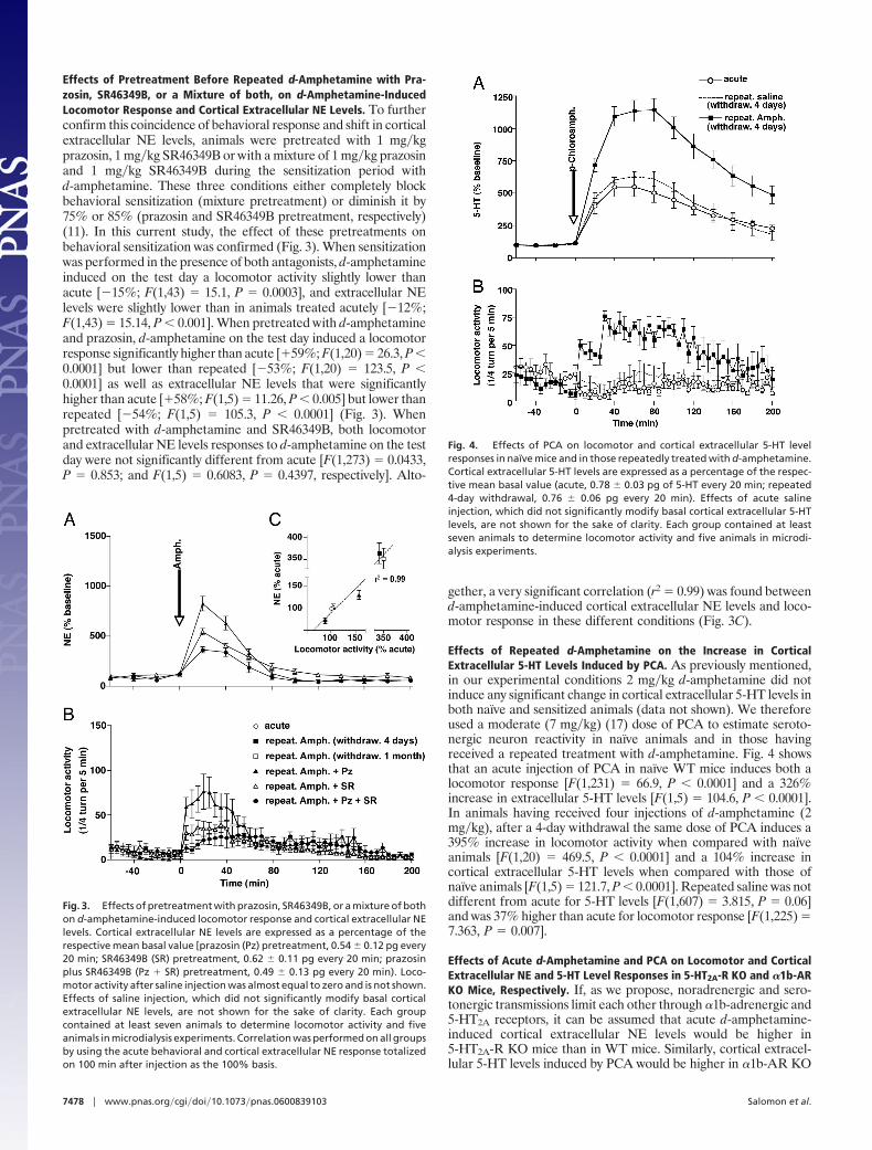
Effects of Pretreatment Before Repeated d-Amphetamine with Pra-zosin, SR46349B, or a Mixture of both, on d-Amphetamine-InducedLocomotor Response and Cortical Extracellular NE Levels. To furtherconfirm this coincidence of behavioral response and shift in corticalextracellular NE levels, animals were pretreated with 1 mg�kgprazosin, 1 mg�kg SR46349B or with a mixture of 1 mg�kg prazosinand 1 mg�kg SR46349B during the sensitization period withd-amphetamine. These three conditions either completely blockbehavioral sensitization (mixture pretreatment) or diminish it by75% or 85% (prazosin and SR46349B pretreatment, respectively)(11). In this current study, the effect of these pretreatments onbehavioral sensitization was confirmed (Fig. 3). When sensitizationwas performed in the presence of both antagonists, d-amphetamineinduced on the test day a locomotor activity slightly lower thanacute [�15%; F(1,43) � 15.1, P � 0.0003], and extracellular NElevels were slightly lower than in animals treated acutely [�12%;F(1,43) � 15.14, P � 0.001]. When pretreated with d-amphetamineand prazosin, d-amphetamine on the test day induced a locomotorresponse significantly higher than acute [�59%; F(1,20) � 26.3, P �0.0001] but lower than repeated [�53%; F(1,20) � 123.5, P �0.0001] as well as extracellular NE levels that were significantlyhigher than acute [�58%; F(1,5) � 11.26, P � 0.005] but lower thanrepeated [�54%; F(1,5) � 105.3, P � 0.0001] (Fig. 3). Whenpretreated with d-amphetamine and SR46349B, both locomotorand extracellular NE levels responses to d-amphetamine on the testday were not significantly different from acute [F(1,273) � 0.0433,P � 0.853; and F(1,5) � 0.6083, P � 0.4397, respectively]. Alto-
gether, a very significant correlation (r2 � 0.99) was found betweend-amphetamine-induced cortical extracellular NE levels and loco-motor response in these different conditions (Fig. 3C).
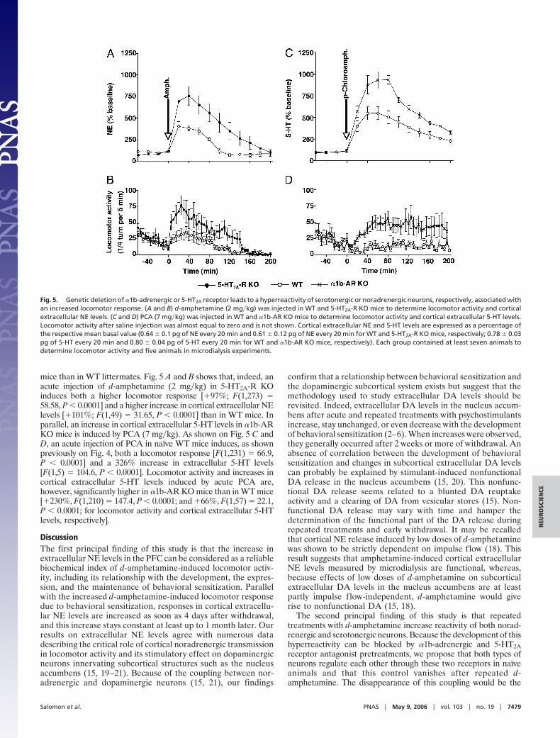
Effects of Repeated d-Amphetamine on the Increase in CorticalExtracellular 5-HT Levels Induced by PCA. As previously mentioned,in our experimental conditions 2 mg�kg d-amphetamine did notinduce any significant change in cortical extracellular 5-HT levels inboth naıve and sensitized animals (data not shown). We thereforeused a moderate (7 mg�kg) (17) dose of PCA to estimate seroto-nergic neuron reactivity in naıve animals and in those havingreceived a repeated treatment with d-amphetamine. Fig. 4 showsthat an acute injection of PCA in naıve WT mice induces both alocomotor response [F(1,231) � 66.9, P � 0.0001] and a 326%increase in extracellular 5-HT levels [F(1,5) � 104.6, P � 0.0001].In animals having received four injections of d-amphetamine (2mg�kg), after a 4-day withdrawal the same dose of PCA induces a395% increase in locomotor activity when compared with naıveanimals [F(1,20) � 469.5, P � 0.0001] and a 104% increase incortical extracellular 5-HT levels when compared with those ofnaıve animals [F(1,5) � 121.7, P � 0.0001]. Repeated saline was notdifferent from acute for 5-HT levels [F(1,607) � 3.815, P � 0.06]and was 37% higher than acute for locomotor response [F(1,225) �7.363, P � 0.007].
Effects of Acute d-Amphetamine and PCA on Locomotor and CorticalExtracellular NE and 5-HT Level Responses in 5-HT2A-R KO and �1b-ARKO Mice, Respectively. If, as we propose, noradrenergic and sero-tonergic transmissions limit each other through �1b-adrenergic and5-HT2A receptors, it can be assumed that acute d-amphetamine-induced cortical extracellular NE levels would be higher in5-HT2A-R KO mice than in WT mice. Similarly, cortical extracel-lular 5-HT levels induced by PCA would be higher in �1b-AR KO
Fig. 3. Effects of pretreatment with prazosin, SR46349B, or a mixture of bothon d-amphetamine-induced locomotor response and cortical extracellular NElevels. Cortical extracellular NE levels are expressed as a percentage of therespective mean basal value [prazosin (Pz) pretreatment, 0.54 � 0.12 pg every20 min; SR46349B (SR) pretreatment, 0.62 � 0.11 pg every 20 min; prazosinplus SR46349B (Pz � SR) pretreatment, 0.49 � 0.13 pg every 20 min). Loco-motor activity after saline injection was almost equal to zero and is not shown.Effects of saline injection, which did not significantly modify basal corticalextracellular NE levels, are not shown for the sake of clarity. Each groupcontained at least seven animals to determine locomotor activity and fiveanimals in microdialysis experiments. Correlation was performed on all groupsby using the acute behavioral and cortical extracellular NE response totalizedon 100 min after injection as the 100% basis.
Fig. 4. Effects of PCA on locomotor and cortical extracellular 5-HT levelresponses in naıve mice and in those repeatedly treated with d-amphetamine.Cortical extracellular 5-HT levels are expressed as a percentage of the respec-tive mean basal value (acute, 0.78 � 0.03 pg of 5-HT every 20 min; repeated4-day withdrawal, 0.76 � 0.06 pg every 20 min). Effects of acute salineinjection, which did not significantly modify basal cortical extracellular 5-HTlevels, are not shown for the sake of clarity. Each group contained at leastseven animals to determine locomotor activity and five animals in microdi-alysis experiments.
7478 � www.pnas.org�cgi�doi�10.1073�pnas.0600839103 Salomon et al.
mice than in WT littermates. Fig. 5 A and B shows that, indeed, anacute injection of d-amphetamine (2 mg�kg) in 5-HT2A-R KOinduces both a higher locomotor response [�97%; F(1,273) �58.58, P � 0.0001] and a higher increase in cortical extracellular NElevels [�101%; F(1,49) � 31.65, P � 0.0001] than in WT mice. Inparallel, an increase in cortical extracellular 5-HT levels in �1b-ARKO mice is induced by PCA (7 mg�kg). As shown on Fig. 5 C andD, an acute injection of PCA in naıve WT mice induces, as shownpreviously on Fig. 4, both a locomotor response [F(1,231) � 66.9,P � 0.0001] and a 326% increase in extracellular 5-HT levels[F(1,5) � 104.6, P � 0.0001]. Locomotor activity and increases incortical extracellular 5-HT levels induced by acute PCA are,however, significantly higher in �1b-AR KO mice than in WT mice[�230%, F(1,210) � 147.4, P � 0.0001; and �66%, F(1,57) � 22.1,P � 0.0001; for locomotor activity and cortical extracellular 5-HTlevels, respectively].
DiscussionThe first principal finding of this study is that the increase inextracellular NE levels in the PFC can be considered as a reliablebiochemical index of d-amphetamine-induced locomotor activ-ity, including its relationship with the development, the expres-sion, and the maintenance of behavioral sensitization. Parallelwith the increased d-amphetamine-induced locomotor responsedue to behavioral sensitization, responses in cortical extracellu-lar NE levels are increased as soon as 4 days after withdrawal,and this increase stays constant at least up to 1 month later. Ourresults on extracellular NE levels agree with numerous datadescribing the critical role of cortical noradrenergic transmissionin locomotor activity and its stimulatory effect on dopaminergicneurons innervating subcortical structures such as the nucleusaccumbens (15, 19–21). Because of the coupling between nor-adrenergic and dopaminergic neurons (15, 21), our findings
confirm that a relationship between behavioral sensitization andthe dopaminergic subcortical system exists but suggest that themethodology used to study extracellular DA levels should berevisited. Indeed, extracellular DA levels in the nucleus accum-bens after acute and repeated treatments with psychostimulantsincrease, stay unchanged, or even decrease with the developmentof behavioral sensitization (2–6). When increases were observed,they generally occurred after 2 weeks or more of withdrawal. Anabsence of correlation between the development of behavioralsensitization and changes in subcortical extracellular DA levelscan probably be explained by stimulant-induced nonfunctionalDA release in the nucleus accumbens (15, 20). This nonfunc-tional DA release seems related to a blunted DA reuptakeactivity and a clearing of DA from vesicular stores (15). Non-functional DA release may vary with time and hamper thedetermination of the functional part of the DA release duringrepeated treatments and early withdrawal. It may be recalledthat cortical NE release induced by low doses of d-amphetaminewas shown to be strictly dependent on impulse flow (18). Thisresult suggests that amphetamine-induced cortical extracellularNE levels measured by microdialysis are functional, whereas,because effects of low doses of d-amphetamine on subcorticalextracellular DA levels in the nucleus accumbens are at leastpartly impulse flow-independent, d-amphetamine would giverise to nonfunctional DA (15, 18).
The second principal finding of this study is that repeatedtreatments with d-amphetamine increase reactivity of both norad-renergic and serotonergic neurons. Because the development of thishyperreactivity can be blocked by �1b-adrenergic and 5-HT2Areceptor antagonist pretreatments, we propose that both types ofneurons regulate each other through these two receptors in naıveanimals and that this control vanishes after repeated d-amphetamine. The disappearance of this coupling would be the
Fig. 5. Genetic deletion of �1b-adrenergic or 5-HT2A receptor leads to a hyperreactivity of serotonergic or noradrenergic neurons, respectively, associated withan increased locomotor response. (A and B) d-amphetamine (2 mg�kg) was injected in WT and 5-HT2A-R KO mice to determine locomotor activity and corticalextracellular NE levels. (C and D) PCA (7 mg�kg) was injected in WT and �1b-AR KO mice to determine locomotor activity and cortical extracellular 5-HT levels.Locomotor activity after saline injection was almost equal to zero and is not shown. Cortical extracellular NE and 5-HT levels are expressed as a percentage ofthe respective mean basal value (0.64 � 0.1 pg of NE every 20 min and 0.61 � 0.12 pg of NE every 20 min for WT and 5-HT2A-R KO mice, respectively; 0.78 � 0.03pg of 5-HT every 20 min and 0.80 � 0.04 pg of 5-HT every 20 min for WT and �1b-AR KO mice, respectively). Each group contained at least seven animals todetermine locomotor activity and five animals in microdialysis experiments.
Salomon et al. PNAS � May 9, 2006 � vol. 103 � no. 19 � 7479
NEU
ROSC
IEN
CE
cause of a higher reactivity of noradrenergic and serotonergicneurons and thus the expression of behavioral sensitization throughan increased reactivity of dopaminergic neurons. Together withd-amphetamine-induced behavioral sensitization, increased reac-tivity of noradrenergic neurons to d-amphetamine can be blockedpartly or completely by prazosin, SR46349B, or a mixture of bothantagonists used for pretreatment. These data indicate that thestimulation of both receptors, �1b-adrenergic and 5-HT2A, is im-plicated in the increased reactivity of noradrenergic neurons due torepeated d-amphetamine. Although not all of these experimentshave been reproduced with PCA, it is very likely that the sameeffects of antagonists would occur. Indeed, we show here theinhibiting influence of each receptor on noradrenergic and sero-tonergic transmissions with the demonstration, in �1b-AR KO and5-HT2A-R KO mice, of a behavioral and biochemical hyperreac-tivity to the acute injection of the indirect agonist of the comple-mentary neurons, (i.e., d-amphetamine and NE release in5-HT2A-R KO mice and PCA and 5-HT release in �1b-AR KOmice). However, the ‘‘constitutive’’ behavioral sensitization thatknockout mice exhibit appears, at least for 5-HT2A-R KO mice, tobe only partial. There could be two reasons for this: first, onereceptor is missing in these mutant mice, and it is likely that eachreceptor has its part in the behavioral activation; second, becauseanimals were not treated repeatedly, the remaining receptor wasnot repeatedly stimulated, and this could hamper the developmentof a complete behavioral sensitization.
The dopaminergic D1 receptor is another monoaminergic re-ceptor whose blockade was shown to inhibit the development andexpression of behavioral sensitization to d-amphetamine (22). Wepretreated animals with systemic SCH23390, a D1 antagonist,before d-amphetamine repeated injections and found that, after a4-day withdrawal period, both cortical d-amphetamine-inducedextracellular NE levels and PCA-induced extracellular 5-HT levelswere identical to those observed in naıve animals (Fig. 6, which ispublished as supporting information on the PNAS web site). Thisfinding confirms that these biochemical indexes covary with be-havioral sensitization to d-amphetamine and also indicates that D1receptor stimulation participates in noradrenergic and serotonergicneuron regulation. We also tested the biochemical consequence ofthe blockade by SCH23390 of the expression of behavioral sensi-tization to d-amphetamine. SCH23390 was injected, in animalspreviously sensitized to d-amphetamine, 30 min before an injectionof either d-amphetamine or PCA. In these conditions, locomotorresponse to both compounds was completely blocked, and corticald-amphetamine-induced extracellular NE level increases stayed3-fold above those obtained after an acute injection, whereasPCA-induced extracellular 5-HT levels became identical to those ofacutely treated animals (Fig. 7, which is published as supportinginformation on the PNAS web site). These results may suggest thatthe noradrenergic neurons are engaged upstream to D1 transmis-sion whereas serotonergic cells would act downstream. However,besides its anti-D1 property, SCH23390 is also a potent 5HT2Cagonist (23), and this characteristic may limit the reactivity ofnoradrenergic and serotonergic neurons to d-amphetamine andPCA, respectively. Obviously, this issue needs further investigation.
Although the precise mechanism responsible for the dysregula-tion of noradrenergic and serotonergic neurons after repeatedstimulations of �1b-adrenergic and 5-HT2A receptors is not yetknown, anatomical relationships between the dorsal raphe and thepontine noradrenergic nuclei suggest the existence of a functionalinteraction between noradrenergic and serotonergic neurons. Bothneuronal groups are REM-off (24, 25), and the discharge rate ofserotonergic neurons is under the excitatory control of �1-adrenergic receptors (26, 27). Conversely, raphe nuclei serotonergiccells may contact through 5-HT2A receptors GABAergic interneu-rons that hyperpolarize noradrenergic cells in the locus coeruleus(28). Another nonexclusive possibility is that coupling between bothneurotransmitter systems occurs in the PFC, where �1b-adrenergic
and 5-HT2A receptors are colocalized. In that case, increasedcortical extracellular NE and 5-HT levels would stimulate gluta-matergic pyramidal cells (29) that excite ventral tegmental area(VTA) dopaminergic neurons. However, although local injection ofprazosin into the PFC blocked amphetamine-induced locomotorresponse (15), local bilateral injections of SR46349B into the PFCor into the VTA have indicated that only those injections done intothe VTA could counteract d-amphetamine-induced locomotoractivity (30), thus suggesting that the 5-HT2A receptors implicatedin the effects we observe are preferentially located in the VTA.Altogether, one can postulate that in naıve animals the activationof noradrenergic cells by external stimuli is immediately attenuatedby serotonergic cells whose activation is itself triggered by norad-renergic neurons. Preliminary data obtained in the laboratoryindicate that, similarly to d-amphetamine, repeated morphineinduces the same uncoupling between noradrenergic and seroto-nergic neurons. Finally, we propose that this long-term uncouplingbetween noradrenergic and serotonergic neurons may explain theextreme sensitivity to emotions described by human addicts duringwithdrawal. Moreover, it must be recalled that stressful situationscross-sensitize with effects of psychostimulants or opiates on be-havioral sensitization. Chronic stress may therefore also induce anuncoupling between noradrenergic and serotonergic systems andthus be one source of mental illnesses such as bipolar disorder.
Materials and MethodsAnimals. WT mice were C57BL6 male adults (25–35 g). Micelacking the �1b-adrenergic receptor (�1b-AR KO) (12) or the5HT2A receptor (5HT2A-R KO) (13) were backcrossed on aC57BL6 genetic background for at least seven generations. Theywere maintained on a 12-h light�dark cycle (lights on at 0700 hours)with food and water freely available.
Autoradiography. Brains were rapidly removed after animal deathand frozen in isopentane (�40°C). Sections (20 �m) were cut witha cryostat, mounted onto gelatin-coated glass slides, and stored at�20°C until incubation. For �1b-AR binding sites, sections wereincubated at 20°C with tritiated prazosin (1 nM) for 30 min in a 50mM Tris�HCl (pH 7.4) buffer, washed five times in ice-cold buffer,and dried. For 5HT2A-R binding sites, sections were incubated at20°C with tritiated ketanserin (2 nM) for 60 min in a 170 mMTris�HCl, 10 mM pargyline, 4 mM CaCl2, and 0.01% ascorbic acid(pH 7.4) buffer, washed five times in ice-cold buffer, and dried.Specificity was tested by adding during incubation 1 �M prazosinfor �1b-AR binding sites or 1 �M SR46349B for 5HT2A-R bindingsites. All slides were exposed to tritiated Hyperfilm for 45 days.Autoradiograms were revealed, digitized, and quantified by usingIMAGEJ software.
Drugs. d-Amphetamine sulfate and PCA hydrochloride (SigmaAldrich) were dissolved in saline. Prazosin hydrochloride (SigmaAldrich) was sonicated in water. SR46349B hemifumarate[(1Z,2E)-1-(2-fluoro-phenyl)-3-(4-hydroxyphenyl)-prop-2-en-one-O-(2-dimethylamino-ethyl)-oxime hemifumarate] was a generousgift from Sanofi-Synthelabo Research (Montpellier, France). It wasdissolved with a drop of lactic acid, neutralized with 1 M NaOH, andsonicated in saline. All drugs were injected i.p. (0.3 ml per 100 g).Doses are expressed as salts. d-Amphetamine was given at 2 mg�kg,and PCA was given at 7 mg�kg (17). Doses of prazosin (1 mg�kgi.p.) and SR46349B (1 mg�kg i.p.) were kept identical to previousexperiments (11).
Locomotor Activity. Acute treatment. Mice were introduced in acircular corridor (4.5-cm width, 17-cm external diameter) crossedby four infrared beams (1.5 cm above the base) placed at every 90°(Imetronic, Pessac, France). The locomotor activity was countedwhen animals interrupted two successive beams and thus hadtraveled a quarter of the circular corridor. Spontaneous activity was
7480 � www.pnas.org�cgi�doi�10.1073�pnas.0600839103 Salomon et al.
recorded for 60 min (habituation to the experimental procedure),and then mice were injected i.p. with d-amphetamine or PCA, andlocomotor responses were recorded for an additional 200-minperiod.Repeated treatment. Mice were injected on 4 consecutive days withd-amphetamine, and their locomotor activity was recorded after ad-amphetamine injection after a 4-day withdrawal with the sameprotocol as for an acute treatment. To test the effect of thepretreatment on the development of behavioral sensitization, eachday mice received a pretreatment (saline, prazosin, SR46349B, orprazosin plus SR46349B) 30 min before the injection of d-amphetamine. When animals received prazosin as a pretreatment,two subsequent injections of prazosin were administered at 60 and150 min after d-amphetamine injection because of the short half-life(100 min) of prazosin (11). Finally, locomotor responses to d-amphetamine were tested either 4 days or 1 month after the lastd-amphetamine injection. Control mice were injected on 4 consec-utive days with 0.9% saline (0.1 ml per injection), and theirlocomotor response to d-amphetamine (or PCA) was recorded 4days later.
Surgery. Mice were anesthetized with sodium pentobarbital (60mg�kg; Sanofi Sante Animale, Libourne, France) and placed in astereotaxic frame (Kopf Instruments). Unilateral permanent can-nula (CMA�7 guide cannula; Microdialysis, Solna, Sweden) wasplaced at the edge of the PFC and was secured on the skull withscrew and dental cement. The coordinates for the guide cannula tipwere as follows: anteroposterior, �2.6 relative to bregma; medio-lateral, �0.5; dorsoventral, 0 mm from dura (31).
After surgery, mice were placed in individual plastic cages andallowed to recover for at least 4 days.
Microdialysis Experiment. Acute treatment. The day of the experiment,the microdialysis probe was inserted in the PFC (CMA�7; mem-brane length, 2 mm; diameter, 0.24 mm; cutoff, 6,000 Da; Micro-dialysis). Artificial CSF (147 mM NaCl�3.5 mM KCl�1 mM CaCl2�1.2 mM MgCl2�1 mM NaH2PO4�25 mM NaHCO3, pH 7.6) wasperfused with a CMA100 microinjection pump through the probeat a rate of 1 �l�min via fluorinated ethylene propylene (FEP)catheter (internal diameter, 0.12 mm) connected to a fluid swivel.Adequate steady state of monoamines levels in perfusate sampleswas reached 140 min after probe insertion, and samples werecollected in 300-�l vials placed in a refrigerated, computer-con-trolled fraction collector (CMA�170). Samples (20 �l every 20 min)were collected during 100 min to determine basal extracellularmonoamine values. After d-amphetamine or PCA injection, sam-ples were collected for 200 min. Samples were analyzed the day ofthe experiment.Repeated treatment. Mice received four consecutive daily injectionsof pretreatment plus d-amphetamine and waited 4 days or 1 monthbefore the dialysis experiment. Every day after d-amphetamineinjection, mice were immediately placed for 2 h in the cylindrical
compartment used to perform microdialysis. Control mice receivedfour consecutive daily injections of 0.9% saline (0.1 ml per injection)and waited 4 days before the dialysis experiment.
Biochemistry. Dialysate samples were completed to 30 �l with theadapted mobile phase and placed into a refrigerated automaticinjector (Triathlon; Spark Holland, Emmen, The Netherlands).Twenty-five microliters of the sample were injected every 30 minthrough a rheodyne valve in the mobile phase circuit. High-performance liquid chromatography was performed with a reverse-phase column (80 � 4.6 mm, 3 �m particle size, HR-80, ESA,Chelmsford, MA). Mobile phase (for NE analysis: 0.1 MNaH2PO4�0.1 mM EDTA�3.8 mM octane sulfonic acid�0.25 mMtriethylamine�10% methanol, pH 2.9; for 5-HT analysis: 0.1 MNaH2PO4�0.1 mM EDTA�1.5 mM octane sulfonic acid�0.25 mMtriethylamine�15% methanol�5% acetonitrile, pH 2.9) was deliv-ered at 0.7 ml�min by an ESA-580 pump (ESA). Electrochemicaldetection was performed with an ESA coulometric detector (Cou-lochem II 5100A, with a 5014B analytical cell; Eurosep, Cergy,France). The conditioning electrode was set at �0.175 mV, and thedetecting electrode was set at �0.175 mV, allowing a good signal-to-noise ratio. External standards were regularly injected to deter-mine the stability of the sensitivity (0.2 pg for NE and 0.3 pg for5-HT).
Histology. At the end of the experiment, brains were put into aformaldehyde solution and cut on a microtome in serial coronalslices according to the atlas of Paxinos and Franklin (31). Histo-logical examination of cannula tip placement was subsequentlymade on 100-�m safranine-stained coronal sections.
Statistics. Statistical analysis was performed by using PRISM 3.0software (GraphPad, San Diego). Data from microdialysis andlocomotor activity experiments were described as a function oftime. Data from microdialysis were expressed as a percentage of therespective mean basal value. The extracellular monoamines leveland the locomotor activity obtained after the d-amphetamine orPCA injection (100-min analysis) were compared and analyzed withtwo-way ANOVA (repeated measures). Pharmacological treat-ments correspond to independent groups of animals. Opticaldensity data of the different mice were compared by using atwo-tailed unpaired Student t test. Significant differences were setat P � 0.05.
We thank S. Cotecchia (Faculte de Pharmacologie et de Toxicologie,Lausanne, Switzerland) for �1b-AR KO mice; L. Lanfumey and M. Hamon[Institut National de la Sante de la Recherche Medicale (INSERM), UniteMixte de Recherche 677, Universite Pierre et Marie Curie, Faculte PitieSalpetriere, Paris] for 5-HT2A-R KO mice; Y. Torrens, G. Blanc, and G.Godeheu for skillful technical assistance; and P. Tierney for careful readingof the manuscript. This work was supported by INSERM and MissionInterministerielle de Lutte Contre les Drogues et les Toxicomanies.
1. Paulson, P. E., Camp, D. M. & Robinson, T. E. (1991) Psychopharmacology 103, 480–492.2. Kuczenski, R. & Segal, D. S. (1992) Synapse 11, 164–169.3. Segal, D. S. & Kuczenski, R. (1992) Brain Res. 571, 330–337.4. Segal, D. S. & Kuczenski, R. (1992) Brain Res. 577, 351–355.5. Paulson, P. E. & Robinson, T. E. (1995) Synapse 19, 56–65.6. Cadoni, C., Solinas, M. & Di Chiara, G. (2000) Eur. J. Pharmacol. 388, 69–76.7. Robinson, T. E. & Becker, J. B. (1986) Brain Res. 396, 157–198.8. Di Chiara, G. & Imperato, A. (1988) Proc. Natl. Acad. Sci. USA 85, 5274–5278.9. Wise, R. A. (2004) Nat. Rev. Neurosci. 5, 483–494.
10. Johnson, S. W. & North, R. A. (1992) J. Neurosci. 12, 483–488.11. Auclair, A., Drouin, C., Cotecchia, S., Glowinski, G. & Tassin, J. P. (2004) Eur. J. Neurosci.
20, 3073–3084.12. Cavalli, A., Lattion, A. L., Hummler, E., Nenniger, M., Pedrazzini, T., Aubert, J. F., Michel, M. C.,
Yang, M., Lembo, G., Vecchione, C., et al. (1997) Proc. Natl. Acad. Sci. USA 94, 11589–11594.13. Fiorica-Howells, E., Hen, R., Gingrich, J., Li, Z. & Gershon, M. D. (2002) Am. J. Physiol.
282, G877–G893.14. Drouin, C., Darracq, L., Trovero, F., Blanc, G., Glowinski, G., Cotecchia, S. & Tassin, J. P.
(2002) J. Neurosci. 22, 2873–2884.15. Darracq, L., Blanc, G., Glowinski, G. & Tassin, J. P. (1998) J. Neurosci. 18, 2729–2739.16. Pazos, A., Cortes, R. & Palacios, J. M. (1985) Brain Res. 346, 231–249.
17. Itzhak, Y., Achat-Mendes, C. N., Ali, S. F. & Anderson, K. L. (2004) Neuropharmacology46, 74–84.
18. Florin, S. M., Kuczenski, R. & Segal, D. S. (1994) Brain Res. 654, 53–62.19. Shi, W. X., Pun, C. L., Zhang, X. X., Jones, M. D. & Bunney, B. S. (2000) J. Neurosci. 20, 3504–3511.20. Auclair, A., Cotecchia, S., Glowinski, G. & Tassin, J. P. (2002) J. Neurosci. 22, 9150–9154.21. Ventura, R., Cabib, S., Alcaro, A., Orsini, C. & Puglisi-Allegra, S. (2003) J. Neurosci. 23,
1879–1885.22. Vezina, P. (1996) J. Neurosci. 16, 2411–2420.23. Millan, M. J., Newman-Tancredi, A., Quentric, Y. & Cussac, D. (2001) Psychopharmacology
156, 58–62.24. Aston-Jones, G., Shipley, M. T., Chouvet, G., Ennis, M., Van Bockstaele, E., Pieribone, V.,
Shiekhattar, R., Akaoka, H., Drolet, G. & Astier, B. (1991) Prog. Brain Res. 88, 47–75.25. Jacobs, B. L. & Azmitia, E. C. (1992) Physiol. Rev. 72, 165–229.26. Baraban, J. M. & Aghajanian, G. K. (1980) Neuropharmacology 19, 355–363.27. Bortolozzi, A. & Artigas, F. (2003) Neuropsychopharmacology 28, 421–434.28. Szabo, S. T. & Blier, P. (2001) Brain Res. 922, 9–20.29. Sesack, S. R. & Pickel, V. M. (1992) J. Comp. Neurol. 320, 145–160.30. Auclair, A., Blanc, G., Glowinski, J. & Tassin, J. P. (2004) J. Neurochem. 91, 318–326.31. Paxinos, G. & Franklin, K. B. J. (1997) The Mouse Brain in Stereotaxic Coordinates
(Academic, New York), 2nd Ed.
Salomon et al. PNAS � May 9, 2006 � vol. 103 � no. 19 � 7481
NEU
ROSC
IEN
CE