Embed Size (px)
Citation preview
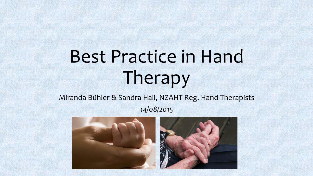
Best Practice in Hand Therapy
Miranda Bűhler & Sandra Hall, NZAHT Reg. Hand Therapists
14/08/2015
Purpose
• Introduction to Hand Therapy
• Overview of (non-pharmacological) conservative management of base-of-thumb osteoarthritis (BTOA)
• Overview of conservative management of carpal tunnel syndrome
• Resources to support best practice
What is Hand Therapy?
• The specialty of hand therapy has emerged from the professions of Occupational Therapy and Physiotherapy.
• This was in response to advances in surgical techniques that enabled greater functional restoration of injured and diseased upper extremities.
• Has built a body of knowledge and expertise that can contribute to improved outcomes from a range of acute and chronic conditions affecting the hand and upper limb.
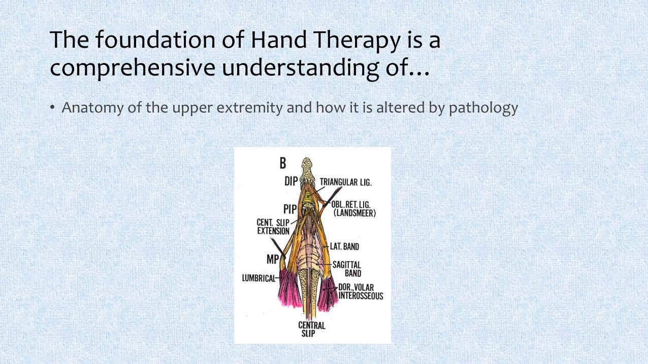
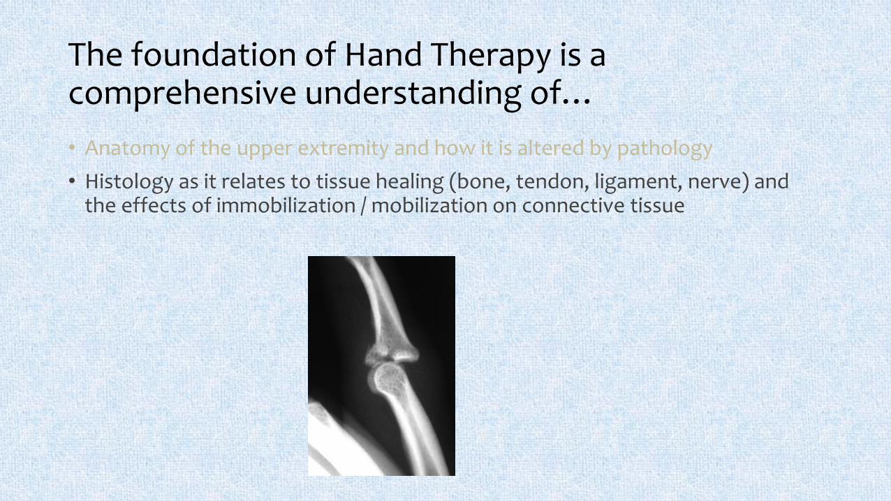
The foundation of Hand Therapy is a comprehensive understanding of…
• Anatomy of the upper extremity and how it is altered by pathology
The foundation of Hand Therapy is a comprehensive understanding of…
• Anatomy of the upper extremity and how it is altered by pathology
• Histology as it relates to tissue healing (bone, tendon, ligament, nerve) and the effects of immobilization / mobilization on connective tissue
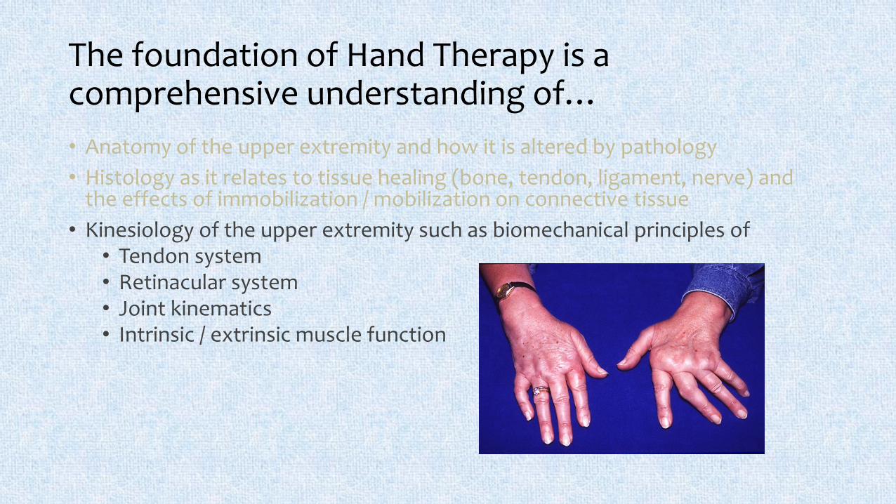
The foundation of Hand Therapy is a comprehensive understanding of…
• Anatomy of the upper extremity and how it is altered by pathology
• Histology as it relates to tissue healing (bone, tendon, ligament, nerve) and the effects of immobilization / mobilization on connective tissue
• Kinesiology of the upper extremity such as biomechanical principles of• Tendon system• Retinacular system• Joint kinematics• Intrinsic / extrinsic muscle function
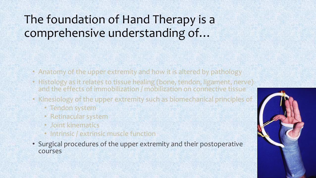
• Surgical procedures of the upper extremity and their postoperative courses
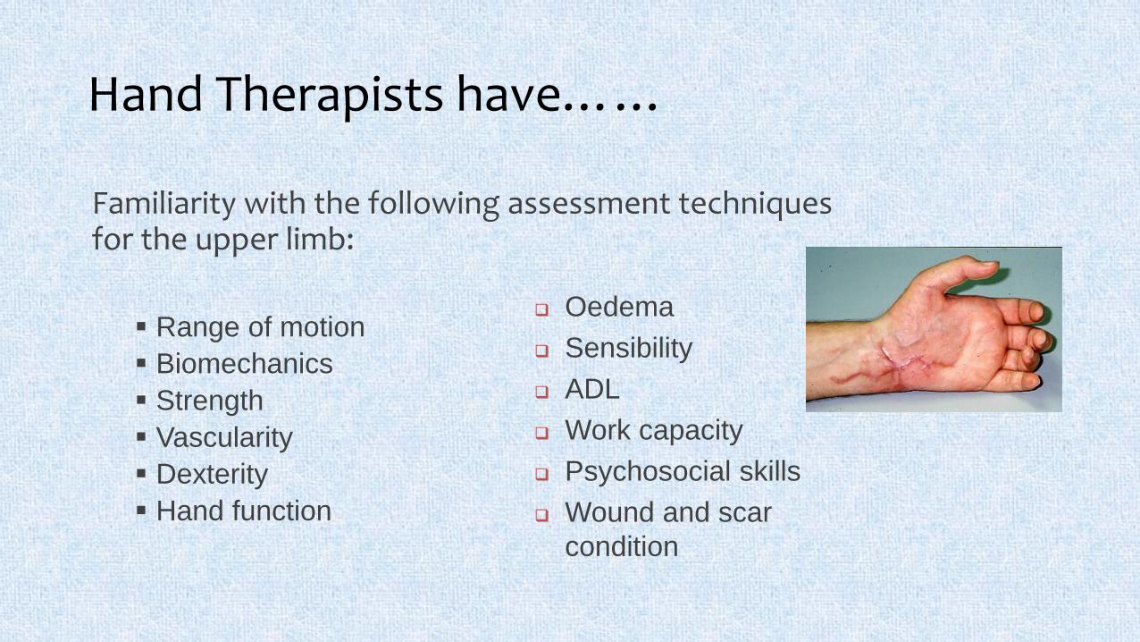
Hand Therapists have……
Familiarity with the following assessment techniques for the upper limb:
Range of motion
Biomechanics
Strength
Vascularity
Dexterity
Hand function
Oedema
Sensibility
ADL
Work capacity
Psychosocial skills
Wound and scar
condition
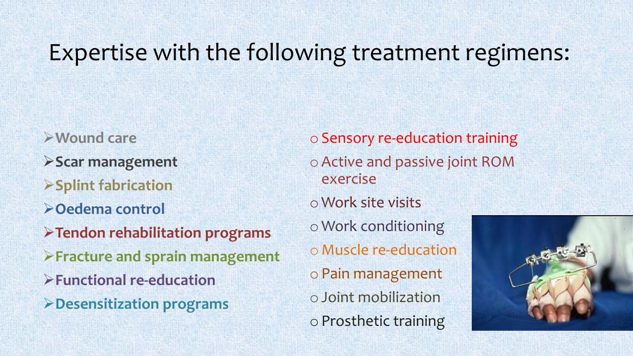
Expertise with the following treatment regimens:
Wound care
Scar management
Splint fabrication
Oedema control
Tendon rehabilitation programs
Fracture and sprain management
Functional re-education
Desensitization programs
oSensory re-education training
oActive and passive joint ROM exercise
oWork site visits
oWork conditioning
oMuscle re-education
oPain management
o Joint mobilization
oProsthetic training
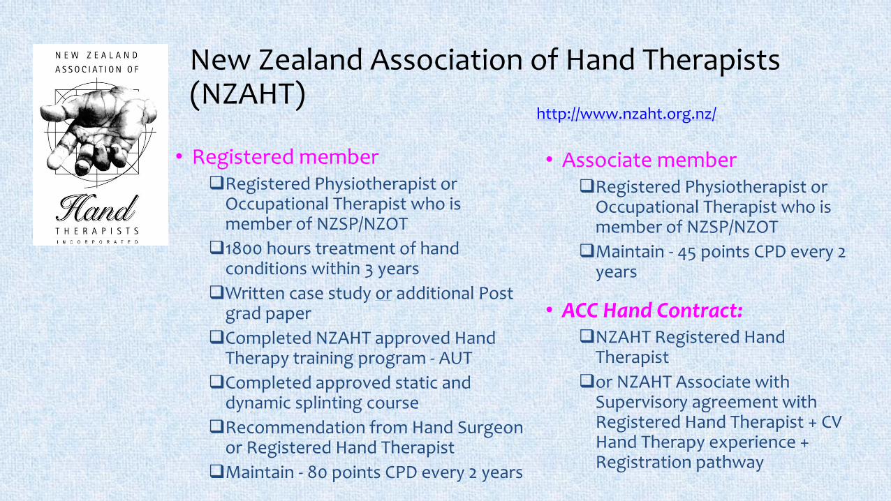
New Zealand Association of Hand Therapists (NZAHT)
• Associate memberRegistered Physiotherapist or
Occupational Therapist who is member of NZSP/NZOT
Maintain - 45 points CPD every 2 years
• ACC Hand Contract:NZAHT Registered Hand
Therapist
or NZAHT Associate with Supervisory agreement with Registered Hand Therapist + CV Hand Therapy experience + Registration pathway
• Registered member Registered Physiotherapist or
Occupational Therapist who is member of NZSP/NZOT
1800 hours treatment of hand conditions within 3 years
Written case study or additional Post grad paper
Completed NZAHT approved Hand Therapy training program - AUT
Completed approved static and dynamic splinting course
Recommendation from Hand Surgeon or Registered Hand Therapist
Maintain - 80 points CPD every 2 years
http://www.nzaht.org.nz/
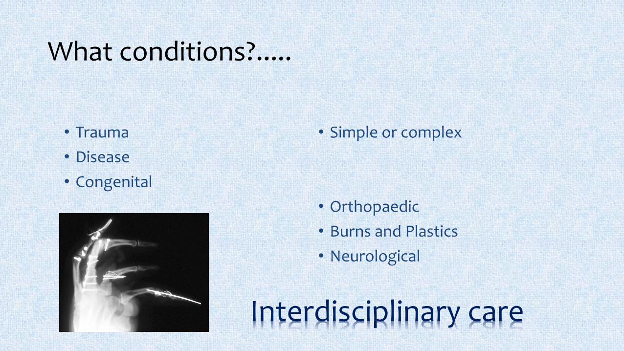
What conditions?.....
• Trauma
• Disease
• Congenital
• Simple or complex
• Orthopaedic
• Burns and Plastics
• Neurological
Interdisciplinary care
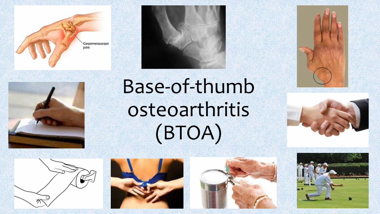
Base-of-thumb osteoarthritis
(BTOA)
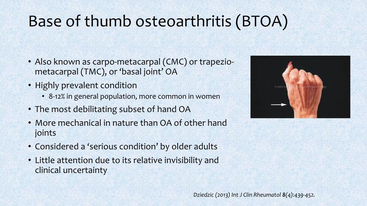
Base of thumb osteoarthritis (BTOA)
• Also known as carpo-metacarpal (CMC) or trapezio-metacarpal (TMC), or ‘basal joint’ OA
• Highly prevalent condition • 8-12% in general population, more common in women
• The most debilitating subset of hand OA
• More mechanical in nature than OA of other hand joints
• Considered a ‘serious condition’ by older adults
• Little attention due to its relative invisibility and clinical uncertainty
Dziedzic (2013) Int J Clin Rheumatol 8(4):439-452.
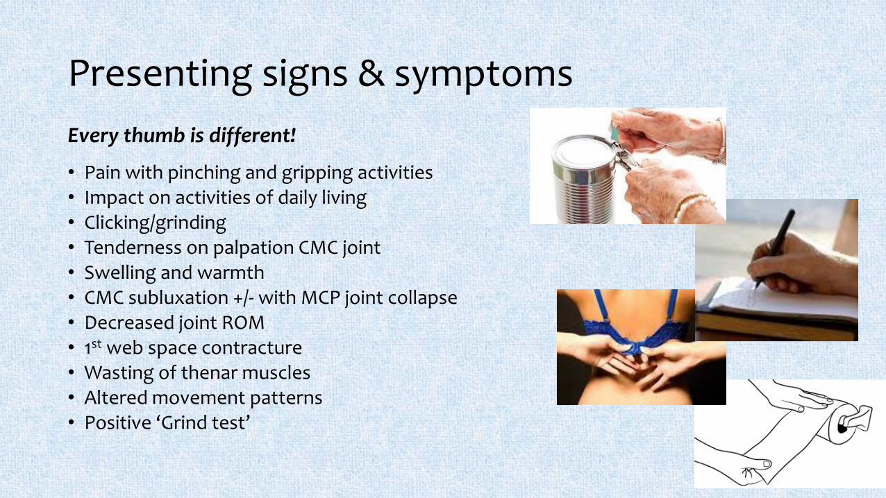
Presenting signs & symptoms
Every thumb is different!
• Pain with pinching and gripping activities• Impact on activities of daily living• Clicking/grinding• Tenderness on palpation CMC joint• Swelling and warmth• CMC subluxation +/- with MCP joint collapse• Decreased joint ROM• 1st web space contracture• Wasting of thenar muscles• Altered movement patterns• Positive ‘Grind test’
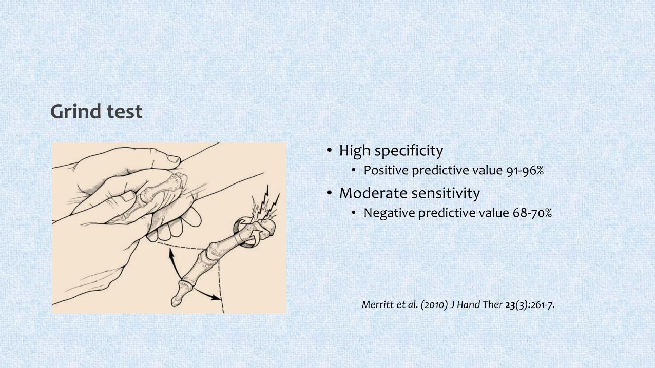
Grind test
• High specificity• Positive predictive value 91-96%
• Moderate sensitivity• Negative predictive value 68-70%
Merritt et al. (2010) J Hand Ther 23(3):261-7.
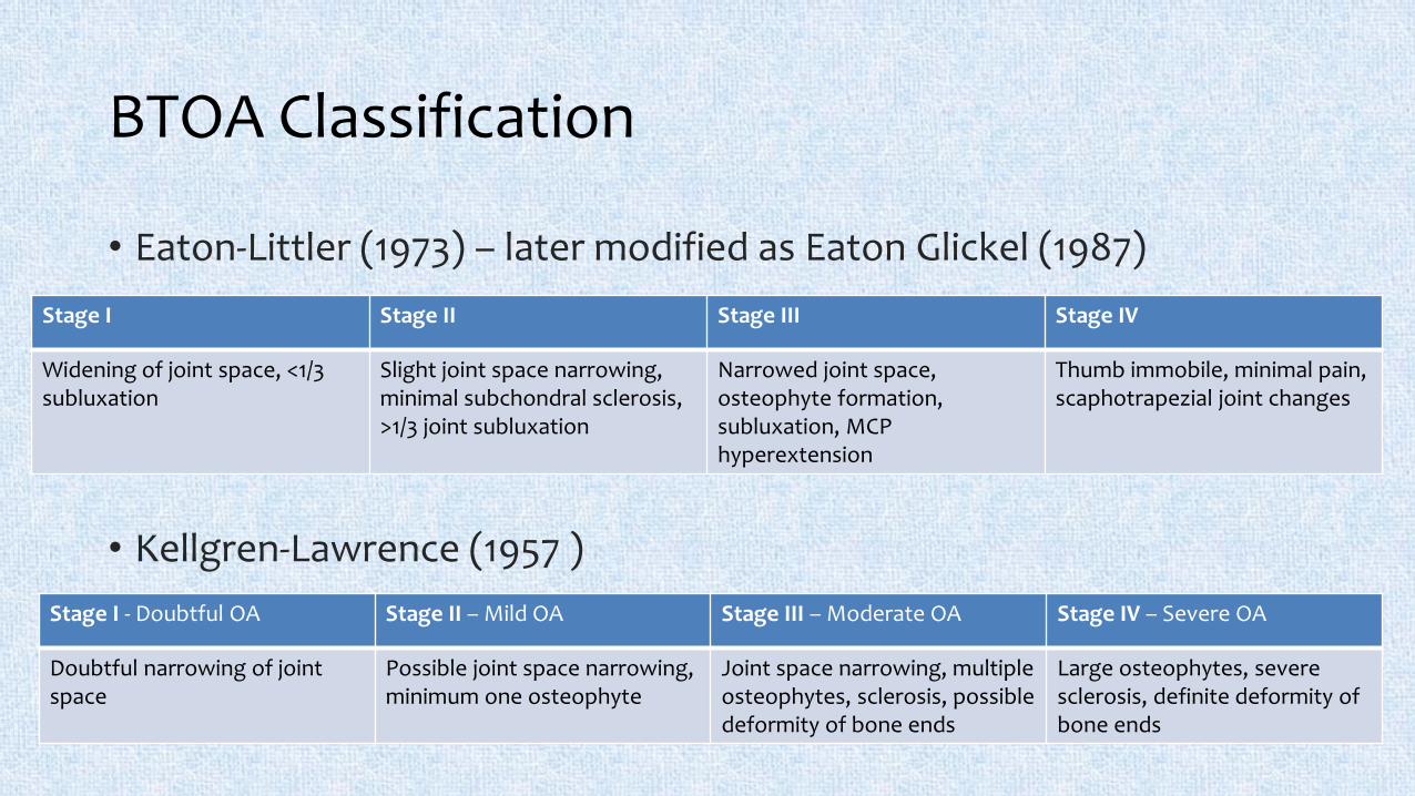
BTOA Classification
• Eaton-Littler (1973) – later modified as Eaton Glickel (1987)
• Kellgren-Lawrence (1957 )
Stage I Stage II Stage III Stage IV
Widening of joint space, <1/3 subluxation
Slight joint space narrowing, minimal subchondral sclerosis, >1/3 joint subluxation
Narrowed joint space, osteophyte formation, subluxation, MCP hyperextension
Thumb immobile, minimal pain, scaphotrapezial joint changes
Stage I - Doubtful OA Stage II – Mild OA Stage III – Moderate OA Stage IV – Severe OA
Doubtful narrowing of joint space
Possible joint space narrowing,minimum one osteophyte
Joint space narrowing, multipleosteophytes, sclerosis, possible deformity of bone ends
Large osteophytes, severe sclerosis, definite deformity of bone ends
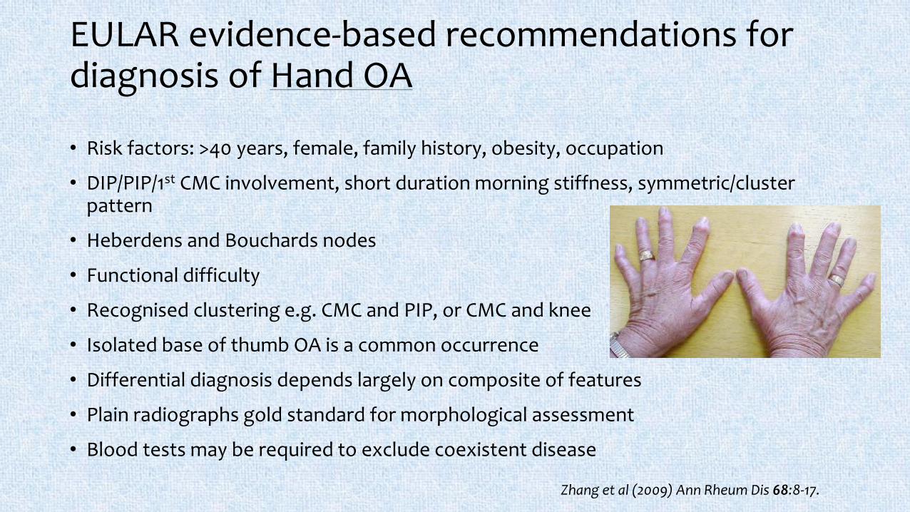
EULAR evidence-based recommendations for diagnosis of Hand OA
• Risk factors: >40 years, female, family history, obesity, occupation
• DIP/PIP/1st CMC involvement, short duration morning stiffness, symmetric/cluster pattern
• Heberdens and Bouchards nodes
• Functional difficulty
• Recognised clustering e.g. CMC and PIP, or CMC and knee
• Isolated base of thumb OA is a common occurrence
• Differential diagnosis depends largely on composite of features
• Plain radiographs gold standard for morphological assessment
• Blood tests may be required to exclude coexistent disease
Zhang et al (2009) Ann Rheum Dis 68:8-17.
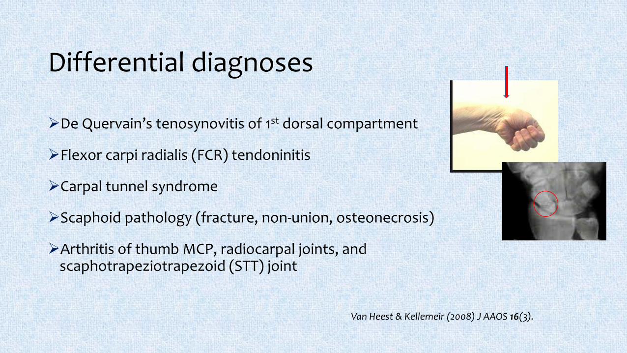
Differential diagnoses
De Quervain’s tenosynovitis of 1st dorsal compartment
Flexor carpi radialis (FCR) tendoninitis
Carpal tunnel syndrome
Scaphoid pathology (fracture, non-union, osteonecrosis)
Arthritis of thumb MCP, radiocarpal joints, and scaphotrapeziotrapezoid (STT) joint
Van Heest & Kellemeir (2008) J AAOS 16(3).
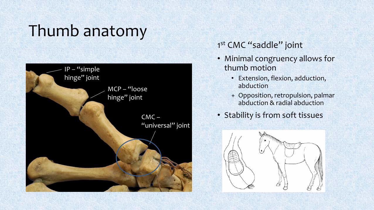
Thumb anatomy1st CMC “saddle” joint
• Minimal congruency allows for thumb motion• Extension, flexion, adduction,
abduction
+ Opposition, retropulsion, palmar abduction & radial abduction
• Stability is from soft tissues
IP – “simple hinge” joint
MCP – “loose hinge” joint
CMC –“universal” joint
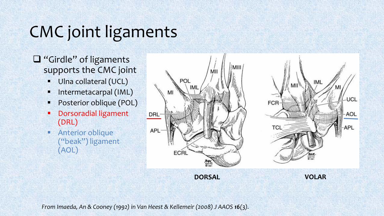
CMC joint ligaments
“Girdle” of ligaments supports the CMC joint Ulna collateral (UCL)
Intermetacarpal (IML)
Posterior oblique (POL)
Dorsoradial ligament (DRL)
Anterior oblique (“beak”) ligament (AOL)
DORSAL VOLAR
From Imaeda, An & Cooney (1992) in Van Heest & Kellemeir (2008) J AAOS 16(3).
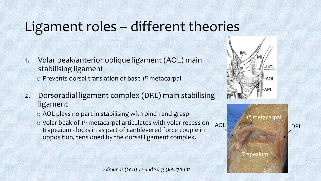
Ligament roles – different theories
1. Volar beak/anterior oblique ligament (AOL) main stabilising ligamento Prevents dorsal translation of base 1st metacarpal
2. Dorsoradial ligament complex (DRL) main stabilising ligamento AOL plays no part in stabilising with pinch and grasp
o Volar beak of 1st metacarpal articulates with volar recess on trapezium - locks in as part of cantilevered force couple in opposition, tensioned by the dorsal ligament complex.
Edmunds (2011) J Hand Surg 36A:170-182.
Trapezium
1st metacarpal
AOL DRL
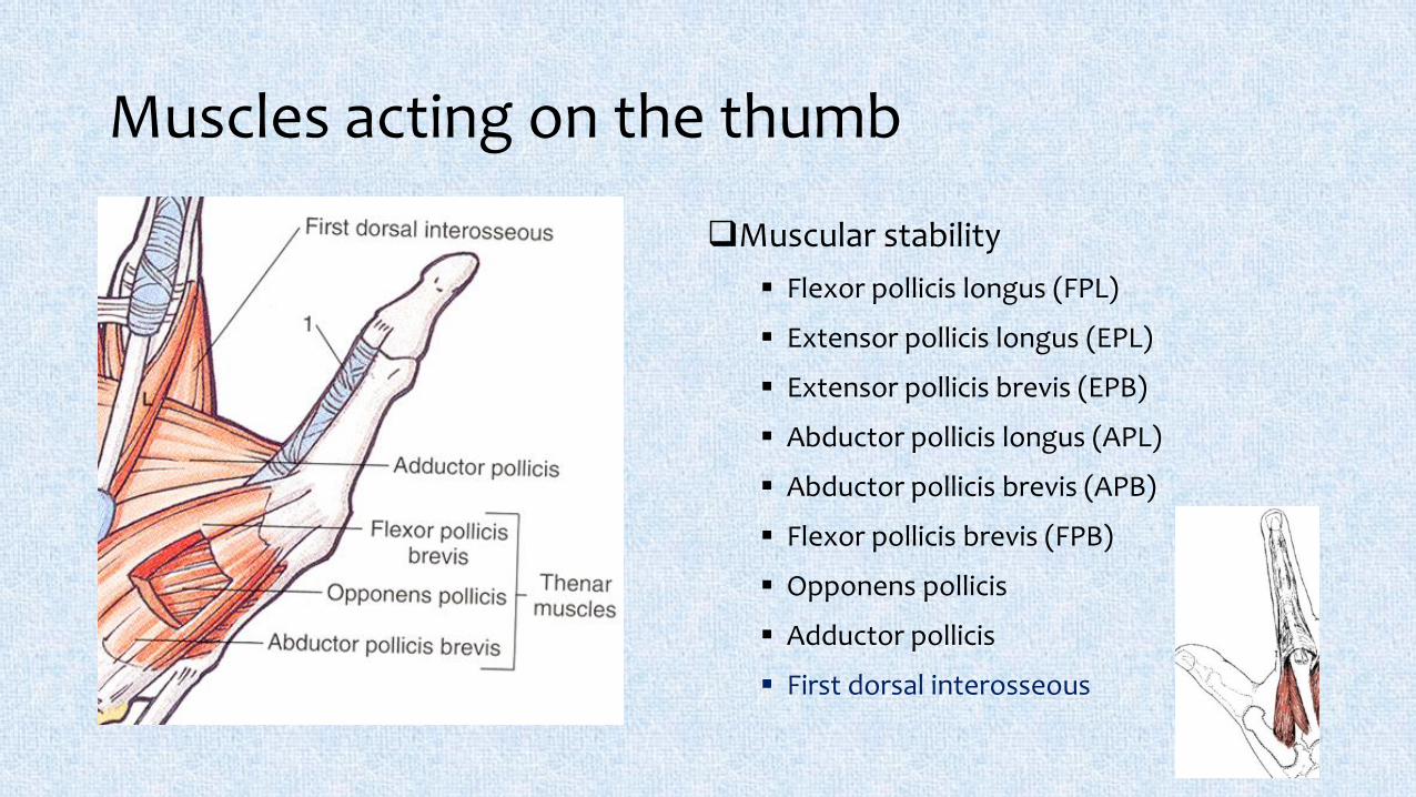
Muscles acting on the thumb
Muscular stability
Flexor pollicis longus (FPL)
Extensor pollicis longus (EPL)
Extensor pollicis brevis (EPB)
Abductor pollicis longus (APL)
Abductor pollicis brevis (APB)
Flexor pollicis brevis (FPB)
Opponens pollicis
Adductor pollicis
First dorsal interosseous
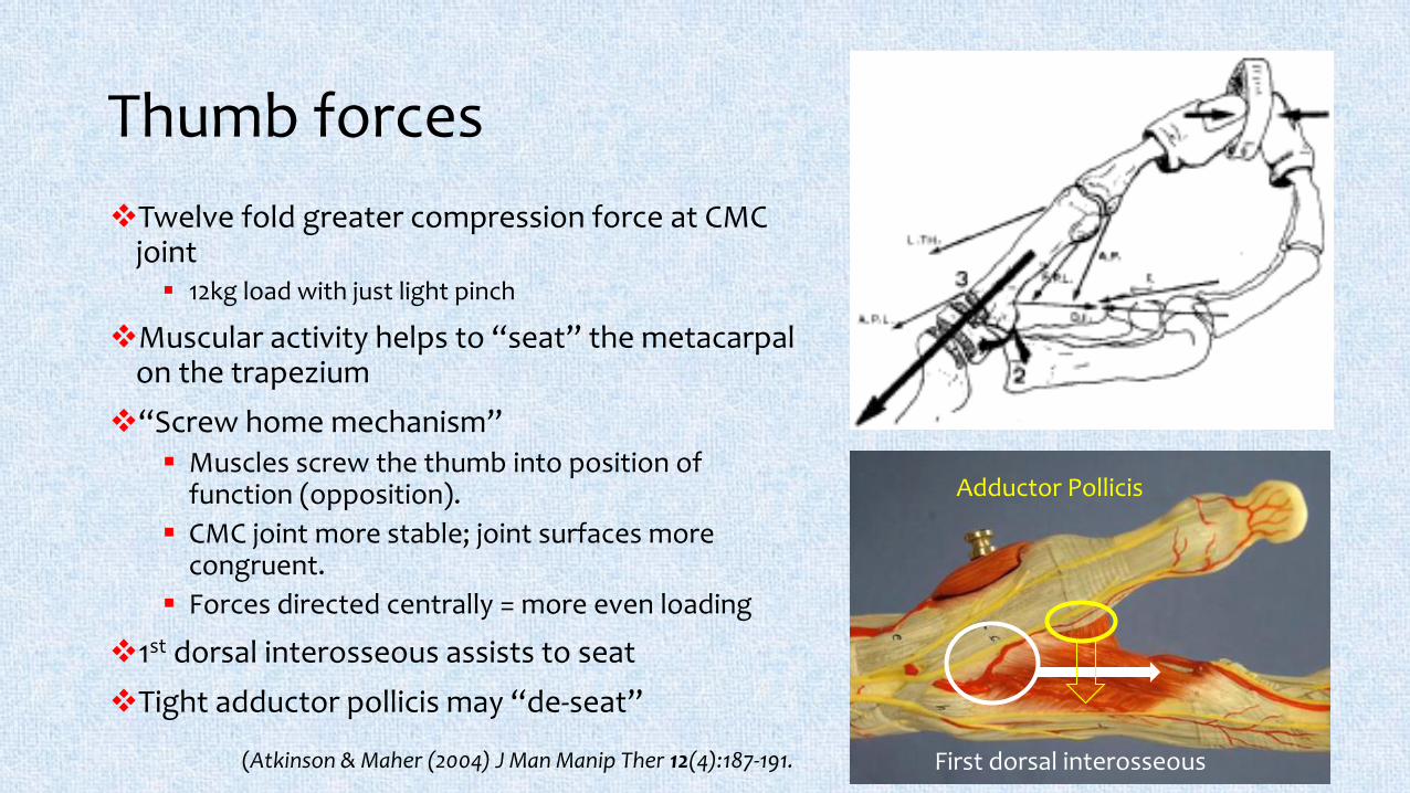
Thumb forces
Twelve fold greater compression force at CMC joint 12kg load with just light pinch
Muscular activity helps to “seat” the metacarpal on the trapezium
“Screw home mechanism” Muscles screw the thumb into position of
function (opposition).
CMC joint more stable; joint surfaces more congruent.
Forces directed centrally = more even loading
1st dorsal interosseous assists to seat
Tight adductor pollicis may “de-seat”
(Atkinson & Maher (2004) J Man Manip Ther 12(4):187-191. First dorsal interosseous
Adductor Pollicis
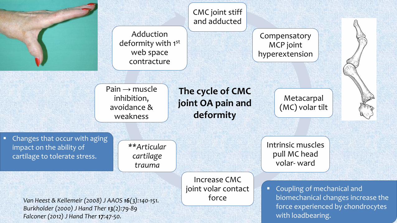
CMC joint stiff and adducted
Compensatory MCP joint
hyperextension
Metacarpal (MC) volar tilt
Intrinsic muscles pull MC head volar- ward
Increase CMC joint volar contact
force
**Articular cartilage trauma
Pain → muscle inhibition,
avoidance & weakness
Adduction deformity with 1st
web space contracture
Van Heest & Kellemeir (2008) J AAOS 16(3):140-151.Burkholder (2000) J Hand Ther 13(2):79-89Falconer (2012) J Hand Ther 17:47-50.
The cycle of CMC joint OA pain and
deformity
Coupling of mechanical and biomechanical changes increase the force experienced by chondrocytes with loadbearing.
Changes that occur with aging impact on the ability of cartilage to tolerate stress.

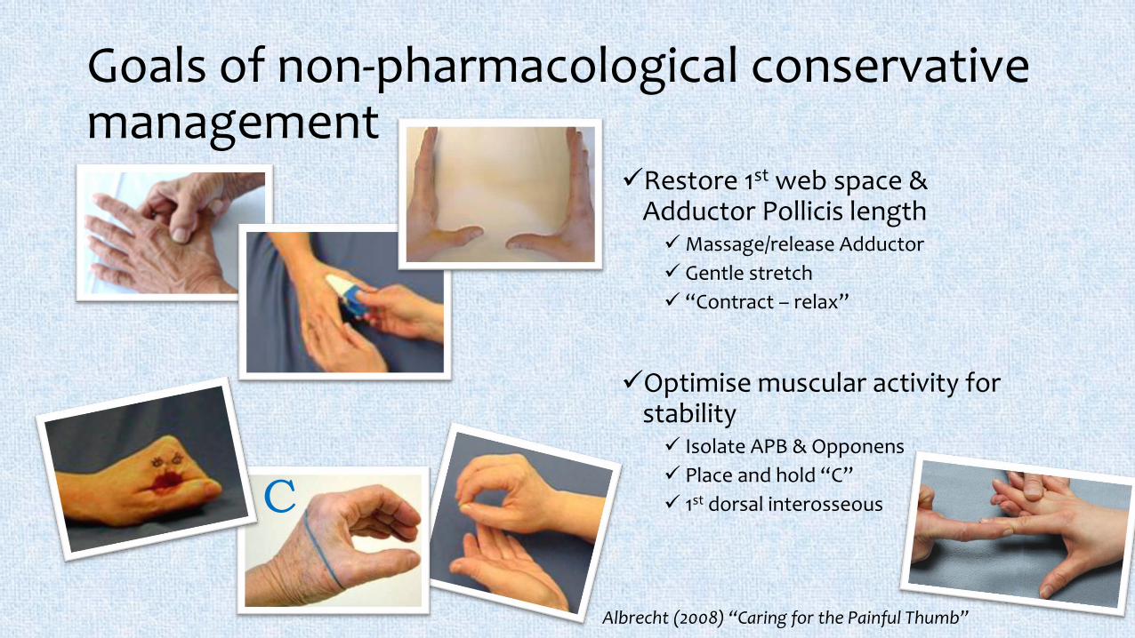
Goals of non-pharmacological conservative management
Restore 1st web space & Adductor Pollicis lengthMassage/release Adductor
Gentle stretch
“Contract – relax”
Optimise muscular activity for stability Isolate APB & Opponens
Place and hold “C”
1st dorsal interosseousC
Albrecht (2008) “Caring for the Painful Thumb”
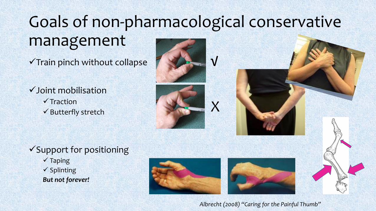
Goals of non-pharmacological conservative managementTrain pinch without collapse
Joint mobilisationTraction
Butterfly stretch
Support for positioning Taping
Splinting
But not forever!
√
X
Albrecht (2008) “Caring for the Painful Thumb”
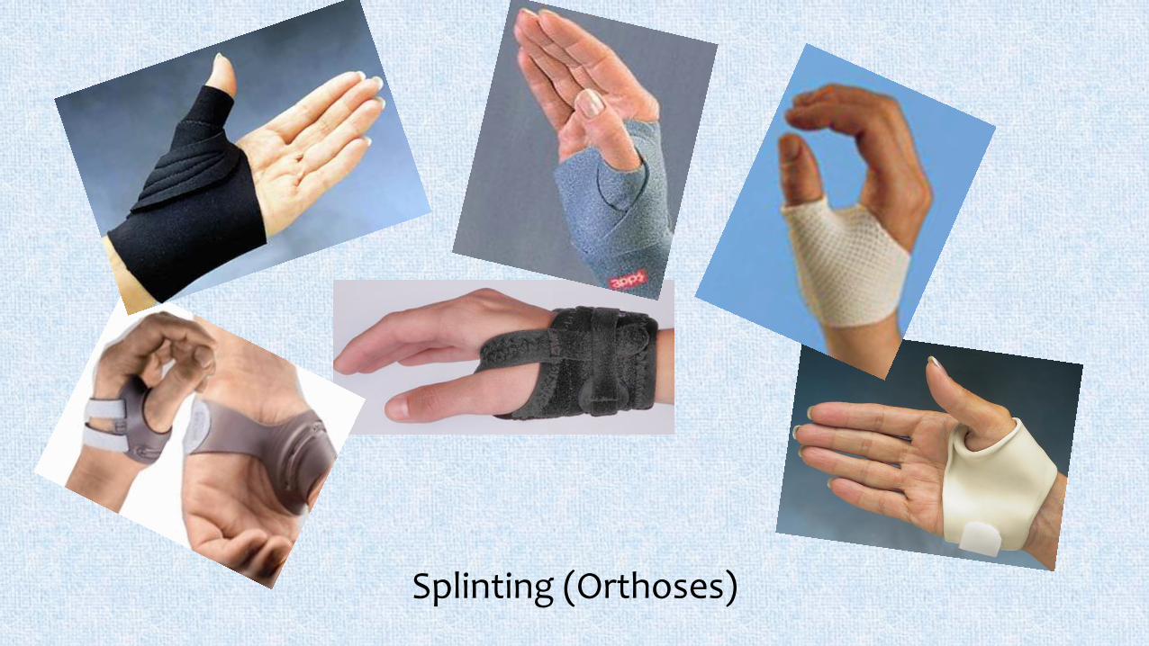
Splinting (Orthoses)
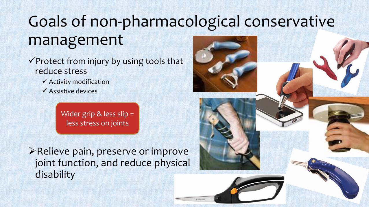
Goals of non-pharmacological conservative managementProtect from injury by using tools that
reduce stress Activity modification
Assistive devices
Relieve pain, preserve or improve joint function, and reduce physical disability
Wider grip & less slip = less stress on joints
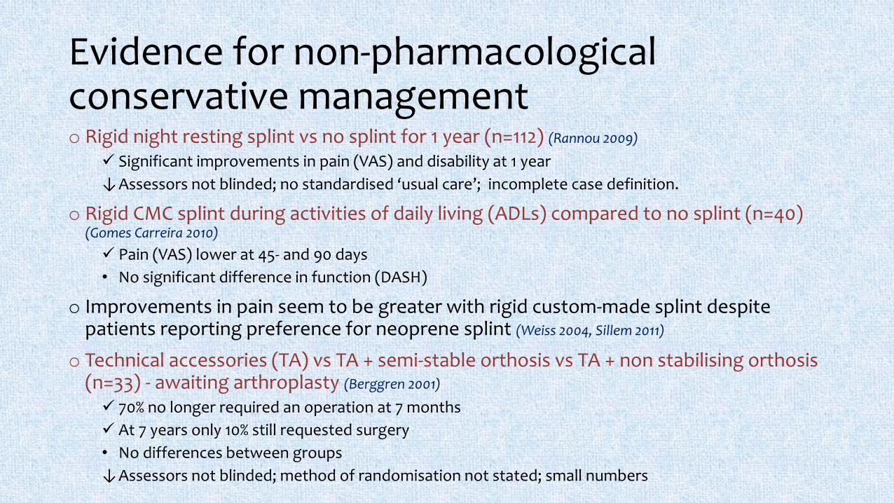
Evidence for non-pharmacological conservative managemento Rigid night resting splint vs no splint for 1 year (n=112) (Rannou 2009)
Significant improvements in pain (VAS) and disability at 1 year
↓Assessors not blinded; no standardised ‘usual care’; incomplete case definition.
o Rigid CMC splint during activities of daily living (ADLs) compared to no splint (n=40) (Gomes Carreira 2010)
Pain (VAS) lower at 45- and 90 days
• No significant difference in function (DASH)
o Improvements in pain seem to be greater with rigid custom-made splint despite patients reporting preference for neoprene splint (Weiss 2004, Sillem 2011)
o Technical accessories (TA) vs TA + semi-stable orthosis vs TA + non stabilising orthosis (n=33) - awaiting arthroplasty (Berggren 2001)
70% no longer required an operation at 7 months
At 7 years only 10% still requested surgery
• No differences between groups
↓Assessors not blinded; method of randomisation not stated; small numbers
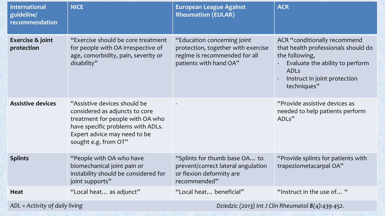
International guideline/ recommendation
NICE European League Against Rheumatism (EULAR)
ACR
Exercise & joint protection
“Exercise should be core treatment for people with OA irrespective of age, comorbidity, pain, severity or disability”
“Education concerning joint protection, together with exerciseregime is recommended for all patients with hand OA”
ACR “conditionally recommend that health professionals should do the following,- Evaluate the ability to perform
ADLs- Instruct in joint protection
techniques”
Assistive devices “Assistive devices should be considered as adjuncts to core treatment for people with OA who have specific problems with ADLs. Expert advice may need to be sought e.g. from OT”
- “Provide assistive devices as needed to help patients perform ADLs”
Splints “People with OA who have biomechanical joint pain or instability should be considered for joint supports”
“Splints for thumb base OA… to prevent/correct lateral angulation or flexion deformity are recommended”
“Provide splints for patients with trapeziometacarpal OA”
Heat “Local heat… as adjunct” “Local heat… beneficial” “Instruct in the use of… “
ADL = Activity of daily living Dziedzic (2013) Int J Clin Rheumatol 8(4):439-452.
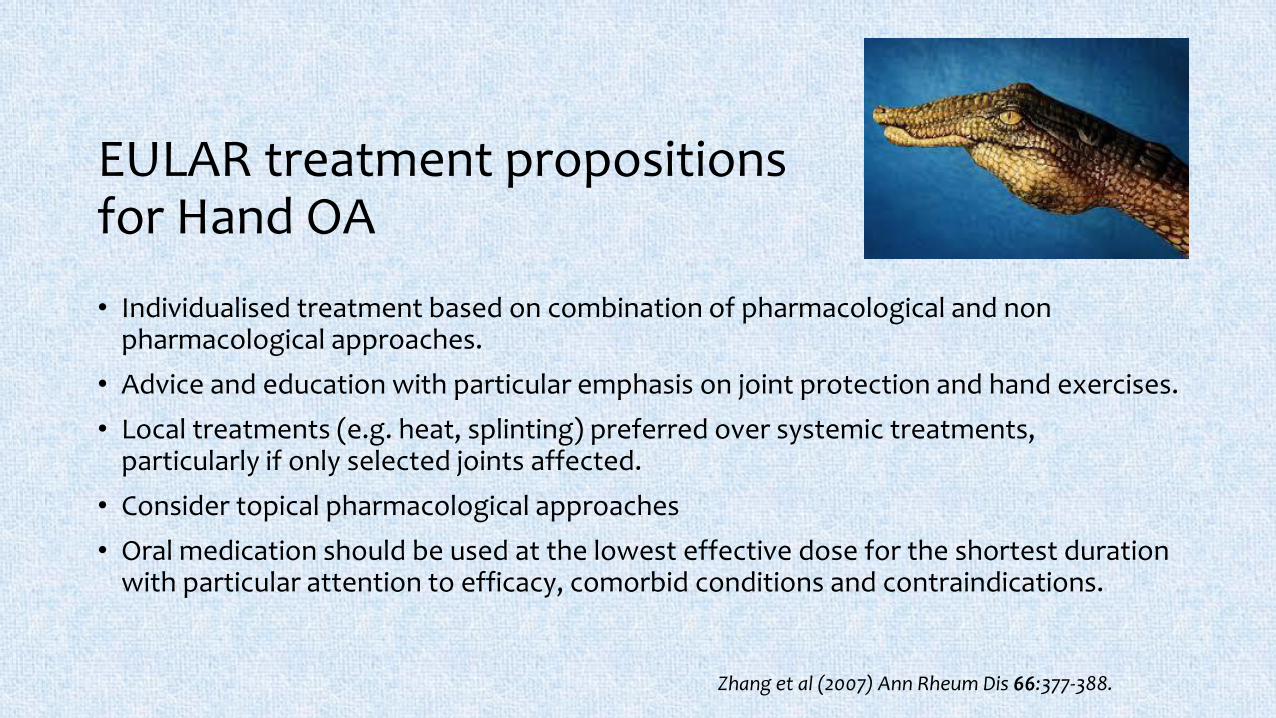
EULAR treatment propositionsfor Hand OA
• Individualised treatment based on combination of pharmacological and non pharmacological approaches.
• Advice and education with particular emphasis on joint protection and hand exercises.
• Local treatments (e.g. heat, splinting) preferred over systemic treatments, particularly if only selected joints affected.
• Consider topical pharmacological approaches
• Oral medication should be used at the lowest effective dose for the shortest duration with particular attention to efficacy, comorbid conditions and contraindications.
Zhang et al (2007) Ann Rheum Dis 66:377-388.
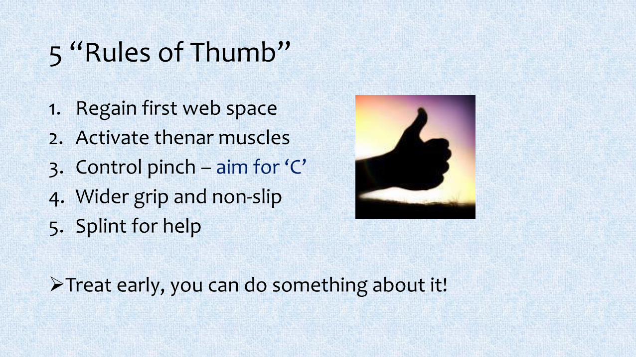
5 “Rules of Thumb”
1. Regain first web space
2. Activate thenar muscles
3. Control pinch – aim for ‘C’
4. Wider grip and non-slip
5. Splint for help
Treat early, you can do something about it!