Embed Size (px)
Citation preview

8/3/2019 Beta and Gamma Rays
http://slidepdf.com/reader/full/beta-and-gamma-rays 1/10
Beta particle
Beta particles are high-energy, high-speed electrons or positrons emitted by certain types of
radioactive nuclei such as potassium-40. The beta particles emitted are a form of ionizing radiation
also known as beta rays. The production of beta particles is termed beta decay. They are designatedby the Greek letter beta (β). There are two forms of beta decay, β
− and β+, which respectively give
rise to the electron and the positron.
β− decay (electron emission)
Further information: Beta decay
Beta decay
An unstable atomic nucleus with an excess of neutrons may undergo β− decay, where aneutron is converted into a proton, an electron and an electron-type antineutrino (theantiparticle of the neutrino):
n → p + e− + νe
This process is mediated by the weak interaction. The neutron turns into a proton through theemission of a virtual W− boson. At the quark level, W− emission turns a down-type quark intoan up-type quark, turning a neutron (one up quark and two down quarks) into a proton (twoup quarks and one down quark). The virtual W− boson then decays into an electron and anantineutrino.
Beta decay commonly occurs among the neutron-rich fission byproducts produced in nuclearreactors. Free neutrons also decay via this process. This is the source of the copious amountof electron antineutrinos produced by fission reactors.
[edit ] β+ decay (positron emission)
Further information: Beta decay
Unstable atomic nuclei with an excess of protons may undergo β+ decay, also called positrondecay, where a proton is converted into a neutron, a positron and an electron-type neutrino:
p → n + e+ + νe
8/3/2019 Beta and Gamma Rays
http://slidepdf.com/reader/full/beta-and-gamma-rays 2/10
Beta plus decay can only happen inside nuclei when the absolute value of the binding energy of the daughter nucleus is higher than that of the mother nucleus.
[edit ] Interaction with other matter
Of the three common types of radiation given off by radioactive materials, alpha, beta andgamma, beta has the medium penetrating power and the medium ionising power. Althoughthe beta particles given off by different radioactive materials vary in energy, most betaparticles can be stopped by a few millimeters of aluminum. Being composed of chargedparticles, beta radiation is more strongly ionising than gamma radiation. When passingthrough matter, a beta particle is decelerated by electromagnetic interactions and may give off bremsstrahlung x-rays.
[edit ] Uses
Beta particles can be used to treat health conditions such as eye and bone cancer, and are alsoused as tracers. Strontium-90 is the material most commonly used to produce beta particles.Beta particles are also used in quality control to test the thickness of an item, such as paper,coming through a system of rollers. Some of the beta radiation is absorbed while passingthrough the product. If the product is made too thick or thin, a correspondingly differentamount of radiation will be absorbed. A computer program monitoring the quality of themanufactured paper will then move the rollers to change the thickness of the final product.The well-known 'betalight' contains tritium and a phosphor.
Beta plus(or positron) decay of a radioactive tracer isotope is the source of the positrons usedin positron emission tomography (PET scan).
[edit ] History
Henri Becquerel, while experimenting with fluorescence, accidentally found out thatUranium exposed a black paper wrapped photographic plate with some unknown radiation that could not be turned off like X-rays. Ernest Rutherford continued these experiments anddiscovered two different kinds of radiation:
alpha particles that did not show up on the Becquerel plates because they were easily
absorbed by the black wrapping paper
beta particles which are 100 times more penetrating than alpha particles.
He published his results in 1897.
[edit ] Health
Beta particles are able to penetrate living matter to a certain extent and can change thestructure of struck molecules. In most cases such change can be considered as damage withresults possibly as severe as cancer and death. If the struck molecule is DNA it can show aspontaneous mutation.
Beta sources can be used in radiation therapy to kill cancer cells.

8/3/2019 Beta and Gamma Rays
http://slidepdf.com/reader/full/beta-and-gamma-rays 3/10
Gamma ray
Gamma radiation, also known as gamma rays (denoted as γ), is electromagnetic radiation of high frequency (very short wavelength). They are produced by sub-atomic particle
interactions such as electron-positron annihilation, neutral pion decay, radioactive decay, fusion, fission or inverse Compton scattering in astrophysical processes. Gamma raystypically have frequencies above 1019 Hz, and therefore have energies above 100 keV andwavelength less than 10 picometers, often smaller than an atom. Gamma rays fromradioactive decay commonly have energies of a few hundred keV, and are almost always lessthan 10 MeV in energy.
Because they are a form of ionizing radiation, gamma rays can cause serious damage whenabsorbed by living tissue, and are therefore a health hazard.
Paul Villard, a French chemist and physicist, discovered gamma radiation in 1900, while
studying radiation emitted from radium.[1] Alpha and beta "rays" had already been separatedand named by the work of Ernest Rutherford in 1899, and in 1903 Rutherford namedVillard's distinct new radiation "gamma rays."
In the past, the distinction between X-rays and gamma rays was based on energy (orequivalently frequency or wavelength), with gamma rays being considered a higher-energyversion of X-rays. However, modern high-energy (megavoltage) X-rays produced by linearaccelerators ("linacs") for megavoltage treatment, in cancer radiotherapy usually have higherenergy than gamma rays produced by radioactive gamma decay. Conversely, one of the mostcommon gamma-ray emitting isotopes used in diagnostic nuclear medicine, technetium-99m, produces gamma radiation of about the same energy (140 KeV) as produced by a diagnosticX-ray machine, and significantly lower energy than therapeutic photons from linacs. Becauseof this broad overlap in energy ranges, the two types of electromagnetic radiation are nowusually defined by their origin: X-rays are emitted by electrons (either in orbitals outside of the nucleus, or while being accelerated to produce Bremsstrahlung-type radiation), whilegamma rays are emitted by the nucleus or from other particle decays or annihilation events.There is no lower limit to the energy of photons produced by nuclear reactions, and thusultraviolet and even lower energy photons produced by these processes would also be definedas "gamma rays".[2]
In certain fields such as astronomy, gamma rays and X-rays are still sometimes defined by
energy, or used interchangeably, since the processes which produce them may be uncertain.
Units of measure and exposure
The measure of gamma rays' ionizing ability is called the exposure:
The coulomb per kilogram (C/kg) is the SI unit of ionizing radiation exposure, and is the
amount of radiation required to create 1 coulomb of charge of each polarity in 1 kilogram of
matter.
The röntgen (R) is an obsolete traditional unit of exposure, which represented the amount of
radiation required to create 1 esu of charge of each polarity in 1 cubic centimeter of dry air.
1 röntgen = 2.58×10⁻⁴ C/kg
8/3/2019 Beta and Gamma Rays
http://slidepdf.com/reader/full/beta-and-gamma-rays 4/10
However, the effect of gamma and other ionizing radiation on living tissue is more closelyrelated to the amount of energy deposited rather than the charge. This is called the absorbeddose:
The gray (Gy), which has units of (J/kg), is the SI unit of absorbed dose, and is the amount of
radiation required to deposit 1 joule of energy in 1 kilogram of any kind of matter. The rad is the (obsolete) corresponding traditional unit, equal to 0.01 J deposited per kg. 100
rad = 1 Gy.
The equivalent dose is the measure of the biological effect of radiation on human tissue. Forgamma rays it is equal to the absorbed dose.
The sievert (Sv) is the SI unit of equivalent dose, which for gamma rays is numerically equal
to the gray (Gy).
The rem is the traditional unit of equivalent dose. For gamma rays it is equal to the rad or
0.01 J of energy deposited per kg. 1 Sv = 100 rem.
[edit ] Properties
[edit ] Shielding
Shielding from gamma rays requires large amounts of mass. They are better absorbed bymaterials with high atomic numbers and high density, although neither effect is importantcompared to the total mass per area in the path of the gamma ray. For this reason, a leadshield is only modestly better (20-30%) as a gamma shield than an equal mass of anothershielding material such as aluminium, concrete, or soil; the lead's major advantage is itsdensity.
The higher the energy of the gamma rays, the thicker the shielding required. Materials forshielding gamma rays are typically measured by the thickness required to reduce the intensityof the gamma rays by one half (the half value layer or HVL). For example gamma rays thatrequire 1 cm (0.4″) of lead to reduce their intensity by 50% will also have their intensityreduced in half by 4.1 cm of granite rock, 6 cm (2½″) of concrete, or 9 cm (3½″) of packedsoil. However, the mass of this much concrete or soil is only 20 – 30% larger than that of thisamount of lead. Depleted uranium is used for shielding in portable gamma ray sources, butagain the savings in weight over lead is modest, and the main effect is to reduce shieldingbulk.
[edit ] Matter interaction
8/3/2019 Beta and Gamma Rays
http://slidepdf.com/reader/full/beta-and-gamma-rays 5/10
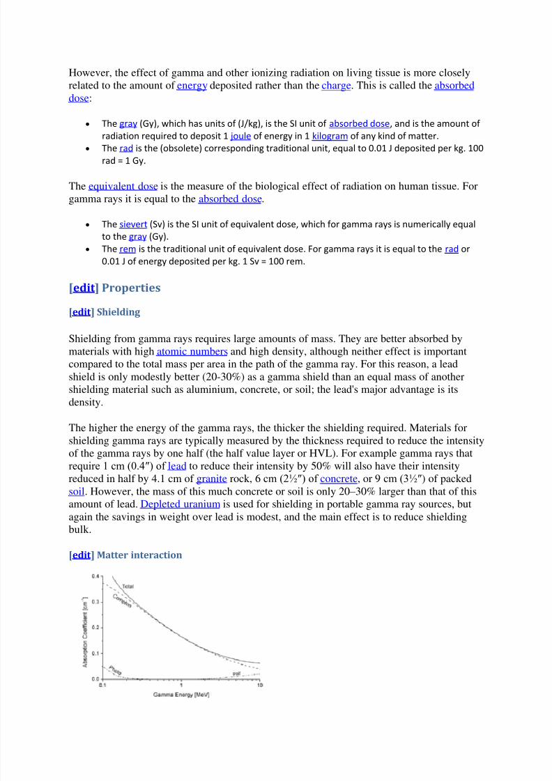
The total absorption coefficient of aluminium (atomic number 13) for gamma rays, plotted versus
gamma energy, and the contributions by the three effects. Over most of the energy region shown,
the Compton effect dominates.
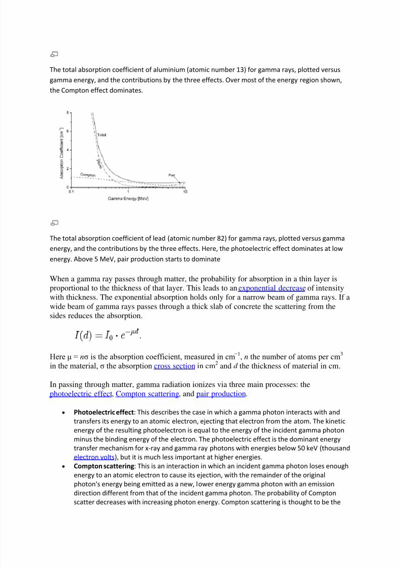
The total absorption coefficient of lead (atomic number 82) for gamma rays, plotted versus gamma
energy, and the contributions by the three effects. Here, the photoelectric effect dominates at low
energy. Above 5 MeV, pair production starts to dominate
When a gamma ray passes through matter, the probability for absorption in a thin layer isproportional to the thickness of that layer. This leads to an exponential decrease of intensity
with thickness. The exponential absorption holds only for a narrow beam of gamma rays. If awide beam of gamma rays passes through a thick slab of concrete the scattering from thesides reduces the absorption.
Here μ = nσ is the absorption coefficient, measured in cm−1, n the number of atoms per cm3 in the material, σ the absorption cross section in cm2 and d the thickness of material in cm.
In passing through matter, gamma radiation ionizes via three main processes: the
photoelectric effect, Compton scattering, and pair production.
Photoelectric effect: This describes the case in which a gamma photon interacts with and
transfers its energy to an atomic electron, ejecting that electron from the atom. The kinetic
energy of the resulting photoelectron is equal to the energy of the incident gamma photon
minus the binding energy of the electron. The photoelectric effect is the dominant energy
transfer mechanism for x-ray and gamma ray photons with energies below 50 keV (thousand
electron volts), but it is much less important at higher energies.
Compton scattering: This is an interaction in which an incident gamma photon loses enough
energy to an atomic electron to cause its ejection, with the remainder of the original
photon's energy being emitted as a new, lower energy gamma photon with an emission
direction different from that of the incident gamma photon. The probability of Compton
scatter decreases with increasing photon energy. Compton scattering is thought to be the
8/3/2019 Beta and Gamma Rays
http://slidepdf.com/reader/full/beta-and-gamma-rays 6/10
principal absorption mechanism for gamma rays in the intermediate energy range 100 keV
to 10 MeV. Compton scattering is relatively independent of the atomic number of the
absorbing material, which is why very dense metals like lead are only modestly better
shields, on a per weight basis, than are less dense materials.
Pair production: This becomes possible with gamma energies exceeding 1.02 MeV, and
becomes important as an absorption mechanism at energies over about 5 MeV (seeillustration at right, for lead). By interaction with the electric field of a nucleus, the energy of
the incident photon is converted into the mass of an electron-positron pair. Any gamma
energy in excess of the equivalent rest mass of the two particles (1.02 MeV) appears as the
kinetic energy of the pair and the recoil nucleus. At the end of the positron's range, it
combines with a free electron. The entire mass of these two particles is then converted into
two gamma photons of at least 0.51 MeV energy each (or higher according to the kinetic
energy of the annihilated particles).
The secondary electrons (and/or positrons) produced in any of these three processesfrequently have enough energy to produce much ionization themselves.
[edit ] Light interaction
High-energy (from 80 to 500 GeV) gamma rays arriving from far far-distant quasars are usedto estimate the extragalactic background light in the universe: The highest-energy raysinteract more readily with the background light photons and thus their density may beestimated by analyzing the incoming gamma-ray spectrums.[3]
[edit ] Gamma ray production
See also: Gamma-ray generation
Gamma rays are often produced alongside other forms of radiation such as alpha or beta. When a nucleus emits an α or β particle, the daughter nucleus is sometimes left in an excitedstate. It can then jump down to a lower energy state by emitting a gamma ray, in much thesame way that an atomic electron can jump to a lower energy state by emitting infrared, visible, or ultraviolet light.
8/3/2019 Beta and Gamma Rays
http://slidepdf.com/reader/full/beta-and-gamma-rays 7/10
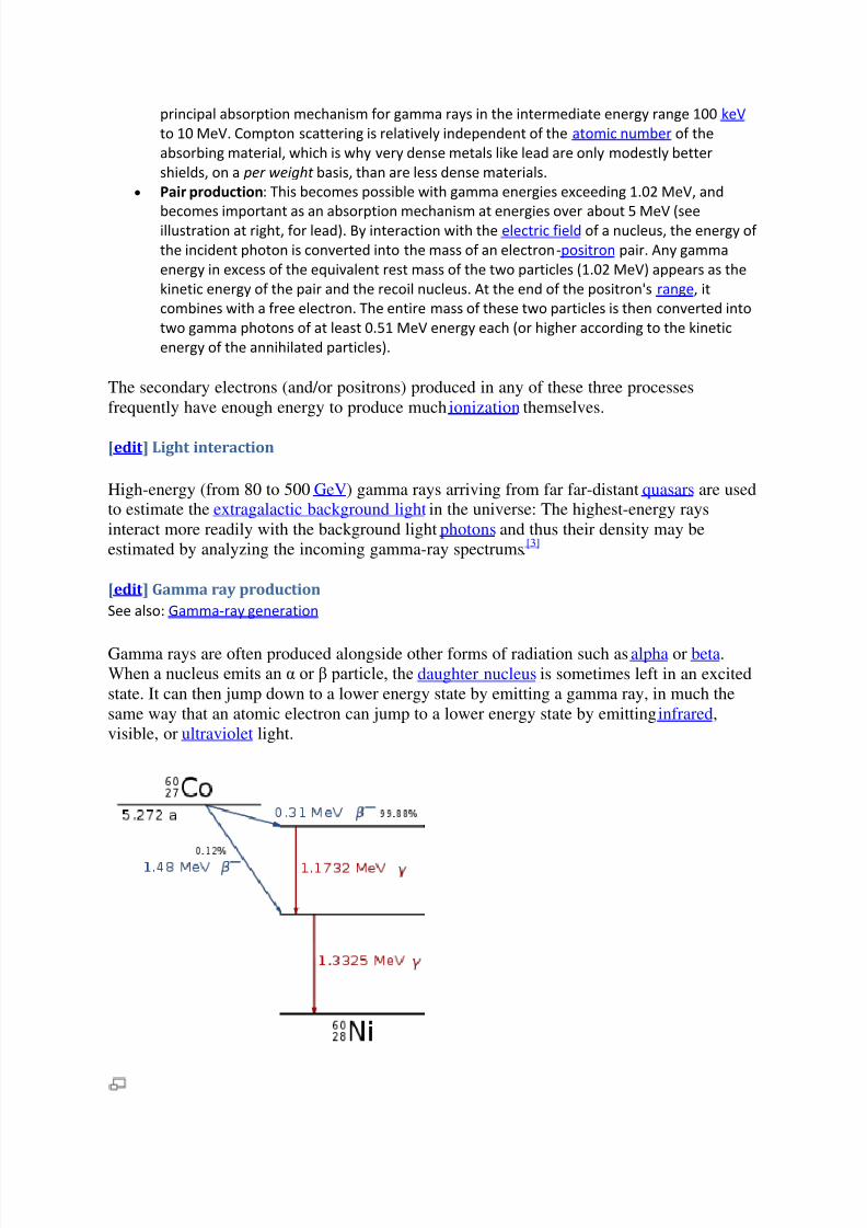
Decay scheme of 60
Co
Gamma rays, x-rays, visible light, and radio waves are all forms of electromagnetic radiation. The only difference is the frequency and hence the energy of the photons. Gamma rays arethe most energetic. An example of gamma ray production follows.
First 60Co decays to excited 60Ni by beta decay. Then the 60Ni drops down to the ground state(see nuclear shell model) by emitting two gamma rays in succession (1.17 MeV then 1.33MeV):
6027Co → 6028Ni* + e− + νe + γ + 1.17 MeV
6028Ni* → 6028Ni + γ + 1.33 MeV
Another example is the alpha decay of
241
Am to form
237
Np; this alpha decay is accompaniedby gamma emission. In some cases, the gamma emission spectrum for a nucleus (daughternucleus) is quite simple, (e.g. 60Co/ 60Ni) while in other cases, such as with (241Am/ 237Np and192Ir / 192Pt), the gamma emission spectrum is complex, revealing that a series of nuclearenergy levels can exist. The fact that an alpha spectrum can have a series of different peakswith different energies reinforces the idea that several nuclear energy levels are possible.
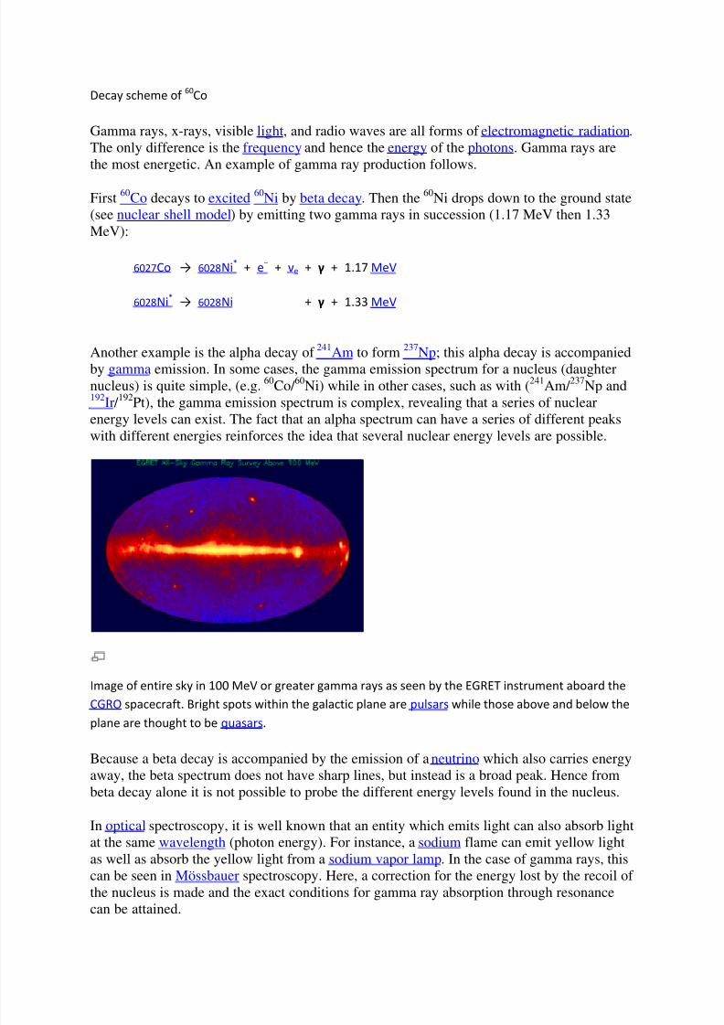
Image of entire sky in 100 MeV or greater gamma rays as seen by the EGRET instrument aboard the
CGRO spacecraft. Bright spots within the galactic plane are pulsars while those above and below the
plane are thought to be quasars.
Because a beta decay is accompanied by the emission of a neutrino which also carries energyaway, the beta spectrum does not have sharp lines, but instead is a broad peak. Hence frombeta decay alone it is not possible to probe the different energy levels found in the nucleus.
In optical spectroscopy, it is well known that an entity which emits light can also absorb lightat the same wavelength (photon energy). For instance, a sodium flame can emit yellow lightas well as absorb the yellow light from a sodium vapor lamp. In the case of gamma rays, thiscan be seen in Mössbauer spectroscopy. Here, a correction for the energy lost by the recoil of
the nucleus is made and the exact conditions for gamma ray absorption through resonancecan be attained.
8/3/2019 Beta and Gamma Rays
http://slidepdf.com/reader/full/beta-and-gamma-rays 8/10
This is similar to the Franck Condon effects seen in optical spectroscopy.
[edit ] Health effects
All ionizing radiation causes similar damage at a cellular level, but because rays of alpha
particles and beta particles are relatively non-penetrating, external exposure to them causesonly localized damage, e.g. radiation burns to the skin. Gamma rays and neutrons are morepenetrating, causing diffuse damage throughout the body (e.g. radiation sickness, increasedincidence of cancer) rather than burns. External radiation exposure should also bedistinguished from internal exposure, due to ingested or inhaled radioactive substances,which, depending on the substance's chemical nature, can produce both diffuse and localizedinternal damage. The most biological damaging forms of gamma radiation occur in thegamma ray window, between 3 and 10 MeV, with higher energy gamma rays being lessharmful because the body is relatively transparent to them. See cobalt-60.
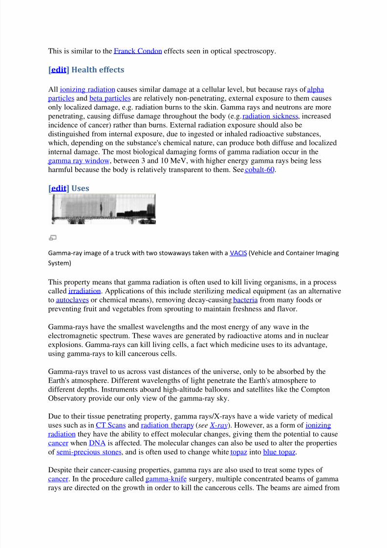
[edit ] Uses
Gamma-ray image of a truck with two stowaways taken with a VACIS (Vehicle and Container Imaging
System)
This property means that gamma radiation is often used to kill living organisms, in a process
called irradiation. Applications of this include sterilizing medical equipment (as an alternativeto autoclaves or chemical means), removing decay-causing bacteria from many foods orpreventing fruit and vegetables from sprouting to maintain freshness and flavor.
Gamma-rays have the smallest wavelengths and the most energy of any wave in theelectromagnetic spectrum. These waves are generated by radioactive atoms and in nuclearexplosions. Gamma-rays can kill living cells, a fact which medicine uses to its advantage,using gamma-rays to kill cancerous cells.
Gamma-rays travel to us across vast distances of the universe, only to be absorbed by the
Earth's atmosphere. Different wavelengths of light penetrate the Earth's atmosphere todifferent depths. Instruments aboard high-altitude balloons and satellites like the ComptonObservatory provide our only view of the gamma-ray sky.
Due to their tissue penetrating property, gamma rays/X-rays have a wide variety of medicaluses such as in CT Scans and radiation therapy (see X-ray). However, as a form of ionizingradiation they have the ability to effect molecular changes, giving them the potential to causecancer when DNA is affected. The molecular changes can also be used to alter the propertiesof semi-precious stones, and is often used to change white topaz into blue topaz.
Despite their cancer-causing properties, gamma rays are also used to treat some types of
cancer. In the procedure called gamma-knife surgery, multiple concentrated beams of gammarays are directed on the growth in order to kill the cancerous cells. The beams are aimed from
8/3/2019 Beta and Gamma Rays
http://slidepdf.com/reader/full/beta-and-gamma-rays 9/10
different angles to concentrate the radiation on the growth while minimizing damage to thesurrounding tissues. (As an illustration of the radiation origin-process contributing to itsname, a similar technique which uses photons from linacs rather than cobalt gamma decay, iscalled "Cyberknife").
Gamma rays are also used for diagnostic purposes in nuclear medicine. Several gamma-emitting radioisotopes are used, one of which is technetium-99m. When administered to apatient, a gamma camera can be used to form an image of the radioisotope's distribution bydetecting the gamma radiation emitted. Such a technique can be employed to diagnose a widerange of conditions (e.g. spread of cancer to the bones).
In the US, gamma ray detectors are beginning to be used as part of the Container SecurityInitiative (CSI). These US$5 million machines are advertised to scan 30 containers per hour.The objective of this technique is to screen merchant ship containers before they enter USports.
[edit ] Body response
After gamma-irradiation, and the breaking of DNA double-strands, a cell can repair thedamaged genetic material to the limit of its capability. However, a study of Rothkamm andLobrich has shown that the repairing process works well after high-dose exposure but ismuch slower in the case of a low-dose exposure.[4]
[edit ] Risk assessment
The natural outdoor exposure in Great Britain ranges from 2 × 10⁻⁷ to 4 × 10⁻⁷ cSv/h
(centisieverts per hour).
[5]
Natural exposure to gamma rays is about 0.1 to 0.2 cSv per year,and the average total amount of radiation received in one year per inhabitant in the USA is0.36 cSv.[6] There is a small increase in the dose, due to naturally occurring gamma-radiation,around small particles of high atomic number materials in the human body caused by thephotoelectric effect[7]
By comparison, the radiation dose from chest radiography is a fraction of the annual naturallyoccurring background radiation dose,[8] and the dose from fluoroscopy of the stomach is, atmost, 5 cSv on the skin of the back.
For acute full-body equivalent dose, 100 cSv causes slight blood changes; 200 – 350 cSv
causes nausea, hair loss, hemorrhaging and will cause death in a sizable number of cases(10% – 35%) without medical treatment; 500 cSv is considered approximately the LD50 (lethaldose for 50% of exposed population) for an acute exposure to radiation even with standardmedical treatment; more than 500 cSv brings an increasing chance of death; eventually,above 750 – 1000 cSv, even extraordinary treatment, such as bone-marrow transplants, willnot prevent the death of the individual exposed (see Radiation poisoning).[clarification
needed ][citation needed ]
For low dose exposure, for example among nuclear workers, who receive an average yearlyradiation dose of 1.9 cSv,[clarification needed ] the risk of dying from cancer (excluding leukemia) increases by 2 percent. For a dose of 10 cSv, that risk increase is at 10 percent. By
comparison, risk of dying from cancer was increased by 32 percent for the survivors of theatomic bombing of Hiroshima and Nagasaki.[9]

8/3/2019 Beta and Gamma Rays
http://slidepdf.com/reader/full/beta-and-gamma-rays 10/10