Embed Size (px)
Citation preview
RESEARCH Open Access
Betaine supplement alleviates hepatic triglycerideaccumulation of apolipoprotein E deficient micevia reducing methylation of peroxisomalproliferator-activated receptor alpha promoterLijun Wang1, Li Chen1, Yaozong Tan1, Jun Wei1, Ying Chang1, Tianru Jin2 and Huilian Zhu1*
Abstract
Background: Betaine is a methyl donor and has been considered as a lipotropic effect substance. But itsmechanism remains unclear. Hepatic steatosis is associated with abnormal expression of genes involved in hepaticlipid metabolism. DNA methylation contributes to the disregulation of gene expression. Here we hypothesized thatbetaine supplement and subsequent DNA methylation modifications alter the expression of genes that are involvedin hepatic lipid metabolism and hence alleviate hepatic triglyceride accumulation.
Methods: Male wild-type (WT) C57BL/6 mice (n = 6) were fed with the AIN-93 G diet. ApoE−/− mice (n = 12),weight-matched with the WT mice, were divided into two groups (n = 6 per group), and fed with the AIN-93 G dietand AIN-93 G supplemented with 2% betaine/100 g diet. Seven weeks after the intervention, mice were sacrificed.Liver betaine, choline, homocysteine concentration were measured by HPLC. Liver oxidants activity and triglyceridelevel were assessed by ultraviolet spectrophotometry. Finally, hepatic PPAR alpha gene and its target genesexpression levels and the methylation status of the PPAR alpha gene were determined.
Results: ApoE−/− mice had higher hepatic triglyceride and lower GSH-Px activity when compared with the WTmice. Betaine intervention reversed triglyceride deposit, enhanced SOD and GSH-Px activity in the liver.Interestingly, mice fed on betaine-supplemented diet showed a dramatic increase of hepatic choline concentrationand a decrease of betaine and homocysteine concentration relative to the WT mice and the ApoE−/− mice absentwith betaine intervention. Expression of PPAR alpha and CPT1 were decreased and expression of FAS was markedlyincreased in ApoE−/− mice. In parallel, PPAR alpha promoter methylation level were slightly increased in ApoE−/−
mice though without significance. Betaine supplement upregulated expression of PPAR alpha and its target genes(CPT1, CYP2E1) and reversed hypermethylation of PPAR alpha promoter of ApoE−/− mice. Furthermore, PPAR alphamethylation was positively correlated with hepatic betaine concentration.
Conclusions: Our findings indicate that betaine supplement could alleviate hepatic triglyceride accumulation andimprove antioxidant capacity by decreasing PPAR alpha promoter methylation and upregulating PPAR alpha and itstarget genes mRNA expression.
Keywords: Betaine, PPAR alpha, Fatty liver, DNA methylation
* Correspondence: [email protected] Provincial Key Laboratory of Food, Nutrition and Health, SunYat-Sen University, 74th Zhongshan Road II, 510080 Guangzhou, People’sRepublic of ChinaFull list of author information is available at the end of the article
© 2013 Wang et al.; licensee BioMed Central Ltd. This is an Open Access article distributed under the terms of the CreativeCommons Attribution License (http://creativecommons.org/licenses/by/2.0), which permits unrestricted use, distribution, andreproduction in any medium, provided the original work is properly cited.
Wang et al. Lipids in Health and Disease 2013, 12:34http://www.lipidworld.com/content/12/1/34
BackgroundTriglyceride (TG) accumulation in hepatocytes is consid-ered the primary manifestation of fatty liver disease [1].In addition, the increase of liver triglyceride deposit isassociated with obesity, hepatocellular carcinoma [2] andatherosclerosis [3]. Therefore, relieving hepatic lipid accu-mulation is viewed as potentially promising strategy for theprevention of many chronic diseases. The pathogenesis ofhepatic TG deposit is incompletely understood. A previousstudy by others has shown that upregulation of genesexpression for de novo lipogenesis (FAS, fatty acid synthase;ACC, acetyl-CoA carboxylase) and downregulation ofgenes expression for fatty acid oxidation (PPARα,peroxisomal proliferator-activated receptor alpha; CPT1,carnitine palmitoyl transferase I; UCP2, uncouplingproteins; ACOX, acyl-CoA oxidase; CYP2E1, cyto-chromeP450 2E1) are involved in the onsets of triglycerideaccumulation in the liver [4].Betaine, a methyl donor, is a naturally occurring com-
pound in common foods, such as wheat bran, wheat germ,spinach, pretzels, shrimp and wheat bread etc. [5]. In vivo,betaine can also be produced by oxidation of choline, and itserves as an effective methyl donor for remethylatinghomocysteine (Hcy) into methionine (Met). DNA methy-lation in the CpG islands is one of the epigenetic mecha-nisms to regulate gene expression. Choline and methioninedeficiency in diet are closely associated with biological func-tions by differentially methylated and inversely expressedgenes in the tissue [6-8]. Mice with hyperhomocysteinemia,induced by heterozygous cystathionine β-synthasedeficiency, show diminished methylation capacity andhypermethylation silencing of Fads2 mRNA expres-sion which contribute to the impaired transport ofTG [9]. High-fat diet can exacerbate methyl donors defi-ciency [10] and strikingly produce high level of serum Hcy,which may promote hypermethylation of MTTP gene anddown-regulation of its expression, resulting in the hin-drance to assembly lipoprotein and export lipid from liver[11]. It has become clear that PPARα can regulate the tran-scription of a suite of genes encoding enzymes in hepaticmitochondrial (CPT1, UCP2) and extramitochondrial(ACOX, CYP2E) fatty acid oxidation. Mice deficient inPPARα is demonstrated as a useful mouse model of fattyliver because of its important role in fatty acid oxidationand alleviation of hepatic TG [12]. Although An accumulat-ing clinical and experimental evidences suggest that betaineis a lipotropic substance [13-15], the DNA methylationmechanism remains to be clearly defined.In the present study, We attempt to investigate
betaine supplement undergoing improvement on lipidmetabolism and antioxidant capacity through changesin methylation level of PPARα promoter and expres-sion of PPARα and its target genes(CPT1, UCP2,ACOX, CYP2E) in ApoE−/− mice .
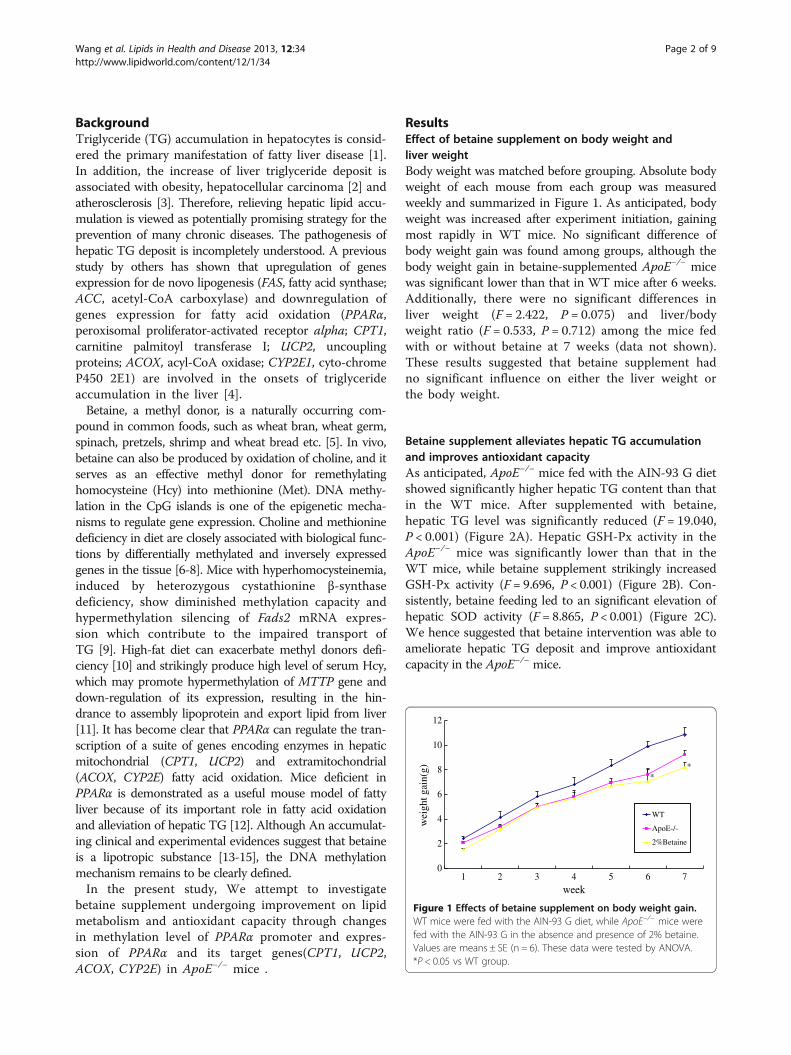
ResultsEffect of betaine supplement on body weight andliver weightBody weight was matched before grouping. Absolute bodyweight of each mouse from each group was measuredweekly and summarized in Figure 1. As anticipated, bodyweight was increased after experiment initiation, gainingmost rapidly in WT mice. No significant difference ofbody weight gain was found among groups, although thebody weight gain in betaine-supplemented ApoE−/− micewas significant lower than that in WT mice after 6 weeks.Additionally, there were no significant differences inliver weight (F = 2.422, P = 0.075) and liver/bodyweight ratio (F = 0.533, P = 0.712) among the mice fedwith or without betaine at 7 weeks (data not shown).These results suggested that betaine supplement hadno significant influence on either the liver weight orthe body weight.
Betaine supplement alleviates hepatic TG accumulationand improves antioxidant capacityAs anticipated, ApoE−/− mice fed with the AIN-93 G dietshowed significantly higher hepatic TG content than thatin the WT mice. After supplemented with betaine,hepatic TG level was significantly reduced (F = 19.040,P < 0.001) (Figure 2A). Hepatic GSH-Px activity in theApoE−/− mice was significantly lower than that in theWT mice, while betaine supplement strikingly increasedGSH-Px activity (F = 9.696, P < 0.001) (Figure 2B). Con-sistently, betaine feeding led to an significant elevation ofhepatic SOD activity (F = 8.865, P < 0.001) (Figure 2C).We hence suggested that betaine intervention was able toameliorate hepatic TG deposit and improve antioxidantcapacity in the ApoE−/− mice.
0
2
4
6
8
10
12
1 2 3 4 5 6 7
**
week
wei
ght g
ain(
g)
WT
ApoE-/-
2%Betaine
Figure 1 Effects of betaine supplement on body weight gain.WT mice were fed with the AIN-93 G diet, while ApoE−/− mice werefed with the AIN-93 G in the absence and presence of 2% betaine.Values are means ± SE (n = 6). These data were tested by ANOVA.*P < 0.05 vs WT group.
Wang et al. Lipids in Health and Disease 2013, 12:34 Page 2 of 9http://www.lipidworld.com/content/12/1/34
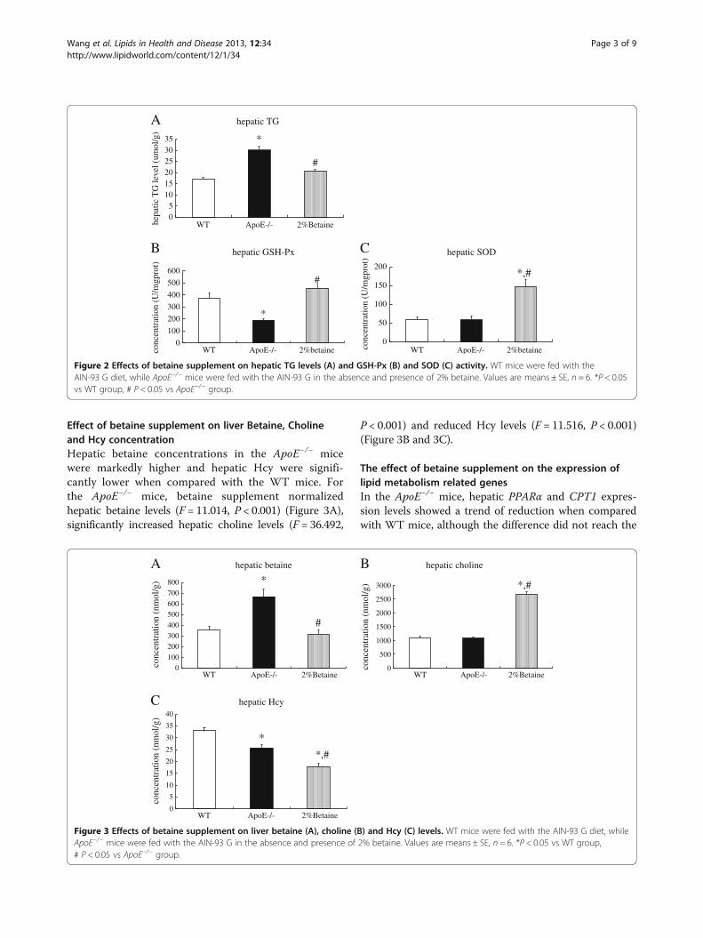
Effect of betaine supplement on liver Betaine, Cholineand Hcy concentrationHepatic betaine concentrations in the ApoE−/− micewere markedly higher and hepatic Hcy were signifi-cantly lower when compared with the WT mice. Forthe ApoE−/− mice, betaine supplement normalizedhepatic betaine levels (F = 11.014, P < 0.001) (Figure 3A),significantly increased hepatic choline levels (F = 36.492,
P < 0.001) and reduced Hcy levels (F = 11.516, P < 0.001)(Figure 3B and 3C).
The effect of betaine supplement on the expression oflipid metabolism related genesIn the ApoE−/− mice, hepatic PPARα and CPT1 expres-sion levels showed a trend of reduction when comparedwith WT mice, although the difference did not reach the
A
05
101520253035
WT ApoE-/- 2%Betaine
hepatic TG
*
#he
patic
TG
leve
l (um
ol/g
)
B
0
100
200
300
400
500
600
WT ApoE-/- 2%betaine
hepatic GSH-Px
*
#
conc
entr
atio
n (U
/mgp
rot)
C
0
50
100
150
200
WT ApoE-/- 2%betaine
hepatic SOD
*,#
conc
entr
atio
n (U
/mgp
rot)
Figure 2 Effects of betaine supplement on hepatic TG levels (A) and GSH-Px (B) and SOD (C) activity. WT mice were fed with theAIN-93 G diet, while ApoE−/− mice were fed with the AIN-93 G in the absence and presence of 2% betaine. Values are means ± SE, n = 6. *P < 0.05vs WT group, # P < 0.05 vs ApoE−/− group.
A
0
100
200
300
400
500
600
700
800
WT ApoE-/- 2%Betaine
hepatic betaine
*
#
B
0
500
1000
1500
2000
2500
3000
WT ApoE-/- 2%Betaine
hepatic choline
*,#
*,#
conc
entr
atio
n (n
mol
/g)
C
0
5
10
15
20
25
30
35
40
WT ApoE-/- 2%Betaine
hepatic Hcy
*
conc
entr
atio
n (n
mol
/g)
conc
entr
atio
n (n
mol
/g)
Figure 3 Effects of betaine supplement on liver betaine (A), choline (B) and Hcy (C) levels. WT mice were fed with the AIN-93 G diet, whileApoE−/− mice were fed with the AIN-93 G in the absence and presence of 2% betaine. Values are means ± SE, n = 6. *P < 0.05 vs WT group,# P < 0.05 vs ApoE−/− group.
Wang et al. Lipids in Health and Disease 2013, 12:34 Page 3 of 9http://www.lipidworld.com/content/12/1/34
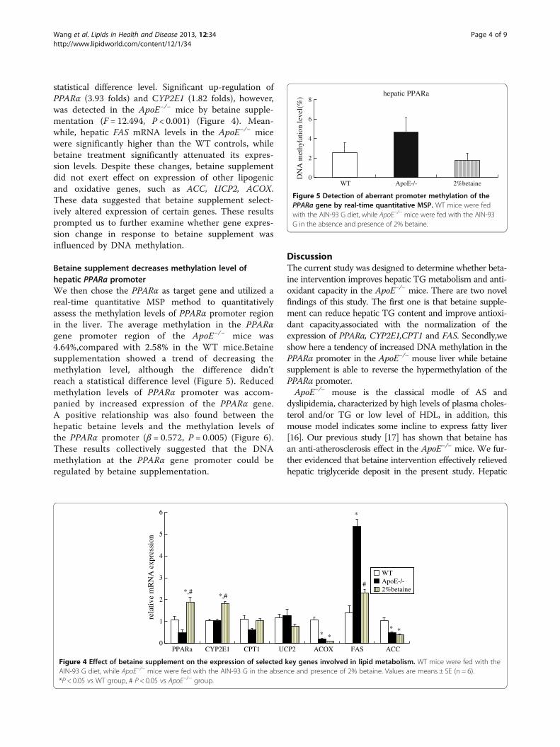
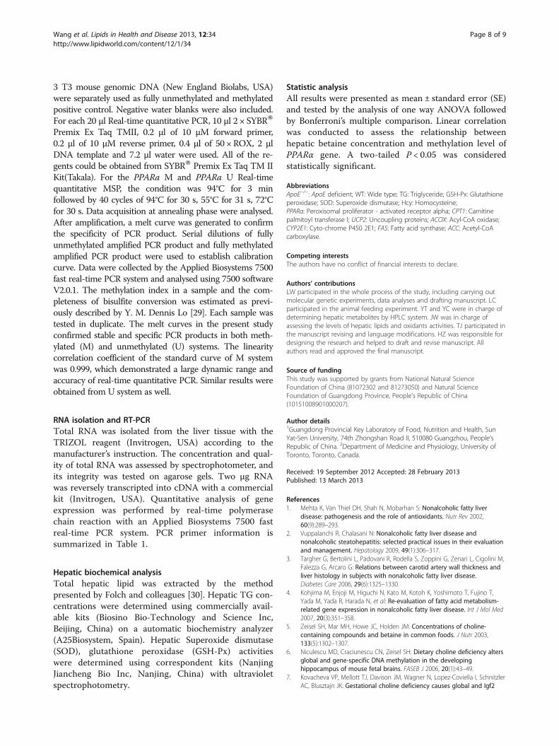
statistical difference level. Significant up-regulation ofPPARα (3.93 folds) and CYP2E1 (1.82 folds), however,was detected in the ApoE−/− mice by betaine supple-mentation (F = 12.494, P < 0.001) (Figure 4). Mean-while, hepatic FAS mRNA levels in the ApoE−/− micewere significantly higher than the WT controls, whilebetaine treatment significantly attenuated its expres-sion levels. Despite these changes, betaine supplementdid not exert effect on expression of other lipogenicand oxidative genes, such as ACC, UCP2, ACOX.These data suggested that betaine supplement select-ively altered expression of certain genes. These resultsprompted us to further examine whether gene expres-sion change in response to betaine supplement wasinfluenced by DNA methylation.
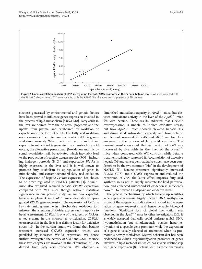
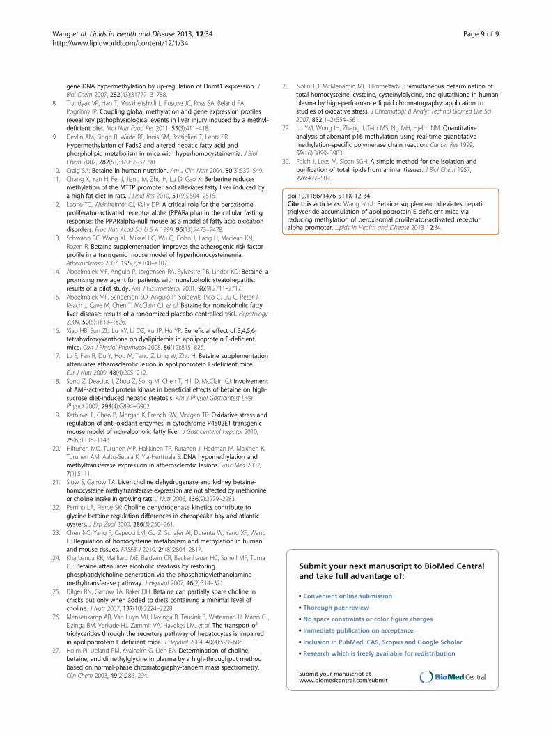
Betaine supplement decreases methylation level ofhepatic PPARa promoterWe then chose the PPARα as target gene and utilized areal-time quantitative MSP method to quantitativelyassess the methylation levels of PPARα promoter regionin the liver. The average methylation in the PPARαgene promoter region of the ApoE−/− mice was4.64%,compared with 2.58% in the WT mice.Betainesupplementation showed a trend of decreasing themethylation level, although the difference didn’treach a statistical difference level (Figure 5). Reducedmethylation levels of PPARα promoter was accom-panied by increased expression of the PPARα gene.A positive relationship was also found between thehepatic betaine levels and the methylation levels ofthe PPARα promoter (β = 0.572, P = 0.005) (Figure 6).These results collectively suggested that the DNAmethylation at the PPARα gene promoter could beregulated by betaine supplementation.
DiscussionThe current study was designed to determine whether beta-ine intervention improves hepatic TG metabolism and anti-oxidant capacity in the ApoE−/− mice. There are two novelfindings of this study. The first one is that betaine supple-ment can reduce hepatic TG content and improve antioxi-dant capacity,associated with the normalization of theexpression of PPARα, CYP2E1,CPT1 and FAS. Secondly,weshow here a tendency of increased DNA methylation in thePPARα promoter in the ApoE−/− mouse liver while betainesupplement is able to reverse the hypermethylation of thePPARα promoter.ApoE−/− mouse is the classical modle of AS and
dyslipidemia, characterized by high levels of plasma choles-terol and/or TG or low level of HDL, in addition, thismouse model indicates some incline to express fatty liver[16]. Our previous study [17] has shown that betaine hasan anti-atherosclerosis effect in the ApoE−/− mice. We fur-ther evidenced that betaine intervention effectively relievedhepatic triglyceride deposit in the present study. Hepatic
0
1
2
3
4
5
6
PPARa CYP2E1 CPT1 UCP2 ACOX FAS ACC
*
*
*
*,#*,#
*
#
*
rela
tive
mR
NA
exp
ress
ion
WTApoE-/-2%betaine
Figure 4 Effect of betaine supplement on the expression of selected key genes involved in lipid metabolism. WT mice were fed with theAIN-93 G diet, while ApoE−/− mice were fed with the AIN-93 G in the absence and presence of 2% betaine. Values are means ± SE (n = 6).*P < 0.05 vs WT group, # P < 0.05 vs ApoE−/− group.
0
2
4
6
8
WT ApoE-/- 2%betaine
hepatic PPARa
DN
A m
ethy
latio
n le
vel(
%)
Figure 5 Detection of aberrant promoter methylation of thePPARa gene by real-time quantitative MSP. WT mice were fedwith the AIN-93 G diet, while ApoE−/− mice were fed with the AIN-93G in the absence and presence of 2% betaine.
Wang et al. Lipids in Health and Disease 2013, 12:34 Page 4 of 9http://www.lipidworld.com/content/12/1/34
steatosis generated by environmental and genetic factorshave been proved to influence genes expression involved inthe process of lipid metabolism [4,8,9,11,18]. Fatty acids inthe liver are derived from the de novo lipogenesis and theuptake from plasma, and catabolized by oxidation orexportation in the form of VLDL-TG. Fatty acid oxidationoccurs mainly in the mitochondria, in which ATP is gener-ated simultaneously. When the impairment of antioxidantcapacity in mitochondria generated by excessive fatty acidoccurs, the alternative peroxisomal β-oxidation and micro-somal ω-oxidation will be activated which inevitably leadto the production of reactive oxygen species (ROS), includ-ing hydrogen peroxide (H2O2) and superoxide. PPARα ishighly expressed in the liver and it is well-known topromote fatty catabolism by up-regulation of genes inmitochondral and extramitochondral fatty acid oxidation.The expression of hepatic PPARα expression has shownto be down-regulated in NAFLD patients [4], ApoE−/−
mice also exhibited reduced hepatic PPARα expressioncompared with WT mice though without statisticalsignificance in our present study. As we have expected,betaine supplement in ApoE−/− mice dramatically upre-gulated PPARα gene expression. The expression of CPT1, akey rate-limiting enzyme in mitochondral oxidation, justmirrored the alteration of PPARα expression in response tobetaine treatment. CYP2E1 is one of the targets of PPARα,a key enzyme in the microsomal ω-oxidation. CYP2E1overexpression in the liver is a definite factor for oxidativestress [19]. In the current study, we found that betainetreatment increased CYP2E1 expression which wasparalleled by increased PPARα expression. We hencefurther investigated the activity of SOD and GSH-Px sincethese two enzymes are involved in the elimination of ROSderived from fatty acid oxidation. We observed a
diminished antioxidant capacity in ApoE−/− mice, but ele-vated antioxidant activity in the liver of the ApoE−/− micefed with betaine. These results indicated that CYP2E1overexpression is unable to induce oxidative stress,but how ApoE−/− mice showed elevated hepatic TGand diminished antioxidant capacity and how betainesupplement reversed it? FAS and ACC are two keyenzymes in the process of fatty acid synthesis. Thecurrent results revealed that expression of FAS wasincreased by five folds in the liver of the ApoE−/−
mice when compared with WT controls, while betainetreatment strikingly repressed it. Accumulation of excessivehepatic TG and consequent oxidative stress have been con-firmed to be the two common “hits” in the development ofNAFLD [1]. Betaine treatment significantly increasedPPARα, CPT1 and CYP2E1 expression and reduced theexpression of FAS, the latter effect impaires fatty acidsynthesis so as not to supply substrate for lipid peroxida-tion, and enhanced mitochondrial oxidation is sufficientlypowerful to prevent TG deposit and oxidative stress.The precise mechanisms by which betaine up regulates
gene expression remain largely unclear. DNA methylationis one of the epigenetic modifications involved in the regu-lation of gene expression and hence versatile biologicalfunctions. Significant loss of global methylation wasobserved in the ApoE−/− mice by other investigators [20]. Itis widely accepted that cells could undergo global DNAhypomethylation but simultaneously possess hyperme-thylation of a specific gene promoter, while the expressionof a gene is usually silenced or attenuated when its pro-moter is heavily methylated. Methyl-deficient diet has beenevidenced to exhibit hypermethylation promoter of genesinvolved in lipid metabolism which has inverse relationshipwith gene expression [8]. Betaine with its three chemically
0.00
2.00
4.00
6.00
8.00
10.00
12.00
0.00 200.00 400.00 600.00 800.00 1,000.00 1,200.00 1,400.00
hepatic betaine level(nmol/g)
hepa
tic P
PAR
a m
ethy
latio
n le
vel(
%)
Figure 6 Linear correlation analysis of DNA methylation level of PPARα promoter vs the hepatic betaine levels. WT mice were fed withthe AIN-93 G diet, while ApoE−/− mice were fed with the AIN-93 G in the absence and presence of 2% betaine.
Wang et al. Lipids in Health and Disease 2013, 12:34 Page 5 of 9http://www.lipidworld.com/content/12/1/34
reactive methyl groups could serve as a methyl donor forDNA, RNA, histone and proteins, and its supplement mayreverse alterations in DNA methylation induced by methyldeficiency. Here we investigated for the first time whetherPPARα promoter methylation and corresponding geneexpression can be affected by betaine supplement.The results of quantitative MSP assay showed thatthe methylation level of PPARα promoter was higherin the ApoE−/− mice when compared with that of WTmice, while betaine treatment showed a tendency toattenuate it. In attempt to explore the underlyingmechanism, we did further analysis and found outthat PPARα promoter methylation level was positivelycorrelated with hepatic betaine concentration.In present study, betaine supplement significantly
decreased Hcy concentration and increased hepatic cholineconcentration. Unexpectedly, betaine treatment decreasedhepatic betaine concentration as well. Endogenous betaineis oxidized from choline by two steps. Choline dehydrogen-ase (CHDH) mediates the first committed step for the for-mation of betaine aldehyde from choline, which is furthercatalyzed into betaine by betaine aldehyde dehydrogenase(BADH). Once betaine is formed, it serves as methyl donorfor remethylating Hcy into Met by betaine-homocysteinemethyltransferase (BHMT). Hepatic CHDH is rarelyaffected by dietary methionine and choline [21], but it hasbeen shown to be competitively inhibited by both betainealdehyde and betaine [22]. BHMT is expressed primarily inthe liver of humans and mice [23]. Betaine-alone supple-mentation results in a 2-fold elevation of hepatic BHMTlevels and activities [24]. Thus, betaine-elicited repressionof CHDH as well as of activation BHMT can collectivelyincrease the catabolism but decrease the synthesis of beta-ine in the liver. Our finding that betaine treatmentdramaticly increased choline level and reduced Hcy level inthe liver also confirmed this effect. This is among potentialexplainations why liver betaine levels in the betaine-supplemented ApoE−/− mice dropped to the levels thatwere comparable with the levels in the WT mice. Wehence suggest that hepatic betaine depletion plays a role inthe hypomethylation of PPARα promoter.On the other hand, choline, another methyl donor,
was observed to dramaticly increase by betaine intake.Betaine has been proved to have choline-sparing effect[25]. Choline is a basic component for the synthesis ofPhosphatidylcholine (PC) which takes part in the assemblyof mature VLDL. Betaine-containing diet could enhancethe synthesis of hepatic SAM as well as the activity of phos-phatidylethanolamine methyltransferase (PEMT), whichsubsequently facilitates the metabolic conversion of phos-phatidylethanolamine (PE) to form PC [24]. ApoE−/− miceis characterized by lipid accumulation and the developmentof hepatic steatosis. As a result, more choline are availableto increase PC synthesis and PC:PE ratio in VLDL, thereby
promoting VLDL secretion and lipid transport [26]. Theseresults suggest that betaine supplement exert importanteffects on the pool of related metabolites in the liver,whereby reduced betaine concentration in the liver connotsupply enough substrate for DNA methylated modificationand may selectively decrease the methylation statue ofcertain gene involved in lipid metabolism.
ConclusionTaken together, the present study reports for the first timethat betaine intervention reduces hepatic TG content andimproves antioxidant capacity in the ApoE−/− mouse model.This is partially achieved by upregulating PPARα geneexpression, as a result of hypomethylation of its promoter.These findings provided a novel insight into the importantlipotropic effect of betaine. Due to its fundamental impor-tant role as a methyl-donor, betaine is gradually become anew target for the intervention of many chronic metabolicdiseases. The hepatic and vascular protective effects ofbetaine we found provide promising insight into the humannutrition. Our present study has also build up a strongbasis for further evaluation of potential mechanisms ofbetaine in epigenetic studies.
MethodsChemicalsCholine chloride, Betaine glycine, d9-Betaine (internalstandard), d9-choline (internal standard), trichloroaceticacid (TCA), tris (2-carboxylethyl) phosphine (TCEP),ammonium-7- fluorobenzo-2-oxa-1, 3-diazole-4-sulfonicacid (SBD-F), mercaptopropionylglycine (MPG), mineraloil were purchased from Sigma Chemicals. Methanol,chloroform, acetonitrile, formic acid were HPLC gradeand obtained from Merck Chemical.
Animals and dietMale wild-type (WT) C57BL/6 mice (n = 6) and ApoE−/−
mice on the C57BL/6 background (n = 12) were obtainedfrom the Jackson Laboratories (Bar Harbor, ME, USA).All mice were acclimated on a standard AIN-93 G dietfor one week. The ApoE−/− mice were weight-matchedand divided into two groups (n = 6 per group) feedingwith the AIN-93 G diets supplemented with 0, 2 gbetaine/100 g diet and the WT mice continue toconsume the AIN-93 G diet. All mice were housed at aconstant temperature, humidity and 12 hR light/darkcycle, and freely accessed to food and water for 7 weeks.Body weight and consumption of diet were recordedweekly. Mice were anesthetized and sacrificed after theexperiment, liver were excised and stored at −80°C. Thisstudy was approved by the Institutional Animal Careand Use Committee at Sun Yat-Sen University.
Wang et al. Lipids in Health and Disease 2013, 12:34 Page 6 of 9http://www.lipidworld.com/content/12/1/34
Determination of betaine and choline concentration inthe animal tissueOne hundred mg frozen liver tissue were homogenized in600 μl methanol/chloroform (2:1,V/V) using an UltrasonicCell Disruptor (PRO scientific Inc. USA). The mixture wasvortex-mixed vigorously and left at −20°C overnight. Afterthe extraction, the mixture was centrifuged at 1500 g for5 min. The supernatants were drawn and transferred tonew tubes. The residuals were mixed with 300 μl metha-nol/chloroform/water (2:1:0.8,V/V/V) and centrifuged at1500 g for 5 min. The supernatants from the two extrac-tions were pooled. One hundred μl chloroform and 100 μlwater were added into the mixture to form two phases.After a centrifugation, the aqueous phase was taken by asyringe, dried by N2 Blowing Concentrator (OrganomationAssociates Inc, America) and dissolved in 20 μl water.The dissolved water were treated with 800 μl methanol,100 μl acetonitrile containing 10 μM internal standardand centrifuged at 6000 g for 2 min to precipitate protein.Finally the supernatants were injected into high perfor-mance liquid chromatography-tandem mass spectrometry(HPLC-MS) to analysis [27].
Determination of Hcy concentration in the animal tissueTwenty five mg frozen liver tissue was homogenized in200 μl cold PBS using an Ultrasonic Cell Disruptor (PROscientific Inc, USA). The homogenized tissue suspensionwas treated with 50 μl TCEP (120 mg/ml) and 60 μl MPG(10 μM), vortex-mixed thoroughly, and incubated at 37°Cfor 30 min. After adding 125 μl TCA (100 g/L with 1 mMEDTA) and vortex-mixing thoroughly, the precipitatedproteins were immediately centrifugated at 13000 g for10 min. One hundred μl supernatant was incubated with125 μl borate buffer (0.125 M, pH 9.5, with 4 mM EDTA),10 μl NaOH solution (1.55 M) and 50 μl SBD-F solution(1 g/L in borate buffer) at 60°C for 60 min, placed in theroom temperature to cool down, and then samples (10 μl)were injected into HPLC system to analysis [28].
Bisulfite conversion of DNAGenomic DNA was isolated from the liver tissue with theTIANamp Genomic DNA Kit (TIANGEN, Beijing, China).Treatment of DNA with bisulfite would result in the con-version of unmethylated cytosines to uracils, while methyl-ated cytosines would remain unaltered. Briefly, 5.5 μL of3 M NaOH was added into 1 ~ 2 μg of DNA (50 μL vol-ume) and incubated at 42°C for 20 min. Following the incu-bation step, 520 μL of 3.6 M NaHSO3 (pH 5.0) and 30 μL10 mM hydroquinone were added to each sample (freshand avoiding exposure to light). All reagents were softlymixed, centrifuged and covered by mineral oil, then incu-bated at 53°C for 16 h. The bisulfite-converted DNA waspurified by Wizard DNA Clean-Up System (Promega,USA). Finally DNA was eluated with 54 μL DDW at 80°C.
6 μL of 3 M NaOH was added into each sample and incu-bated at 42°C for 15 min. Following the incubation, DNAwas precipitated using 6 μl of 3 M sodium acetate and30 μl 100% cold ethanol at −80°C for 3 h. Each sample wascentrifuged 30 min at 12,000 g and the supernatant liquidwas poured out. The bisulfite-converted DNA was washedby 70% ethanol, dried and finally resuspended in a total vol-ume of 10 μl.
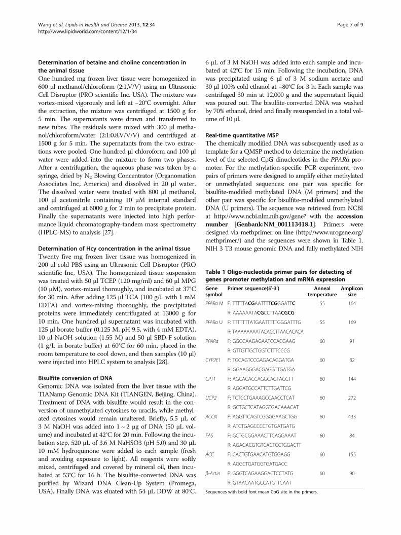
Real-time quantitative MSPThe chemically modified DNA was subsequently used as atemplate for a QMSP method to determine the methylationlevel of the selected CpG dinucleotides in the PPARα pro-moter. For the methylation-specific PCR experiment, twopairs of primers were designed to amplify either methylatedor unmethylated sequences: one pair was specific forbisulfite-modified methylated DNA (M primers) and theother pair was specific for bisulfite-modified unmethylatedDNA (U primers). The sequence was retrieved from NCBIat http://www.ncbi.nlm.nih.gov/gene? with the accessionnumber [Genbank:NM_001113418.1]. Primers weredesigned via methprimer on line (http://www.urogene.org/methprimer/) and the sequences were shown in Table 1.NIH 3 T3 mouse genomic DNA and fully methylated NIH
Table 1 Oligo-nucleotide primer pairs for detecting ofgenes promoter methylation and mRNA expression
Genesymbol
Primer sequence(50-30) Annealtemperature
Ampliconsize
PPARa M F: TTTTTACGAATTTTCGGGATTC 55 164
R: AAAAAATACGCCTTAACGCG
PPARa U F: TTTTTTTATGAATTTTTGGGATTTG 55 169
R: TAAAAAAAATACACCTTAACACACA
PPARa F: GGGCAAGAGAATCCACGAAG 60 91
R: GTTGTTGCTGGTCTTTCCCG
CYP2E1 F: TGCAGTCCGAGACAGGATGA 60 82
R: GGAAGGGACGAGGTTGATGA
CPT1 F: AGCACACCAGGCAGTAGCTT 60 144
R: AGGATGCCATTCTTGATTCG
UCP2 F: TCTCCTGAAAGCCAACCTCAT 60 272
R: GCTGCTCATAGGTGACAAACAT
ACOX F: AGGTTCAGTCGGGGAAGCTGG 60 433
R: ATCTGAGCCCCTGTGATGATG
FAS F: GCTGCGGAAACTTCAGGAAAT 60 84
R: AGAGACGTGTCACTCCTGGACTT
ACC F: CACTGTGAACATGTGGAGG 60 155
R: AGGCTGATGGTGATGACC
β-Actin F: GGGTCAGAAGGACTCCTATG 60 90
R: GTAACAATGCCATGTTCAAT
Sequences with bold font mean CpG site in the primers.
Wang et al. Lipids in Health and Disease 2013, 12:34 Page 7 of 9http://www.lipidworld.com/content/12/1/34
3 T3 mouse genomic DNA (New England Biolabs, USA)were separately used as fully unmethylated and methylatedpositive control. Negative water blanks were also included.For each 20 μl Real-time quantitative PCR, 10 μl 2 × SYBRW
Premix Ex Taq TMII, 0.2 μl of 10 μM forward primer,0.2 μl of 10 μM reverse primer, 0.4 μl of 50 × ROX, 2 μlDNA template and 7.2 μl water were used. All of the re-gents could be obtained from SYBRW Premix Ex Taq TM IIKit(Takala). For the PPARa M and PPARa U Real-timequantitative MSP, the condition was 94°C for 3 minfollowed by 40 cycles of 94°C for 30 s, 55°C for 31 s, 72°Cfor 30 s. Data acquisition at annealing phase were analysed.After amplification, a melt curve was generated to confirmthe specificity of PCR product. Serial dilutions of fullyunmethylated amplified PCR product and fully methylatedamplified PCR product were used to establish calibrationcurve. Data were collected by the Applied Biosystems 7500fast real-time PCR system and analysed using 7500 softwareV2.0.1. The methylation index in a sample and the com-pleteness of bisulfite conversion was estimated as previ-ously described by Y. M. Dennis Lo [29]. Each sample wastested in duplicate. The melt curves in the present studyconfirmed stable and specific PCR products in both meth-ylated (M) and unmethylated (U) systems. The linearitycorrelation coefficient of the standard curve of M systemwas 0.999, which demonstrated a large dynamic range andaccuracy of real-time quantitative PCR. Similar results wereobtained from U system as well.
RNA isolation and RT-PCRTotal RNA was isolated from the liver tissue with theTRIZOL reagent (Invitrogen, USA) according to themanufacturer’s instruction. The concentration and qual-ity of total RNA was assessed by spectrophotometer, andits integrity was tested on agarose gels. Two μg RNAwas reversely transcripted into cDNA with a commercialkit (Invitrogen, USA). Quantitative analysis of geneexpression was performed by real-time polymerasechain reaction with an Applied Biosystems 7500 fastreal-time PCR system. PCR primer information issummarized in Table 1.
Hepatic biochemical analysisTotal hepatic lipid was extracted by the methodpresented by Folch and colleagues [30]. Hepatic TG con-centrations were determined using commercially avail-able kits (Biosino Bio-Technology and Science Inc,Beijing, China) on a automatic biochemistry analyzer(A25Biosystem, Spain). Hepatic Superoxide dismutase(SOD), glutathione peroxidase (GSH-Px) activitieswere determined using correspondent kits (NanjingJiancheng Bio Inc, Nanjing, China) with ultravioletspectrophotometry.
Statistic analysisAll results were presented as mean ± standard error (SE)and tested by the analysis of one way ANOVA followedby Bonferroni’s multiple comparison. Linear correlationwas conducted to assess the relationship betweenhepatic betaine concentration and methylation level ofPPARα gene. A two-tailed P < 0.05 was consideredstatistically significant.
AbbreviationsApoE−/−: ApoE deficient; WT: Wide type; TG: Triglyceride; GSH-Px: Glutathioneperoxidase; SOD: Superoxide dismutase; Hcy: Homocysteine;PPARα: Peroxisomal proliferator - activated receptor alpha; CPT1: Carnitinepalmitoyl transferase I; UCP2: Uncoupling proteins; ACOX: Acyl-CoA oxidase;CYP2E1: Cyto-chrome P450 2E1; FAS: Fatty acid synthase; ACC: Acetyl-CoAcarboxylase.
Competing interestsThe authors have no conflict of financial interests to declare.
Authors’ contributionsLW participated in the whole process of the study, including carrying outmolecular genetic experiments, data analyses and drafting manuscript. LCparticipated in the animal feeding experiment. YT and YC were in charge ofdetermining hepatic metabolites by HPLC system. JW was in charge ofassessing the levels of hepatic lipids and oxidants activities. TJ participated inthe manuscript revising and language modifications. HZ was responsible fordesigning the research and helped to draft and revise manuscript. Allauthors read and approved the final manuscript.
Source of fundingThis study was supported by grants from National Natural ScienceFoundation of China (81072302 and 81273050) and Natural ScienceFoundation of Guangdong Province, People’s Republic of China(10151008901000207).
Author details1Guangdong Provincial Key Laboratory of Food, Nutrition and Health, SunYat-Sen University, 74th Zhongshan Road II, 510080 Guangzhou, People’sRepublic of China. 2Department of Medicine and Physiology, University ofToronto, Toronto, Canada.
Received: 19 September 2012 Accepted: 28 February 2013Published: 13 March 2013
References1. Mehta K, Van Thiel DH, Shah N, Mobarhan S: Nonalcoholic fatty liver
disease: pathogenesis and the role of antioxidants. Nutr Rev 2002,60(9):289–293.
2. Vuppalanchi R, Chalasani N: Nonalcoholic fatty liver disease andnonalcoholic steatohepatitis: selected practical issues in their evaluationand management. Hepatology 2009, 49(1):306–317.
3. Targher G, Bertolini L, Padovani R, Rodella S, Zoppini G, Zenari L, Cigolini M,Falezza G, Arcaro G: Relations between carotid artery wall thickness andliver histology in subjects with nonalcoholic fatty liver disease.Diabetes Care 2006, 29(6):1325–1330.
4. Kohjima M, Enjoji M, Higuchi N, Kato M, Kotoh K, Yoshimoto T, Fujino T,Yada M, Yada R, Harada N, et al: Re-evaluation of fatty acid metabolism-related gene expression in nonalcoholic fatty liver disease. Int J Mol Med2007, 20(3):351–358.
5. Zeisel SH, Mar MH, Howe JC, Holden JM: Concentrations of choline-containing compounds and betaine in common foods. J Nutr 2003,133(5):1302–1307.
6. Niculescu MD, Craciunescu CN, Zeisel SH: Dietary choline deficiency altersglobal and gene-specific DNA methylation in the developinghippocampus of mouse fetal brains. FASEB J 2006, 20(1):43–49.
7. Kovacheva VP, Mellott TJ, Davison JM, Wagner N, Lopez-Coviella I, SchnitzlerAC, Blusztajn JK: Gestational choline deficiency causes global and Igf2
Wang et al. Lipids in Health and Disease 2013, 12:34 Page 8 of 9http://www.lipidworld.com/content/12/1/34
gene DNA hypermethylation by up-regulation of Dnmt1 expression. JBiol Chem 2007, 282(43):31777–31788.
8. Tryndyak VP, Han T, Muskhelishvili L, Fuscoe JC, Ross SA, Beland FA,Pogribny IP: Coupling global methylation and gene expression profilesreveal key pathophysiological events in liver injury induced by a methyl-deficient diet. Mol Nutr Food Res 2011, 55(3):411–418.
9. Devlin AM, Singh R, Wade RE, Innis SM, Bottiglieri T, Lentz SR:Hypermethylation of Fads2 and altered hepatic fatty acid andphospholipid metabolism in mice with hyperhomocysteinemia. J BiolChem 2007, 282(51):37082–37090.
10. Craig SA: Betaine in human nutrition. Am J Clin Nutr 2004, 80(3):539–549.11. Chang X, Yan H, Fei J, Jiang M, Zhu H, Lu D, Gao X: Berberine reduces
methylation of the MTTP promoter and alleviates fatty liver induced bya high-fat diet in rats. J Lipid Res 2010, 51(9):2504–2515.
12. Leone TC, Weinheimer CJ, Kelly DP: A critical role for the peroxisomeproliferator-activated receptor alpha (PPARalpha) in the cellular fastingresponse: the PPARalpha-null mouse as a model of fatty acid oxidationdisorders. Proc Natl Acad Sci U S A 1999, 96(13):7473–7478.
13. Schwahn BC, Wang XL, Mikael LG, Wu Q, Cohn J, Jiang H, Maclean KN,Rozen R: Betaine supplementation improves the atherogenic risk factorprofile in a transgenic mouse model of hyperhomocysteinemia.Atherosclerosis 2007, 195(2):e100–e107.
14. Abdelmalek MF, Angulo P, Jorgensen RA, Sylvestre PB, Lindor KD: Betaine, apromising new agent for patients with nonalcoholic steatohepatitis:results of a pilot study. Am J Gastroenterol 2001, 96(9):2711–2717.
15. Abdelmalek MF, Sanderson SO, Angulo P, Soldevila-Pico C, Liu C, Peter J,Keach J, Cave M, Chen T, McClain CJ, et al: Betaine for nonalcoholic fattyliver disease: results of a randomized placebo-controlled trial. Hepatology2009, 50(6):1818–1826.
16. Xiao HB, Sun ZL, Lu XY, Li DZ, Xu JP, Hu YP: Beneficial effect of 3,4,5,6-tetrahydroxyxanthone on dyslipidemia in apolipoprotein E-deficientmice. Can J Physiol Pharmacol 2008, 86(12):815–826.
17. Lv S, Fan R, Du Y, Hou M, Tang Z, Ling W, Zhu H: Betaine supplementationattenuates atherosclerotic lesion in apolipoprotein E-deficient mice.Eur J Nutr 2009, 48(4):205–212.
18. Song Z, Deaciuc I, Zhou Z, Song M, Chen T, Hill D, McClain CJ: Involvementof AMP-activated protein kinase in beneficial effects of betaine on high-sucrose diet-induced hepatic steatosis. Am J Physiol Gastrointest LiverPhysiol 2007, 293(4):G894–G902.
19. Kathirvel E, Chen P, Morgan K, French SW, Morgan TR: Oxidative stress andregulation of anti-oxidant enzymes in cytochrome P4502E1 transgenicmouse model of non-alcoholic fatty liver. J Gastroenterol Hepatol 2010,25(6):1136–1143.
20. Hiltunen MO, Turunen MP, Hakkinen TP, Rutanen J, Hedman M, Makinen K,Turunen AM, Aalto-Setala K, Yla-Herttuala S: DNA hypomethylation andmethyltransferase expression in atherosclerotic lesions. Vasc Med 2002,7(1):5–11.
21. Slow S, Garrow TA: Liver choline dehydrogenase and kidney betaine-homocysteine methyltransferase expression are not affected by methionineor choline intake in growing rats. J Nutr 2006, 136(9):2279–2283.
22. Perrino LA, Pierce SK: Choline dehydrogenase kinetics contribute toglycine betaine regulation differences in chesapeake bay and atlanticoysters. J Exp Zool 2000, 286(3):250–261.
23. Chen NC, Yang F, Capecci LM, Gu Z, Schafer AI, Durante W, Yang XF, WangH: Regulation of homocysteine metabolism and methylation in humanand mouse tissues. FASEB J 2010, 24(8):2804–2817.
24. Kharbanda KK, Mailliard ME, Baldwin CR, Beckenhauer HC, Sorrell MF, TumaDJ: Betaine attenuates alcoholic steatosis by restoringphosphatidylcholine generation via the phosphatidylethanolaminemethyltransferase pathway. J Hepatol 2007, 46(2):314–321.
25. Dilger RN, Garrow TA, Baker DH: Betaine can partially spare choline inchicks but only when added to diets containing a minimal level ofcholine. J Nutr 2007, 137(10):2224–2228.
26. Mensenkamp AR, Van Luyn MJ, Havinga R, Teusink B, Waterman IJ, Mann CJ,Elzinga BM, Verkade HJ, Zammit VA, Havekes LM, et al: The transport oftriglycerides through the secretory pathway of hepatocytes is impairedin apolipoprotein E deficient mice. J Hepatol 2004, 40(4):599–606.
27. Holm PI, Ueland PM, Kvalheim G, Lien EA: Determination of choline,betaine, and dimethylglycine in plasma by a high-throughput methodbased on normal-phase chromatography-tandem mass spectrometry.Clin Chem 2003, 49(2):286–294.
28. Nolin TD, McMenamin ME, Himmelfarb J: Simultaneous determination oftotal homocysteine, cysteine, cysteinylglycine, and glutathione in humanplasma by high-performance liquid chromatography: application tostudies of oxidative stress. J Chromatogr B Analyt Technol Biomed Life Sci2007, 852(1–2):554–561.
29. Lo YM, Wong IH, Zhang J, Tein MS, Ng MH, Hjelm NM: Quantitativeanalysis of aberrant p16 methylation using real-time quantitativemethylation-specific polymerase chain reaction. Cancer Res 1999,59(16):3899–3903.
30. Folch J, Lees M, Sloan SGH: A simple method for the isolation andpurification of total lipids from animal tissues. J Biol Chem 1957,226:497–509.
doi:10.1186/1476-511X-12-34Cite this article as: Wang et al.: Betaine supplement alleviates hepatictriglyceride accumulation of apolipoprotein E deficient mice viareducing methylation of peroxisomal proliferator-activated receptoralpha promoter. Lipids in Health and Disease 2013 12:34.
Submit your next manuscript to BioMed Centraland take full advantage of:
• Convenient online submission
• Thorough peer review
• No space constraints or color figure charges
• Immediate publication on acceptance
• Inclusion in PubMed, CAS, Scopus and Google Scholar
• Research which is freely available for redistribution
Submit your manuscript at www.biomedcentral.com/submit
Wang et al. Lipids in Health and Disease 2013, 12:34 Page 9 of 9http://www.lipidworld.com/content/12/1/34