Embed Size (px)
Citation preview

Running title: 89Zr-Bevacizumab distribution in brainstem glioma xenografts
1
Bevacizumab targeting diffuse intrinsic pontine glioma: results of 89Zr-bevacizumab PET
imaging in brain tumor models
Marc H.A. Jansen*1, Tonny Lagerweij*2,5, A. Charlotte P. Sewing*1,2, Danielle J. Vugts3, Dannis
G. van Vuurden1,2, Carla F.M. Molthoff3, Viola Caretti1,2,4, Susanna J.E. Veringa1,2, Naomi
Petersen2, Angel Montero-Carcaboso8, David P. Noske2,5, W. Peter Vandertop5, Pieter
Wesseling 2,6,7, Guus A.M.S. van Dongen3, Gertjan J.L. Kaspers1, Esther Hulleman1,2
* These authors contributed equally
1Department of Pediatrics, Pediatric Hematology and Oncology, 2Neuro-oncology Research Group,
Cancer Center Amsterdam, 3Department of Radiology & Nuclear Medicine VU University Medical
Center Amsterdam, The Netherlands, 4Departments of Neurology, Pediatrics and Neurosurgery,
Stanford University School of Medicine, USA, 5Department of Neurosurgery VU University Medical
Center and Academic Medical Center, Amsterdam, the Netherlands, 6Department of Pathology VU
University Medical Center Amsterdam, the Netherlands, 7Department of Pathology, Radboud
University Medical Center, Nijmegen, The Netherlands, 8Preclinical Therapeutics and Drug Delivery
Research Program, Department of Oncology, Hospital Sant Joan de Déu Barcelona, Spain
Running title: 89Zr-Bevacizumab distribution in brainstem glioma xenografts
Key words: Diffuse intrinsic pontine glioma; Brain Stem Neoplasms; Vascular Endothelial
Growth Factor; Positron-Emission Tomography; 89Zirconium-bevacizumab, Animal Models
Abbreviations:
VEGF: vascular endothelial growth factor
DIPG: diffuse intrinsic pontine glioma
89Zr: zirconium-89
BLI: bioluminescence imaging
MRI: Magnetic Resonance Imaging
PET: Positron Emitting Tomography
ISH: in situ hybridization
on October 13, 2018. © 2016 American Association for Cancer Research. mct.aacrjournals.org Downloaded from
Author manuscripts have been peer reviewed and accepted for publication but have not yet been edited. Author Manuscript Published OnlineFirst on June 20, 2016; DOI: 10.1158/1535-7163.MCT-15-0558
Running title: 89Zr-Bevacizumab distribution in brainstem glioma xenografts
2
BBB: blood brain barrier
HGG: high-grade gliomas
LG-BSG: low grade brainstem glioma
GBM: glioblastoma multiforme
IHC: immunohistochemical
N-suc-Df: N-succinyl-desferrioxamine
EDTA: ethylenediaminetetraacetic acid
iLTC: instant thin-layer chromatography
FLUC: firefly luciferase
TSM: tumor stem medium
PBS: phosphate buffered saline
IP: intraperitoneally
PBMCs: peripheral blood mononuclear cells
Financial support: E. Hulleman received funding from Semmy Foundation and Stichting Kika
(Children-Cancer-free, project 69) M.H. Jansen received funding from Stichting Egbers. A.
Montero-Carcaboso acknowledges funding from ISCIII-FEDER (CP13/00189).
Reprint requests should be send to the corresponding author:
Esther Hulleman, PhD
Cancer Center Amsterdam, Neuro-oncology Research Group
De Boelelaan 1117, 1081 HV Amsterdam, Netherlands, CCA room 3.36
T/ +31(0)20-4447909 [email protected]
Conflict of Interest: We have no conflicts of interest to disclose.
Word count: 3968, total number of figures: 7
on October 13, 2018. © 2016 American Association for Cancer Research. mct.aacrjournals.org Downloaded from
Author manuscripts have been peer reviewed and accepted for publication but have not yet been edited. Author Manuscript Published OnlineFirst on June 20, 2016; DOI: 10.1158/1535-7163.MCT-15-0558

Running title: 89Zr-Bevacizumab distribution in brainstem glioma xenografts
3
Abstract
The role of the vascular endothelial growth factor (VEGF)-inhibitor bevacizumab in the
treatment of diffuse intrinsic pontine glioma (DIPG) is unclear. We aim to study the
biodistribution and uptake of zirconium-89 (89Zr)-labeled bevacizumab in DIPG mouse
models.
Human E98-FM,U251-FM glioma cells and HSJD-DIPG-007-FLUC primary DIPG cells were
injected into the subcutis, pons, or striatum of nude mice. Tumor growth was monitored by
bioluminescence imaging (BLI) and visualized by Magnetic Resonance Imaging (MRI).
Seventy-two to 96 hours after 89Zr-bevacizumab injections, mice were imaged by Positron
Emitting Tomography (PET) and biodistribution was analyzed ex vivo.
High VEGF expression in human DIPG was confirmed in a publically available mRNA
database, but no significant 89Zr-bevacizumab uptake could be detected in xenografts
located in the pons and striatum at an early or late stage of the disease. The E98-FM, and to
a lesser extent the U251-FM and HSJD-DIPG-007 subcutaneous tumors, showed high
accumulation of 89Zr-bevacizumab. VEGF expression could not be demonstrated in the
intracranial tumors by in situ hybridization (ISH), but was clearly present in the perinecrotic
regions of subcutaneous E98-FM tumors.
The poor uptake of 89Zr-bevacizumab in xenografts located in the brain suggests that VEGF
targeting with bevacizumab has limited efficacy for diffuse infiltrative parts of glial brain
tumors in mice. Translating these results to the clinic would imply that treatment with
bevacizumab in DIPG patients is only justified after targeting of VEGF has been
demonstrated by 89Zr-bevacizumab immuno-PET. We aim to confirm this observation in a
clinical PET study with DIPG patients.
on October 13, 2018. © 2016 American Association for Cancer Research. mct.aacrjournals.org Downloaded from
Author manuscripts have been peer reviewed and accepted for publication but have not yet been edited. Author Manuscript Published OnlineFirst on June 20, 2016; DOI: 10.1158/1535-7163.MCT-15-0558
Running title: 89Zr-Bevacizumab distribution in brainstem glioma xenografts
4
Introduction
Recent advances in molecular and cellular cancer biology have resulted in the identification
of critical molecular tumor targets involved in the different phases of tumor growth and
spreading. This knowledge has boosted the rational design of novel drugs, especially
monoclonal antibodies and tyrosine kinase inhibitors. However, broad application of these
targeted therapies to treat brain tumors has lagged behind in daily clinical practice. By using
molecular positron emission tomography (PET) imaging, target expression, bio-distribution
can be studied concurrently in a relatively non-invasive manner. It potentially allows the
identification of those patients who may, or may not, benefit from targeted therapy.
A disease that may benefit from molecular imaging is diffuse intrinsic pontine glioma
(DIPG), a childhood malignancy located in the brainstem. In the past 40 years the outcome of
patients with DIPG has remained unchanged, with less than 10% of the patients being alive
two years from diagnosis (1,2). Given the lack of gadolinium uptake on MRI in DIPG tumors
(3,4), it is plausible that the blood brain barrier (BBB) in DIPG often remains intact which
might explain the resistance to systemic chemotherapy in these patients.
A well-studied drug target in gliomas is the vascular endothelial growth factor
(VEGF), a signal protein stimulating angiogenesis and increasing vessel permeability (5).
Overexpression of VEGF-A, its receptor VEGFR2, or both, have been implicated as poor
prognostic markers in various clinical studies (5,6). Bevacizumab is a recombinant humanized
monoclonal antibody that selectively binds with high affinity to all isoforms of human VEGF-
A, and neutralizes their biologic activity (5,7). Studies in adult patients with recurrent high-
grade gliomas (HGG) reported high radiological response rates with bevacizumab treatment
(7–9) but recently two large phase III randomized studies showed no improvement in overall
survival with bevacizumab treatment in an upfront setting (10–12). Bevacizumab has been
studied in a number of non-randomized trials in pediatric brain tumor patients. Efficacy in
these trials has been variable, with a subset of patients showing clear radiological and/or
clinical improvement (13,14). The role of bevacizumab in the treatment of DIPG patients is
even less clear, but is currently studied in several trials (15–18), (NCT00890786 and
NCT01182350; clinicaltrials.gov; NTR2391 Trialregister.nl).
on October 13, 2018. © 2016 American Association for Cancer Research. mct.aacrjournals.org Downloaded from
Author manuscripts have been peer reviewed and accepted for publication but have not yet been edited. Author Manuscript Published OnlineFirst on June 20, 2016; DOI: 10.1158/1535-7163.MCT-15-0558

Running title: 89Zr-Bevacizumab distribution in brainstem glioma xenografts
5
No validated methods are available to identify patients who may potentially benefit
from bevacizumab treatment. Lack of clinical effect may be due to either poor transport of
bevacizumab into the tumor microenvironment due to an intact BBB, or a lack of VEGF
expression. In this study we analyzed VEGF(R) expression in adult and childhood high-grade
gliomas, including DIPG tumors. Furthermore, we studied bevacizumab distribution in vivo
using molecular PET imaging with 89Zirconium-labeled bevacizumab in murine DIPG models
(19).
Materials and Methods
VEGF-A and VEGFR2 mRNA expression profiles
VEGF-A and VEGFR2 (KDR) mRNA expression in DIPG (n=27) and pediatric high grade glioma
(pHGG) (n=53) were determined in silico, using publicly available datasets, and compared to
a dataset of non-malignant brain tissue (n=44), low grade brainstem glioma (n= 6) and adult
HGG (n=284). These datasets include tumor material from biopsy, resection and autopsy
(DIPG). Differences were analysed by two way ANOVA and a p < 0.01 was considered
significant. As a validation of these findings, VEGF associated gene expression was studied in
normal brain, low grade brainstem glioma (LG-BSG), DIPG and GBM by creating a heat map
using K-means clustering. All expression analyses were performed using R2, a web-based
microarray analysis and visualization platform (http://r2.amc.nl).
Immunohistochemistry and in situ hybridization
Formalin-fixed paraffin-embedded slides were sectioned from xenograft tumors and brain
tissue and subjected to immunohistochemical (IHC) staining. Briefly, after deparaffinization
and heat induced antigen retrieval, sections were incubated with primary mouse anti-Ki67
antibodies (clone MIB-1, DAKO) overnight at 4°C. Thereafter slides were washed and
incubated with HRP-conjugated EnVision (DAKO) and subsequently stained by DAB with
hematoxylin counterstaining.
For in situ hybridization (ISH), tumors were cut in 5 µm slices and incubated with
VEGF probes against the human VEGF coding sequence using a previously described protocol
(20). Samples were evaluated by microscopy with a Zeiss Axioskip microscope (HBO100W/Z),
equipped with a Canon digital camera and imaging software (Canon PowerShot A640, Canon
Utilities, ZoomBrowser Ex. 5.7, Canon Inc., Tokyo, Japan).
on October 13, 2018. © 2016 American Association for Cancer Research. mct.aacrjournals.org Downloaded from
Author manuscripts have been peer reviewed and accepted for publication but have not yet been edited. Author Manuscript Published OnlineFirst on June 20, 2016; DOI: 10.1158/1535-7163.MCT-15-0558

Running title: 89Zr-Bevacizumab distribution in brainstem glioma xenografts
6
Labeling and quality control of 89Zr-bevacizumab
Bevacizumab was labeled with zirconium-89 using N-succinyl-desferrioxamine (N-suc-Df) as
described previously (21). In short: the chelator, desferrioxamine, was succinylated to N-suc-
Df. Next, the hydroxamate groups were blocked with iron and the succinyl group was
activated as its TFP-ester (Fe-N-suc-Df-TFP ester). Bevacizumab (6 mg/mL) was reacted with
two equivalents of Fe-N-suc-Df-TFP ester at pH 9 for 30 min at room temperature. Hereafter,
iron was removed at pH 4.2-4.5 with an excess of ethylenediaminetetraacetic acid (EDTA) for
30 min at 35°C and N-suc-Df-bevacizumab was purified by size exclusion chromatography
using a PD-10 column. Radiolabeling of N-suc-Df-bevacizumab was performed in HEPES
buffer: to 200 μL 89Zr in 1M oxalic acid 90 μL 2M Na2CO3 was added. After 3 min 300 μL 0.5
M HEPES, N-suc-Df-bevacizumab and 700 μL 0.5 M HEPES were added. After 60 minutes
reaction time 89Zr-N-suc-Df-bevacizumab was purified by PD10 using 5 mg/mL gentisic acid
in 0.9% NaCl (pH 4.9-5.4) as the mobile phase.
Radiochemical purity and antibody integrity were determined using instant thin-layer
chromatography (iTLC), high-performance liquid chromatography (HPLC) and sodium
dodecylsulfate-polyacrylamide gel electrophoresis followed by phosphor imager analysis. For
analysis of immunoreactivity an ELISA based assay was used. iTLC analysis of 89Zr-
bevacizumab was performed on TEC control chromatography strips (Biodex). As the mobile
phase, citrate buffer (20 mmol/L, pH 5.0) containing 10% acetonitrile was used. HPLC
analyses of bevacizumab modification and radiolabeling were performed using a Jasco HPLC
system equipped with a SuperdexTM 200 10/30 GL size exclusion column (GE healthcare Life
sciences) using a mixture of 0.05 M sodium phosphate, 0.15 M sodium chloride (pH 6.8) and
0.01 M NaN3 as the eluent at a flow rate of 0.5 mL/min. The radioactivity of the eluate was
monitored using an inline NaI(Tl) radiodetector (RaytestSockett).
Cell lines and animal models
Animal experiments were performed in accordance with the European Community Council
Directive 2010/63/EU for laboratory animal care and the Dutch Law on animal
experimentation. The experimental protocol was validated and approved by the local
committee on animal experimentation of the VU University Medical Center. Athymic Nude-
on October 13, 2018. © 2016 American Association for Cancer Research. mct.aacrjournals.org Downloaded from
Author manuscripts have been peer reviewed and accepted for publication but have not yet been edited. Author Manuscript Published OnlineFirst on June 20, 2016; DOI: 10.1158/1535-7163.MCT-15-0558
Running title: 89Zr-Bevacizumab distribution in brainstem glioma xenografts
7
Foxn1nu mice (six weeks old) were purchased from Harlan/Envigo (Horst, The Netherlands)
and kept under filter top conditions and received food and water ad libitum.
The primary HSJD-DIPG-007 cell line was established from DIPG tumor material obtained at
Hospital Sant Joan de Deu (Barcelona, Spain) after autopsy from a 6-year-old patient and
was confirmed to have a H3F3A (K27M) and ACVR1 (R206H) mutation (22). The E98 cell line
was obtained from Radboud University Medical Center Nijmegen (23), the U251 glioma cell
line from ATCC. All cell lines were transduced in our laboratory to express firefly luciferase
(FLUC) and/or mCherry (23,24). Cell lines were mycoplasm negative and were authenticated
by STR-analysis modified from De Weger et al. (25)
E98-FM cells were injected subcutaneously in female athymic nude mice (7-9 weeks of age)
to expand the number of cells (19). When the subcutaneous tumor reached a diameter of 1
cm, the tumor was removed and a single cell suspension was prepared by mechanical
disruption through a 100 µm nylon cell strainer. HSJD-DIPG-007-FLUC was cultured in tumor
stem medium (TSM) (26), U251-FM in DMEM supplemented with 10% FCS and penicillin/
streptomycin. Shortly before stereotactic injection, cells were washed once with phosphate
buffered saline (PBS) and concentrated to 1x10⁵ cells per µl. Mice were stereotactically
injected with 5x10⁵ cells in a final volume of 5 µL into either the pons or striatum, or injected
subcutaneously with 3x10⁶ cells in a final volume of 100 µl/flank. Coordinates used for
intracranial injections were -1.0 mm X, -0.8 mm Y, 4,5 mm Z from the lambda for pontine
tumors and 0.5 mm X, 2 mm Y, −2 mm Z from the bregma for the striatum tumors.
Coordinates were previously validated and based on “The mouse brain in stereotaxic
coordinates” by Franklin and Paxinos (27). Tumor engraftment was monitored by
bioluminescence measurement of the Fluc signal. For E98-FM, early stage tumors in the pons
(n=8), striatum (n=7) and s.c. xenografts (n=3) were allowed to grow for 18 days and late
stage tumors (n=9, striatum; n=3, subcutaneous) for 35 days after xenograft injection. E98
xenograft tumors in the pons were not available 35 days post injection, because injection of
E98FM cells in the pons would result in death of the mice due to tumor growth within three
weeks. For HSJD-DIPG-007-FLUC and U251-FM tumors (pons, striatum, subcutaneous) were
evaluated, at days 78 and 22 respectively (Figure 2A)
on October 13, 2018. © 2016 American Association for Cancer Research. mct.aacrjournals.org Downloaded from
Author manuscripts have been peer reviewed and accepted for publication but have not yet been edited. Author Manuscript Published OnlineFirst on June 20, 2016; DOI: 10.1158/1535-7163.MCT-15-0558

Running title: 89Zr-Bevacizumab distribution in brainstem glioma xenografts
8
Two to three days before the endpoint of the study, mice were injected intraperitoneally
(i.p.) with 89Zr-labeled bevacizumab (40 µg, 5 MBq for PET analysis and ex vivo
biodistribution or 185 kBq for ex vivo biodistribution only).
PET imaging and ex vivo analysis
The distribution of 89Zr-bevacizumab was determined 72 hours (ex vivo only) or 96 hours
(PET followed by ex vivo analysis) after administration, as a minimum of 72 hours interval
between injection and scanning has previously been shown to achieve optimal tumor-to-
nontumor ratios (28). Scanning of E98-FM tumors was performed using a LSO/LYSO double
layer ECAT High Resolution Research Tomograph (HRRT, CTI/Siemens, Knoxville, TN, USA): a
small animal and human brain 3-dimensional (3D) scanner with high spatial resolution (2.3–
3.4 mm full width at half maximum) and high sensitivity (29). Mice were anesthetized by
isoflurane inhalation anesthesia (1.5 L 02/minute and 2.5% isoflurane) before positioning in
the HRRT. A static transmission scan (6 min) using a rotating 740 MBq 137Cs point source was
performed. Prior to positioning the mice in the HRRT, a canula was placed i.p. to enable later
injection of 18F(-). Static images of 60 minutes acquisition time were obtained. Immediately
thereafter 18F(-) was injected i.p (10 MBq/mouse) for co-localization of bone structures
(static images of 60 minutes acquisition). Body temperature was controlled with a heated
platform (kept at 37 °C).
Mice with HSJD-DIPG-007-FLUC or U251-FM tumors and where tumor growth was confirmed
by increase of BLI signal were imaged using another, preclinical, PET system, (Nanoscan PET-
CT, MEDISO, Budapest, Hungary), 72 hours after 89Zr-bevacizumab injection. PET images
were analyzed using AMIDE software. (Amide’s a Medical Image Data Examiner, version
1.0.1) (30)
Magnetic Resonance Imaging (MRI)
Mice with representative HSJD-DIPG-007-FLUC or U251-FM tumors were selected for MRI
based on the intensity of the bioluminescence signal. Gadolinium (750 µmol, Dotarem) was
administered i.v. immediately before imaging. Mice were anesthetized by isoflurane
inhalation anaesthesia (1.5 L 02/minute and 2.5% isoflurane), placed in a preclinical PET-MRI
system (Nanoscan system, MEDISO, Budapest, Hungary) and T1 and T2 weighed images with
on October 13, 2018. © 2016 American Association for Cancer Research. mct.aacrjournals.org Downloaded from
Author manuscripts have been peer reviewed and accepted for publication but have not yet been edited. Author Manuscript Published OnlineFirst on June 20, 2016; DOI: 10.1158/1535-7163.MCT-15-0558
Running title: 89Zr-Bevacizumab distribution in brainstem glioma xenografts
9
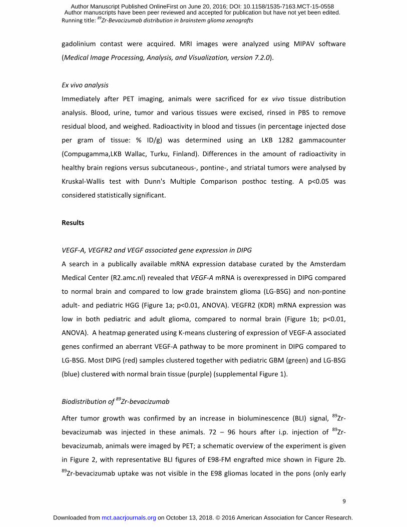
gadolinium contast were acquired. MRI images were analyzed using MIPAV software
(Medical Image Processing, Analysis, and Visualization, version 7.2.0).
Ex vivo analysis
Immediately after PET imaging, animals were sacrificed for ex vivo tissue distribution
analysis. Blood, urine, tumor and various tissues were excised, rinsed in PBS to remove
residual blood, and weighed. Radioactivity in blood and tissues (in percentage injected dose
per gram of tissue: % ID/g) was determined using an LKB 1282 gammacounter
(Compugamma,LKB Wallac, Turku, Finland). Differences in the amount of radioactivity in
healthy brain regions versus subcutaneous-, pontine-, and striatal tumors were analysed by
Kruskal-Wallis test with Dunn's Multiple Comparison posthoc testing. A p<0.05 was
considered statistically significant.
Results
VEGF-A, VEGFR2 and VEGF associated gene expression in DIPG
A search in a publically available mRNA expression database curated by the Amsterdam
Medical Center (R2.amc.nl) revealed that VEGF-A mRNA is overexpressed in DIPG compared
to normal brain and compared to low grade brainstem glioma (LG-BSG) and non-pontine
adult- and pediatric HGG (Figure 1a; p<0.01, ANOVA). VEGFR2 (KDR) mRNA expression was
low in both pediatric and adult glioma, compared to normal brain (Figure 1b; p<0.01,
ANOVA). A heatmap generated using K-means clustering of expression of VEGF-A associated
genes confirmed an aberrant VEGF-A pathway to be more prominent in DIPG compared to
LG-BSG. Most DIPG (red) samples clustered together with pediatric GBM (green) and LG-BSG
(blue) clustered with normal brain tissue (purple) (supplemental Figure 1).
Biodistribution of 89Zr-bevacizumab
After tumor growth was confirmed by an increase in bioluminescence (BLI) signal, 89Zr-
bevacizumab was injected in these animals. 72 – 96 hours after i.p. injection of 89Zr-
bevacizumab, animals were imaged by PET; a schematic overview of the experiment is given
in Figure 2, with representative BLI figures of E98-FM engrafted mice shown in Figure 2b. 89Zr-bevacizumab uptake was not visible in the E98 gliomas located in the pons (only early
on October 13, 2018. © 2016 American Association for Cancer Research. mct.aacrjournals.org Downloaded from
Author manuscripts have been peer reviewed and accepted for publication but have not yet been edited. Author Manuscript Published OnlineFirst on June 20, 2016; DOI: 10.1158/1535-7163.MCT-15-0558
Running title: 89Zr-Bevacizumab distribution in brainstem glioma xenografts
10
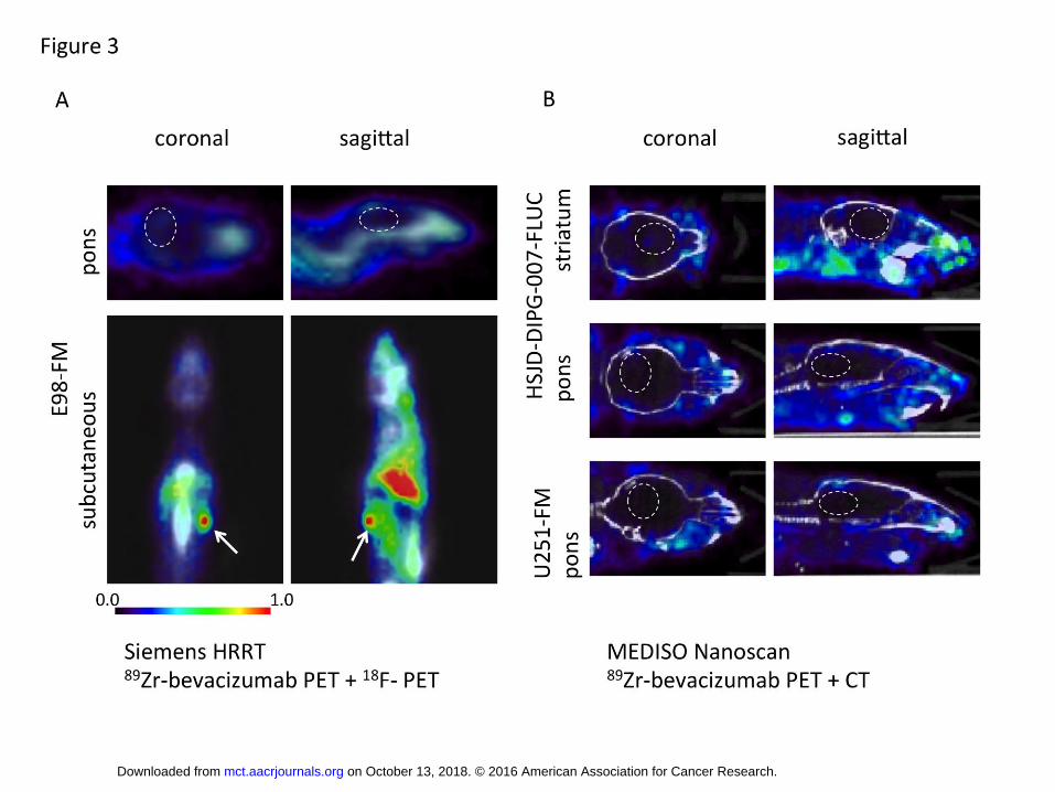
stage, Figure 3a) or in the striatum at early or late stage. The rest of the brain showed no
uptake of 89Zr-bevacizumab either, independently of the presence of a tumor. However,
subcutaneous E98 tumors showed high accumulation of 89Zr-bevacizumab indicated by a red
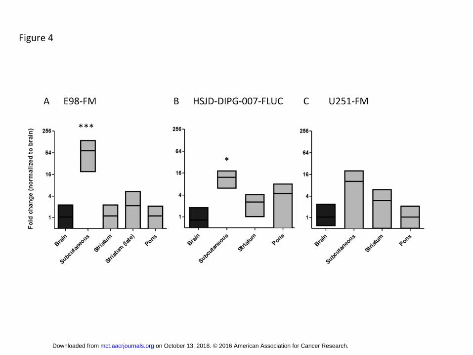
hotspot (Figure 3a). Ex vivo tissue distribution measurements confirmed that no significant 89Zr-bevacizumab uptake was detected in the brain or brain tumor at any stage of disease,
while there was high uptake in the subcutaneous tumor (p < 0.01) (Figure 4a). Besides
accumulation in the subcutaneous tumor with an average level of 50% ID/g, 89Zr-
bevacizumab was observed in all animals (to a lesser extent) in the blood pool and in well-
perfused organs, such as the liver, spleen and lungs (Figure 5). Experiments using the two
other cell lines (U251FM and HSJD-DIPG-007-FLUC) showed comparable results. Using these
cell lines, no uptake of 89Zr-bevacizumab was visualized on PET in any of the xenografts
(Figure 3b). Ex vivo biodistribution analysis showed higher 89Zr-bevacizumab uptake in
subcutaneous HSJD-DIPG-007-FLUC tumors (p<0.05) as compared to brain (areas) without
tumor (Figure 4b). The subcutaneous U251FM tumors showed no significant increase in 89Zr-
bevacizumab uptake (Figure 4c) as compared to non-tumor brain. Of note, both HSJD-DIPG-
007-FLUC and U251FM cells formed only very small subcutaneous tumors during the time
window in which mice baring intracranial tumors had reached human endpoints (78 vs 22
days after tumor injection).
In situ-hybridization of VEGF
To determine whether the differences in 89Zr-bevacizumab uptake in the subcutaneous and
the intracranial tumors were due to an impaired distribution of 89Zr-bevacizumab into the
brain or to a differential expression of its target (or both), VEGF expression was analyzed in
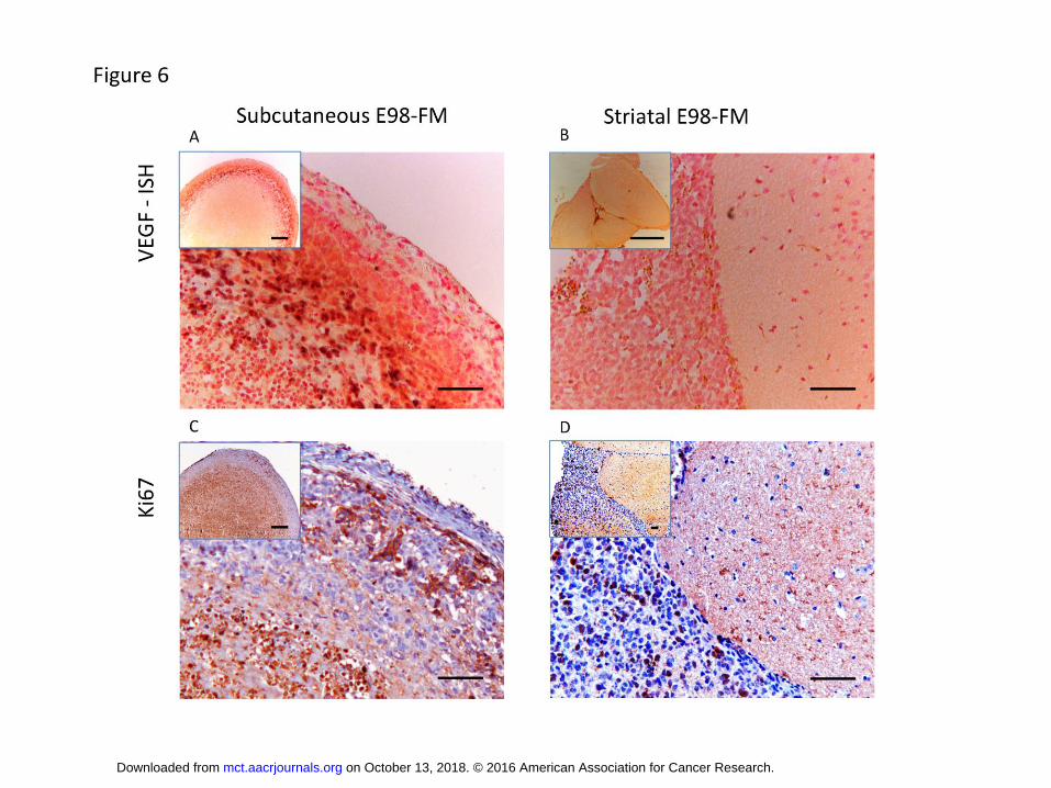
the different xenografts. In situ hybridization confirmed expression of VEGF in the
subcutaneous E98FM tumor (Figure 6a), while VEGF expression was absent in E98FM-brain
tumors (Figure 6b) and brain tissue without a xenograft. Of note, in the subcutaneous
tumors, VEGF was preferentially expressed in perinecrotic areas, while necrosis was absent
in all of the striatal and pontine tumors. A Ki67 (Mib-1) staining was performed to confirm
presence of proliferating tumor tissue (Figures 6c,d). In HSJD-DIPG-007-FLUC and U251FM
tumors, VEGF mRNA expression was not detectable in striatal and pontine gliomas. The
subcutaneous tumors were too small to adequately perform VEGF ISH and therefore no
conclusions could be drawn regarding VEGF expression in these s.c. tumors.
on October 13, 2018. © 2016 American Association for Cancer Research. mct.aacrjournals.org Downloaded from
Author manuscripts have been peer reviewed and accepted for publication but have not yet been edited. Author Manuscript Published OnlineFirst on June 20, 2016; DOI: 10.1158/1535-7163.MCT-15-0558

Running title: 89Zr-Bevacizumab distribution in brainstem glioma xenografts
11
Magnetic Resonance Imaging.
To visualize disruption of the blood-brain-barrier, mice with HSJD-DIPG-007-FLUC and U251-
FM intracranial tumors were imaged by MRI after intravenous administration of gadolinium.
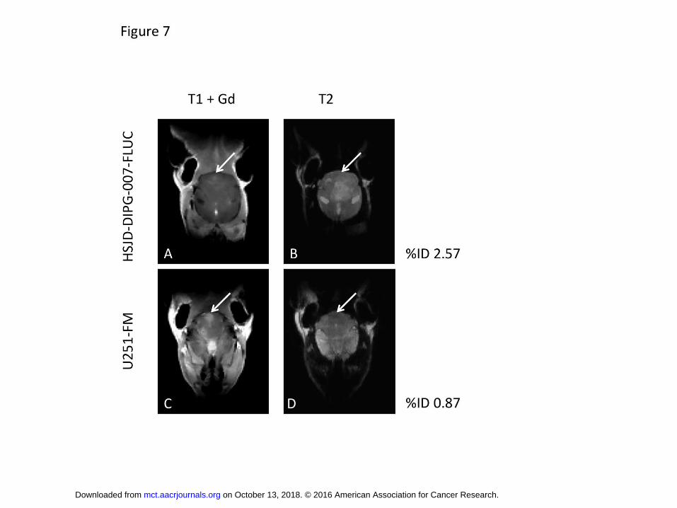
In the diffusely growing HSJD-DIPG-007-FLUC tumor, gadolinium enhancement on T1
weighed images was limited (Figure 7a, arrow), whereas gadolinium enhancement was
clearly visible in the U251-FM tumor (Figure 7c, arrow). On T2 weighed images, tumors were
not clearly visible (Figures 7b, d; arrow).
Discussion
The potential benefit of bevacizumab in the treatment of DIPG is unclear, as efficacy
depends on expression of VEGF-A as well as appropriate drug distribution (31). We used
molecular PET imaging to study the influence of location and stage of disease on
biodistribution of 89Zr-bevacizumab in three glioma mouse models (pontine, striatal,
subcutaneous) using three different cell lines. The E98-FM pontine and striatal and HSJD-
DIPG-007 pontine xenograft models have previously been described to resemble the diffuse
phenotype of human DIPG and other diffuse high grade gliomas (19,32,33). U251 has been
described as an intracranial murine tumor model that recapitulates most of the key figures
of adult GBM (34). We found no significant uptake of 89Zr-bevacizumab in the intracranial
tumor models at any stage of the disease, nor in the normal/non-neoplastic surrounding
brain. In contrast, high accumulation of 89Zr–bevacizumab was observed in the subcutaneous
E98-xenograft and moderate uptake in the subcutaneous HSJD-DIPG-007-FLUC.
We initially hypothesized that lack of 89Zr–bevacizumab uptake could be explained
solely by poor distribution into the brain, as large molecules like monoclonal antibodies may
not be able to pass the BBB. This hypothesis is supported by the absence of enhancement of
the tumor on MRI after administration of gadolinium in animals with HSJD-DIPG-007-FLUC
pontine tumors. However, MRI analysis of U251-FM tumors in the brainstem of mice showed
clear gadolinium enhancement, which is indicative of “leaky” blood vessels in the tumor.
Furthermore, VEGF expression of the E98FM glioma cells - analyzed by ISH – also differed
between tumor locations: E98FM gliomas in both striatum and pons appeared VEGF-
negative, while the s.c. E98FM tumors were partly VEGF-positive. The differences in VEGF
expression of the tumors in distinct locations originating from the same cell line, confirms
that the orthotopic microenvironment and the resulting growth pattern significantly
on October 13, 2018. © 2016 American Association for Cancer Research. mct.aacrjournals.org Downloaded from
Author manuscripts have been peer reviewed and accepted for publication but have not yet been edited. Author Manuscript Published OnlineFirst on June 20, 2016; DOI: 10.1158/1535-7163.MCT-15-0558

Running title: 89Zr-Bevacizumab distribution in brainstem glioma xenografts
12
influence gene expression in glioma cells, a phenomenon that has been described previously
(19, 20). Moreover, it has been shown that in GBM, VEGF is predominantly overexpressed in
hypoxic, perinecrotic cells (35,36). Indeed, in our study the VEGF expression in subcutaneous
E98FM tumors was especially present around areas of necrosis, whereas in intracranial
E98FM tumors necrosis and VEGF expression were lacking and this also coincided with lack
of bevacizimab uptake studied ex vivo and by PET. Of note, bevacizumab does not bind to
murine VEGF-A (37), but as typically the neoplastic cells are upregulating VEGF expression in
tumor angiogenesis we consider it unlikely that stromal cell-derived mouse VEGF-A plays an
important role in this particular xenograft model. (33)
In contrast to our preclinical findings, the in silico analysis that we performed in this
study, indicates that human DIPG tumors have relatively high expression levels of VEGF
mRNA. However, the majority of tumors (23 out of 27) used for the microarray experiments
were collected post-mortem (38,39) and therefore these samples represent the end-stage of
the disease, and are post-radiation therapy. In the end-stage of the disease, DIPG is known
to have necrotic areas with microvascular proliferations and blood-brain barrier disruption,
compatible with the histology of a GBM, which is associated with high VEGF expression.
Although the numbers are low, it is important to point out that VEGF-A expression levels in
samples obtained pre-treatment were low compared to expression in post-mortem/end-
stage samples (supplemental Figure 2). In addition, biopsy samples that were included in the
analysis are frequently directed at contrast-enhancing regions, and this 'biased sampling'
may well lead to overestimation of the role of VEGF in the tumor as a whole. We are
currently studying the differences in VEGF-A expression in autopsy-derived DIPG tissue
between the perinecrotic areas and the more diffusely growing tumor parts without
necrosis.
Experimental and clinical research in both adult and pediatric high grade glioma and
DIPG has suggested that there is a complex relation between a histologically diffuse growth
pattern of braintumors, VEGF expression and availability and BBB integrity (20,40–44).
Traditionally, VEGF is viewed as the main cause of increased BBB permeability in CNS tumors
as represented by contrast-enhancing lesions on MRI (45). More recently, anti-VEGF therapy
is thought to potentially induce a more diffuse and distant spread of tumor cells (41,42). In
contrast to adult high grade gliomas, gadolinium contrast enhancement on MRI in DIPGs at
diagnosis is generally limited, with 50% of the patients showing no enhancement at all (3).
on October 13, 2018. © 2016 American Association for Cancer Research. mct.aacrjournals.org Downloaded from
Author manuscripts have been peer reviewed and accepted for publication but have not yet been edited. Author Manuscript Published OnlineFirst on June 20, 2016; DOI: 10.1158/1535-7163.MCT-15-0558

Running title: 89Zr-Bevacizumab distribution in brainstem glioma xenografts
13
The lack of gadolinium enhancement suggests an intact BBB, at least for large molecules, in a
substantial percentage of the patients, coinciding with low VEGF expression and inability of
bevacizumab to target the tumor (46).
Results from our preclinical PET, MRI with gadolinium contrast and ISH studies
suggest that this relation is not so straightforward, pressing the need for studying VEGF
targeting in patients treated with anti-VEGF therapy. One could however argue, whether
high local tumor accumulation of bevacizumab is at all needed to obtain potential
therapeutic effects in DIPG. Bevacizumab is capable of decreasing VEGF levels in blood to
undetectable range in less than 20 days in a large cohort of adult cancer patients (47), but
only a subgroup of patients responded to anti-VEGF therapy. Also in DIPG, decreased
phosphoVEGR2 levels in peripheral blood mononuclear cells (PBMCs) did not correlate with
treatment response (15). In the E98 xenograft model used in this study, treatment with
bevacizumab did not increase survival nor did it influence the growth pattern in the diffusely
growing parts of the tumor (36). This suggest simply decreasing VEGF in the blood-pool is, at
least for the tumor types studied and our xenograft model, often insufficient for adequate
tumor targeting and instead, local bevacizumab accumulation seems needed (47–49).
The results of immuno-PET imaging and VEGF-ISH in these DIPG models are in line
with the poor clinical response rates thus far obtained with bevacizumab in children with
DIPG (15). The data presented suggest that no adequate uptake of bevacizumab will occur in
diffusely growing gliomas, which present with BBB disruption but without drastically
increased VEGF expression. Therefore we suggest that bevacizumab treatment is only
justified if targeting of VEGF by bevacizumab has been visualized by immuno-PET scan. We
aim to confirm this hypothesis in a clinical PET study with DIPG patients.
Future directions
This study underlies the importance of using strong biological and biodistributional rationale
before using any therapy in any patient. Following the results of this study, we developed a
molecular drug imaging trial with 89Zr-bevacizumab in children with DIPG (study number
NTR3518 www.trialregister.nl). This technique aims to further unravel the role of
bevacizumab treatment in DIPG. Ideally, such molecular imaging is combined with VEGF-A
and VEGFR2 expression analysis on tumor tissue originating from biopsies taken from several
on October 13, 2018. © 2016 American Association for Cancer Research. mct.aacrjournals.org Downloaded from
Author manuscripts have been peer reviewed and accepted for publication but have not yet been edited. Author Manuscript Published OnlineFirst on June 20, 2016; DOI: 10.1158/1535-7163.MCT-15-0558

Running title: 89Zr-Bevacizumab distribution in brainstem glioma xenografts
14
(contrast-enhancing and non-enhancing) parts of the tumor. Because DIPG can generally be
diagnosed based on its typical radiological presentation and the delicate nature of the brain
involved, taking biopsies from DIPGs is still no common practice and sampling of multiple
regions is even more cumbersome. In general, integrating molecular imaging with
radiolabelled drugs (classic cytostatic agents, small molecules, other monoclonal antibodies)
in the treatment of childhood brain cancer provides an insight in drug targeting and might
help to personalize treatment and thereby to avoid unnecessary side effects of drugs that do
not reach the tumor.
Acknowledgements
We are thankful to Ricardo Vos, Iris Mes and Alex Poot (Department of Radiology &
Nuclear Medicine, VUmc) for technical support with labelling of PET tracers and PET imaging.
Piotr Waranecki and Yanyan Veldman (Neuro-oncology Research Group, VUmc) for technical
assistance and animal handling. DIPG research at the VUmc has been made possible by the
invaluable support of the Semmy Foundation, Stichting Egbers and of Stichting Kika
(Children-Cancer-free, project 69). Angel Montero-Carcaboso form Hospital Sant Joan de
Deu acknowledges funding from ISCIII-FEDER (CP13/00189).
REFERENCES
1. Hargrave D, Bartels U, Bouffet E. Diffuse brainstem glioma in children: critical review of clinical trials. Lancet Oncol. 2006;7:241–8.
2. Jansen MHA, van Vuurden DG, Vandertop WP, Kaspers GJL. Diffuse intrinsic pontine gliomas: a systematic update on clinical trials and biology. Cancer Treat Rev. 2012;38:27–35.
3. Hargrave D, Chuang N, Bouffet E. Conventional MRI cannot predict survival in childhood diffuse intrinsic pontine glioma. J Neurooncol. 2008;86:313–9.
4. Jansen MH, Veldhuijzen van Zanten SE, Sanchez Aliaga E, Heymans MW, Warmuth-Metz M, Hargrave D, et al. Survival prediction model of children with diffuse intrinsic pontine glioma based on clinical and radiological criteria. Neuro Oncol. 2015;17:160–6.
5. Ferrara N. Vascular endothelial growth factor: basic science and clinical progress. Endocr Rev. 2004;25:581–611.
6. Shibuya M. Differential roles of vascular endothelial growth factor receptor-1 and receptor-2 in angiogenesis. J Biochem Mol Biol. 2006;39:469–78.
on October 13, 2018. © 2016 American Association for Cancer Research. mct.aacrjournals.org Downloaded from
Author manuscripts have been peer reviewed and accepted for publication but have not yet been edited. Author Manuscript Published OnlineFirst on June 20, 2016; DOI: 10.1158/1535-7163.MCT-15-0558
Running title: 89Zr-Bevacizumab distribution in brainstem glioma xenografts
15
7. Friedman HS, Prados MD, Wen PY, Mikkelsen T, Schiff D, Abrey LE, et al. Bevacizumab alone and in combination with irinotecan in recurrent glioblastoma. J Clin Oncol. 2009;27:4733–40.
8. Vredenburgh JJ, Desjardins A, Herndon JE, Dowell JM, Reardon DA, Quinn JA, et al. Phase II trial of bevacizumab and irinotecan in recurrent malignant glioma. Clin Cancer Res. 2007;13:1253–9.
9. Vredenburgh JJ, Desjardins A, Herndon JE, Marcello J, Reardon DA, Quinn JA, et al. Bevacizumab plus irinotecan in recurrent glioblastoma multiforme. J Clin Oncol. 2007;25:4722–9.
10. Chinot OL, Wick W, Mason W, Henriksson R, Saran F, Nishikawa R, et al. Bevacizumab plus radiotherapy-temozolomide for newly diagnosed glioblastoma. N Engl J Med. 2014;370:709–22.
11. Gilbert MR, Dignam JJ, Armstrong TS, Wefel JS, Blumenthal DT, Vogelbaum MA, et al. A randomized trial of bevacizumab for newly diagnosed glioblastoma. N Engl J Med. 2014;370:699–708.
12. Khasraw M, Ameratunga MS, Grant R, Wheeler H, Pavlakis N. Antiangiogenic therapy for high-grade glioma. Cochrane database Syst Rev. 2014;9:CD008218.
13. Narayana A, Kunnakkat S, Chacko-Mathew J, Gardner S, Karajannis M, Raza S, et al. Bevacizumab in recurrent high-grade pediatric gliomas. Neuro Oncol. 2010;12:985–90.
14. Parekh C, Jubran R, Erdreich-Epstein A, Panigrahy A, Bluml S, Finlay J, et al. Treatment of children with recurrent high grade gliomas with a bevacizumab containing regimen. J Neurooncol. 2011;103:673–80.
15. Gururangan S, Chi SN, Young Poussaint T, Onar-Thomas A, Gilbertson RJ, Vajapeyam S, et al. Lack of efficacy of bevacizumab plus irinotecan in children with recurrent malignant glioma and diffuse brainstem glioma: a Pediatric Brain Tumor Consortium study. J Clin Oncol. 2010;28:3069–75.
16. Aguilera DG, Mazewski C, Hayes L, Jordan C, Esiashivilli N, Janns A, et al. Prolonged survival after treatment of diffuse intrinsic pontine glioma with radiation, temozolamide, and bevacizumab: report of 2 cases. J Pediatr Hematol Oncol. 2013;35:e42–6.
17. Zaky W, Wellner M, Brown RJ, Blüml S, Finlay JL, Dhall G. Treatment of children with diffuse intrinsic pontine gliomas with chemoradiotherapy followed by a combination of temozolomide, irinotecan, and bevacizumab. Pediatr Hematol Oncol. 2013;30:623–32.
18. Hummel TR, Salloum R, Drissi R, Kumar S, Sobo M, Goldman S, et al. A pilot study of bevacizumab-based therapy in patients with newly diagnosed high-grade gliomas and diffuse intrinsic pontine gliomas. J Neurooncol. 2016;127:53–61.
19. Caretti V, Zondervan I, Meijer DH, Idema S, Vos W, Hamans B, et al. Monitoring of tumor growth and post-irradiation recurrence in a diffuse intrinsic pontine glioma mouse model. Brain Pathol. 2011;21:441–51.
20. Roodink I, van der Laak J, Kusters B, Wesseling P, Verrijp K, de Waal R, et al. Development of the tumor vascular bed in response to hypoxia-induced VEGF-A differs from that in tumors with constitutive VEGF-A expression. Int J Cancer.
on October 13, 2018. © 2016 American Association for Cancer Research. mct.aacrjournals.org Downloaded from
Author manuscripts have been peer reviewed and accepted for publication but have not yet been edited. Author Manuscript Published OnlineFirst on June 20, 2016; DOI: 10.1158/1535-7163.MCT-15-0558

Running title: 89Zr-Bevacizumab distribution in brainstem glioma xenografts
16
2006;119:2054–62. 21. Verel I, Visser GWM, Boellaard R, Stigter-van Walsum M, Snow GB, van Dongen
GAMS. 89Zr immuno-PET: comprehensive procedures for the production of 89Zr-labeled monoclonal antibodies. J Nucl Med. 2003;44:1271–81.
22. Taylor KR, Mackay A, Truffaux N, Butterfield YS, Morozova O, Philippe C, et al. Recurrent activating ACVR1 mutations in diffuse intrinsic pontine glioma. Nat Genet. Nature Publishing Group, a division of Macmillan Publishers Limited. All Rights Reserved.; 2014;46:457–61.
23. Bernsen HJ, Rijken PF, Peters H, Bakker H, van der Kogel AJ. The effect of the anti-angiogenic agent TNP-470 on tumor growth and vascularity in low passaged xenografts of human gliomas in nude mice. J Neurooncol. 1998;38:51–7.
24. Mir SE, De Witt Hamer PC, Krawczyk PM, Balaj L, Claes A, Niers JM, et al. In silico analysis of kinase expression identifies WEE1 as a gatekeeper against mitotic catastrophe in glioblastoma. Cancer Cell. 2010;18:244–57.
25. de Weger RA, Tilanus MG, Scheidel KC, van den Tweel JG, Verdonck LF. Monitoring of residual disease and guided donor leucocyte infusion after allogeneic bone marrow transplantation by chimaerism analysis with short tandem repeats. Br J Haematol. 2000;110:647–53.
26. Grasso CS, Tang Y, Truffaux N, Berlow NE, Liu L, Debily M-A, et al. Functionally defined therapeutic targets in diffuse intrinsic pontine glioma. Nat Med. 2015;21:555–9.
27. Paxinos GKF. The Mouse Brain in Stereotaxic Coordinates, Compact, 3rd Edition | George Paxinos, Keith Franklin | ISBN 9780123742445 [Internet]. Mouse Brain Stereotaxic Coord. Compact. 3rd Ed. 2008. Available from: http://store.elsevier.com/product.jsp?isbn=9780123742445&pagename=search
28. Verel I, Visser GWM, Boellaard R, Boerman OC, van Eerd J, Snow GB, et al. Quantitative 89Zr immuno-PET for in vivo scouting of 90Y-labeled monoclonal antibodies in xenograft-bearing nude mice. J Nucl Med. 2003;44:1663–70.
29. de Jong HWAM, van Velden FHP, Kloet RW, Buijs FL, Boellaard R, Lammertsma AA. Performance evaluation of the ECAT HRRT: an LSO-LYSO double layer high resolution, high sensitivity scanner. Phys Med Biol. 2007;52:1505–26.
30. Loening AM, Gambhir SS. AMIDE: a free software tool for multimodality medical image analysis. Mol Imaging. 2003;2:131–7.
31. Papadopoulos N, Martin J, Ruan Q, Rafique A, Rosconi MP, Shi E, et al. Binding and neutralization of vascular endothelial growth factor (VEGF) and related ligands by VEGF Trap, ranibizumab and bevacizumab. Angiogenesis. 2012;15:171–85.
32. Boult JKR, Taylor KR, Vinci M, Popov S, Jury A, Molinari V, et al. Abstract 3271: Novel orthotopic pediatric high grade glioma xenografts evaluated with magnetic resonance imaging mimic human disease. Cancer Res. 2015;75:3271–3271.
33. Claes A, Schuuring J, Boots-Sprenger S, Hendriks-Cornelissen S, Dekkers M, van der Kogel AJ, et al. Phenotypic and genotypic characterization of orthotopic human glioma models and its relevance for the study of anti-glioma therapy. Brain Pathol. 2008;18:423–33.
34. Jacobs VL, Valdes PA, Hickey WF, De Leo JA. Current review of in vivo GBM rodent models: emphasis on the CNS-1 tumour model. ASN Neuro. 2011;3:e00063.
on October 13, 2018. © 2016 American Association for Cancer Research. mct.aacrjournals.org Downloaded from
Author manuscripts have been peer reviewed and accepted for publication but have not yet been edited. Author Manuscript Published OnlineFirst on June 20, 2016; DOI: 10.1158/1535-7163.MCT-15-0558

Running title: 89Zr-Bevacizumab distribution in brainstem glioma xenografts
17
35. Johansson M, Brännström T, Bergenheim AT, Henriksson R. Spatial expression of VEGF-A in human glioma. J Neurooncol. 2002;59:1–6.
36. Navis AC, Hamans BC, Claes A, Heerschap A, Jeuken JWM, Wesseling P, et al. Effects of targeting the VEGF and PDGF pathways in diffuse orthotopic glioma models. J Pathol. 2011;223:626–34.
37. Yu L, Wu X, Cheng Z, Lee C V, LeCouter J, Campa C, et al. Interaction between bevacizumab and murine VEGF-A: a reassessment. Invest Ophthalmol Vis Sci. 2008;49:522–7.
38. Paugh BS, Broniscer A, Qu C, Miller CP, Zhang J, Tatevossian RG, et al. Genome-wide analyses identify recurrent amplifications of receptor tyrosine kinases and cell-cycle regulatory genes in diffuse intrinsic pontine glioma. J Clin Oncol. 2011;29:3999–4006.
39. Paugh BS, Qu C, Jones C, Liu Z, Adamowicz-Brice M, Zhang J, et al. Integrated molecular genetic profiling of pediatric high-grade gliomas reveals key differences with the adult disease. J Clin Oncol. 2010;28:3061–8.
40. Zhao L, Yang Z, Liu Y, Ying H, Zhang H, Xue Y. Vascular endothelial growth factor increases permeability of the blood-tumor barrier via caveolae-mediated transcellular pathway. J Mol Neurosci. 2011;44:122–9.
41. Pàez-Ribes M, Allen E, Hudock J, Takeda T, Okuyama H, Viñals F, et al. Antiangiogenic therapy elicits malignant progression of tumors to increased local invasion and distant metastasis. Cancer Cell. 2009;15:220–31.
42. Salloum R, DeWire M, Lane A, Goldman S, Hummel T, Chow L, et al. Patterns of progression in pediatric patients with high-grade glioma or diffuse intrinsic pontine glioma treated with Bevacizumab-based therapy at diagnosis. J Neurooncol. 2015;121:591–8.
43. Gomez-Manzano C, Holash J, Fueyo J, Xu J, Conrad CA, Aldape KD, et al. VEGF Trap induces antiglioma effect at different stages of disease. Neuro Oncol. 2008;10:940–5.
44. Wesseling P, Ruiter DJ, Burger PC. Angiogenesis in brain tumors; pathobiological and clinical aspects. J Neurooncol. 1997;32:253–65.
45. Gagner J-P, Law M, Fischer I, Newcomb EW, Zagzag D. Angiogenesis in gliomas: imaging and experimental therapeutics. Brain Pathol. 2005;15:342–63.
46. Jansen MHA, Kaspers GJ. A new era for children with diffuse intrinsic pontine glioma: hope for cure? Expert Rev Anticancer Ther. 2012;12:1109–12.
47. Jubb AM, Harris AL. Biomarkers to predict the clinical efficacy of bevacizumab in cancer. Lancet Oncol. 2010;11:1172–83.
48. Jubb AM. Impact of Vascular Endothelial Growth Factor-A Expression, Thrombospondin-2 Expression, and Microvessel Density on the Treatment Effect of Bevacizumab in Metastatic Colorectal Cancer. J Clin Oncol. 2005;24:217–27.
49. Shih T, Lindley C. Bevacizumab: an angiogenesis inhibitor for the treatment of solid malignancies. Clin Ther. 2006;28:1779–802.
Figure legends
on October 13, 2018. © 2016 American Association for Cancer Research. mct.aacrjournals.org Downloaded from
Author manuscripts have been peer reviewed and accepted for publication but have not yet been edited. Author Manuscript Published OnlineFirst on June 20, 2016; DOI: 10.1158/1535-7163.MCT-15-0558

Running title: 89Zr-Bevacizumab distribution in brainstem glioma xenografts
18
Figure 1: Expression profiles of VEGF-A and VEGFR2
1A: Box-plots representing relative median mRNA expression in diffuse intrinsic pontine
glioma (DIPG) (n=27) and pediatric high grade glioma (pHGG) (n=53), versus datasets of
normal brain tissue (n=44, blue), low grade brainstem glioma (n= 6) and adult HGG (n=284).
VEGF-A overexpression is shown in DIPG compared to normal brain and compared to adult
glioma. 1B: VEGFR2 is not overexpressed in DIPG and pediatric glioma compared to normal
brain. * P<0.01, ANOVA
Figure 2A: Schematic overview of the experiment. Cells are injected at day 0 in the pons,
striatum or subcutis (early stage tumors) or the striatum and subcutis (late stage E98-FM
tumors). Tumor progression was monitored by bioluminescent imaging. The time point for 89Zr-bevacizumab injection was dependent on the growth speed of used cell lines. This
injection was done 72-96 hours before PET scanning. MRI, PET or PET/CT imaging was
followed by ex vivo measurement of 89Zr-bevacizumab accumulation in tissues (Bio-
distribution) . 2B: Charged couple device (CCD) camera images of mice bearing E98-FM
tumors. BLI images were obtained at the study endpoint, day 18 (early stage, upper panel)
and day 35 (late stage, lower panel).
Figure 3A: 89Zr-PET combined with 18F- imaging of mice with an E98 tumor located in the
pons (upper panels), or subcutaneous (bottom panels). White circles indicate expected
location of intracranial tumors. While no uptake of 89Zr-bevacizumab is observed in the
pontine tumor, the subcutaneous tumor shows a PET hot spot reflecting 89Zr-bevacizumab
accumulation in the tumor (arrow). In all animals, high intensity (visualized by red color
code) is seen in the heart and liver of the mice, reflecting 89Zr-bevacizumab in the blood
pool. Images acquired with Siemens HRRT. 3B: Immuno-PET scanning of mice with HSJD-
DIPG-007-Fluc (upper and middle panels) or U251FM tumors (bottom panels) confirmed the
lack of bevacizumab uptake in intracranial tumors. Image acquired with MEDISO Nanoscan
PET-CT system. White circles indicate expected location of intracranial tumors.
Figure 4: 89Zr-bevacizumab measured ex vivo by a gamma-counter and normalized to counts
found in healthy brain tissue (brain tissue of animals without xenografted brain tumors). 4A:
Uptake is significantly higher in the subcutaneous E98-FM tumors (*** p<0.01), but there is
on October 13, 2018. © 2016 American Association for Cancer Research. mct.aacrjournals.org Downloaded from
Author manuscripts have been peer reviewed and accepted for publication but have not yet been edited. Author Manuscript Published OnlineFirst on June 20, 2016; DOI: 10.1158/1535-7163.MCT-15-0558

Running title: 89Zr-Bevacizumab distribution in brainstem glioma xenografts
19
no significant difference in uptake in pontine or striatal xenografts at any stage of the
disease compared to normal brain. 4B: Uptake in subcutaneous DIPG7-Fluc tumors is higher
than in normal brain (*p<0.05). 4C: No significant differences between U251-FM tumors (s.c.
or intracranial) versus normal brain.
Figure 5: 89Zr-bevacizumab uptake measured ex vivo by radioactivity of the specific organs
and the tumor after dissection, expressed as percentage of the injected dose per gram tissue
in E98-FM tumor bearing animals. Note the high uptake in subcutaneous tumors, compared
to the negligible uptake in the intracerebral tumors.
Figure 6: VEGF expression in DIPG tumors as assessed by VEGF in situ hybridization (ISH). 6A:
Positive VEGF staining in subcutaneous E98-FM tumor cells (magn. 20x, insert 2,5x). The
tumor shows extensive necrosis surrounded by VEGF positive cells. 6B: No VEGF mRNA is
detected in the E98-FM tumor tissue in the striatum (magn. 20x, insert 1.25x). 6C: Ki67
staining shows the presence of proliferating tumor cells in the subcutaneous tumor tissue
(magn. 20x, insert 2.5x). 6D: Ki67 positive tumor cells in the striatal E98-FM tumor (magn.
20x, insert 10x)
Figure 7: MRI scans of pontine tumors. The 89Zr-bevacizumab uptake in the tumors of these
two mice is presented as %ID/g. 7A: Limited gadolinium contrast enhancement in the tumor
area (arrow) on T1 scan. 7B: HSJD-DIPG-007-FLUC tumor is poorly visible on T2 weighed MRI
image (arrow). 7C: Clear gadolinium contrast enhancement in the U251-FM tumor area (T1
weighed MRI scan) indicates disruption of the blood-brain-barrier. 7D: U251-FM tumor is
visible on T2 weighed MRI scan.
on October 13, 2018. © 2016 American Association for Cancer Research. mct.aacrjournals.org Downloaded from
Author manuscripts have been peer reviewed and accepted for publication but have not yet been edited. Author Manuscript Published OnlineFirst on June 20, 2016; DOI: 10.1158/1535-7163.MCT-15-0558

Figure 2
on October 13, 2018. © 2016 American Association for Cancer Research. mct.aacrjournals.org Downloaded from
Author manuscripts have been peer reviewed and accepted for publication but have not yet been edited. Author Manuscript Published OnlineFirst on June 20, 2016; DOI: 10.1158/1535-7163.MCT-15-0558

Published OnlineFirst June 20, 2016.Mol Cancer Ther Marc H.A. Jansen, Tonny Lagerweij, A Charlotte P Sewing, et al. of 89Zr-bevacizumab PET imaging in brain tumor modelsBevacizumab targeting diffuse intrinsic pontine glioma: results
Updated version
10.1158/1535-7163.MCT-15-0558doi:
Access the most recent version of this article at:
Material
Supplementary
http://mct.aacrjournals.org/content/suppl/2016/06/18/1535-7163.MCT-15-0558.DC1
Access the most recent supplemental material at:
Manuscript
Authoredited. Author manuscripts have been peer reviewed and accepted for publication but have not yet been
E-mail alerts related to this article or journal.Sign up to receive free email-alerts
Subscriptions
Reprints and
To order reprints of this article or to subscribe to the journal, contact the AACR Publications
Permissions
Rightslink site. Click on "Request Permissions" which will take you to the Copyright Clearance Center's (CCC)
.http://mct.aacrjournals.org/content/early/2016/06/18/1535-7163.MCT-15-0558To request permission to re-use all or part of this article, use this link
on October 13, 2018. © 2016 American Association for Cancer Research. mct.aacrjournals.org Downloaded from
Author manuscripts have been peer reviewed and accepted for publication but have not yet been edited. Author Manuscript Published OnlineFirst on June 20, 2016; DOI: 10.1158/1535-7163.MCT-15-0558