Embed Size (px)
Citation preview
314
The Korean Journal of Pathology2008; 42: 314-6
A 40-year-old woman underwent surgery to remove tender bilateral vulvar masses. The masseswere gray/brown, well circumscribed, non-encapsulated, and were composed of an increasednumber of ducts and acini with a normal lobular architecture and a duct-acinar relationship.This appearance was consistent with Bartholin’s gland hyperplasia (BGH). Bilateral Bartholin’sgland cysts were also associated with BGH. Benign tumors and tumor-like conditions of Bar-tholin’s gland are uncommon, and only a few cases of BGH have been reported in the litera-ture. Hyperplasia is a rare etiology for an enlarged Bartholin’s gland, and must be distin-guished histologically from adenoma.
Key Words : Bartholin’s gland; Hyperplasia; Adenoma; Cyst
Hyun-Soo Kim∙∙Gou Young KimSung-Jig Lim∙∙Eun-Hee You1
Youn Wha Kim2
314
Bilateral Bartholin’s Gland Hyperplasia Associated with Bartholin’s Gland
Cyst: A Brief Case Report
314 314
Corresponding AuthorGou Young Kim, M.D.Department of Pathology, East-West Neo MedicalCenter, School of Medicine, Kyung Hee University,149 Sangil-dong, Gangdong-gu, Seoul 134-727,KoreaTel: 02-440-7551Fax: 02-440-7564E-mail: [email protected]
Departments of Pathology and 1Woman’sMedicine Center, East-West Neo Medical Center, Seoul; 2Department of Pathology, Kyung Hee Medical Center,School of Medicine, Kyung Hee University, Seoul, Korea
Received : June 4, 2008Accepted : July 25, 2008
The major vestibular glands of Bartholin are bilateral, race-mose and tubuloalveolar. Each gland is made up of acini com-posed of simple, columnar, mucus-secreting epithelium and aduct lined by transitional epithelium. Benign tumors and tumor-like conditions of Bartholin’s glands include adenoma, hyper-plasia, adenomyoma, hamartoma, leiomyoma, mucinous cys-tadenoma, cellular angiofibroma, mixed tumors, angiomyxo-ma, and papilloma.1,2 Adenoma and hyperplasia are two of theless common lesions and a systemic way of distinguishing bet-ween them has not been well defined until recently.2,3 Here, wepresent a case of bilateral Bartholin’s gland hyperplasia (BGH)associated with bilateral Bartholin’s gland cysts (BGCs).
CASE REPORT
A 40-year-old woman presented with a complaint of non-spe-cific discomfort in the perineal region for one month. She had a
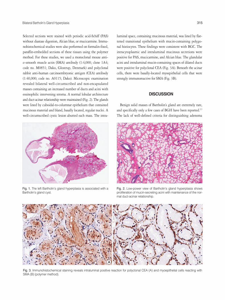
cesarean delivery 13 years previously and a vaginal delivery with-out episiotomy. She denied having associated vaginal discharge,dyspareunia, or pruritus. Physical examination revealed bilater-al firm and tender masses that measured 3×2 cm in both loweraspects of the labia minora. A clinical diagnosis of bilateral BGCswas made, and the masses were excised for analysis. The massfrom the left side measured 3×2.5×1 cm. It was not encapsu-lated and composed of a yellow/white firm mass of 1.8×1.2×1cm and a tan/brown cystic lesion of 1×1×0.8 cm (Fig. 1). Thecut surface showed a pale brownish solid tissue with a whorledappearance attached to a cyst that contained golden yellow gelati-nous material. The mass from the right side measured 1.7×1.2×1 cm and was brown in color without clear encapsulation. Itscut surface had a solid fibrous and whorled appearance. A tan/brown cystic lesion of 1×0.7×0.7 cm was also identified inthe mass from right-side.
These specimens were submitted in their entirety for histo-logical examination by hematoxylin and eosin (H&E) staining.
Bilateral Bartholin’s Gland Hyperplasia 315
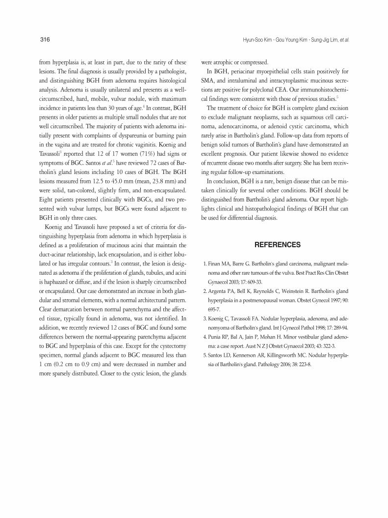
Selected sections were stained with periodic acid-Schiff (PAS)without diastase digestion, Alcian blue, or mucicarmine. Immu-nohistochemical studies were also performed on formalin-fixed,paraffin-embedded sections of these tissues using the polymermethod. For these studies, we used a monoclonal mouse anti-α-smooth muscle actin (SMA) antibody (1:4,000; clone 1A4;code no. M0851; Dako, Glostrup, Denmark) and polyclonalrabbit anti-human carcinoembryonic antigen (CEA) antibody(1:40,000; code no. A0115; Dako). Microscopic examinationrevealed bilateral well-circumscribed and non-encapsulatedmasses containing an increased number of ducts and acini witheosinophilic intervening stroma. A normal lobular architectureand duct-acinar relationship were maintained (Fig. 2). The glandswere lined by cuboidal-to-columnar epithelium that containedmucinous material and bland, basally located, regular nuclei. Awell-circumscribed cystic lesion abutted each mass. The intra-
luminal space, containing mucinous material, was lined by flat-tened transitional epithelium with mucin-containing polygo-nal histiocytes. These findings were consistent with BGC. Theintracytoplasmic and intraluminal mucinous secretions werepositive for PAS, mucicarmine, and Alcian blue. The glandularacini and intraluminal mucin-containing spaces of dilated ductswere positive for polyclonal CEA (Fig. 3A). Beneath the acinarcells, there were basally-located myoepithelial cells that werestrongly immunoreactive for SMA (Fig. 3B).
DISCUSSION
Benign solid masses of Bartholin’s gland are extremely rare,and specifically only a few cases of BGH have been reported.2,3
The lack of well-defined criteria for distinguishing adenoma
Fig. 1. The left Bartholin’s gland hyperplasia is associated with aBartholin’s gland cyst.
Fig. 2. Low-power view of Bartholin’s gland hyperplasia showsproliferation of mucin-secreting acini with maintenance of the nor-mal duct-acinar relationship.
Fig. 3. Immunohistochemical staining reveals intraluminal positive reaction for polyclonal CEA (A) and myoepithelial cells reacting withSMA (B) (polymer method).
A B
316 Hyun-Soo Kim∙Gou Young Kim∙Sung-Jig Lim, et al.
from hyperplasia is, at least in part, due to the rarity of theselesions. The final diagnosis is usually provided by a pathologist,and distinguishing BGH from adenoma requires histologicalanalysis. Adenoma is usually unilateral and presents as a well-circumscribed, hard, mobile, vulvar nodule, with maximumincidence in patients less than 30 years of age.4 In contrast, BGHpresents in older patients as multiple small nodules that are notwell circumscribed. The majority of patients with adenoma ini-tially present with complaints of dyspareunia or burning painin the vagina and are treated for chronic vaginitis. Koenig andTavassoli3 reported that 12 of 17 women (71%) had signs orsymptoms of BGC. Santos et al.5 have reviewed 72 cases of Bar-tholin’s gland lesions including 10 cases of BGH. The BGHlesions measured from 12.5 to 45.0 mm (mean, 23.8 mm) andwere solid, tan-colored, slightly firm, and non-encapsulated.Eight patients presented clinically with BGCs, and two pre-sented with vulvar lumps, but BGCs were found adjacent toBGH in only three cases.
Koenig and Tavassoli have proposed a set of criteria for dis-tinguishing hyperplasia from adenoma in which hyperplasia isdefined as a proliferation of mucinous acini that maintain theduct-acinar relationship, lack encapsulation, and is either lobu-lated or has irregular contours.3 In contrast, the lesion is desig-nated as adenoma if the proliferation of glands, tubules, and aciniis haphazard or diffuse, and if the lesion is sharply circumscribedor encapsulated. Our case demonstrated an increase in both glan-dular and stromal elements, with a normal architectural pattern.Clear demarcation between normal parenchyma and the affect-ed tissue, typically found in adenoma, was not identified. Inaddition, we recently reviewed 12 cases of BGC and found somedifferences between the normal-appearing parenchyma adjacentto BGC and hyperplasia of this case. Except for the cystectomyspecimen, normal glands adjacent to BGC measured less than1 cm (0.2 cm to 0.9 cm) and were decreased in number andmore sparsely distributed. Closer to the cystic lesion, the glands
were atrophic or compressed.In BGH, periacinar myoepithelial cells stain positively for
SMA, and intraluminal and intracytoplasmic mucinous secre-tions are positive for polyclonal CEA. Our immunohistochemi-cal findings were consistent with those of previous studies.5
The treatment of choice for BGH is complete gland excisionto exclude malignant neoplasms, such as squamous cell carci-noma, adenocarcinoma, or adenoid cystic carcinoma, whichrarely arise in Bartholin’s gland. Follow-up data from reports ofbenign solid tumors of Bartholin’s gland have demonstrated anexcellent prognosis. Our patient likewise showed no evidenceof recurrent disease two months after surgery. She has been receiv-ing regular follow-up examinations.
In conclusion, BGH is a rare, benign disease that can be mis-taken clinically for several other conditions. BGH should bedistinguished from Bartholin’s gland adenoma. Our report high-lights clinical and histopathological findings of BGH that canbe used for differential diagnosis.
REFERENCES
1. Finan MA, Barre G. Bartholin’s gland carcinoma, malignant mela-
noma and other rare tumours of the vulva. Best Pract Res Clin Obstet
Gynaecol 2003; 17: 609-33.
2. Argenta PA, Bell K, Reynolds C, Weinstein R. Bartholin’s gland
hyperplasia in a postmenopausal woman. Obstet Gynecol 1997; 90:
695-7.
3. Koenig C, Tavassoli FA. Nodular hyperplasia, adenoma, and ade-
nomyoma of Bartholin’s gland. Int J Gynecol Pathol 1998; 17: 289-94.
4. Punia RP, Bal A, Jain P, Mohan H. Minor vestibular gland adeno-
ma: a case report. Aust N Z J Obstet Gynaecol 2003; 43: 322-3.
5. Santos LD, Kennerson AR, Killingsworth MC. Nodular hyperpla-
sia of Bartholin’s gland. Pathology 2006; 38: 223-8.