Embed Size (px)
Citation preview
*Corresponding author email: [email protected] Group
Symbiosis www.symbiosisonline.org www.symbiosisonlinepublishing.com
Bilateral Gynecomastia Associated with Accessory Mammary Gland: Sporadic Clinical
Finding in a Patient with Acne Vulgaris Tchernev G1*, James W. Patterson2, Chokoeva AA3, Wollina U4
1Policlinic for Dermatology and Venereology, University Hospital Lozenetz, Sofia, Bulgaria2Department of Pathology, University of Virginia Health System, Charlottesville.
3Onkoderma-Policlinic for Dermatology and Dermatologic Surgery, General Skobelev, Sofia, Bulgaria4Department of Dermatology and Allergology, Academic Teaching Hospital Dresden-Friedrichstadt, Dresden, Germany
Clinical Research in Dermatology: Open Access Open AccessTherapeutic Hotline Letter
Received: December 15, 2015; Accepted: December 18, 2015; Published: December 20, 2015
*Corresponding author: Assoc. Prof. Georgi Tchernev, Policlinic for Dermatology and Venereology, University Hospital Lozenetz, Koziak street 1, 1407 Sofia, Bulgaria, Tel: +359 885 588 424; E-mail: [email protected]
did not show any abnormalities. Psychological evaluation revealed mild emotional lability with susceptibility to external influence and manipulation. Cardiovascular, lung and abdominal examinations were normal. Ultrasonography and mammography of the male breasts showed bilateral diffuse gynecomasty with enlargement of the breast with diffuse density, but absence of well-defined identifiable mass and secondary signs for malignancy. The patient denied brain MRI to exclude pituitary tumors. Complete blood count and liver function tests were within the normal range. The hormonal blood tests showed high levels of Prolactin – 466.60 mlU/l (referent values: 8 – 324 mlU/l), Progesterone – 4.98 nmol/l (referent values: 0.7 – 4.3 nmol/l) and DHEA–S –19.480 µmol/l (referent values: < 12.65 µmol/l) with normal levels for LH – 3.32 IU/l (1.7 – 8.6 IU/l), FSH – 2.70 IU/l (1.5 – 12.4 IU/l), Estradiol – 80.76 pmol/l (28 – 156 pmol/l), Testosterone – 14.83 nmol/l (9.9 – 27.8 nmol/l), TSH – 3.99 µIU/ml and total hCG – < 0.100 mU/ml (<2).
The causes of gynecomastia could be idiopatic, physiological (neonatal, pubertal, ageing), pathological (congenital/genetical disorders, endocrine causes, tumors, drugs, metabolic, miscellaneous) [1,2]. Different classifications for gynecomastia are described in the literature [3,4].
The whole nude body dermatological examination every time is of important value, because the physician could find untypical, associated or concomitant diseases with different skin expression. It could be important for the early detection of malignant diseases or disease with slow progression but with fatal outcome in different aspects if not diagnosed at time. The most practical one is proposed by Simon (Figure 3) [3]. We determine the patient’s gynecomastia with marked breast enlargement and marked skin redundancy. Differential diagnosis was done with pituitary tumors, adrenal gland tumors, breast cancer, dermoid cyst, hypogonadism, lymphangioma, drug intake. The treatment could be medicinal or surgical with different surgical techniques [5]. Total surgical resection of the accessory mammary gland was
Dear editor,A 18 year-old white male patient presented to our clinic
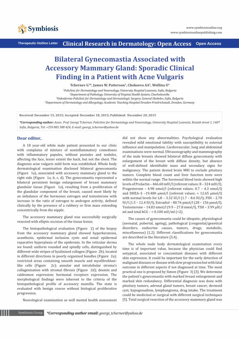
with complains of mixture of noninflammatory comedones with inflammatory papules, without pustules and nodules, affecting the face, lesser extent the back, but not the chest. The diagnosis acne vulgaris mild form was established. Whole body dermatological examination disclosed bilateral gynecomastia (Figure 1a), associated with accessory mammary gland to the right side (Figure 1a, b, c, d). The gynecomastia represented a bilateral persistent benign enlargement of breast mammary glandular tissue (Figure 1a), resulting from a proliferation of the glandular component of the breast, caused most likely by an imbalance of the hormones estrogen and testosterone with increase in the ratio of estrogen to androgen activity, defined clinically by the presence of a rubbery or firm mass extending concentrically from the nipple.
The accessory mammary gland was successfully surgically resected with elliptic excision of the tissue lesion.
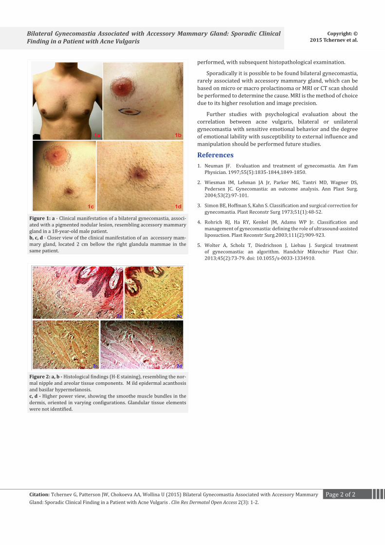
The histopathological evaluation (Figure 2) of the biopsy from the accessory mammary gland showed hyperkeratosis, acanthosis, epidermal inclusion cysts and zonal epidermal reparative hyperplasia of the epidermis. In the reticular derma we found: uniform rounded and spindly cells, distinguished by different wide stripes of hyalinized collagen (Figure 2b), located in different directions in poorly organized bundles (Figure 2a); restricted areas containing smooth muscle and myofibroblast-like cells (Figure 2c); annular and intralobular stroma’s collagenisation with stromal fibrosis (Figure 2d); desmin and caldesmon expression; hormonal receptors expression. The morphological findings were inherent to the criteria of the histopathological profile of accessory mamilla. The state is evaluated with benign course without biological proliferative progression.
Neurological examination as well mental health assessment
Page 2 of 2Citation: Tchernev G, Patterson JW, Chokoeva AA, Wollina U (2015) Bilateral Gynecomastia Associated with Accessory Mammary Gland: Sporadic Clinical Finding in a Patient with Acne Vulgaris . Clin Res Dermatol Open Access 2(3): 1-2.
Bilateral Gynecomastia Associated with Accessory Mammary Gland: Sporadic Clinical Finding in a Patient with Acne Vulgaris
Copyright: ©2015 Tchernev et al.
performed, with subsequent histopathological examination.
Sporadically it is possible to be found bilateral gynecomastia, rarely associated with accessory mammary gland, which can be based on micro or macro prolactinoma or MRI or CT scan should be performed to determine the cause. MRI is the method of choice due to its higher resolution and image precision.
Further studies with psychological evaluation about the correlation between acne vulgaris, bilateral or unilateral gynecomastia with sensitive emotional behavior and the degree of emotional lability with susceptibility to external influence and manipulation should be performed future studies.
References1. Neuman JF. Evaluation and treatment of gynecomastia. Am Fam
Physician. 1997;55(5):1835-1844,1849-1850.
2. Wiesman IM, Lehman JA Jr, Parker MG, Tantri MD, Wagner DS, Pedersen JC. Gynecomastia: an outcome analysis. Ann Plast Surg. 2004;53(2):97-101.
3. Simon BE, Hoffman S, Kahn S. Classification and surgical correction for gynecomastia. Plast Reconstr Surg 1973;51(1):48-52.
4. Rohrich RJ, Ha RY, Kenkel JM, Adams WP Jr. Classification and management of gynecomastia: defining the role of ultrasound-assisted liposuction. Plast Reconstr Surg.2003;111(2):909-923.
5. Wolter A, Scholz T, Diedrichson J, Liebau J. Surgical treatment of gynecomastia: an algorithm. Handchir Mikrochir Plast Chir. 2013;45(2):73-79. doi: 10.1055/s-0033-1334910.
Figure 1: a - Clinical manifestation of a bilateral gynecomastia, associ-ated with a pigmented nodular lesion, resembling accessory mammary gland in a 18-year-old male patient.b, c, d - Closer view of the clinical manifestation of an accessory mam-mary gland, located 2 cm bellow the right glandula mammae in the same patient.
Figure 2: a, b - Histological findings (H-E staining), resembling the nor-mal nipple and areolar tissue components. M ild epidermal acanthosis and basilar hypermelanosis.c, d - Higher power view, showing the smoothe muscle bundles in the dermis, oriented in varying configurations. Glandular tissue elements were not identified.


![Gynecomastia Correction in Individuals with Below Average ...Gynecomastia or enlarged breasts in men is a problem attracting increased attention [1-14]. Gynecomastia has been our primary](https://img.pdfslide.net/doc/110x75/5e7e37f087012e723f742b27/gynecomastia-correction-in-individuals-with-below-average-gynecomastia-or-enlarged.jpg)