Embed Size (px)
Citation preview
Schistosomiasis
(Bilharzia, Snail fever)Trematode (Fluke)
Sina Helbig*, Alia Tayea, Akre M Adja, Neil Arya**
Prepared as part of an education project of the Global Health Education Consortium and
collaborating partners
*First author **Corresponding author
9.1 Epidemiology
• Parasitic helminth endemic to >70 countries
o At least 240 million people affected, and 700 million at risk
o Annual deaths from schistosomiasis difficult to estimate:
disease can kill directly & indirectly (e.g. associated
carcinomas)
o Revised estimates put death toll from disease at over
200,000/yr, mostly in Africa
• In endemic areas usually acquired in childhood
o Infection increases in prevalence with age
o Peak age 15-20 years old
o Risk is related to occupation and exposure routes
Page 2
• Infection intensity decreases with age
o Acquired immunity, change in water contact, change in egg
production by worms
• Although relatively low (direct) case - fatality ratio,
disease has extremely high morbidity and enormous
impacts on health and socioeconomics
o Decreased productivity in chronic disease
o Associated impacts on families of those affected, in the form of
treatment costs and loss of income
• Two main types of disease: Urinary and Intestinal
9.1 Epidemiology
Page 3
9.2 Risk factors
• Poor sanitation practiceso Excretion of eggs into water sources used for drinking,
bathing, domestic use, etc.
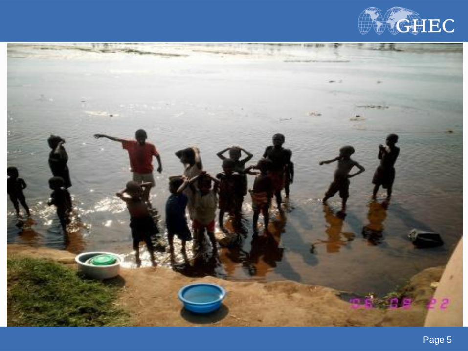
• School-age children (less immunity)
• Fresh-water exposure by swimming or bathing in
water containing infectious cercariae
• Occupational exposure through e.g. irrigation ditches,
rice farming, etc.o Particularly in/near standing or slow-moving water, where the
snail hosts can attach to vegetation
Page 4
Page 5
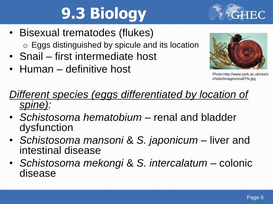
9.3 Biology• Bisexual trematodes (flukes)
o Eggs distinguished by spicule and its location
• Snail – first intermediate host
• Human – definitive host
Different species (eggs differentiated by location of spine):
• Schistosoma hematobium – renal and bladder dysfunction
• Schistosoma mansoni & S. japonicum – liver and intestinal disease
• Schistosoma mekongi & S. intercalatum – colonic disease
Page 6
Photo:http://www.york.ac.uk/res/s
chisto/images/snailTN.jpg
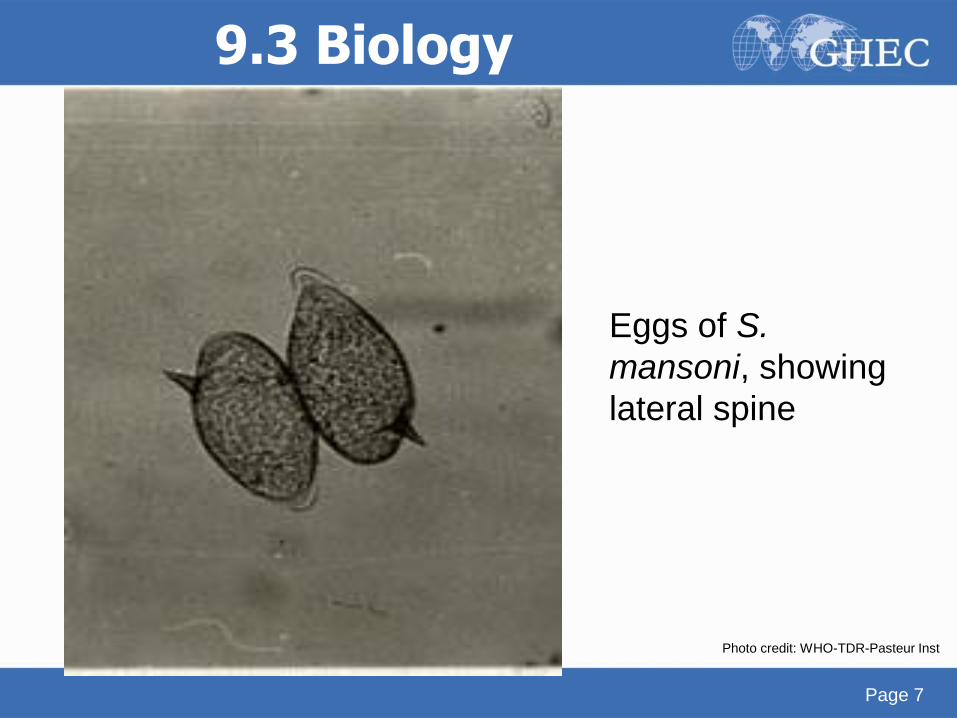
Eggs of S.
mansoni, showing
lateral spine
9.3 Biology
Page 7
Photo credit: WHO-TDR-Pasteur Inst
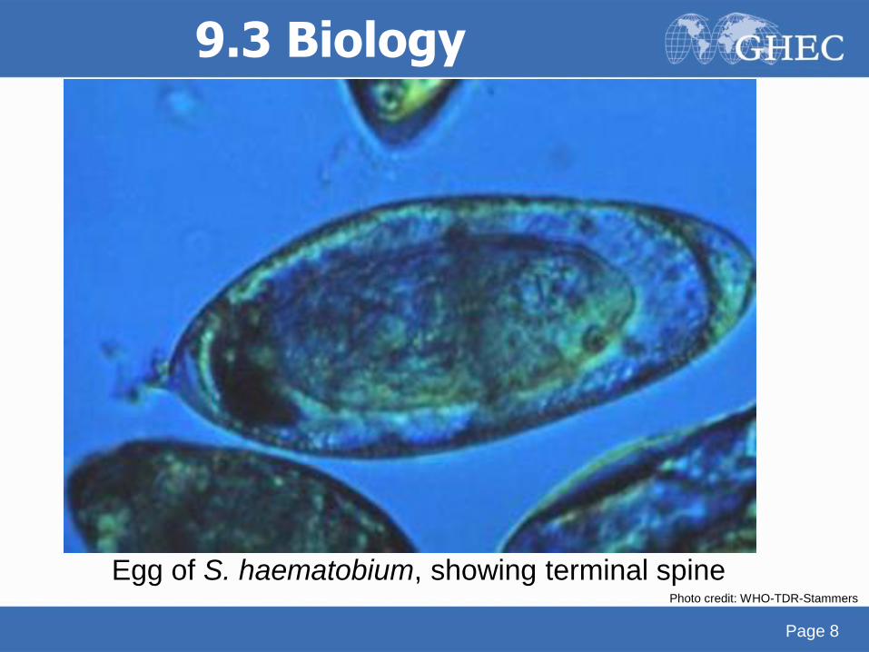
Egg of S. haematobium, showing terminal spine
9.3 Biology
Page 8
Photo credit: WHO-TDR-Stammers
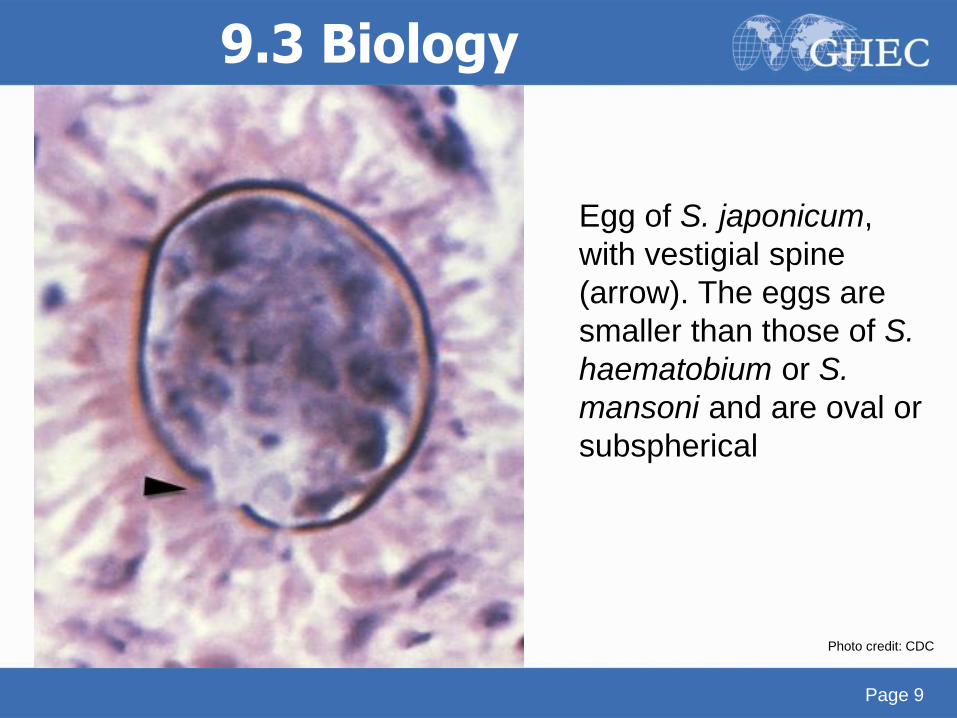
Egg of S. japonicum,
with vestigial spine
(arrow). The eggs are
smaller than those of S.
haematobium or S.
mansoni and are oval or
subspherical
9.3 Biology
Page 9
Photo credit: CDC
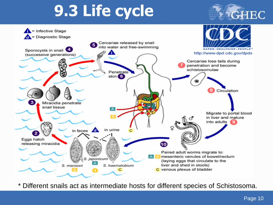
9.3 Life cycle
Page 10
* Different snails act as intermediate hosts for different species of Schistosoma.
9.4 Symptoms (general)• Two syndromes typically seen
o Early/Acute
Cercarial dermatitis, “swimmer’s itch”
Katayama fever/Katayama syndrome
o Chronic disease (various manifestations)
o Pathogenesis primarily through cell mediated immunity
Egg granuloma formation leads to fibrosis
• Cercarial dermatitis
o Result of penetration of cercariae through skin
o Commonly with S. hematobium, S. mansoni
o Pruritis, papular rash at the site of cercarial penetration
o Occurs mostly within 24h of exposure, but reports range from
minutes up to 1 week
Page 11
• Acute schistosomiasis- Katayama fever/syndromeo Early clinical manifestation with 1st infection or heavy reinfection
(primarily S. japonicum/mansoni)
o More likely in traveler or new immigrant to endemic region
o Symptoms 4-8 weeks after exposure with beginning of egg deposition in tissue
o Non-specific symptoms: fever, sweat, chills, cough, diarrhea, headaches
o Lymphadenopathy
o Hepatosplenomegaly
o Urticaria
o Respiratory symptoms – interstitial pneumonitis (more commonly seen in S. haematobium)
o Eosinophilia, hyperglobulinemia
o Immune complex reaction = Serum sickness
9.4 Symptoms- Acute disease
Page 12
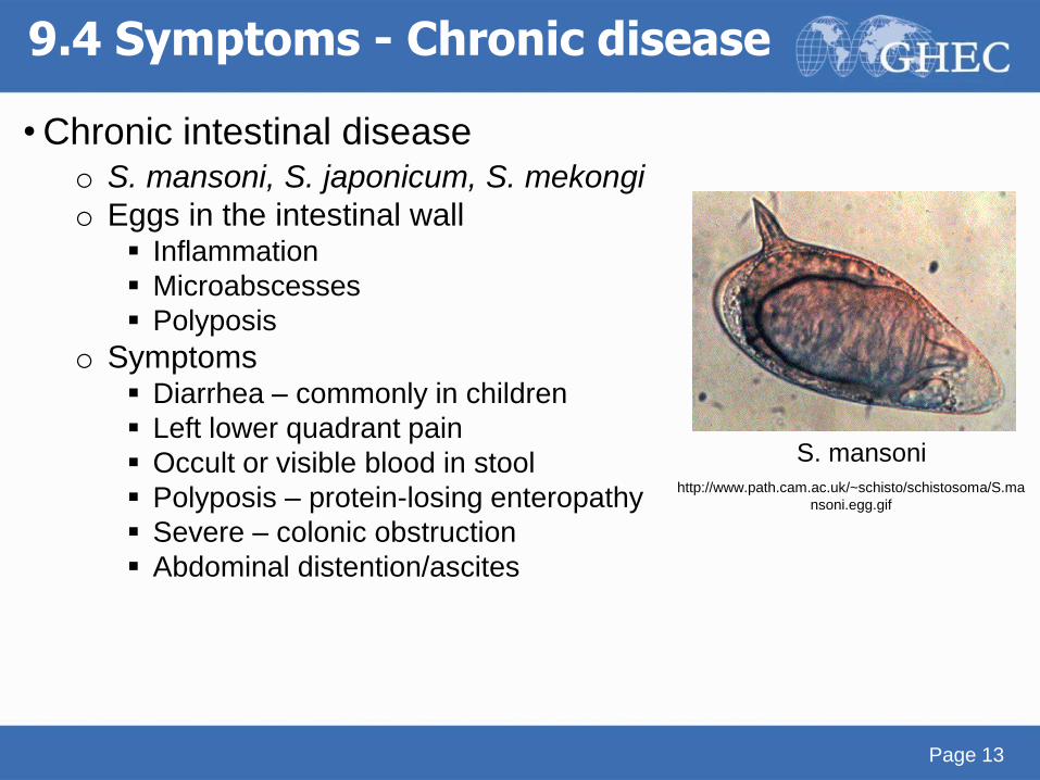
• Chronic intestinal diseaseo S. mansoni, S. japonicum, S. mekongi
o Eggs in the intestinal wall Inflammation
Microabscesses
Polyposis
o Symptoms Diarrhea – commonly in children
Left lower quadrant pain
Occult or visible blood in stool
Polyposis – protein-losing enteropathy
Severe – colonic obstruction
Abdominal distention/ascites
9.4 Symptoms - Chronic disease
Page 13
S. mansoni
http://www.path.cam.ac.uk/~schisto/schistosoma/S.ma
nsoni.egg.gif
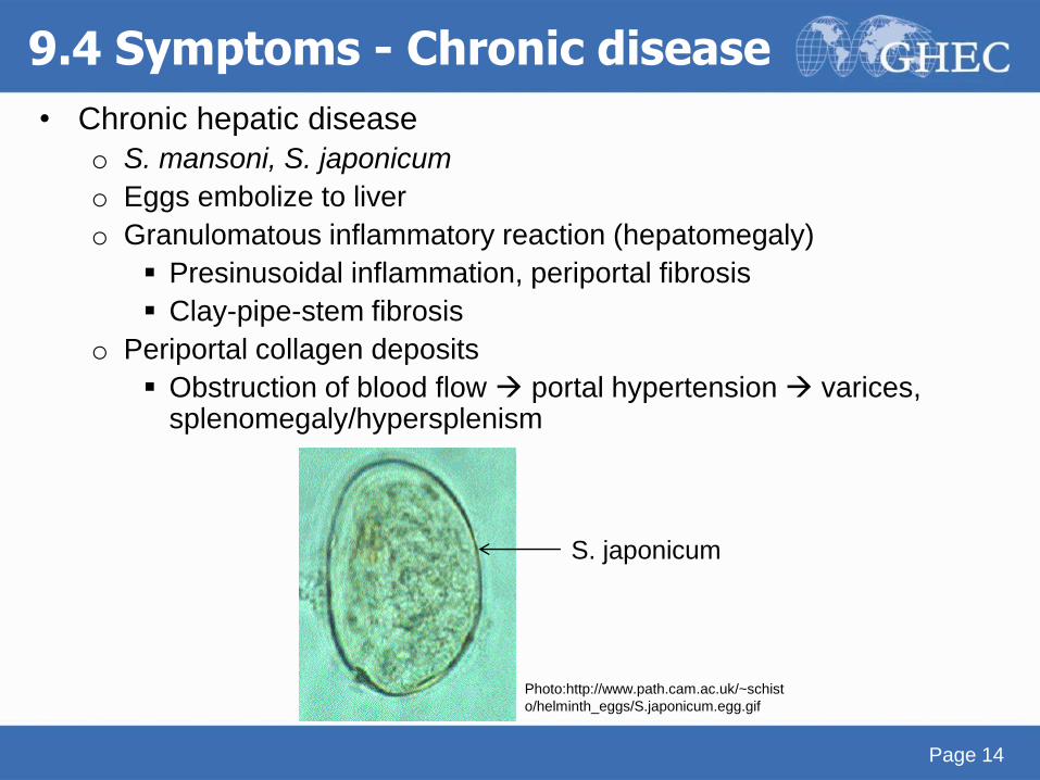
• Chronic hepatic disease
o S. mansoni, S. japonicum
o Eggs embolize to liver
o Granulomatous inflammatory reaction (hepatomegaly)
Presinusoidal inflammation, periportal fibrosis
Clay-pipe-stem fibrosis
o Periportal collagen deposits
Obstruction of blood flow portal hypertension varices, splenomegaly/hypersplenism
9.4 Symptoms - Chronic disease
Page 14
S. japonicum
Photo:http://www.path.cam.ac.uk/~schist
o/helminth_eggs/S.japonicum.egg.gif
• Chronic cardiopulmonary/pulmonary disease
o Cardiopulmonary hypertension
o Associated with hepatosplenic disease
Presinusoidal portal hypertension portocaval shunts eggs
travel and lodge in pulmonary vasculature
o Pulmonary hypertension: antiparasitic treatment = no effect
• Chronic CNS disease
o S. japonicum – brain lesions (seizures)
o S. mansoni – spinal cord lesions (more likely to be
symptomatic, transverse myelitis)
o Ectopic worm or egg dissemination (egg embolizes)
o Diagnosis usually difficult, since rarely systemic symptoms
9.4 Symptoms - Chronic disease
Page 15
• Chronic urinary tract diseaseo Usually in children
o Egg deposits in distal ureter and bladder wall
o Hematuria 10-12 weeks after exposure
o Dysuria
o Late manifestations: Nephrotic range proteinuria
Bladder calcifications
Ureteral obstruction
Renal colic
Renal failure
Secondary bacterial infections
Bladder carcinoma (squamous cell – well differentiated with local spread; accounts for up to 31% of all cancers in Egypt)
9.4 Symptoms - Chronic disease
Page 16
Public health impact of schistosomiasis: disease and mortality. WHO Expert Committee on the Control of Schistosomiasis
Bull World Health Organ 1993, 71(6):657-662.
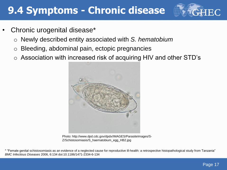
• Chronic urogenital disease*
o Newly described entity associated with S. hematobium
o Bleeding, abdominal pain, ectopic pregnancies
o Association with increased risk of acquiring HIV and other STD’s
9.4 Symptoms - Chronic disease
Page 17
Photo: http://www.dpd.cdc.gov/dpdx/IMAGES/ParasiteImages/S-
Z/Schistosomiasis/S_haematobium_egg_HB2.jpg
* “Female genital schistosomiasis as an evidence of a neglected cause for reproductive ill-health: a retrospective histopathological study from Tanzania”
BMC Infectious Diseases 2006, 6:134 doi:10.1186/1471-2334-6-134
9.5 Diagnosis
• Gold Standard: Eggs in stool of patient
• Intestinal and hepatic schistosomiasis
o Eggs identified in tissue of patient (liver/intestinal biopsy)
o Serology: FAST ELISA No distinction between past and current infection
Useful if negative – rule out
Useful in travelers
• Urinary schistosomiasiso Eggs in urine or on bladder biopsy
o FAST ELISA
o After diagnosis: evaluation of ureters, squamous cell
carcinoma
Page 18
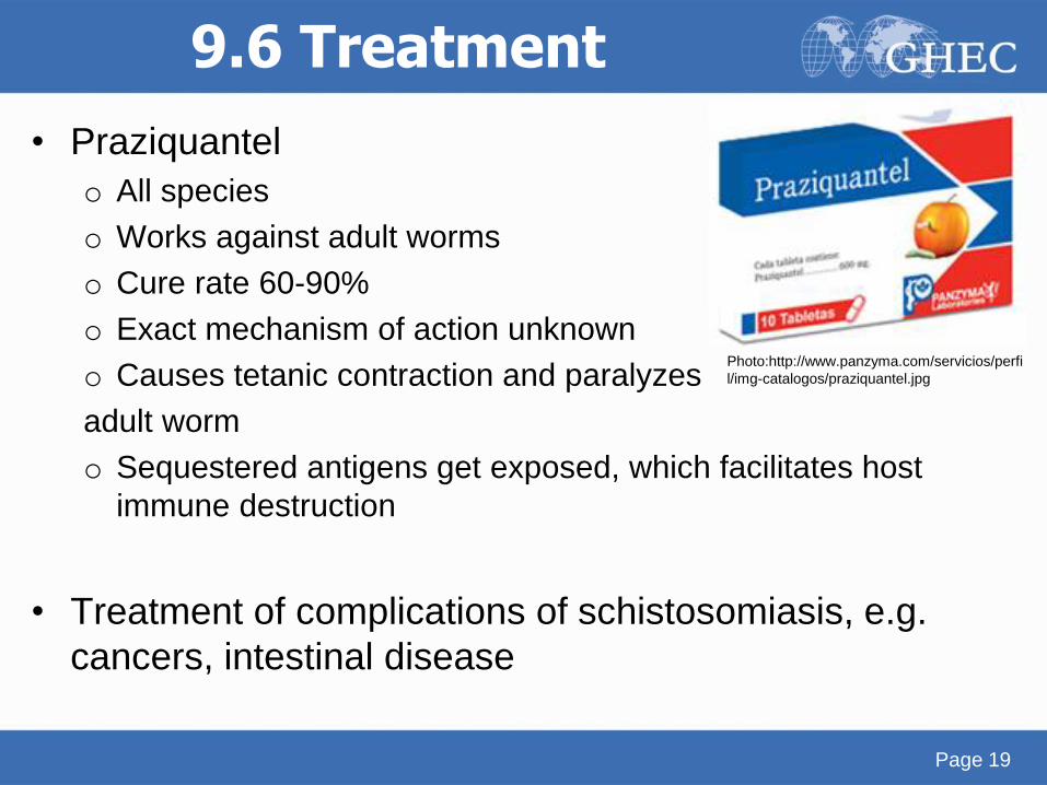
9.6 Treatment
• Praziquantel
o All species
o Works against adult worms
o Cure rate 60-90%
o Exact mechanism of action unknown
o Causes tetanic contraction and paralyzes
adult worm
o Sequestered antigens get exposed, which facilitates host
immune destruction
• Treatment of complications of schistosomiasis, e.g.
cancers, intestinal disease
Page 19
Photo:http://www.panzyma.com/servicios/perfi
l/img-catalogos/praziquantel.jpg
9.7 Control
• Ecological approaches for snail control
o Shown to be successful in Japan, where S. japonicum
largely eliminated from islands; considered eradicated since
1976
o E.g. current speed of rivers, streams, etc.
This has significant effect on snail host density
• Community-based control measures
o Public health education on disease & infection routes
o Improved sanitation and sanitation practices
No urination/ defaecation in water sources!
o Mass chemotherapy (praziquantel)
Page 20
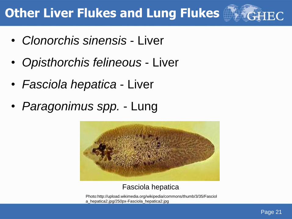
Other Liver Flukes and Lung Flukes
• Clonorchis sinensis - Liver
• Opisthorchis felineous - Liver
• Fasciola hepatica - Liver
• Paragonimus spp. - Lung
Page 21
Fasciola hepaticaPhoto:http://upload.wikimedia.org/wikipedia/commons/thumb/3/35/Fasciol
a_hepatica2.jpg/250px-Fasciola_hepatica2.jpg
Neurocysticercosis
Tapeworm (Cestode)
Page 22
10.1 Epidemiology
• Infection of the CNS by larval form of pork tapeworm
(Taenia solium)
• Estimated 50 million affected worldwide, with 50,000
deaths resulting from the condition
• Prevalence greatly reduced in Western Europe, where
prevalence used to be close to the current prevalence in
Mexico (1.9%)o Remains common in most developing countries
• A major cause of epilepsy in developing countrieso Main cause of acquired epilepsy
Page 23
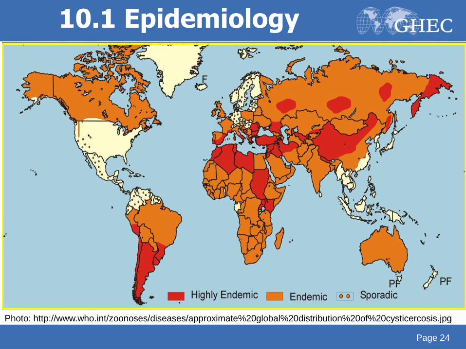
10.1 Epidemiology
Page 24
Photo: http://www.who.int/zoonoses/diseases/approximate%20global%20distribution%20of%20cysticercosis.jpg
10.2 Risk factors• Ingestion of parasite eggs, larvae by any route along the
feacal-oral transmission pathwayo Poor hygiene practices (esp. handwashing) of infected
people, as T. solium eggs excreted in faeces
o During food preparation, handling
o Autoinfection
• However, infection with parasite does not mean person will
develop neurocysticercosis
o Not all persons with taeniasis have CNS involvement
• Vegetarians equally at risk if they are exposed to person
excreting eggs!
*Eating undercooked meat only leads to T. Solium infection – which is asymptomatic for this
person, excreted eggs in feaces of this person are infectious and causes NCC.
Page 25
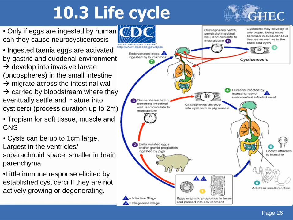
10.3 Life cycle• Only if eggs are ingested by human
can they cause neurocysticercosis
• Ingested taenia eggs are activated
by gastric and duodenal environment
develop into invasive larvae
(oncospheres) in the small intestine
migrate across the intestinal wall
carried by bloodstream where they
eventually settle and mature into
cysticerci (process duration up to 2m)
• Tropism for soft tissue, muscle and
CNS
• Cysts can be up to 1cm large.
Largest in the ventricles/
subarachnoid space, smaller in brain
parenchyma
•Little immune response elicited by
established cysticerci If they are not
actively growing or degenerating.
Page 26
10.4 Symptoms
• Majority of cases are asymptomatic, and can remain so
for decadeso Little immunological reaction from intact cysticercal larvae;
more serious reactions to dying, degenerating cysticerci
• Leads to seizures, headaches (common) and stroke
• Cases with many lesions and brisk inflammatory
response (particularly with degenerating cysts): sub-
acute encephalitis
• If space-occupying lesion: any focal symptomatology
including weakness and extrapyramidal symptoms
Page 27
• Ventricular disease: 15% of cases, obstructive hydrocephalus
o May spontaneously remit due to ball-valve effect upon motion intermittently occluding the ventricular outlet foramina
o Untreated, most cases progress to sustained hydrocephalus
• Spinal disease: cysts in and around the cord have been reported
o Progressive paraplegia developing over period of weeks
• Changes in mental status, cognitive impairment, psychiatric disease, balance problems
• Ocular disease: cysts subretinal or intravitreous
10.4 Symptoms
Page 28
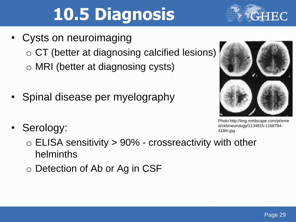
10.5 Diagnosis
• Cysts on neuroimaging
o CT (better at diagnosing calcified lesions)
o MRI (better at diagnosing cysts)
• Spinal disease per myelography
• Serology:
o ELISA sensitivity > 90% - crossreactivity with other
helminths
o Detection of Ab or Ag in CSF
Page 29
Photo:http://img.medscape.com/pi/eme
d/ckb/neurology/1134815-1168784-
418tn.jpg
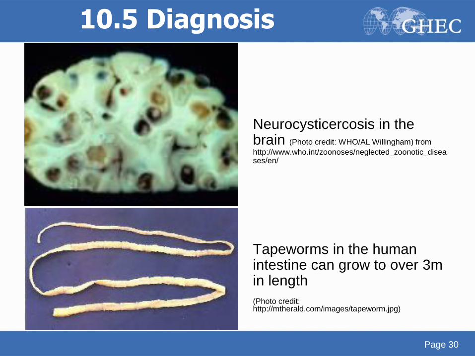
Neurocysticercosis in the brain (Photo credit: WHO/AL Willingham) from
http://www.who.int/zoonoses/neglected_zoonotic_diseases/en/
Tapeworms in the human intestine can grow to over 3m in length
(Photo credit: http://mtherald.com/images/tapeworm.jpg)
10.5 Diagnosis
Page 30
10.6 Treatment
• Treatment is complex and treatment plan controversial
• Some advocate only treating certain cases due to the
adverse reactions associated with immune reaction to
dying parasiteo Most viable cystic lesions either calcify or disappear, in viable
lesions antihelminthic therapy may hasten the death of
parasite, but not change clinical outcome
o Advocate treating with corticosteroids and anti-epileptic
drugs, optimal duration is subject to debate
• Medicationso Praziquantel
o Albendazole
Page 31
• Always pre-treat with corticosteroids, and continue
during treatment
o Decreases inflammatory reaction elicited by dying cysticerci
• Surgical treatment only in selected cases of
ventricular cysts, eye involvement
• Shunt placement in case of hydrocephalus
10.6 Treatment
Page 32
10.7 Control
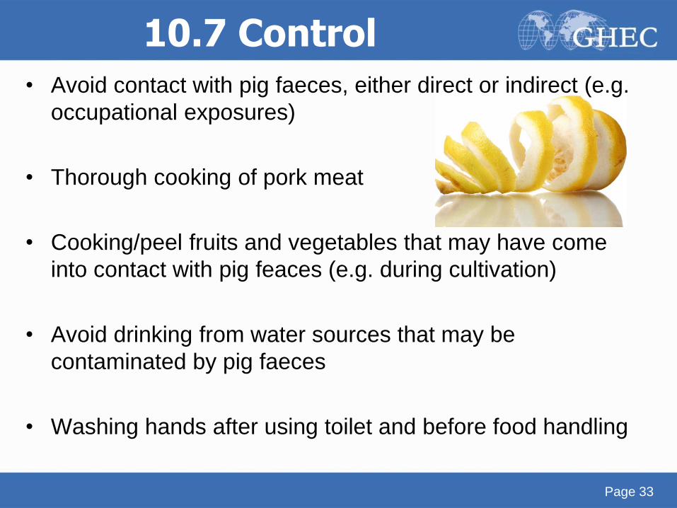
• Avoid contact with pig faeces, either direct or indirect (e.g.
occupational exposures)
• Thorough cooking of pork meat
• Cooking/peel fruits and vegetables that may have come
into contact with pig feaces (e.g. during cultivation)
• Avoid drinking from water sources that may be
contaminated by pig faeces
• Washing hands after using toilet and before food handling
Page 33

Page 34
Acknowledgments
• Thanks to Jenna Kelly, Shazeen Bandukwala and
Melissa Whaling for critical editing.
• We appreciate Tim Brewer and Jackeline Alger for
thoughtful review.
Page 34
Credits
Akre M Adja1, Sina Helbig2, Alia Tayea3, Neil Arya4
1: Institut Pierre Richet, Université de Cocody Abidjan
2: Boston University School of Medicine, Division of
Infectious Diseases, Boston, MA, USA
3: Médecins Sans Frontières
4: Western University, University of Waterloo,
McMaster University
Contact [email protected]
End of module
[Reserved for GHEC notes and acknowledgment of donor organizations]


![The Pathogenesis ef Bilharzia] Hydroureter](https://img.pdfslide.net/doc/110x75/61ffc6f2b5bc566f4e5aa1de/the-pathogenesis-ef-bilharzia-hydroureter.jpg)