Embed Size (px)
Citation preview
Biochemistry 1995,34, 15829- 15837 15829
Binding of Phenylarsenoxide to Arg-tRNA Protein Transferase Is Independent of Vicinal Thiols?
Jun Li$ and Cecile M. Pickart*JsJ
Department of Biochemistry, School of Medicine and Biomedical Sciences, SUNY at Buffalo, Buffalo, New York 14214
Received August 21, 1995@
ABSTRACT: Reversible enzyme inhibition by phenylarsenoxides is generally taken to indicate the presence of functionally important vicinal thiol groups. Arginyl aminoacyl-tRNA transferase from eukaryotes is potently inhibited by phenylarsenoxides and possesses one or more essential sulfhydryl groups [Li, J., & Pickart, C. M. (1995) Biochemistry 34, 139-1471. To map the putative Cys residues that mediate arsenoxide binding to the transferase from Saccharomyces cerevisiae, we systematically mutagenized the 15 Cys residues of the transferase, singly and in combination, to Ala (13 Cys) or Ser (2 Cys). Six mutant enzymes, encompassing all 15 Cys residues of the transferase, were characterized in detail. The results revealed that Cys-20, Cys-23, and Cys-94 and/or Cys-95 were important for activity, since mutations at these positions reduced activity by 1 00-fold (Cys-94 and Cys-95 were mutated simultaneously). Surprisingly, however, all of the mutant enzymes retained the ability to bind a radioiodinated phenylarsenoxide derivative, with undiminished stoichiometry and affinity. All of the mutant enzymes also remained susceptible to irreversible reaction with a bifunctional phenylarsenoxide bearing a para- alkyl halide substituent. Prior reaction of the enzyme with the bifunctional reagent blocked subsequent binding of the radiolabeled phenylarsenoxide, indicating that these two reagents bind at a single common site. These results indicate that high-affinity binding of trivalent arsenicals can occur by a thiol-independent mechanism.
Monosubstituted trivalent arsenicals have long been known to form addition compounds with thiols [reviewed in Webb (1966)l. Alkylarsenoxides (RAs=O) react with vicinal thiol groups to form ring structures as shown in
The crystal structure of the adduct between C H ~ C ~ H ~ A S C I ~ and 2,3-dimercaptopropanol reveals a five-membered ring in which both sulfur atoms are chelated to the arsenic (Adams et al., 1990). Similar cyclic complexes of phenylarsenoxides with small dithiols have been identified by gas chromato- graphic analysis (Hannestad & Sorbo, 1980). As expected on entropic grounds, cyclic dithiolarsinites are markedly more stable than the noncyclic addition products formed from trivalent arsenicals and monothiols (Stocken & Thompson, 1946b).
On the basis of the observation that Lewisite (Cl- CH=CHAsC12) and arsenite (HOAs-0) reacted with thiol groups of reduced keratin in the molar ratio of 1:2, Stocken and Thompson ( 1946a) postulated that arsenic combined with
vicinal Cys residues of proteins to form ring structures analogous to those described above:
HS \ R - A s = O + HS, Protein R - As ' ' Protein + H2O (2)
As expected for this mechanism, vicinal sulfhydryl reagents such as 2,3-dimercaptopropanol and DTTI both prevent and reverse complex formation between Lewisite and arsenoxide- sensitive enzymes, while monothiols are much less effective (Whittaker, 1947; Peters et al., 1945). The protective effects of specific dithiols correlate well with the stabilities of the corresponding cyclic dithiolarsenite adducts (Whittaker, 1947).
For the past 50 years, the ring hypothesis outlined in eq 2 has served as the paradigm for explaining enzyme inhibition by trivalent arsenicals, and inhibition has usually been taken to indicate the presence of a spatially vicinal thiol pair, except in special cases [e.g., Barber and Siege1 (1983)l. For example, arsenoxides inhibit the pyruvate dehydrogenase complex by binding to the vicinal thiols of the reduced lipoamide cofactor (Stevenson et al., 1978; Adamson et al., 1984). However, vicinal thiols need not be chemically vicinal as in lipoamide. They can also be brought into spatial apposition by folding [e.g., Jauhiainen et al. (1988)l.
' S '
+ Supported by a grant from the NIH (DK43769). * Address correspondence to this author at Department of Biochem-
istry, School of Hygiene and Public Health, Johns Hopkins University, 615 North Wolfe Street, Baltimore, MD 21205. Telephone: (410) 614- 4554. Fax: (410) 955-2926.
Present address: Molecular Immunology Department, SmithKline Beecham, King of Prussia, PA 19406.
e Present address: Department of Biochemistry, School of Hygiene and Public Health, Johns Hopkins University, Baltimore, MD 21205.
II Recipient of a Career Development Award from the NIH. @ Abstract published in Advance ACSAbstrucrs, November 15, 1995.
0006-296019510434- 15829$09.0010
' Abbreviations: BSA, bovine serum albumin; PME, P-mercapto- ethanol; BrAcAn, bromoacetyl aniline; BrAcNPAsO, para-[(bro- moacetyl)amino]phenylarsenoxide; DlT, dithiothreitol; E, Arg aminoacyl- tRNA protein transferase; HEPES, (hydroxyethy1)piperazineethane- sulfonate; I and '251-PAs0, para-'251-phenylarsenoxide; MOPS, mor- pholinepropane sulfonate; NEM, N-ethylmaleimide; NPAsO, p-ami- nophenylarsenoxide; PAGE, polyacrylamide gel electrophoresis; PAsO, phenylarsenoxide; PCR, polymerase chain reaction; PMSF, phenyl- methanesulfonyl fluoride; rcmBSA, reduced and carboxymethylated bovine serum albumin; SDS, sodium dodecyl sulfate; TCA, trichloro- acetic acid; TFA, trifluoroacetic acid.
0 1995 American Chemical Society

15830 Biochemistry, Vol. 34, No. 48, 1995
Trivalent arsenicals have been applied to the study of diverse enzyme systems, including the pyruvate dehydroge- nase complex (Stevenson et al., 1978), lecithin-cholesterol acyltransferase (Jauhiainen et al., 1988), tyrosyl phosphatases [e.g., Zhang et al. (1992)], hexose transport systems (Frost & Lane, 1985; Hoffman & Lane, 1992), thioredoxin (Brown et al., 1987), and the ubiquitin-dependent protein degradation pathway (Klemperer & Pickart, 1989; Berleth et al., 1992a). Strong and specific interactions between arsenoxides and proteins have been demonstrated using arsenical affinity chromatography and radioactive aromatic arsenoxides (Zhou et al., 1991; Hoffman & Lane, 1992; Berleth et al., 1992a; Kalef et al., 1993). In a few cases the vicinal thiols believed to mediate these interactions have been identified. Examples include lipoamide in the pyruvate dehydrogenase complex (Stevenson et al., 1978); Cys-31 and Cys-184 in lecithin- cholesterol acyl transferase (Jauhiainen et al., 1988); the redox-active Cys pair in the active site of thioredoxin (Brown et al., 1987); and adjacent Cys residues in the reduced nicotinic acetylcholine receptor (Dou et al., 1994). For the majority of proteins that bind arsenoxides, however, the actual ligands of the arsenic remain unidentified [e.g., Hoffman and Lane (1992) and Kalef et al. (1993)l.
We have been using phenylarsenoxides to study the mechanism of Arg-tRNA protein transferase. This enzyme catalyzes the transfer of Arg from Arg-tRNA to a-amino groups of proteins that initiate with Glu or Asp (Soffer, 1970). Its specific role is to make its targets susceptible to ubiquitin conjugation and thus to degradation by the 26s protease (Bachmair et al., 1986). Both the mammalian and the yeast transferase are inhibited by phenylarsenoxides in a DTT-reversible fashion (Klemperer & Pickart, 1989; Berleth et al., 1992b; Li & Pickart, 1995). We have interpreted this inhibition to indicate the existence of vicinal thiols that function in transferase catalysis or regulation.
In previous efforts to map the arsenoxide binding site, we identified Cys-315 of the yeast transferase as the nucleophile that reacts irreversibly with a bound bifunctional phenyl- arsenoxide (Li & Pickart, 1995). Here we describe the use of Cys to Ala scanning mutagenesis to identify the putative vicinal thiols that mediate arsenoxide binding. Unexpectedly, our results strongly suggest that binding of phenylarsenoxides to the transferase is not mediated by vicinal thiols.
EXPERIMENTAL PROCEDURES Materials. The following were purchased from the
indicated sources: ~-[2,3-~H]Arg (53 Ci/mmol; Dupont- NEN); crude Escherichia coli tRNA (Sigma); crude mixture of E . coli aminoacyl-tRNA synthetases (Sigma); NEM (Sigma); protein A-Sepharose (Sigma); PAsO (Aldrich); and carrier-free Na1251 (Amersham). RcmBSA was synthesized as described (Evans & Wilkinson, 1985). BrAcAn, NPAsO, and BrAcNPAsO were synthesized as described (Berleth et al., 1992a).
Enzyme Pur8cation. Recombinant Arg-tRNA transferase was induced in E. coli strain BL21(DE3)/pLysS (Moffatt & Studier, 1987) harboring the vector pET3d-ATT; as described previously, the wild type enzyme bears a Ser-to-Ala mutation at residue two (Li & Pickart, 1995). Wild type and mutant enzymes were purified by the published procedure (Li & Pickart, 1995). Enzyme concentrations were based on measured protein concentrations, assuming a molecular mass of 58 kDa.
Li and Pickart
Transferase Assay. Incorporation of [3H]Arg into rcmBSA was monitored as the appearance of radioactivity insoluble in TCA, as described (Li & Pickart, 1995). [3H]Arg tRNA was first generated in a preincubation containing: 50 mM Tris-HC1 (24% base), 5 mM MgC12, 2 mM ATP, 10 mM phosphocreatine, 0.6 unit/mL each of creatine phosphokinase and inorganic pyrophosphatase, 1.5 mg/mL E. coli tRNA, 30 pCilmL [3H]Arg, and 130 units/ml E. coli aminoacyl- tRNA synthetases (pH 7.3). After 4 min at 37 "C, this mixture was transferred to ice. To initiate an assay, 4 pL of this mixture was added to 4 pL of a second solution containing the transferase and rcmBSA (1 mg/mL final concentration). After 5 min at 37 OC, the incubation was quenched with 8 pL of 8 M urea and spotted onto Whatman #2 filter paper. Filters were processed as described (Klem- perer & Pickart, 1989), and acid-insoluble radioactivity was determined by liquid scintillation counting. Data were corrected by subtracting a blank obtained by omitting the transferase.
Production and Pur@cation of Polyclonal Antibodies. Inclusion bodies containing the transferase were purified by a previously described method (Lin & Cheng, 1991) and subjected to 10% SDS-PAGE. The transferase band (58 kDa) was visualized by staining with 0.05% Coomassie blue (in water), excised, homogenized, and used to raise antiserum against the transferase in rabbits essentially as described (Haas & Bright, 1985). Antibodies were purified by affinity chromatography, using a column of purified transferase immobilized on activated CH-Sepharose from Pharmacia- LKB Biotech (Haas & Bright, 1985).
PCR Mutagenesis. All single-site mutagenesis was per- formed by a two-step PCR method that permits a mutation to be generated with a single unique primer (Li & Pickart, 1995). The plasmid pET3d-ATT was the template for both PCR reactions. Flanking primers were the same as those used previously (Li & Pickart, 1995). The mutant primer and the flanking primer closest to the mutation site were used in the first PCR reaction (to minimize the size of the first PCR product). This first PCR product was gel-purified and used as one of the primers in the second PCR reaction. The other flanking primer served as the second primer. The final -1550 bp product was digested with NcoI and BgZII and cloned into plasmid pET3d. To make multisite mutants, the template vector contained a previously-mutated trans- ferase gene. In each mutant plasmid, the presence of expected mutation(s) was verified by DNA sequencing (fmol Kit, Promega).
Primers for PCR mutagenesis were as follows (underlined bases induce mutations): Mut- 1 (C3 15A): 5'-ACTGGAT-
CGTGEATACCCACACTTTG; Mut-3 (C94S,C95A): GTGGCTCACAGTAAATATA-3'; Mut-2 (C23A): TTAC-
GTATAACCTACTAGCATTCCTCAACGGG; Mut-4 (c 1 9 3 ~ ) : AAGGGCTTATCAGCTAGGAATCGCTTA; Mut-5 (C233A): TCATAGTAGTTGGCTTCGTGCACAG- GAC; Mut-6 (C298A): TATTGAGGZETCCGAAGAT- GAAT; Mut-7 (A48 1-503): GGGAGATCTCAAATACG- TTTCTTGAGA; Mut-8 (C56A): GCCAATTGTGEGTT- TTCAATCTTTA; Mut-9 (C54A): ATCCCATATTAE CATGCGATCATACG; Mut- 10 (C 1 16A): GCGGCTGATG- - GCTTTTTTCAATTCCT; Mut-1 1 (C 130A): AGCGGCT- GGGGCGTAGTCCTACTT; Mut- 12 (C382A): TGTGG- GAGmGTGCGTITAAATCTG; Mut- 13 (C20S): ATAC- CCACTCTTTGCGGCAGG; Mut- 14 (C48 1A): AAGAAA-

Thiol-Independent Arsenoxide Binding
CGTATTGCTGACGTGAT-ACGG; Mut- 15 (C492A): GGCTTCGAAACTETATGAAAGCCGTCA; AN (AI- 30): GGGAGATCTCCATGGATCAGTTGTTCG.
Preparation of 1251-PAs0. This reagent was synthesized as described by Hoffman and Lane (1992), with the following modifications. (1) The reaction was scaled down to utilize 0.5-1 mCi of Na1251, maintaining the same specific radio- activity. (2) The product was purified only through the ether extraction step, since HPLC analysis showed that the purity at this stage was '90%. 1251-PAs0 was stored in the dark at -20 "C.
Immunoprecipitation assay. Purified transferase (1.4 pM) was incubated with various concentrations of 1251-PAs0 in 50 mM MOPS (pH 7.2) and 2 mM EDTA at 37 "C for 4 min (10 p L volume). Affinity-purified anti-transferase antibodies (4 pL of a 1.2 mg/mL stock) were added, and incubation continued at 5 "C for 30 min. Protein A- Sepharose (40 p L of 25% v/v suspension in dilution buffer) was then added, and the suspension was rotated at 25 "C for 20 min. (Dilution buffer consisted of phosphate-buffered saline supplemented with 2 mg/mL BSA, 5 mM EDTA, 1 % Triton-X-100,O.l mM PMSF, and 0.02% sodium azide.) The suspension was then diluted with 400 p L of dilution buffer and vortexed. The resin was pelleted in a microfuge and washed three times with 400 pL of dilution buffer. The washed pellet was counted in a gamma counter. Data were corrected by subtracting a blank obtained by omitting enzyme from the assay.
Mass Spectrometry. Electrospray ionization mass spec- trometry was carried out at the Harvard Microchemistry facility (Chicz et al., 1993). For this experiment the transferase was purified in the absence of Triton-X-100 and glycerol.
Pantothenate Assay. The transferase was tested for the presence of pantotheine by the procedure of Pugh and Wakil (1965). Bacterium Lactobacillus plantarum (ATCC 8014) was grown at 37 "C for 20 h in 5 mL of pantothenate medium (Bacto AOAC USP) containing 20 ng/mL pantothenic acid. The cells were pelleted, washed once with 5 mL of 0.85% (w/v) NaCI, resuspended in 5 mL of 0.85% NaC1, and diluted 25-fold in 0.85% NaCl. An aliquot of 50 p L was used to inoculate a 5 mL aliquot of pantothenate assay medium that had been mixed with the sample being assayed. (This mixture was autoclaved 15 min prior to innoculation.) After incubation at 37 "C for 24 h, growth was assessed by determination of absorbance at 600 nm. Samples to be assayed for pantothenate were first treated with alkali, then neutralized and treated with alkaline phosphatase (Pugh & Wakil, 1965). When assayed by this procedure, 1.5 nmol of CoA gave the expected positive result (A600 = 0.41, vs 0.014 for no addition). The result obtained with the transferase (2.9 nmol, A600 = 0.01, average of duplicate determinations) was indistinguishable from the negative control. The absence of inhibitory species in the transferase- derived sample was shown by the strong positive signal obtained when this sample was mixed with 0.23 nmol of pantothenic acid (A600 = 0.56).
RESULTS
'2sI-PAs0 as a Probe of the Enzyme-Arsenoxide Interac- tion. In order to monitor directly the interaction between the transferase and phenylarsenoxide, we synthesized 1251-
Biochemistry, Vol. 34, No. 48, 1995 15831
5ot I
25u 2 4 6 8
"M IPASO
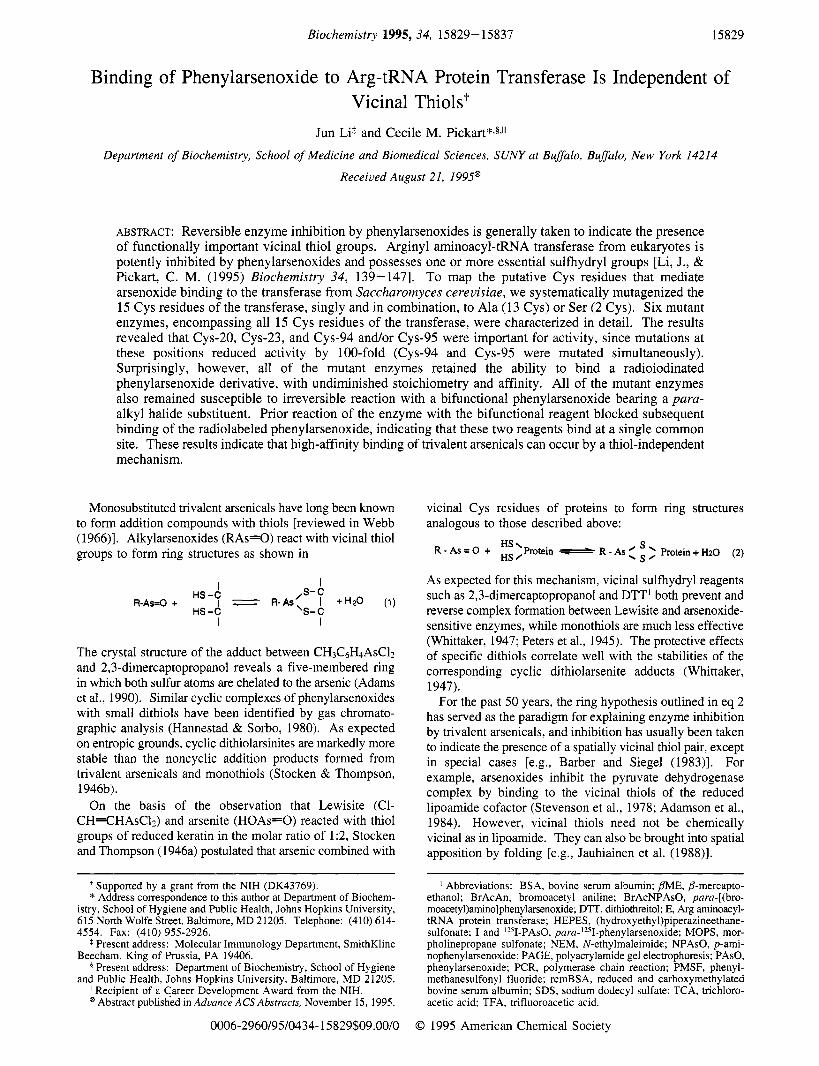
FIGURE 1 : Binding of 1251-PAs0 to the transferase and effect on activity. Wild type ransferase (1.4 pM) was incubated with the indicated concentration of 1251-PAs0 (2094 cpdpmol) for 4 min at 37 "C in 50 mM MOPS (pH 7.2) and 2 mM EDTA. (A) Reversible inhibition. Replicate aliquots of the incubation were diluted 10-fold into 50 mM MOPS (pH 7.2) alone (filled circles) or 50 mM MOPS supplemented with 10 mM DTT (open circles); aliquots of 2 pL were assayed for transferase activity. (B) Binding (immunoprecipitation assay). A 10 pL aliquot of the incubation was analyzed for transferase-bound lZ51-PAs0 by immunoprecipi- tation (Experimental Procedures). (C) Relationship between binding and inhibition. Each parameter is expressed relative to its maximum value, as a function of IZ51-PAs0 concentration: filled circles, binding; open circles, inhibition. The lines in each panel have no theoretical basis.
PAsO by the method of Hoffman and Lane (1992) and developed an immunoprecipitation assay to quantitate binding of this ligand to the transferase under native conditions (Experimental Procedures). The relationship between 1251- PAsO binding and enzyme activity is shown in Figure 1. As expected, the transferase (1.4 pM) was strongly inhibited by '251-PAs0, with K0.5 -2.2 p M (filled circles, Figure 1A). Inhibition was reversed by DTT (open circles, Figure 1A). The concentration dependence of inhibition was similar to that seen previously with PAsO (Ko.5 -1.2 p M at 1.5 p M enzyme; Li & Pickart, 1995), indicating that the iodo moiety did not impede the interaction between enzyme and arse- noxide. This was the expected result, based on the lack of effect of other para-substitutions (Li & Pickart, 1995). Thus, the radiolabeled phenylarsenoxide was a suitable probe for studying phenylarsenoxide binding to the transferase.
Quantitative binding studies were first carried out by the immunoprecipitation assay. 1251-PAs0 binding showed a saturating concentration dependence (Figure 1B). Moreover, there was a linear relationship between binding and inhibition (Figure lC), with a stoichiometry of -0.7 mol Vmol E at a saturating concentration of 6 p M (Figure 1B). These data suggest that the transferase has a single phenylarsenoxide binding site. The observed stoichiometry of 0.7 (rather than

15832 Biochemistry, Vol. 34, No, 48, 1995 Li and Pickart
uM lPAs0 - 2 4 6 8 IO 12 14
uM lPAs0
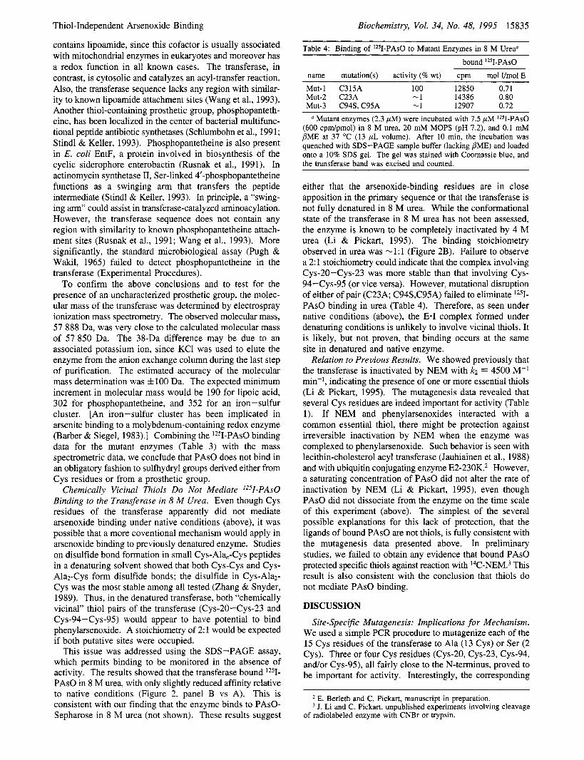
FIGURE 2: Binding of '251-PAs0 to the transferase (SDS-PAGE assay). (A) Native conditions. Fully active mutant enzyme, 0.25 pM, was incubated with the indicated concentration of '251-PAs0 (2370 cpdpmol) in 50 mM HEPES (pH 7.5) and 0.8 mg/mL rcmBSA at 37 "C (10 pL volume; filled circles, Mutd = C298A; open circles, Mut-5 = C233A). After 5 min, the incubation was quenched with 5 p L of 2 x concentrated sample buffer (lacking PME), boiled for 1 min, and loaded onto a 10% SDS gel. The gel was dried and autoradiographed. The band corresponding to the transferase was excised and counted (left axis). The calculated stoichiometry is shown on the right axis. Other experiments showed that rcmBSA did not affect 1251-PAs0 binding and that 6 p M IPAsO was saturating. (B) Denaturing conditions. Fully active transferase enzyme Mut-1 (C315A, 1.3 pM) was incubated with indicated concentrations of 1251-PAs0 (458 cpdpmol) in 40 mM MOPS (pH 7.2) and 8 M urea at 37 "C (13 p L volume). After 10 min, the incubation was quenched with an equal volume of SDS-PAGE sample buffer (lacking BME), boiled for 1 min, and loaded onto a 10% SDS gel. The gel was dried and autoradiographed. The band corresponding to the transferase was excised and counted. The filled circle shows data from an incubation containing 32 pM 1251- PAsO. The lines in each panel have no theoretical basis.
1.0) probably reflects partial loss of the labeled reagent during the extensive washing required by this assay (Ex- perimental Procedures) and perhaps the presence of some inactive oxidized enzyme, as indicated by the finding that DTT stimulated activity by 29% in the absence of '251-PAs0 (Figure 1A).
1251-PAs0 binding was also monitored using an SDS - PAGE assay (Hoffman & Lane, 1992). Here the incubation containing transferase and 1251-PAs0 was quenched in sample buffer lacking mercaptoethanol and electrophoresed; the transferase band was excised and counted. At 6 p M 1251- PAsO, this assay yielded a stoichiometry of -1 mol Ymol E (Figure 2A), comparable to that obtained in the immuno- precipitation assay (Figure 1B). The observed retention of bound 1251-PAs0 during SDS-PAGE and immunoprecipi- tation requires that the rate constant for dissociation of the E.1 complex is extremely slow. Overall, the results of both assays indicate that the transferase is completely inactivated upon binding of 1 mol Umol E. Since the two assays gave comparable results (Figure 1B vs 2A), most subsequent studies employed the simpler SDS-PAGE assay.
Cys to Ala (Ser) Scanning Mutagenesis To Identify Arsenoxide-Binding Residues. Although the extremely slow dissociation kinetics of the E-I complex encouraged us to
attempt identification of the arsenoxide-binding residues by protein chemical approaches, an approach utilizing radiola- beled NEM was unsuccessful (below). We therefore took a mutagenesis approach. There are 15 Cys residues in the transferase sequence, including two closely spaced pairs, Cys- 2O/Cys-23 and Cys-94/Cys-95 (Balzi et al., 1990). Cys residues separated by zero to two intervening amino acids have been shown, or suggested, to serve as arsenoxide binding sites in other proteins [e.g., Hoffman and Lane (1992), Dou et al. (1994), and Brown et al. (1989)l. Arsenoxide binding to closely spaced Cys residues of the transferase could also be consistent with the observed persistence of arsenoxide binding in 8 M urea (below). Thus, we initially mutated these two pairs of Cys residues. Since the resulting mutant enzymes still bound phenylarsenoxide (below), we conducted a more systematic analysis.
Each of the 15 Cys residues of the transferase was mutated singly to Ala, with the following exceptions: Cys-20 and Cys-95 were mutated to Ser (to minimize the number of base pair changes); and Cys-94 and Cys-95 were mutated simultaneously to Ser and Ala, respectively (Table 1). All 14 of these minimally mutated enzymes were expressed in a soluble form, and only three showed altered activity: the specific activities of the C20A, C23A, and C94S,C95A enzymes were reduced by -99% (Table 1; Table 2 below). (Although these three mutant enzymes retained detectable activity, as described below, they will be referred to as "inactive" for the sake of simplicity.) The three or four functionally important Cys residues (C20, C23, and C94 or C95) are all located toward the N-terminus of the protein, implying that this region may be important for structure or catalytic function.
Enzymes 4Mut and MutC bore four and five mutated Cys residues, respectively (Table 1). Both mutant enzymes were soluble and exhibited activities that were only slightly reduced relative to the wild type enzyme (Table 1; Table 2 below). In two cases where seven or more Cys residues were mutated, the enzyme became insoluble (7Mut and MutN, Table 1). This may be due to gross changes in structure caused by the more numerous mutations. Deleting 30 residues from the N-terminus or 15 residues from the C-terminus also resulted in loss of solubility (Table 1). The requirement for the extreme N- and C-termini of the transferase for solubility rules out approaches to the structure- function relationship that involve N- and C-terminal deletion mapping.
Interaction of '2sI-PAs0 with Mutant Enzymes: Cys Residues Do Not Mediate Binding. If the interaction between the transferase and phenylarsenoxides involves vicinal thiols, eliminating even one of the corresponding Cys residues should abolish binding. We purified six mutant enzymes in which one or several Cys residues were mutated to Ala (or Ser), with the combination of mutation sites in the six proteins encompassing all 15 Cys residues. The binding of Iz5I-PAs0 to each mutant enzyme was then assessed.
As shown in Table 2, each of the six mutant enzymes still bound '251-PAs0 with 'the same stoichiometry as the wild type enzyme (0.7- 1 .O mol Umol E). Since the concentration of 1251-PAs0 used in these experiments, 20 pM, was only -8-fold higher than Ko.5 for the wild type enzyme (Figure lA), none of the mutant enzymes had a strongly reduced affinity compared to wild type (see also Table 4 below, in which a lower ligand concentration was used). These results

Thiol-Independent Arsenoxide Binding Biochemistry, Vol. 34, No. 48, 1995 15833
Table 1: Summary of Activities of Mutant Transferase Enzymes
name mutation site(s) activity" MU-1 Mut-2 Mut-13 Mut-213 MU-3 Mut-4 Mut-5 Mut-6 Mut-8 Mut-9 Mut-10 Mut- 1 1 Mut-12 Mut-14 Mut-15 4Mut 7Mut MutN MutC AN Mut-7
C315A C23A c20s C20S,C23A C94S,C95A C193A C233A C298A C56Ab C64Ab C116A C 1 30Ab C382A C48 1 Ab C492Ab C56A,C64A,C 130A,C193A C56A,C64A,C116A,C130A,C193A,C48 1A,C492A C20S ,C23A,C56A,C64A,C94S ,C95A,C 1 16A,C 130A,C 193A,C233A C298A,C315A,C382A,C481A,C492A A1-30 A481-503
+++ -
+++ +++ +++ +++ +++ +++ +++ +++ +++ +++ ++ NS' NS ++ NS NS
~~
a "+++" indicates 80-100% of wild type specific activity; "++" indicates 30-80% of wild type specific activity; "-" indicates '2% of wild type specific activity. See also Table 2. Specific activities estimated from assays of crude lysates of recombinant E. coli cells. The concentration of the transferase is readily estimated by Coomassie staining (Li & Pickart, 1995). e NS, not soluble (hence activities of these enzymes could not be determined).
Table 2: Binding of '2SI-PAs0 to Mutant Enzymes" Iz5I-PAs0 bound
name mutation(s) sv act. (% of wt) vmol E cvm umol stoichiometrv (mol Umol E) Mut-2 C23A -1% 18.8 6322 13.3 0.71 Mut-13 C20S -1% 20.0 7188 15.2 0.76 Mut-3 C94S,C95A -1% 19.3 6175 12.9 0.67 Mut-5 C233A 100% 20.0 6678 14.2 0.7 1 Mut-10 C116A 100% 12.0 5744 12.0 1.0 4Mut C56A,C64A,C 130A,C 193A 30% 29.3 11363 24.0 0.82 MutC C298A,C3 15A,C382A,C48 1A,C492A 70% 19.5 6774 14.2 0.73
To monitor binding, purified transferase (-1.4 pM) was incubated with 20 pM Iz5I-PAs0 (473 cpdpmol) in 40 mM HEPES (50% base, pH 7.5), 160 mM KC1.0.4 mM PME, 0.16 mM PMSF, and 0.8 mM EDTA at 37 "C for 6 min (15 p L volume). After quenching with an equal volume of SDS-PAGE sample buffer (lacking PME), the mixture was loaded onto a 10% SDS gel. The gel was stained with Coomassie, dried, and autoradiographed. The band corresponding to the transfera~e-'~~I-PAsO complex was excised and counted.
Table 3: Irreversible Inactivation of Mutant Enzymes by Bifunctional Arsenoxide observed activity (cvm)b
name mutation(s) re1 act.a (% control) -BrAcNPAsO +BrAcNPAsO % inactivation by BrAcNPAsO Mut-2' C23A 0.6 17 162 1587 95 Mut-3' C94S,C95A 0.6 17 652 2218 88 Mut-13' C20S 0.6 17 351 457 97 4Mut' C56A,C64A,C130A,C193A 140 87 163 9728 89 MutCd C298A,C3 15A,C382A,C48 1A,C492A 106 65 899 7568 89 Mut-lOd C116A 81 50 038 4758 90 Mut-5' C233A 72 26 004 1582 94
a The specific activities of 4Mut, MutC, Mut-10, and Mut-5 were averaged. The specific activities in the assays without BrAcNPAsO are expressed relative to this average. Transferase activity expressed as [3H]Arg incorporation into rcmBSA (Experimental Procedures). Enzyme (6.9 pM) was incubated with 16 pM BrAcNPAsO in 50 mM HEPES (pH 7.5), 0.5 mM PME, 0.35 M KC1, 1 mM EDTA, and 0.2 mM PMSF. After 5 min at 37 "C, the reaction was quenched with 4 mM DTT, either without dilution,' or by making a 50-fold dilutiond into 50 mM HEPES (pH 7.5), 1 mM EDTA, 0.1 M KC1, and 4 mM DTT. An aliquot of 2 pL was then withdrawn for assay (Experimental Procedures). e Enzyme (1.6pM) was incubated with 6.8 pM BrAcNPAsO at 37 "C in 50 mM HEPES (pH 7.5), 1 mM EDTA, 0.5 mM PME, and 0.1 M KC1. After 4 min, the incubation was quenched with 5 mM DTT and diluted 20-fold before assaying for transferase acitivity.
strongly suggest that Cys residues do not mediate the binding of lZ5I-PAs0 to the transferase.
A potential caveat in the interpretation of the results shown in Table 2 is that, in the mutant enzymes, lZ5I-PAs0 could be binding to a "new" site resulting from a mutation-induced structural change. Arguing against this possibility is the finding that the activity of each mutant enzyme, including
those with very low activity, remained sensitive to irrevers- ible inactivation by BrAcNPAsO (Table 3). With the wild type enzyme, the para-alkyl halide moiety of this bifunctional reagent alkylates a nucleophile proximal to the arsenoxide binding site (Stevenson et al., 1978; Li & Pickart, 1995). Irreversible inactivation of the transferase by BrAcNPAsO is strictly dependent upon the presence of the arsenoxide

15834 Biochemistry, Vol. 34, No. 48, 1995 Li and Pickart
A 6 C
1 2 3 4 5 1 2 3 4 5 1 2 3 4 5
FIGURE 3: BrAcNPAsO blocks binding of t251-PAs0 to the transferase (autoradiograph). Transferase (fully active enzyme Mut-6 = C298A, 6.9 pM) was incubated in the absence of arsenoxide (panel A), in the presence of 16 pM PAsO (panel B), or in the presence of 16 pM BrAcNPAsO (panel C) at 37 "C in 50 mM HEPES (pH 7.5), 0.36 M KCl, 0.5 mM PME, 1 mM EDTA, and 0.2 mM PMSF. After 4 min, the mixture was diluted 4-fold into 50 mM HEPES (pH 7.5), 0.1 M KCl, 1 mM EDTA, 0.2 mM PMSF, and 0.5 mM @ME. An aliquot (12 pL) of the dilution was withdrawn and further incubated with different concentrations of 1251-PAs0 (650 cpdpmol) at 37 "C for 6 min (lane 1, 2 pM; lane 2, 4 pM; lane 3, 10 pM; lane 4, 20 pM; lane 5, 50 pM). The mixture was then quenched with SDS-PAGE sample buffer (lacking PME) and loaded onto a 10% SDS-PAGE gel.
moiety and is accompanied by covalent incorporation of 1 mol BrAcNPAsO/mol E (Li & Pickart, 1995). The data shown in Table 3 indicate that the bifunctional arsenoxide bound to a catalytically relevant site in each of the mutant enzymes. To test definitively whether 1251-PAs0 bound to the same functionally relevant site in the inactive mutant enzymes, we tested whether prior modification of these enzymes with BrAcNPAsO blocked the binding of 1251- PAsO.
Figure 3 shows the positive control for these studies. In this experiment, a fully active transferase enzyme (Mut-6, C298A, Table 1) was preincubated with a saturating con- centration of BrAcNPAsO and then diluted into incubations containing increasing concentrations of '251-PAs0. Pretreat- ment with BrAcNPAsO almost completely blocked binding of even high concentrations of 1251-PAs0 (Figure 3, panel A vs C). This was due to covalent alkylation of the enzyme by BrAcNPAsO, since preincubation with PAsO failed to block binding of all but the lowest concentrations of 1251- PAsO (Figure 3, panel A vs B). Moreover, covalent reaction of BrAcNPAsO required the presence of arsenoxide moiety, since pretreatment with BrAcAn failed to block '251-PAs0 binding (lanes 1 vs 2, Figure 4). BrAcNPAsO reacts with the transferase at a specific site that has been partially mapped (below). The results shown in Figures 3 and 4 thus indicate that 1251-PAs0 binds with similar specificity to the same site. BrAcNPAsO also reacts with 1 : 1 stoichiometry (Li & Pickart, 1995), consistent with the stoichiometry observed for 1251-PAs0 (Figure 1B). Finally, the finding that modification of the transferase with BrAcNPAsO completely blocks subsequent binding of 1251-PAs0 excludes the unlikely possibility that the SDS-PAGE assay monitors binding at a functionally irrelevant site resulting from rearrangement in SDS.
Repeating this experiment with the three inactive mutant enzymes gave results similar to those obtained with the active enzyme (Figure 5 vs Figure 3). Taken together, these results
1 2 3
FIGURE 4: BrAcAn does not block binding of 1251-PAs0 to the transferase (autoradiograph). Transferase (fully active enzyme Mut-5 = C233A, 1.4 pM) was incubated alone (lane l), with 3 pM BrAcAn (lane 2), or with 3 pM BrAcNPAsO (lane 3) in 50 mM MOPS (pH 7.2) and 1 mM EDTA (10 p L volume). After 5 min on ice, 1251-PAs0 (700 cpdpmol) was added to a concentration of 5.5 pM. After 5 min incubation at 37 "C, incubations were quenched with SDS-PAGE sample buffer (lacking PME) and loaded onto a 10% SDS gel.
A 0 C
1 2 3 1 2 3 1 2 3
FIGURE 5: Bifunctional but not monofunctional phenylarsenoxide blocks 1251-PAs0 binding to inactive mutant enzymes (auto- radiographs): (A) Mut-2 (C23A); (B) Mut-3 (C94S,C95A); (C) Mut-13 (C20S). The procedures were the same as described in the legend to Figure 3, except that only one (saturating) concentra- tion of 1251-PAs0 (20 pM), was used in the final incubation. Lane 1, enzyme preincubated alone; lane 2, preincubated with a saturating concentration of PAsO; lane 3, preincubated with a saturating concentration of BrAcNPAsO.
indicate that arsenoxide binds at the same site in the inactive and active enzymes. This site is identical to that occupied by BrAcNPAsO. In the wild type enzyme, and by extension in the mutant enzymes, BrAcNPAsO binds in a highly specific fashion, as shown by the finding that bound BrAcNPAsO alkylates a weakly nucleophilic Glu side chain when the primary site of alkylation is eliminated by making a Cys-to-Ala mutation at position 315 (Li & Pickart, 1995). On the basis of these considerations, we conclude that arsenoxide binding is not mediated by Cys residues of the transferase (see Discussion).
The Transferase Lacks Thiol-Containing Cofactors. Given the unexpected nature of this conclusion, we considered whether the transferase might possess thiol groups not documented in its primary structure. Thiol-containing prosthetic groups can be sites of arsenoxide binding to enzymes, for example, as seen in pyruvate dehydrogenase (Stevenson et al., 1978). It is unlikely that the transferase
Thiol-Independent Arsenoxide Binding
contains lipoamide, since this cofactor is usually associated with mitochondrial enzymes in eukaryotes and moreover has a redox function in all known cases. The transferase, in contrast, is cytosolic and catalyzes an acyl-transfer reaction. Also, the transferase sequence lacks any region with similar- ity to known lipoamide attachment sites (Wang et al., 1993). Another thiol-containing prosthetic group, phosphopanteth- eine, has been localized in the center of bacterial multifunc- tional peptide antibiotic synthetases (Schlumbohn et al., 1991; Stindl & Keller, 1993). Phosphopantetheine is also present in E. coli EntF, a protein involved in biosynthesis of the cyclic siderophore enterobactin (Rusnak et al., 1991). In actinomycin synthetase 11, Ser-linked 4’-phosphopantetheine functions as a swinging arm that transfers the peptide intermediate (Stindl & Keller, 1993). In principle, a “swing- ing arm” could assist in transferase-catalyzed aminoacylation. However, the transferase sequence does not contain any region with similarity to known phosphopantetheine attach- ment sites (Rusnak et al., 1991; Wang et al., 1993). More significantly, the standard microbiological assay (Pugh & Wakil, 1965) failed to detect phosphopantetheine in the transferase (Experimental Procedures).
To confirm the above conclusions and to test for the presence of an uncharacterized prosthetic group, the molec- ular mass of the transferase was determined by electrospray ionization mass spectrometry. The observed molecular mass, 57 888 Da, was very close to the calculated molecular mass of 57 850 Da. The 38-Da difference may be due to an associated potassium ion, since KC1 was used to elute the enzyme from the anion exchange column during the last step of purification. The estimated accuracy of the molecular mass determination was f l O O Da. The expected minimum increment in molecular mass would be 190 for lipoic acid, 302 for phosphopantetheine, and 352 for an iron-sulfur cluster. [An iron-sulfur cluster has been implicated in arsenite binding to a molybdenum-containing redox enzyme (Barber & Siegel, 1983).] Combining the 1251-PAs0 binding data for the mutant enzymes (Table 3) with the mass spectrometric data, we conclude that PAsO does not bind in an obligatory fashion to sulfhydryl groups derived either from Cys residues or from a prosthetic group.
Chemically Vicinal Thiols Do Not Mediate ‘z51-PAs0 Binding to the Transferase in 8 A4 Urea. Even though Cys residues of the transferase apparently did not mediate arsenoxide binding under native conditions (above), it was possible that a more coventional mechanism would apply in arsenoxide binding to previously denatured enzyme. Studies on disulfide bond formation in small Cys-Ala,-Cys peptides in a denaturing solvent showed that both Cys-Cys and Cys- Alaz-Cys form disulfide bonds; the disulfide in Cys-Alaz- Cys was the most stable among all tested (Zhang & Snyder, 1989). Thus, in the denatured transferase, both “chemically vicinal” thiol pairs of the transferase (Cys-20-Cys-23 and Cys-94-Cys-95) would appear to have potential to bind phenylarsenoxide. A stoichiometry of 2: 1 would be expected if both putative sites were occupied.
This issue was addressed using the SDS-PAGE assay, which permits binding to be monitored in the absence of activity. The results showed that the transferase bound lz5I- PAsO in 8 M urea, with only slightly reduced affinity relative to native conditions (Figure 2, panel B vs A). This is consistent with our finding that the enzyme binds to PAsO- Sepharose in 8 M urea (not shown). These results suggest
Biochemistry, Vol. 34, No. 48, 1995 15835
Table 4: Binding of Iz5I-PAs0 to Mutant Enzymes in 8 M Urea“
bound ‘2SI-PAs0 name mutation(s) activity (% wt) cpm mol I/mol E Mut-1 C315A 100 12850 0.7 1 Mut-2 C23A -1 14386 0.80 Mut-3 C94S, C95A - 1 12907 0.72
a Mutant enzymes (2.3 pM) were incubated with 7.5 pM ‘251-PAs0 (600 cpdpmol) in 8 M urea, 20 mM MOPS (pH 7.2), and 0.1 mM PME at 37 OC (13 pL volume). After 10 min, the incubation was quenched with SDS-PAGE sample buffer (lacking PME) and loaded onto a 10% SDS gel. The gel was stained with Coomassie blue, and the transferase band was excised and counted.
either that the arsenoxide-binding residues are in close apposition in the primary sequence or that the transferase is not fully denatured in 8 M urea. While the conformational state of the transferase in 8 M urea has not been assessed, the enzyme is known to be completely inactivated by 4 M urea (Li & Pickart, 1995). The binding stoichiometry observed in urea was - 1: 1 (Figure 2B). Failure to observe a 2: 1 stoichiometry could indicate that the complex involving Cys-20-Cys-23 was more stable than that involving Cys- 94-Cys-95 (or vice versa). However, mutational disruption of either of pair (C23A; C94S,C95A) failed to eliminate Iz5I- PAsO binding in urea (Table 4). Therefore, as seen under native conditions (above), the E.1 complex formed under denaturing conditions is unlikely to involve vicinal thiols. It is likely, but not proven, that binding occurs at the same site in denatured and native enzyme.
Relation to Previous Results. We showed previously that the transferase is inactivated by NEM with kZ = 4500 M-’ min-I, indicating the presence of one or more essential thiols (Li & Pickart, 1995). The mutagenesis data revealed that several Cys residues are indeed important for activity (Table 1). If NEM and phenylarsenoxides interacted with a common essential thiol, there might be protection against irreversible inactivation by NEM when the enzyme was complexed to phenylarsenoxide. Such behavior is seen with lecithin-cholesterol acyl transferase (Jauhiainen et al., 1988) and with ubiquitin conjugating enzyme E2-230K.2 However, a saturating concentration of PAsO did not alter the rate of inactivation by NEM (Li & Pickart, 1995), even though PAsO did not dissociate from the enzyme on the time scale of this experiment (above). The simplest of the several possible explanations for this lack of protection, that the ligands of bound PAsO are not thiols, is fully consistent with the mutagenesis data presented above. In preliminary studies, we failed to obtain any evidence that bound PAsO protected specific thiols against reaction with ‘‘C-NEM.3 This result is also consistent with the conclusion that thiols do not mediate PAsO binding.
DISCUSSION
Site-Specific Mutagenesis: Implications for Mechanism. We used a simple PCR procedure to mutagenize each of the 15 Cys residues of the transferase to Ala (13 Cys) or Ser (2 Cys). Three or four Cys residues (Cys-20, Cys-23, Cys-94, and/or Cys-95), all fairly close to the N-terminus, proved to be important for activity. Interestingly, the corresponding
E. Berleth and C. Pickart, manuscript in preparation. J. Li and C. Pickart, unpublished experiments involving cleavage
of radiolabeled enzyme with CNBr or trypsin.

15836 Biochemistry, Vol. 34, No. 48, 1995
mutant enzymes (C20S, C23A, and C94S,C95A) all exhib- ited low but detectable activity, -1% relative to wild type enzyme (Table 2). The importance of these Cys residues for catalytic activity is unlikely to derive from their involve- ment in disulfide bond(s), since the transferase is fully active in the presence of 5 mM DTT (Li & Pickart, 1995) and moreover is a cytosolic enzyme. Nor is any of these Cys residues likely to function as an obligatory nucleophilic element (i.e., site of formation of an arginyl thiol ester), since repeated efforts have failed to reveal any credible arginyl- enzyme intermediate in the trnasferase reaction? Moreover, if any of these Cys residues functioned as nucleophilic elements, the mutations would probably have decreased activity by more than 100-fold [e.g., Carter and Wells (1988)l. It is unlikely that these mutant enzymes utilize a different mechanism than the wild type enzyme, both a priori and because the residual activity remains arsenoxide-sensitive (Table 3). Further work will be necessary to understand the functions of these Cys residues.
Mechanistic Basis of Arsneoxide Inhibition. Our previous results suggested that irreversible inactivation by the bifunc- tional reagent BrAcNPAsO was due to steric blockade of a functionally important site by the bulky phenyl group. Thus, mutagenesis of the primary alkylation site, Cys-315 (to Ala), had no affect on enzyme activity, indicating that the alkylated residue did not serve a catalytic function (Li & Pickart, 1995). Steric blockade of an essential site may similarly underlie reversible inhibition by the monofunctional reagent PAsO and its para-iodo derivative. The mechanistic function of this site remains unclear. Phenylarsenoxides do not appear to inhibit by competing with substrates, since high concen- trations of rcmBSA and tRNA did not diminish the binding of 1251-PAs0 (not shown).
Mechanism of Arsenoxide Binding. '251-PAs0 and BrAcN- PAsO bound to the transferase at the same site, with - 1: 1 stoichiometry in each case (Figures 3 and 5; Li & Pickart, 1995). These two results are most simply explained if there is a single phenylarsenoxide binding site on the transferase. Surprisingly, no Cys residue proved to be essential for phenylarsenoxide (or BrAcNPAsO) binding (Tables 2 and 3). Our mutagenesis was rationalized on the assumption that arsenoxides bind to vicinal thiols (see the introduction). We thus began with the expectation that single mutation of each of the two vicinal Cys residues would eliminate binding. The failure of any Cys mutation to affect binding is thus a very strong negative result. However, given the unexpected nature of the conclusion that thiols do not mediate binding, several potential caveats need to be considered.
One way in which our results could be misleading would be if there were two negatively interacting arsenoxide binding sites. In this case, the 1:l stoichiometry could result from mutually exclusive binding, and removal of one site by mutagenesis might allow the other to be occupied. If this rather complex model is correct, then each of the two sites must have a proximal nucleophile, since in no case did Cys mutagenesis abolish irreversible reaction of the bifunctional reagent BrAcNPAsO (Table 3). However, labeling by BrAcNPAsO defined a single locus (Li & Pickart, 1995).
A second potential complication derives from the nature of two of the mutations. Small-molecule studies in the gas
Li and Pickart
J. Li and C. Pickart, unpublished experiments.
phase indicate that a hydroxyl and a sulfhydryl have some potential to form a ring structure with phenylarsenoxide (Hannestad & Sorbo, 1980). Such complexes must be very weak in solution, since modest concentrations of mercapto- ethanol do not relieve inhibition of the transferase or other arsenoxide-inhibited enzymes. However, these consider- ations raise the possibility that mutating Cys to Ser might not abolish binding. In our studies, Cys-20 and Cys-94 were mutated to Ser (Table 2). Thus if these two residues constituted the vicinal Cys pair, the C20S and C94S,C95A mutant enzymes could have given misleading binding results. However, it seems unlikely that changing the arsenoxide ligand from a thiol to a hydroxyl would not change binding affinity. Thus, we tentatively exclude this model on the basis of the finding that the 1251-PAs0 titration curves of the C20S and C94S,C95A mutant enzymes were similar to that of the wild type enzyme (not shown).
A final possibility is that there is redundancy in the abilities of Cys residues of the transferase to bind arsenoxide. In a complex situation where several Cys residues are spatially vicinal and any two of them can bind arsenoxide, single- Cys mutations might fail to reveal an essential role for Cys residues in binding. This possibility cannot be rigorously excluded, but is unlikely. Thus, several multiple mutants retained full binding capacity (Table 2). Furthermore, the high specificity with which BrAcNPAsO interacts with the transferase (Li & Pickart, 1995) tends to argue against much flexibility in the transferase-arsenoxide interaction.
Our unexpected results challenge the tranditional view of the mechanism of enzyme inhibition by arsenoxides. While there is no question that vicinal thiols interact strongly with arsenoxides, we note that there is actually little direct evidence to support the proposal that vicinal thiols are the only, or even the predominant, site of arsenoxide binding to proteins. The thermodynamic stability of the cyclic dithiol- arsenite complex provides the basis for reversal of enzymic inhibition by chemical dithiols (see the introduction). But relief of inhibition is in fact a mechanistically neutral obser- vation: as long as binding to the enzyme is reversible, a sufficient concentration of dithiol will relieve inhibition by thermodynamic competition, independent of how the arse- noxide binds to the enzyme. Nonetheless, for many arse- noxide-interacting proteins, reversal of the interaction by DTT is the sole basis for postulating the existence of vicinal thiols. Further characterization of arsenoxide binding to the transferase should provide insight into the spectrum of binding modes available to arsenoxides.
ACKNOWLEDGMENT
We are grateful to Dan Kosman for a critical reading of the manuscript and to the personnel of the Harvard Micro- chemistry facility, especially Roman Chicz, for skilled analyses and hepful discussions.
REFERENCES
Adams, E., Jeter, D., Cordes, A. W., & Kolis, J. W. (1990) Znorg.
Adamson, S. R., Robinson, J. A., & Stevenson, K. J. (1984)
Bachmair, A., Finley, D., & Varshavsky, A. (1986) Science 234,
Balzi, E., Choder, M., Chen, W., Varshavsky, A., & Goffeau, A.
Chem. 29, 1500-1503.
Biochemistry 23, 1269-1274.
179-186.
(1990) J . Bioi. Chem. 265, 7464-7471.
Thiol-Independent Arsenoxide Binding
Barber, M. J., & Siegel, C. M. (1983) Biochemistry 22, 618-624. Berleth, E. S., Kasperek, E. M., Grill, S. P., Braunscheidel, J. A,,
Graziani, L. A., & Pickart, C. M. (1992a) J . Biol. Chem. 267, 16403- 1641 1.
Berleth, E. S . , Li, J., Braunscheidel, J. A., & Pickart, C. M. (1992b) Arch. Biochem. Biophys. 298, 498-504.
Brown, S. B., Turner, R. J., Roche, R. S., & Stevenson, K. J. (1989) Biochem. Cell Biol. 67, 25-33.
Carter, P., & Wells, J. A. (1988) Nature 332, 564-568. Chicz, R. M., Urban, R. G., Gorga, J. C., Vignali, D. A. A., Lane,
W. S., & Strominger, J. L. (1993) J . Exp. Med. 178, 27-47. Dou, Y., McHugh, T., Lane, W. V., Rossant, C. J., & Loring, R.
H. (1994) J . Biol. Chem. 269, 20410-20416. Evans, A. C., & Wilkinson, K. D. (1985) Biochemistry 24, 2915-
2923. Frost, S . C., & Lane, M. D. (1985) J . Biol. Chem. 260, 2646-
2652. Haas, A. L., & Bright, P. M. (1985) J . Biol. Chem. 260, 12464-
12473. Hannestad, U., & Sorbo, B. (1980) J . Chromatogr. 200, 171-177. Hoffman, R. D., & Lane, M. D. (1992) J . Biol. Chem. 267, 14005-
Jauhiainen, M., Stevenson, K. J., & Dolphin, P. J. (1988) J . Biol.
Kalef, E., Walfish, P. G., & Gitler, C. (1993) Anal. Biochem. 212,
Klemperer, N. S., & Pickart, C. M. (1989) J . Biol. Chem. 264,
Li, J., & Pickart, C. M. (1995) Biochemistry 34, 139-147. Lin, K., & Cheng, S. (1991) Biotechniques 11, 748-753. Moffatt, B. A., & Studier, F. W. (1987) Cell 49, 221-227.
14011.
Chem. 263, 6525-6533.
325-334.
19245-19252.
Biochemistry, Vol. 34, No. 48, 1995 15837
Peters, R. A., Stocken, L. A,, & Thompson, R. H. S. (1945) Nature 156, 616-619.
Pugh, E. L., & Wakil, S . J. (1965) J . Biochem. (Tokyo) 240,4727- 4733.
Rusnak, F., Sakaitani, M., Drueckhammer, D., Reichert, J., & Walsh, C. T. (1991) Biochemistry 30, 2916-2927.
Schlumbohn, W., Stein, J., Ullrich, L., Vater, J., Krause, M., Marahiel, M. A., Kruft, V., & Wittmann-Liebold, B. (1991) J . Biol. Chem. 266, 23135-23141.
Soffer, R. L. (1970) J. Biol. Chem. 245, 731-737. Stevenson, K. J., Hale, G., & Perham, R. N. (1978) Biochemistry
Stindl, A., & Keller, U. (1993) J . Biol. Chem. 268, 10612-10620. Stocken, L. A., & Thompson, R. H. S. (1946a) Biochem. J . 40,
Stocken, L. A., & Thompson, R. H. S. (1946b) Biochem. J . 40,
Wang, G.-F., Kuriki, T., Roy, K. L., & Kareda, T. (1993) Eur. J .
Webb, J. L. (1966) in Enzyme and Metabolic Inhibitors, Vol. 3,
Whittaker, V. P. (1947) Biochem. J . 41, 56-62. Zhang, R., & Snyder, G. H. (1989) J . Biol. Chem. 264, 18472-
17, 2189-2192.
529-535.
535-548.
Biochem. 213, 1091-1099.
Chapter 6, Academic Press, New York.
18479. Zhang, Z.-Y., Davis, J. P., & Van Etten, R. L. (1992) Biochemistry
31, 1701-1711. Zhou, G. Y., Jauhiainen, M., Stevenson, K., & Dolphin, P. J. (1991)
J . Chromatogr. 568, 69-83.
BI95 1977X