Embed Size (px)
Citation preview
UPTEC X 05 044 ISSN 1401-2138 JUN 2005
GRETA HULTQVIST
Binding site of arachidonic acid derivatives on glycine transporters
Master’s degree project
Molecular Biotechnology Programme Uppsala University School of Engineering
1
UPTEC X 05 044 Date of issue 2005-08 Author
Greta Hultqvist Title (English)
Binding site of arachidonic acid derivatives on glycine transporters
Title (Swedish) Abstract The arachidonic acid derivative, N-Arachidonyl glycine (NAG), was recently found to be an inhibitor of the glycine transporter GlyT2a but does not interact with the GlyT1b transporter. Previously arachidonic acid and anandamide have been shown to have effects on the GlyT1b transporter but not the GlyT2a transporter. The binding site of these compounds on the glycine transporters was studied by expressing mutated transporters in X. laevis oocytes and measuring the activity of the transporters using the 2- electrode voltage clamp technique. It was found that amino acid 532, predicted to be located in an extracellular loop in GlyT2a, probably is not involved in the binding of NAG, but that further experiments, such as for example to produce a chimera between the GlyT2a and GlyT1b, needs to be performed to increase the understanding for these important transporters. Keywords Glycine, Glycine transporters, arachidonic acid, anandamide, N-arichidonyl glycine, N- arichidonyl- L- alanine
Supervisors Associate Professor Robert Vandenberg
Department of Pharmacology, Sydney University, Australia
Scientific reviewer Professor Fred Nyberg
Department of Pharmacology, Uppsala University
Project name Sponsors Language
English
Security
ISSN 1401-2138
Classification
Supplementary bibliographical information Pages 24
Biology Education Centre Biomedical Center Husargatan 3 Uppsala Box 592 S-75124 Uppsala Tel +46 (0)18 4710000 Fax +46 (0)18 555217
Binding site of arachidonic acid derivatives on glycine transporters
Greta Hultqvist
Sammanfattning
Nerver sänder information till varandra genom att släppa ut ämnen, signalsubstanser, i mellanrummet mellan nervändarna, de så kallade synapserna. Signalerna sänds vidare till hjärnan och vi uppfattar olika saker som till exempel smärta och glädje. Glycin är en av de vanligaste signalsubstanserna i nervsystemet och är bland annat inblandad i styrningen av hur vi uppfattar smärta. När signalen från glycin ska avslutas, transporteras allt glycin bort från synapserna med hjälp av särskilda proteiner, glycin transportörer. Man har nyligen funnit att dessa transportörer får en nedsatt funktion om vissa fettliknande molekyler finns närvarande. Den här undersökningen gick ut på att hitta exakt var på transportörerna dessa fettliknande molekyler binder. Detta kan göras genom att göra väldigt små förändringar i proteinet, så kallade mutationer. Om de muterade transportörerna sedan inte beter sig på samma sätt när de fettliknande molekylerna finns närvarande, kan man starkt misstänka att den muterade regionen är den som är ansvarig för bindningen av de fettliknande molekylerna. I detta examensarbete fann vi att den undersökta regionen troligen inte påverkar transporten av de fettliknande molekylerna. Ytterligare experiment som kompletterar de beskrivna i denna rapport kommer nu att göras i laboratoriet.
Examensarbete 20 p i Molekylär bioteknikprogrammet
6
Uppsala universitet augusti 2005
TABLE OF CONTENTS
1. Introduction............................................................................................................6
1.1 The functions of glycine in the nervous system.............................................6 1.2 Glycine transporters .......................................................................................6 1.3 Arachidonic acid and its derivatives ..............................................................8
1.3.1 Arachidonic acid (AA)...........................................................................8 1.3.2 Anandamide (AEA) ...............................................................................9 1.3.3 N- Arachidonyl Glycine (NAG) ............................................................9 1.3.4 N- Arachidonyl- L- Alanine (NALA)..................................................10
1.4 The arachidonic acid derivatives and the glycine transporters ....................10 1.4.1 The effects of arachidonic acid derivatives on the glycine transporters 10 1.4.2 GlyT1 modulators ................................................................................10 1.4.3 GlyT2 modulators ................................................................................11
1.5 Aims of this study ........................................................................................11 2. Methods................................................................................................................12
2.1 Materials ......................................................................................................12 2.2 Construction of vectors with wild type and altered transporters .................12 2.3 Xenopus laevis oocytes as an expression system.........................................12 2.4 Expression of Transporters in X. laevis Oocytes .........................................12 2.5 Two- Electrode Voltage Clamp ...................................................................13 2.6 Electrophysiological recordings...................................................................13
3. Results..................................................................................................................14 3.1 Recordings with wild type transporters .......................................................14 3.2 Potential binding sites for NAG and NALA at GlyT2a...............................14 3.3 Identification of the derivatives of arachidonic acids binding residues in GlyT2a and GlyT1b .................................................................................................16
4. Discussion ............................................................................................................18 4.1 Conflicting results when recording on wild type transporters .....................18 4.2 Residue 532 probably does not form part of NAG’s binding site on the GlyT2a transporter ...................................................................................................18 4.3 Future experiments.......................................................................................19
5. Ackwnoledgements..............................................................................................21 6. References............................................................................................................22 7. Appendix 1...........................................................................................................25
7
Abbreviations
∆9-THC = ∆9-tetrahydrocannabinol 2-EVC = Two- electrode voltage clamp AA = Arachidonic acid AEA = Anandamide A = Alanine (ala) CNS = Central nervous system COX-2 = Cyclooxygenase- 2 DAT = Dopamine transporter EL = Extracellular loop FAAH = Fatty acid amide hydrolase GABA = γ- aminobutyric acid GAT = GABA transporter Gly = Glycine GlyT1 = Glycine transporter I = Isoleucine (ile) K = Lysine (lys) NAG = N- arachidonyl glycine NALA = N- arachidonyl- L- alanine NMDA = N-methyl-D-aspartate pOTV = oocyte transcription vector R = Arginine (arg) R1MAEA = R- 1 methanandamide SERT = serotonin transporter
8
1. INTRODUCTION 1.1 The functions of glycine in the nervous system
Glycine is a non- essential amino acid, commonly acting as a signal substance in the nervous system, a neurotransmitter. It has been found to work as both an excitatory(1) and inhibitory neurotransmitter but is most frequently found at the inhibitory synapses. As an inhibitory transmitter it is one of the most important in the mammalian CNS. It is present in the spinal cord, the brain stem and in the retina(2, 3). By activating strychnine sensitive Cl- permeable ligand gated ionotropic glycine receptors (GlyR), glycine hyperpolarizes post synaptic neurons and thus prevents them from firing. The glycine signal is terminated by uptake of glycine through glycine transporters that are located on both glial cells and neurons. Glycine works as a high- affinity co-agonist with glutamate at the excitatory N-Methyl-D-Aspartate (NMDA) receptors. So far no evidence of co-release of glycine and glutamate has been found, and one therefore assumes that that the occupancy of glycine at the NMDA receptor relies on the resting concentration of glycine in the synapse. It has also been suggested that reverse uptake of glycine by glycine transporters located on glial cells in the NMDA receptor surroundings might be a means of control of activity of the NMDA receptor(4). 1.2 Glycine transporters
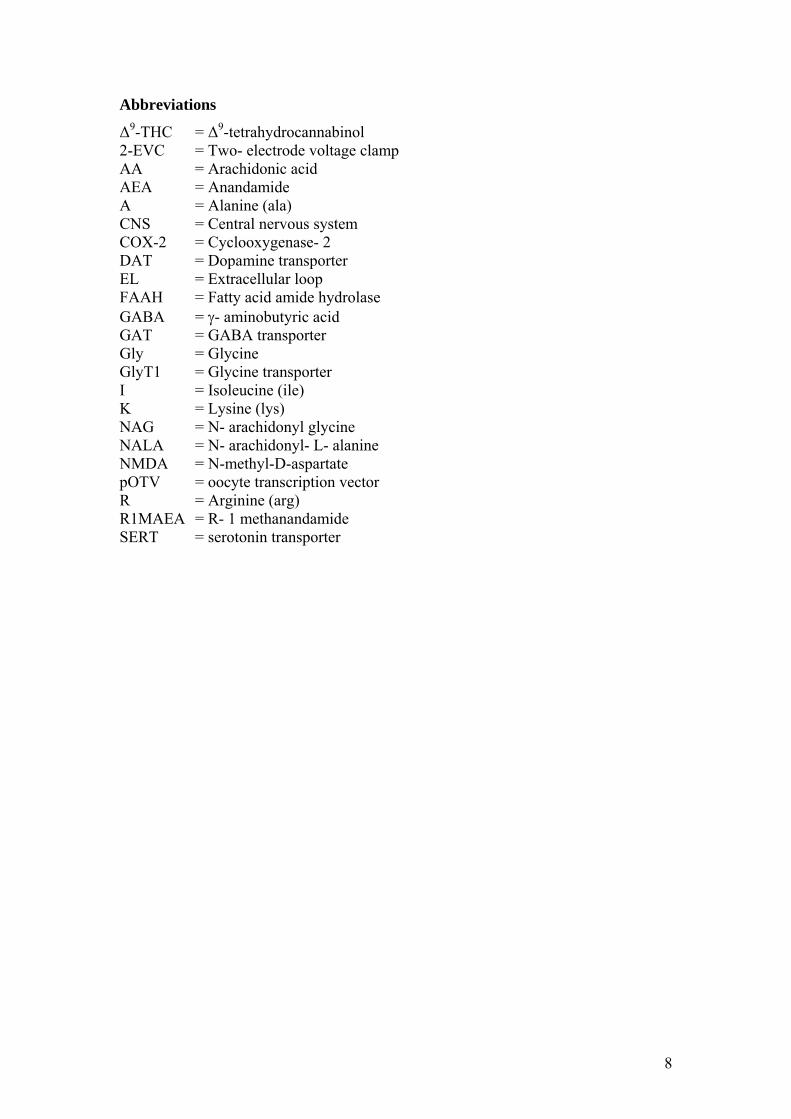
Glycine transporters belong to the family of Na+/ Cl- coupled co-transporters that contain 12 conserved putative transmembrane segments. Other members of this family are the dopamine transporter (DAT), serotonin transporter (SERT), and the GABA transporter GAT. Two different glycine transporters, transcribed from different genes, have been found(5-7), GlyT1 and GlyT2, which both have various isoforms. The different transporter isoforms arise either by the transcription from different promoters or by different splicing patterns. So far, the GlyT1 transporter has been found in five different versions, (a, b, c, e and f) differing in their N- and C- terminus(5, 6, 8, 9). The GlyT2 has been described in two different versions (a and b) which both have an additional ~200 amino acid long N terminus compared to the GlyT1 and all other Na+/ Cl- coupled co-transporters, as seen in figure 1. They also have an extended external loop (EL) 2(10). The GlyT2b isoform differ from the 2a by five amino acids in the N-terminal and it does not transport glycine(11). Between the overlapping sequences of GlyT1 and GlyT2 there is a 48% amino acid sequence identity.
6
N-term.
N-term.
Figure 1. Topology of GlyT1b and GlyT2a. All Na+/ Cl- coupled co-transporters have 12 transmembrane segments. The differences between the GlyT1b and GlyT2a transporters are highlighted with red. Unique for the GlyT2a transporter is the long extension at the N-terminus. The cellular and tissue locations of GlyT1 and GlyT2 have been found to be different. GlyT1 is often found on glial cells (12) while GlyT2 is found at the pre-synaptic terminals of glycinergic neurons (13-15) (see figure 2). GlyT2 expression has so far only been found in the inhibitory system(12, 13) while GlyT1 expression also has been found in areas completely lacking GlyRs where its function is to control the concentration of glycine at the NMDA receptor(6, 12). The GlyT2 transporter is more energetically coupled than the GlyT1 transporter. Each glycine molecule transported is co-transported with three Na+ and one Cl-, while GlyT1 only transports two Na+ and one Cl- per glycine(16, 17). Small changes in the Na+ gradient can cause GlyT1 to function in reverse, whereas GlyT2 is unlikely to operate in reverse (18, 19).
The GlyT2 transporter is more energetically coupled than the GlyT1 transporter. Each glycine molecule transported is co-transported with three Na
+ and one Cl-, while GlyT1 only transports two Na+ and one Cl- per glycine(16, 17). Small changes in the Na+ gradient can cause GlyT1 to function in reverse, whereas GlyT2 is unlikely to operate in reverse (18, 19).
It has been shown that the two transporters differ in their sensitivity to sarcosine (a N-methyl-glycine)(3) which is a substrate of GlyT1b(4) but does not interact with GlyT2a. GlyT2a, on the other hand, has been shown to be inhibited by ethanol and the anti-depressant amoxapine, an inhibition which is not seen to the same extent with GlyT1b(20, 21).
It has been shown that the two transporters differ in their sensitivity to sarcosine (a N-methyl-glycine)(3) which is a substrate of GlyT1b(4) but does not interact with GlyT2a. GlyT2a, on the other hand, has been shown to be inhibited by ethanol and the anti-depressant amoxapine, an inhibition which is not seen to the same extent with GlyT1b(20, 21). Furthermore, glycine uptake by GlyT1 is inhibited by acidic pH (22) and the two different transporters are differently affected by arachidonic acid (23) and their derivatives (Wiles and Vandenberg, unpublished results).
Furthermore, glycine uptake by GlyT1 is inhibited by acidic pH (22) and the two different transporters are differently affected by arachidonic acid (23) and their derivatives (Wiles and Vandenberg, unpublished results).
7
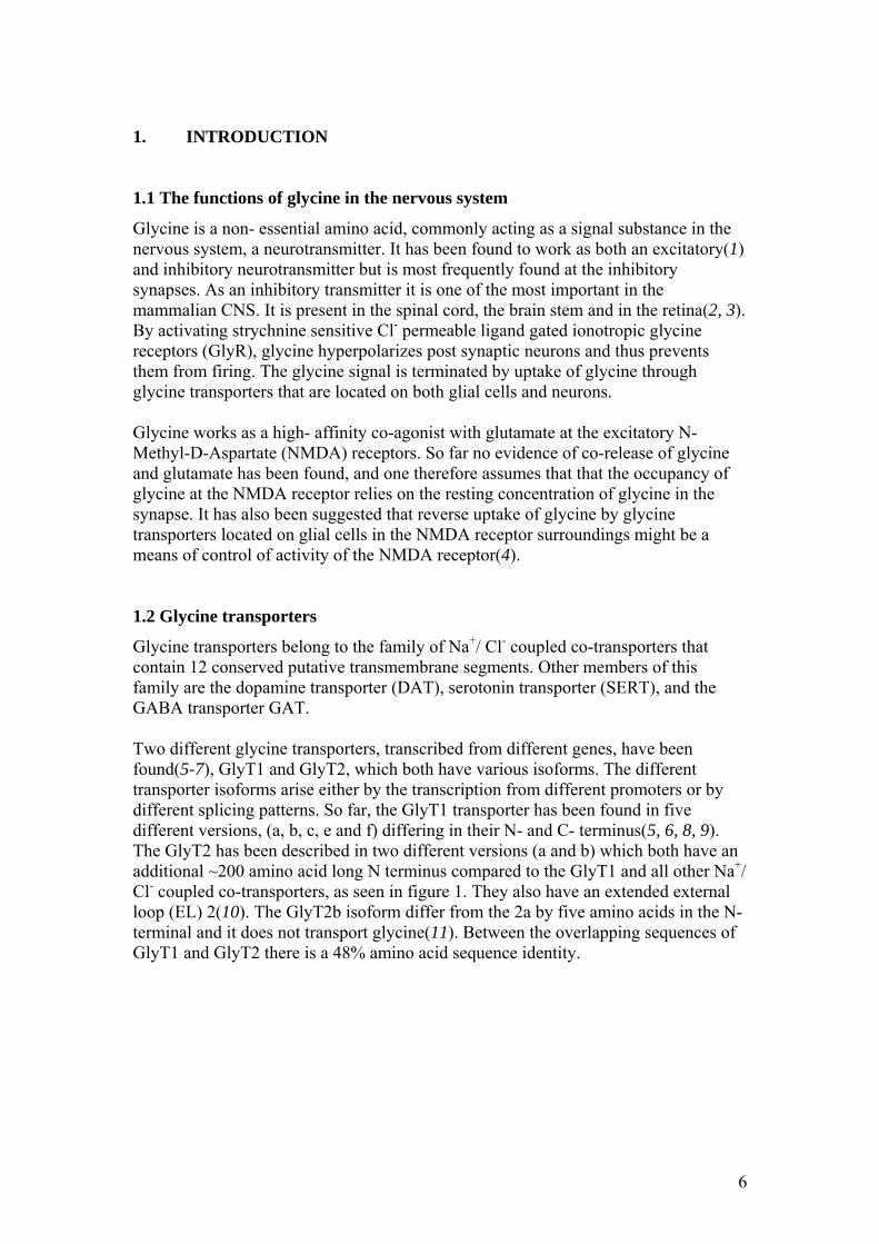
Figure 2. Location and function of glycine transporters. Both GlyT1b and GlyT2a assist in the removal of extracellular glycine at the glycinergic synapses and thus restrict the amount that might be spilled over to neighbouring synapses. Since GlyT2 is highly energetically coupled, a very high concentration of glycine can be maintained and this provides glycine for transport into synaptic vesicles. It is suggested that the GlyT1 receptor is less energetically coupled so to allow some spill over to neighbouring excitatory synapses but enough to keep the glycine concentration under the saturation level for the NMDA receptor. Because of GlyT1’s capability to function as a reverse transporter it has further been suggested that they might act as a transport system between glial cells and neurons. Figure adapted from Ref (16). 1.3 Arachidonic acid and its derivatives
A number of lipids serve as endogenous signalling molecules in the mammalian CNS. Examples include arachidonic acid, anandamide, N- arachidonyl glycine and N- arachidonyl- L- alanine (see figure 3). 1.3.1 Arachidonic acid (AA) One very common example of a lipid-signalling molecule is AA. AA is released through a phospholipase A2 stimulated process(24) and has been found to be active in several neurotransmitter systems for instance the glutamatergic, serotonergic and dopaminergic systems (24-26) but also serves as a precursor to many other signalling molecules. It is thought that AA is effective either through a direct binding with an enzyme or through a second messenger system where it is the metabolites of AA that cause the effect (26, 27). AA has for a long time been known to potentiate the action of glutamate at the NMDA receptor(28)and to inhibit the uptake of glutamate(29). Both these findings indicate an increase in the activity of the NMDA receptor at the excitatory synapses. It has been found that the uptake of glycine, the required co-agonist of the NMDA receptor, is reversibly inhibited through a direct binding of arachidonic acid to the GlyT1 transporter(23). Under the same conditions, AA has no effect on GlyT2( (23).
8
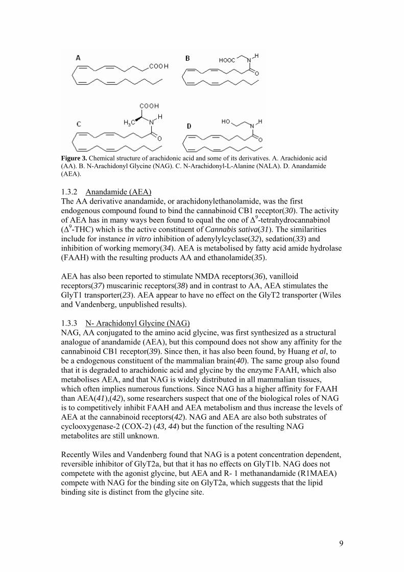
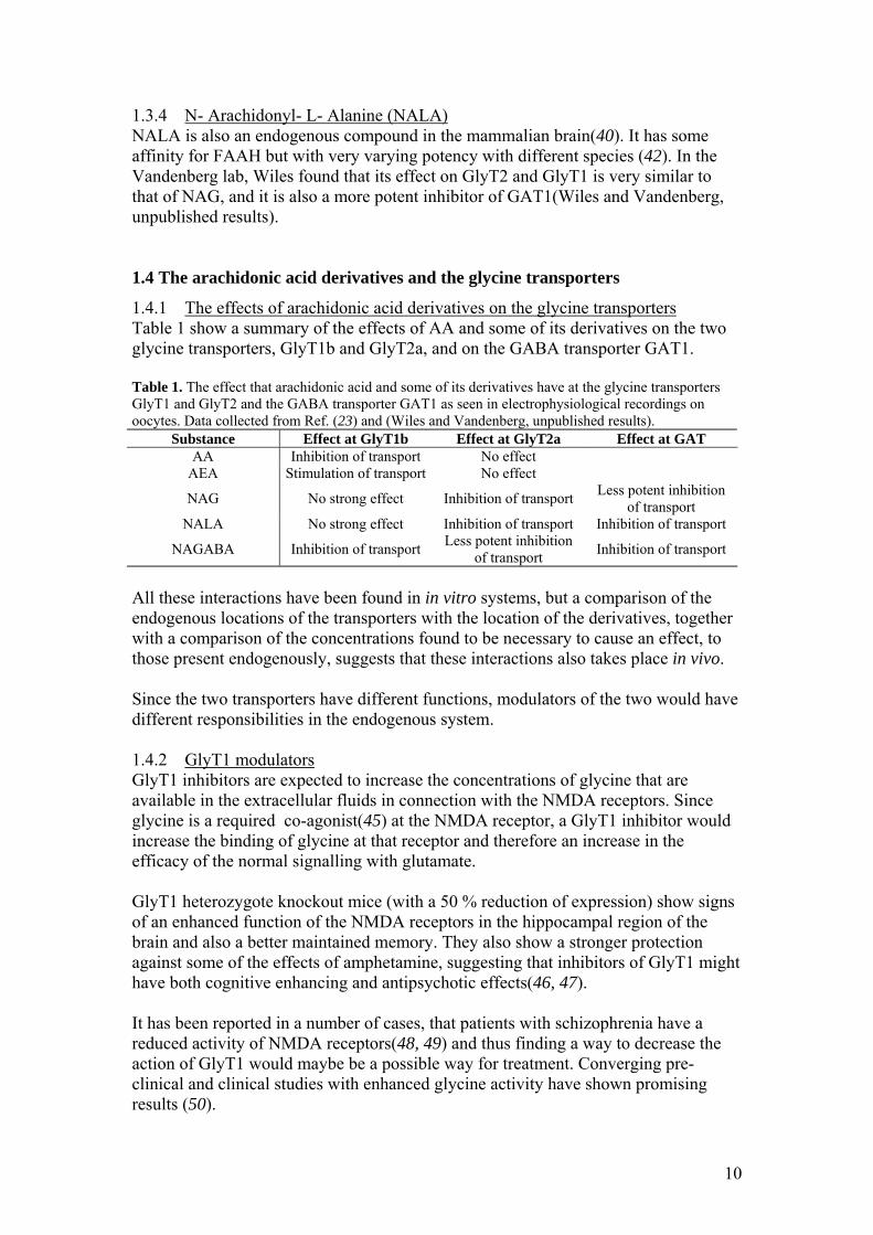
Figure 3. Chemical structure of arachidonic acid and some of its derivatives. A. Arachidonic acid (AA). B. N-Arachidonyl Glycine (NAG). C. N-Arachidonyl-L-Alanine (NALA). D. Anandamide (AEA). 1.3.2 Anandamide (AEA) The AA derivative anandamide, or arachidonylethanolamide, was the first endogenous compound found to bind the cannabinoid CB1 receptor(30). The activity of AEA has in many ways been found to equal the one of ∆9-tetrahydrocannabinol (∆9-THC) which is the active constituent of Cannabis sativa(31). The similarities include for instance in vitro inhibition of adenylylcyclase(32), sedation(33) and inhibition of working memory(34). AEA is metabolised by fatty acid amide hydrolase (FAAH) with the resulting products AA and ethanolamide(35). AEA has also been reported to stimulate NMDA receptors(36), vanilloid receptors(37) muscarinic receptors(38) and in contrast to AA, AEA stimulates the GlyT1 transporter(23). AEA appear to have no effect on the GlyT2 transporter (Wiles and Vandenberg, unpublished results). 1.3.3 N- Arachidonyl Glycine (NAG) NAG, AA conjugated to the amino acid glycine, was first synthesized as a structural analogue of anandamide (AEA), but this compound does not show any affinity for the cannabinoid CB1 receptor(39). Since then, it has also been found, by Huang et al, to be a endogenous constituent of the mammalian brain(40). The same group also found that it is degraded to arachidonic acid and glycine by the enzyme FAAH, which also metabolises AEA, and that NAG is widely distributed in all mammalian tissues, which often implies numerous functions. Since NAG has a higher affinity for FAAH than AEA(41),(42), some researchers suspect that one of the biological roles of NAG is to competitively inhibit FAAH and AEA metabolism and thus increase the levels of AEA at the cannabinoid receptors(42). NAG and AEA are also both substrates of cyclooxygenase-2 (COX-2) (43, 44) but the function of the resulting NAG metabolites are still unknown. Recently Wiles and Vandenberg found that NAG is a potent concentration dependent, reversible inhibitor of GlyT2a, but that it has no effects on GlyT1b. NAG does not competete with the agonist glycine, but AEA and R- 1 methanandamide (R1MAEA) compete with NAG for the binding site on GlyT2a, which suggests that the lipid binding site is distinct from the glycine site.
9
1.3.4 N- Arachidonyl- L- Alanine (NALA) NALA is also an endogenous compound in the mammalian brain(40). It has some affinity for FAAH but with very varying potency with different species (42). In the Vandenberg lab, Wiles found that its effect on GlyT2 and GlyT1 is very similar to that of NAG, and it is also a more potent inhibitor of GAT1(Wiles and Vandenberg, unpublished results). 1.4 The arachidonic acid derivatives and the glycine transporters
1.4.1 The effects of arachidonic acid derivatives on the glycine transporters Table 1 show a summary of the effects of AA and some of its derivatives on the two glycine transporters, GlyT1b and GlyT2a, and on the GABA transporter GAT1. Table 1. The effect that arachidonic acid and some of its derivatives have at the glycine transporters GlyT1 and GlyT2 and the GABA transporter GAT1 as seen in electrophysiological recordings on oocytes. Data collected from Ref. (23) and (Wiles and Vandenberg, unpublished results).
Substance Effect at GlyT1b Effect at GlyT2a Effect at GAT AA Inhibition of transport No effect
AEA Stimulation of transport No effect
NAG No strong effect Inhibition of transport Less potent inhibition of transport
NALA No strong effect Inhibition of transport Inhibition of transport
NAGABA Inhibition of transport Less potent inhibition of transport Inhibition of transport
All these interactions have been found in in vitro systems, but a comparison of the endogenous locations of the transporters with the location of the derivatives, together with a comparison of the concentrations found to be necessary to cause an effect, to those present endogenously, suggests that these interactions also takes place in vivo. Since the two transporters have different functions, modulators of the two would have different responsibilities in the endogenous system. 1.4.2 GlyT1 modulators GlyT1 inhibitors are expected to increase the concentrations of glycine that are available in the extracellular fluids in connection with the NMDA receptors. Since glycine is a required co-agonist(45) at the NMDA receptor, a GlyT1 inhibitor would increase the binding of glycine at that receptor and therefore an increase in the efficacy of the normal signalling with glutamate. GlyT1 heterozygote knockout mice (with a 50 % reduction of expression) show signs of an enhanced function of the NMDA receptors in the hippocampal region of the brain and also a better maintained memory. They also show a stronger protection against some of the effects of amphetamine, suggesting that inhibitors of GlyT1 might have both cognitive enhancing and antipsychotic effects(46, 47). It has been reported in a number of cases, that patients with schizophrenia have a reduced activity of NMDA receptors(48, 49) and thus finding a way to decrease the action of GlyT1 would maybe be a possible way for treatment. Converging pre-clinical and clinical studies with enhanced glycine activity have shown promising results (50).
10
1.4.3 GlyT2 modulators GlyT2 inhibitors are expected to increase the concentrations of glycine that are available in the extracellular fluids in connection with the GlyRs. A more frequent binding to the GlyR will result in a more frequent hyperpolarized post-synaptic membrane and thus the post-synaptic neurone will be less prone to be activated. Inhibitory signals control the transmission of pain in the spinal cord and it is therefore thought that GlyT2 inhibitors might be involved in analgesia. This was first supported by the fact that inhibiting the GlyR with strychnine caused an increase in the response to noxious stimuli (51, 52) but also that the GlyT2 inhibitor, NAG, has been shown to cause analgesia in mice exposed to a hot plate test which is a way to study the reactions of painful stimuli, (35) and a reduction of the pain behaviour during the tonic pain phase in mice injected with formalin(40). The conclusion of this was that NAG has anti- inflammatory and anti- nociceptive (reducing sensitivity to painful stimuli) effects if administered peripherally(40). The mechanisms of how NAG produce analgesia is still unknown but from the statements above one can speculate that it is by the inhibition of GlyT2 that NAG causes its effect.
1.5 Aims of this study
There is very little known about binding sites for arachidonic acid and its various derivatives on membrane proteins. Characterization of the binding site on glycine transporters would provide a lot of additional information about the transporter and may provide information about the mechanism of inhibition. If one would know the binding site of a modulator of an enzyme that would also give insight into how other enzymes interact with this modulator. For instance knowing the binding site of AA derivatives on the GlyT transporters would be a very good indication of whether they also interact with the other transporters in the same family. Mutations of important areas are also an extra confirmation that the effect seen, is caused by the enzyme and not by something that has a similar effect. In the case of the glycine transporters expressed in oocytes, this might be other channels or second messenger systems activated. The aim of this study was to find the site of interaction between the glycine transporters and the arachidonic acid derivatives. This was done by changing the DNA sequences of the GlyT1 and the GlyT2 transporters and expressing the mutated transporters in X. laevis oocytes. The activity of the transporters was then tested for activity with and without the arachidonic acid derivatives using the two- electrode voltage clamp technique to see whether changes in the transport of glycine could be detected.
11
2. METHODS
2.1 Materials
All chemicals were obtained from Sigma (Sydney, Australia) unless otherwise is stated. N-Arachidonyl Glycine (NAG), N-Arachidonyl-γ-Aminobutyric Acid (NAGABA), N-Arachidonyl-L-Alanine (NALA) and Anandamide (AEA) were purchased from Sapphire Biosciences (Crows Nest, N.S.W., Australia). They were stored in ethanol at -20°C and stocks were made up prior to use on the day. Arachidonic Acid was sonicated less than an hour prior to use. The GlyT2a-OTV plasmid was a gift from John Morrow (Organon Laboratories Limited, Newhouse, Lanarkshire, U.K.) and the GlyT1b-OTV plasmid, a gift from Dr Marc Caron (Howard Hughes Medical Institute Research Laboratories, Department of Cell Biology and Medicine, Duke University Medical Center, Durham, NC, U.S.A.).X. laevis frogs were purchased from Xenopus Express (Haute-Loire, France). 2.2 Construction of vectors with wild type and altered transporters
The GlyT1b-OTV and the GlyT2a-OTV were produced by subcloning cDNAs encoding human GlyT1b and human GlyT2a into the pOTV plasmid (Oocyte Transcription Vector). The coding sequences for GlyT1b and GlyT2a were subcloned between the Xenopus β-globin 5’ and 3’ untranslated regions (53). To create the mutated transporters, site- directed mutagenesis was performed using the QuikChange Site-directed Mutagenesis Kit (Stratagene). To create the GlyT1b and GlyT2a chimeras, 5’- end oligonucleotide-directed base insertion was performed, using the ExSite PCR-Based Site-directed Mutagenesis Kit (Stratagene). The modifications were confirmed by DNA sequencing. 2.3 Xenopus laevis oocytes as an expression system
The oocyte from the South African frog X. laevis is a large cell with a diameter of about 1mm which is known to express exogenous RNA species. Because of its size, it is well suited for experiments and has therefore frequently been used as an expression system for different membrane bound transporter proteins. mRNA, prepared in vitro from cDNA, is injected into the cytoplasm of the cell. If the mRNA is correctly capped, it will bind to the ribosome and be translated to a protein. After necessary modifications the protein is transported to and incorporated into the membrane as a functional protein. The activity of the protein can then be measured by techniques such as radiolabelled uptake or, if the activity cause a net movement of current through the membrane, by electrophysiological recordings using the two- electrode voltage clamp technique. 2.4 Expression of Transporters in X. laevis Oocytes
The wild type and altered transporter cDNA plasmids were linearised with SpeI (Fermentas). Their mRNAs were transcribed with T7 RNA polymerase and capped
12
with 5’-7-methyl guanosine using the mMessage mMachine kit (Ambion Inc., TX, U.S.A.) Lobes of oocytes were surgically removed from mature female X. laevis frogs anaesthetised with 0.17% 3-aminobenzoic acid ethyl ester and put into OR2 medium (82.5 mM NaCl, 2 mM KCl, 1 mM MgCl2, 5 mM HEPES, pH 7.5 adjusted with NaOH). After washing, the lobes were dissected into smaller pieces and then incubated with 2mg/ml collagenase A (Boehringer, Mannheim) in OR2 solution at room temperature for approximately 90minutes with shaking. They were then washed thoroughly with OR2, followed by several washes with ND96 (96 mM NaCl, 2 mM KCl, 1 mM MgCl2, 1.8 mM CaCl2, 5 mM HEPES (Hemi-Na) pH 7.55) supplemented with 2.5 mM sodium-pyruvate and 0.5mM theophylline. Stage V oocytes were isolated and within one day, 50 nl of cRNA was microinjected into the oocytes. The injected oocytes were then incubated at 17°C for 2-4 days in ND96 supplemented with 2.5 mM sodium-pyruvate, 0.5 mM theophylline and 50 µg/ml gentamicin with shaking. 2.5 Two- Electrode Voltage Clamp
Two- electrode voltage clamp is a technique used to record movement of ions across a cell membrane. Two sharp glass electrodes are inserted into the X. laevis oocyte. One records the membrane voltage while the second applies a current to maintain the membrane at a constant clamped voltage. In this study the oocytes were clamped at –60mV which is a very common endogenous membrane potential. If the transporters are activated by a substrate of the transporter a net movement of charges across the membrane will occur and more/ less current needs to be applied by the electrode to keep the potential stable. The current applied is recorded which gives a measure of transporter activity. 2.6 Electrophysiological recordings
Two to four days after microinjection of mRNA, expression was sufficient to detect a current through active transporters, with the 2-EVC technique. The oocytes were placed in a bath with ND96 buffer and two glass electrodes filled with 3 M KCl were inserted. One of the electrodes records the membrane voltage while the other passes a current in to the cell to maintain the membrane at a constant command voltage. A holding potential of -60 mV was used for all of our experiments. For the recordings a MacLab/200 chart recorder (ADInstruments, Sydney, Australia) was interfaced with a Geneclamp 500 amplifier (Axon Instruments). Glycine and the AA derivatives were applied in different combinations and at different concentrations to the bath and the transport activity was detected.
13
3. RESULTS
3.1 Recordings with wild type transporters
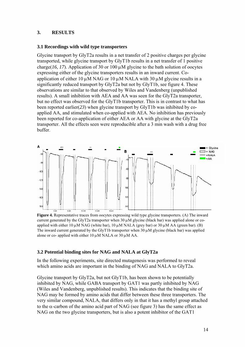
Glycine transport by GlyT2a results in a net transfer of 2 positive charges per glycine transported, while glycine transport by GlyT1b results in a net transfer of 1 positive charge(16, 17). Application of 30 or 100 µM glycine to the bath solution of oocytes expressing either of the glycine transporters results in an inward current. Co-application of either 10 µM NAG or 10 µM NALA with 30 µM glycine results in a significantly reduced transport by GlyT2a but not by GlyT1b, see figure 4. These observations are similar to that observed by Wiles and Vandenberg (unpublished results). A small inhibition with AEA and AA was seen for the GlyT2a transporter, but no effect was observed for the GlyT1b transporter. This is in contrast to what has been reported earlier(23) when glycine transport by GlyT1b was inhibited by co-applied AA, and stimulated when co-applied with AEA. No inhibition has previously been reported for co-application of either AEA or AA with glycine at the GlyT2a transporter. All the effects seen were reproducible after a 3 min wash with a drug free buffer.
Figure 4. Representative traces from oocytes expressing wild type glycine transporters. (A) The inward current generated by the GlyT2a transporter when 30 µM glycine (black bar) was applied alone or co- applied with either 10 µM NAG (white bar), 10 µM NALA (grey bar) or 30 µM AA (green bar). (B) The inward current generated by the GlyT1b transporter when 30 µM glycine (black bar) was applied alone or co- applied with either 10 µM NALA or 30 µM AA. 3.2 Potential binding sites for NAG and NALA at GlyT2a
In the following experiments, site directed mutagenesis was performed to reveal which amino acids are important in the binding of NAG and NALA to GlyT2a. Glycine transport by GlyT2a, but not GlyT1b, has been shown to be potentially inhibited by NAG, while GABA transport by GAT1 was partly inhibited by NAG (Wiles and Vandenberg, unpublished results). This indicates that the binding site of NAG may be formed by amino acids that differ between these three transporters. The very similar compound, NALA, that differs only in that it has a methyl group attached to the α-carbon of the amino acid part of NAG (see figure 3) has the same effect as NAG on the two glycine transporters, but is also a potent inhibitor of the GAT1
14
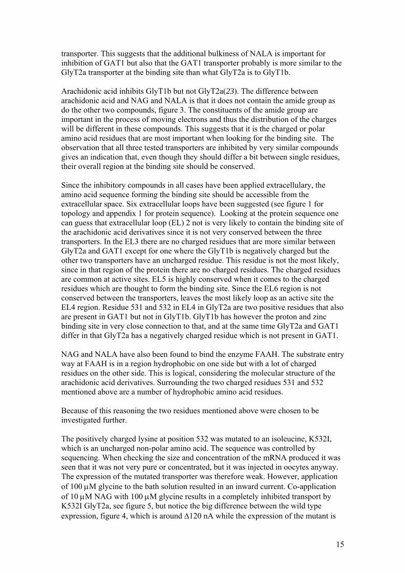
transporter. This suggests that the additional bulkiness of NALA is important for inhibition of GAT1 but also that the GAT1 transporter probably is more similar to the GlyT2a transporter at the binding site than what GlyT2a is to GlyT1b. Arachidonic acid inhibits GlyT1b but not GlyT2a(23). The difference between arachidonic acid and NAG and NALA is that it does not contain the amide group as do the other two compounds, figure 3. The constituents of the amide group are important in the process of moving electrons and thus the distribution of the charges will be different in these compounds. This suggests that it is the charged or polar amino acid residues that are most important when looking for the binding site. The observation that all three tested transporters are inhibited by very similar compounds gives an indication that, even though they should differ a bit between single residues, their overall region at the binding site should be conserved. Since the inhibitory compounds in all cases have been applied extracellulary, the amino acid sequence forming the binding site should be accessible from the extracellular space. Six extracellular loops have been suggested (see figure 1 for topology and appendix 1 for protein sequence). Looking at the protein sequence one can guess that extracellular loop (EL) 2 not is very likely to contain the binding site of the arachidonic acid derivatives since it is not very conserved between the three transporters. In the EL3 there are no charged residues that are more similar between GlyT2a and GAT1 except for one where the GlyT1b is negatively charged but the other two transporters have an uncharged residue. This residue is not the most likely, since in that region of the protein there are no charged residues. The charged residues are common at active sites. EL5 is highly conserved when it comes to the charged residues which are thought to form the binding site. Since the EL6 region is not conserved between the transporters, leaves the most likely loop as an active site the EL4 region. Residue 531 and 532 in EL4 in GlyT2a are two positive residues that also are present in GAT1 but not in GlyT1b. GlyT1b has however the proton and zinc binding site in very close connection to that, and at the same time GlyT2a and GAT1 differ in that GlyT2a has a negatively charged residue which is not present in GAT1. NAG and NALA have also been found to bind the enzyme FAAH. The substrate entry way at FAAH is in a region hydrophobic on one side but with a lot of charged residues on the other side. This is logical, considering the molecular structure of the arachidonic acid derivatives. Surrounding the two charged residues 531 and 532 mentioned above are a number of hydrophobic amino acid residues. Because of this reasoning the two residues mentioned above were chosen to be investigated further. The positively charged lysine at position 532 was mutated to an isoleucine, K532I, which is an uncharged non-polar amino acid. The sequence was controlled by sequencing. When checking the size and concentration of the mRNA produced it was seen that it was not very pure or concentrated, but it was injected in oocytes anyway. The expression of the mutated transporter was therefore weak. However, application of 100 µM glycine to the bath solution resulted in an inward current. Co-application of 10 µM NAG with 100 µM glycine results in a completely inhibited transport by K532I GlyT2a, see figure 5, but notice the big difference between the wild type expression, figure 4, which is around ∆120 nA while the expression of the mutant is
15
only around ∆ 6 nA. That the inhibiting effect remains in the mutant indicate that this residue is not involved in the binding of NAG.
Figure 5. Representative trace from an oocyte expressing K532I mutant GlyT2a. The inward current generated by the mutated transporter when 100µM glycine (black bar) is applied alone or co- applied with 10 µM NAG (white bar).
I also attempted to mutate the arginine residue 531 to an alanine, R531A, which also is an uncharged non- polar amino acid, but even though the sequence received from sequencing had the right sequence the mRNA did not produce a functional transporter. When the mRNA was run on a gel it in some attempts showed a band of the correct size, but in all cases other bands were also present. No further attempt to construct oocytes with mutant 531 residue in EL4 was performed.
3.3 Identification of the derivatives of arachidonic acids binding residues in
GlyT2a and GlyT1b
Since the binding residue could not be found by mutating single residues, it was decided to make a bigger change; to make a chimera of the GlyT2a and GlyT1b transporters. A chimera likely to give a lot of information was a chimera where the EL4 of GlyT1b was replaced with the EL4 of GlyT2a in a GlyT1b mutant, GlyTch12, and also the other way round, the EL4 of GlyT1b in a GlyT2a mutant, GlyTch21. This is based on the arguments presented above when selecting single residues. Two techniques were used to try and produce the chimera. The first one was to introduce restriction sites, cut out the unwanted loop and then introduce the new loop and ligate. No results were obtained with this method and there for it have not been described more. The other technique was to order phosporylated oligos containing the desired loop and run a PCR resulting in linearised fragments with the correct sequence. The final step is then to ligate the linearised fragments to a plasmid. However, I was not successful in generating the two chimeras with neither of the techniques. Although a number of attempts were made to make the chimeras, in each case the results from sequencing was only wild. Two possible causes for the lack of chimeras may be that the oligonucleotides may not have been adequately phosphorylated so that the ligation step was unsuccessful or the plasmid DNA used to make the chimeras from was not sufficiently pure. These two possibilities are currently being investigated.
16

4.
17
DISCUSSION The results from this study shows that NAG probably does not bind to the 532 lysine residue in the GlyT2a transporter. Further attempts to make chimeras are required to address the question as to whether EL4 plays a role in formation of the AA derivatives binding site on GlyT2a and GlyT1b transporters. 4.1 Conflicting results when recording on wild type transporters
To get my own data for a later comparison with the data from the mutated transporters, recordings on the wild type transporters was made. This data did not completely match previous data on wild type transporters. In contrast to previous reports, none of the tested compounds showed stimulatory or inhibitory effect on GLYT1b. Furthermore, GlyT2a was partly inhibited by AA and AEA whereas in previous studies no effects were observed with these compounds. However, I do not put much trust in these results because not many recordings were performed, nor were the any of the oocytes expressing GlyT1b transporters healthy. They were slowly dying, which can be seen from the slowly descending base line (see figure 4). The arachidonic acid is also a very difficult substance to do experiments on since it very easily forms micelles, even at rather low concentrations, and thus the interaction with the transporter could be changed. The stock of AA that was used was also a few years old and therefore it is possible that parts of it were broken down to compounds that interact with the GlyT2a transporter. This may be possible even for AEA. That a stronger effect was seen on the GlyT2a cells is not strange since what has been reported earlier is that a lower concentration of those inhibitors produces a larger inhibition. If the problems when using AA persists one possibility is to instead of applying AA to the bath, to apply melittin which is a substance that releases AA from the membranes in oocytes and therefore one can escape the problems of formation of micelles and the effects of the degradation products of AA. 4.2 Residue 532 probably does not form part of NAG’s binding site on the
GlyT2a transporter
After mutating the potential binding candidate of NAG, residue 532, the inhibition caused by NAG was of equal or higher strength. Since the residue was mutated to an uncharged non-polar amino acid which makes the mutant less reactive but also makes it more similar to GlyT1b, I expected a less potent inhibition to support the hypothesis that this is the binding site. From the result, that the binding was not weaker, I conclude that the residue is not involved in the endogenous binding of NAG to the glycine transporters. Not enough recordings were made to ensure whether the binding was of equal or higher strength. In some cases the data received indicated on a total inhibition of the mutated transporter with 10 µM NAG. This has not previously been found but could be due to the poor expression of the transporter. If, what we saw was an actual increase in the potency of the inhibition, then that gives no information to whether or
18
not the residue is involved in the binding of the wild type transporter. It, however, suggests that a modification of that residue changes the binding properties and thus it is likely that the important residues for the binding of AA to the wild type transporter are in close connection to that residue. It also suggests that if there exists a protein with a sequence similar to that of the mutated EL4, this protein is very likely to be more potently inhibited by NAG than GlyT2a. However, performing a blast search looking for a sequence quite similar to EL4 of Glyt2a but with an uncharged residue at the position corresponding to GlyT2a’s 532 gave no positives. 4.3 Future experiments
Since I did not succeed in producing the chimeras, the obvious continuation of this study is to try to find other ways to produce the chimeras. At the moment we are investigating if the reason for why we did not get any chimeras can be due to that the oligonucleotides might not have been adequately phosphorylated so that the ligation step was unsuccessful or that the plasmid DNA used to make the chimeras from was not sufficiently pure. If one successfully manages to produce a chimera, it can be used to test other things than the binding of the AA derivatives. Not much is know about which residues which are responsible for what in the glycine transporters. A chimera between the two can be used to test if the differences that exist between them are located to the exchanged region. One example is the binding of sarcosine which is a substrate of GlyT1b but not GlyT2a, or the inhibition of GlyT2a with ethanol or amoxapine which have not been seen at the GlyT1b transporter. It is also possible that the coupling of how many ions transported per glycine can be determined with the chimera since the GlyT2a transports 3 Na+ while GlyT1b only transports 2 Na+. If it is found that the EL4 is an important region in the binding of the AA derivatives it is also interesting to test the transporters that have sequences most similar to GlyT2a’s EL4. The norepinephrine transporter NET and the taurine transporter have a sequence very similar to GlyT2a’s EL4. It is in general interesting to test the AA derivatives on more transporters when this would give more information to where one should be looking for the binding sequence in the future. Currently experiments using the taurine transporter are commencing in the lab. If the EL4 is found to be an important region then that would also increase the chance that the inhibition seen by NAG on the 532 mutant was a more potent inhibition compared to the one seen on the wild type transporters, as discussed in 4.2. If this is the case, then it is very likely that the neighbouring residue 531 is important for the binding of NAG and it would be interesting to continue the construction of that mutant. It would also be interesting to see if there are any other AA derivatives that are affected by the mutation. However, if it is found that it probably not is the EL4 that is involved in the binding of the AA derivatives then the other most likely amino acids affecting binding could be located in the end of the EL2 region where GlyT2a and GAT1 has a negatively charged amino acid which are not present in GlyT1b. At the end of that loop the sequences are also quite conserved.
19

In order to fully characterize the binding site of the AA derivatives to the GlyT transporters a fair amount of work remains to be done, but it is an important area of research and therefore a lot of energy should be put into it in the future.
20

5. ACKWNOLEDGEMENTS I want to thank Rob for letting me do my project in his lab and for being such a great supervisor; Ann for helping me when Rob was not there. I want to thank Karin for immediately making me feel like home in the lab and Pengchu for getting my work started. Suzanne for being there all the time and Amy for very many things especially the meatballs. And of course, Christina, for coping with me all the time and Erik, for giving the best possible support from back home. 6.
21
REFERENCES 1. Chatterton, J. E., Awobuluyi, M., Premkumar, L. S., Takahashi, H., Talantova,
M., Shin, Y., Cul, J., Tu, S., Sevarino, K. A., Nakanishi, N., Tong, G., Lipton, S. A., and Zhang, D. (2002) Nature 415, 793-798.
2. Gallagher, M. J., Burgess, L. H., and Brunden, K. R. (1999) Molecular Brain Research 70, 101-15.
3. Supplisson, S., and Roux, M. J. (2002) FEBS Letters 529, 93-101. 4. Supplisson, S., and Bergman, C. (1997) J Neurosci 17, 4580-90. 5. Guastella, J., Brecha, N., Weigmann, C., Lester, H. A., and Davidson, N.
(1992) Proc Natl Acad Sci U S A 89, 7189-93. 6. Smith, K. E., Borden, L. A., Hartig, P. R., Branchek, T., and Weinshank, R. L.
(1992) Neuron 8, 927-35. 7. Liu, Q. R., Nelson, H., Mandiyan, S., Lopez-Corcuera, B., and Nelson, N.
(1992) FEBS Lett 305, 110-4. 8. Hanley, J. G., Jones, E. M., and Moss, S. J. (2000) J Biol Chem 275, 840-6. 9. Kim, K. M., Kingsmore, S. F., Han, H., Yang-Feng, T. L., Godinot, N., Seldin,
M. F., Caron, M. G., and Giros, B. (1994) Molecular Pharmacology 45, 608-17.
10. Liu, Q. R., Lopez-Corcuera, B., Mandiyan, S., Nelson, H., and Nelson, N. (1993) Journal of Biological Chemistry 268, 22802-8.
11. Ponce, J., Poyatos, I., Aragon, C., Gimenez, C., and Zafra, F. (1998) Neuroscience Letters 242, 25-8.
12. Zafra, F., Aragon, C., Olivares, L., Danbolt, N. C., Gimenez, C., and Storm-Mathisen, J. (1995) J Neurosci 15, 3952-69.
13. Jursky, F., and Nelson, N. (1995) Journal of Neurochemistry 64, 1026-33. 14. Spike, R. C., Watt, C., Zafra, F., and Todd, A. J. (1997) Neuroscience 77, 543-
51. 15. Poyatos, I., Ponce, J., Aragon, C., Gimenez, C., and Zafra, F. (1997) Brain Res
Mol Brain Res 49, 63-70. 16. Roux, M. J., and Supplisson, S. (2000) Neuron 25, 373-83. 17. Aragon, M. C., Gimenez, C., and Mayor, F. (1987) FEBS Lett 212, 87-90. 18. Tsen, G., Williams, B., Allaire, P., Zhou, Y. D., Ikonomov, O., Kondova, I.,
and Jacob, M. H. (2000) Nat Neurosci 3, 126-32. 19. Lopez-Corcuera, B., Nunez, E., Martinez-Maza, R., Geerlings, A., and
Aragon, C. (2001) J Biol Chem 276, 43463-70. 20. Nunez, E., Lopez-Corcuera, B., Martinez-Maza, R., and Aragon, C. (2000) Br
J Pharmacol 129, 802-10. 21. Nunez, E., Lopez-Corcuera, B., Vazquez, J., Gimenez, C., and Aragon, C.
(2000) Br J Pharmacol 129, 200-6. 22. Aubrey, K. R., Mitrovic, A. D., and Vandenberg, R. J. (2000) Mol Pharmacol
58, 129-35. 23. Pearlman, R. J., Aubrey, K. R., and Vandenberg, R. J. (2003) Journal of
Neurochemisrty 84, 592-601. 24. Lazarewicz, J. W., Wroblewski, J. T., Palmer, M. E., and Costa, E. (1988)
Neuropharmacology 27, 765-9. 25. Dumuis, A., Pin, J. P., Oomagari, K., Sebben, M., and Bockaert, J. (1990)
Nature 347, 182-4. 26. Attwell, D., Miller, B., and Sarantis, M. (1993) The Neurosciences 5, 159-69.
22
27. Ordway, R. W., Singer, J. J., and Walsh, J. V., Jr. (1991) Trends Neurosci 14, 96-100.
28. Miller, B., Sarantis, M., Traynelis, S. F., and Attwell, D. (1992) Nature 355, 722-5.
29. Volterra, A., Trotti, D., and Racagni, G. (1994) Mol Pharmacol 46, 986-92. 30. Devane, W. A., Hanus, L., Breuer, A., Pertwee, R. G., Stevenson, L. A.,
Griffin, G., Gibson, D., Mandelbaum, A., Etinger, A., and Mechoulam, R. (1992) Science 258, 1946-9.
31. Gaoni, Y., and Mechoulam, R. (1971) Journal of the American Chemical Society 93, 217-24.
32. Vogel, Z., Barg, J., Levy, R., Saya, D., Heldman, E., and Mechoulam, R. (1993) Journal of Neurochemistry 61, 352-5.
33. Fride, E., and Mechoulam, R. (1993) European Journal of Pharmacology 231, 313-4.
34. Mallet, P. E., and Beninger, R. J. (1996) 7, 276-284. 35. Burstein, S. H., Rossetti, R. G., Yagen, B., and Zurier, R. B. (2000)
Prostaglandins & Other Lipid Mediators 61, 29-41. 36. Hampson, A. J., Bornheim, L. M., Scanziani, M., Yost, C. S., Gray, A. T.,
Hansen, B. M., Leonoudakis, D. J., and Bickler, P. E. (1998) J Neurochem 70, 671-6.
37. Vaughan, C. W., Connor, M., Bagley, E. E., and Christie, M. J. (2000) Mol Pharmacol 57, 288-95.
38. Christopoulos, A., and Wilson, K. (2001) Brain Res 915, 70-8. 39. Sheskin, T., Hanus, L., Slager, J., Vogel, Z., and Mechoulam, R. (1997)
Journal of Medicinal Chemisrty 40, 659-67. 40. Huang, S. M., Bisogno, T., Petros, T. J., Chang, S. Y., Zavitsanos, P. A.,
Zipkin, R. E., Sivakumar, R., Coop, A., Maeda, D. Y., De Petrocellis, L., Burstein, S., Di Marzo, V., and Walker, J. M. (2001) Journal of Biological Chemistry 2001, 42639-44.
41. Burstein, S. H., Huang, S. M., Petros, T. J., Rossetti, R. G., Walker, J. M., and Zurier, R. B. (2002) Biochemical Pharmacology 64, 1147-50.
42. Grazia Cascio, M., Minassi, A., Ligresti, A., Appendino, G., Burstein, S., and Di Marzo, V. (2004) Biochemical & Biophysical Research Communications 314, 192-6.
43. Prusakiewicz, J. J., Kingsley, P. J., Kozak, K. R., and Marnett, L. J. (2002) Biochemical & Biophysical Research Communications 296, 612-7.
44. Yu, M., Ives, D., and Ramesha, C. S. (1997) Journal of Biological Chemistry 272, 21181-6.
45. Johnson, J. W., and Ascher, P. (1987) Nature 325, 529-31. 46. Tsai, G., Ralph-Williams, R. J., Martina, M., Bergeron, R., Berger-Sweeney,
J., Dunham, K. S., Jiang, Z., Caine, S. B., and Coyle, J. T. (2004) Proc Natl Acad Sci U S A 101, 8485-90.
47. Martina, M., ME, B. T., Halman, S., Tsai, G., Tiberi, M., Coyle, J. T., and Bergeron, R. (2005) J Physiol 563, 777-93.
48. Mohn, A. R., Gainetdinov, R. R., Caron, M. G., and Koller, B. H. (1999) Cell 98, 427-36.
49. Javitt, D. C., Zylberman, I., Zukin, S. R., Heresco-Levy, U., and Lindenmayer, J. P. (1994) Am J Psychiatry 151, 1234-6.
50. Sur, C., and Kinney, G. G. (2004) Expert Opin Investig Drugs 13, 515-21. 51. Peng, Y. B., Lin, Q., and Willis, W. D. (1996) Brain Res 736, 189-201.
23
52. Yaksh, T. L. (1989) Pain 37, 111-23. 53. Arriza, J. L., Fairman, W. A., Wadiche, J. I., Murdoch, G. H., Kavanaugh, M.
P., and Amara, S. G. (1994) Journal of Neuroscience 14, 5559-69.
24
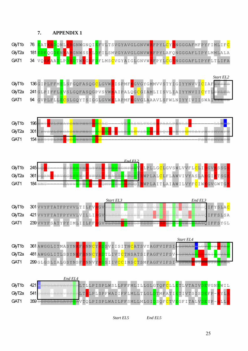
7. APPENDIX 1
GlyT1b 76 EATKRDQNLKRGNWGNQIEFVLTSVGYAVGLGNVWRFPYLCYRNGGGAFMFPYFIMLIFC GlyT2a 181 EDEQGDENKARGNWSSKLDFILSMVGYAVGLGNVWRFPYLAFQNGGGAFLIPYLMMLALA GAT1 34 VQKKAADLPDRDTWKGRFDFLMSCVGYAIGLGNVWRFPYLCGKNGGGAFLIPYFLTLIFA
Start EL2 GlyT1b 136 GIPLFFMELSFGQFASQGCLGVWRISPMFKGVGYGMMVVSTYIGIYYNVVICIAFGlyT2a 241 GLPIFFLEVSLGQFASQGPVSVWKAIPALQGCGIAMLIISVLIAIYYNVIICYTLGAT1 94 GVPLFLLECSLGQYTSIGGLGVWKLAPMFKGVGLAAAVLSFWLNIYYIVIISWAI
GlyT1b 196 GlyT2a 301 GAT1 154
End EL2 GlyT1b 245 VRLPLLGCLGVSWLVVFLCLIRGVKSSGK
GlyT2a 361 IRWPLALCLFLAWVIVYASLAKGIKTSGK
GAT1 184 IRWPLAITLAIAWILVYFCIWKGVGWTGK
Start EL3 End EL3 GlyT1b 301 VVYFTATFPYVVLTILFVRGV QIFYSLAC
GlyT2a 421 VVYFTATFPYVVLVILLIRGV QIFFSLSA
GAT1 239 VVYFSATYPYIMLIILFFRGV QIFFSYGL
Start EL4 GlyT1b 361 AWGGLITMASYNKFHNNCYRDSVIISITNCATSVYAGFVIFSIGlyT2a 481 AWGGLITLSSYNKFHNNCYRDTLIVTCTNSATSIFAGFVIFSVGAT1 299 GLGSLIALGSYNSFHNNVYRDSIIVCCINSCTSMFAGFVIFSI
End EL4 GlyT1b 421 LTLLPISPLWSLLFFFMLILLGLGTQFCLLETLVTAIVDEVGNEWIL
GlyT2a 541 LTRLPLSPFWAIIFFLMLLTLGLDTMFATIETIVTSISDEFP-KYLR
GAT1 359 VTQLPISPLWAILFFSMLLMLGIDSQFCTVEGFITALVDEYP-RLLR
Start EL5 End EL5
25
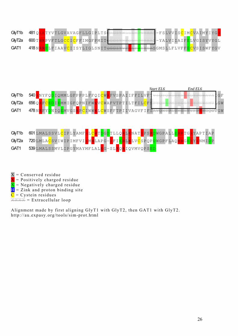
GlyT1b 481 QKKTYVTLGVAVAGFLLGIPLTS S-FSLVVISCIMCVAIMYIYGH
GlyT2a 600 THKPVFTLGCCICFFIMGFPMIT S-YALVIIAIFELVGISYVYGL
GAT1 418 NRRELFIAAVCIISYLIGLSNIT SGMSLLFLVFFECVSISWFYGV
Start EL6 End EL6 GlyT1b 540 RNYFQDIQMMLGFPPPLFFQICWRFVSPAIIFFILVFT IGF
GlyT2a 656 QRFCEDIEMMIGFQPNIFWKVCWAFVTPTILTFILCFS LGW
GAT1 478 NRFYDNIQEMVGSRPCIWWKLCWSFFTPIIVAGVFIFS VGW
GlyT1b 601 LMALSSVLCIPLYAMFRLCRTDGDTLLQRLKNATKPSRDWGPALLEHRTGRYAPTIAP GlyT2a 720 LMLACSVIWIPIMFVIKMHLAPG-RFIERLKLVCSPQPDWGPFLAQHRGERYKNMIDP GAT1 539 LMALSSMVLIPGYMAYMFLALKG-SLKQRIQVMVQPSED X = Conserved residue X = Posi t ively charged residue X = Negat ively charged residue H = Zink and proton binding s i te C = Cystein residues
= Extracel lu lar loop Alignment made by f i rs t a l igning GlyT1 with GlyT2, then GAT1 with GlyT2. ht tp: / /au.expasy.org/ tools /s im-prot .html
26


![Ions channels/transporters and chloroplast regulation · transporters/pumps and secondary transporters (according to the Transport Classification system [1]). Channels transport](https://img.pdfslide.net/doc/110x75/601623c1d6936b1074546c48/ions-channelstransporters-and-chloroplast-transporterspumps-and-secondary-transporters.jpg)

![Research Article Modulation of Arachidonic Acid Metabolism ...downloads.hindawi.com/archive/2014/683508.pdf · metabolism of arachidonic acid to biologically active EETs [ ]. e three](https://img.pdfslide.net/doc/110x75/606ff9bcbd5c0d69301096c4/research-article-modulation-of-arachidonic-acid-metabolism-metabolism-of-arachidonic.jpg)