Embed Size (px)
Citation preview
1
BIO 475 - ParasitologySpring 2009
Stephen M. ShusterNorthern Arizona University
http://www4.nau.edu/isopod
Lecture 13
Important Ordersa. Echinostomatiformes
b. Strigeiformesc. Opisthorchiformesd. Plagiorchiformes
2
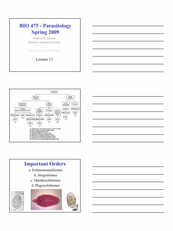
Trematode Phylogeny
Note the sequence of characters
Order Echinostomatiformes1. Redia with
appendages, often with collars.
2. Cercaria encyst"on" things
3. Adults with spines4. Examples:a. liver flukes,
Fasciola
Order EchinostomatiformesFasciola spp.
1. Characteristicsa. large testes and
vitellariab. oral cone and shoulders
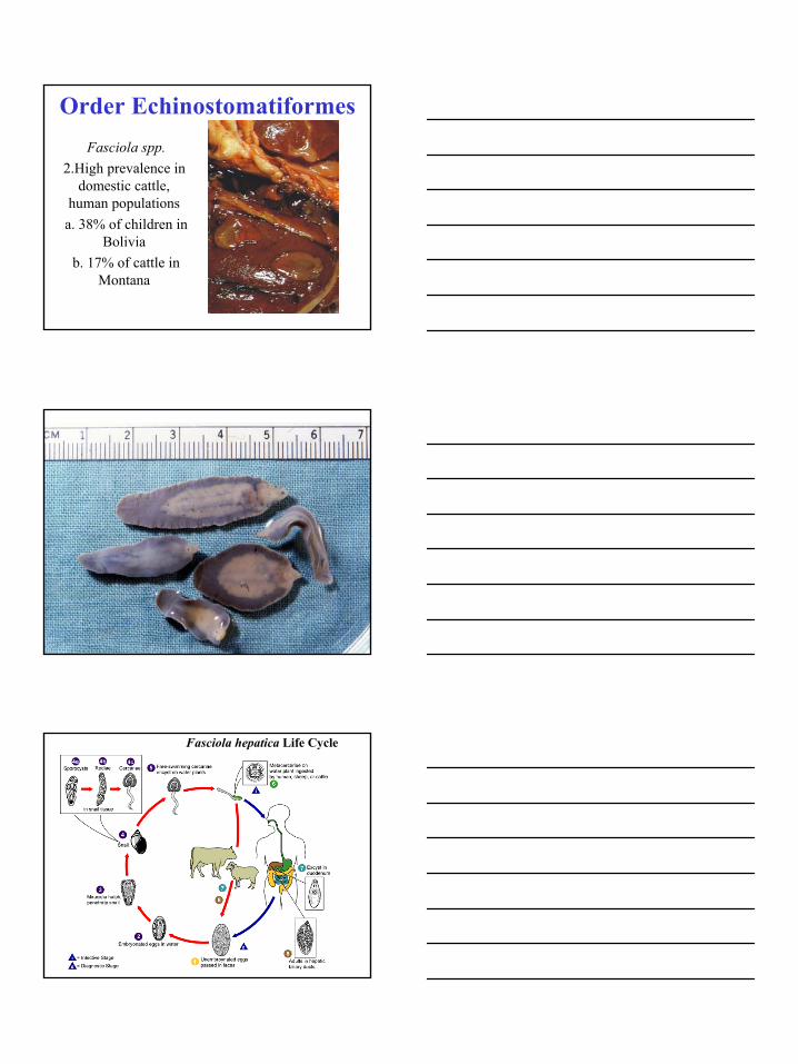
c. life cycle
3
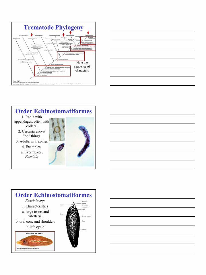
Order EchinostomatiformesFasciola spp.
2.High prevalence in domestic cattle,
human populationsa. 38% of children in
Boliviab. 17% of cattle in
Montana
Fasciola hepatica Life Cycle

4
Limnaea
Order EchinostomatiformesFasciolopsis buski
1. parasite of pigs and humans in orient
2. large, 7.5 cm), causes intestinal obstructions
Order Echinostomatiformesc. Fasciola gigantica
1. very large (7cm long)2. formerly wild ungulates,
now present in domestic species.

5
Order Strigiformes1. cercaria with 2 eyespots, may encyst or burrow into
definitive host.2. adults with spines, occasionally with expanded anterior end.
Order Strigiformes
Alaria spp.1. parasite of foxes; A. americana in NA
2. Slight modifications in life
cycle.a. Mesocercaria in tadpoles, later in
frogs.
6
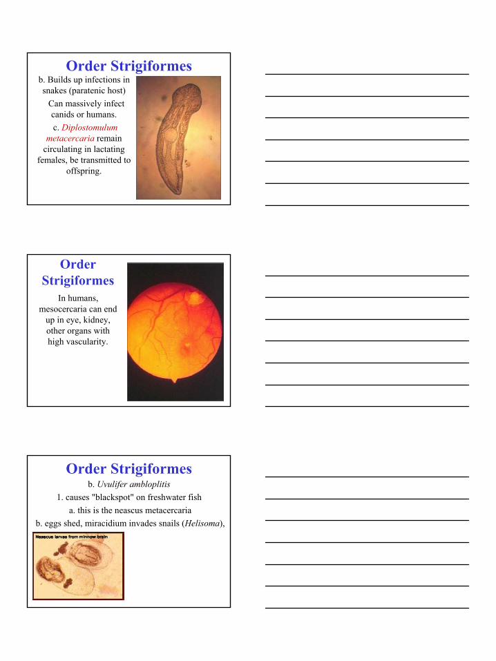
Order Strigiformesb. Builds up infections in snakes (paratenic host)
Can massively infect canids or humans.c. Diplostomulum
metacercaria remain circulating in lactating
females, be transmitted to offspring.
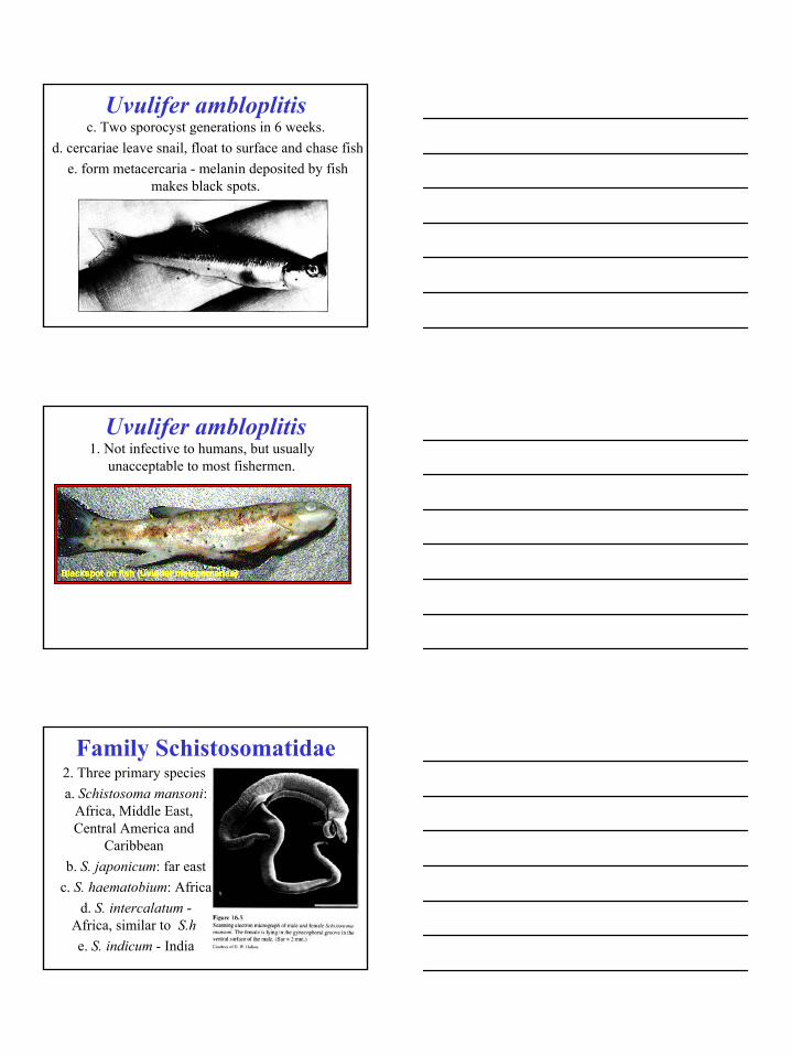
Order Strigiformes
In humans, mesocercaria can end
up in eye, kidney, other organs with high vascularity.
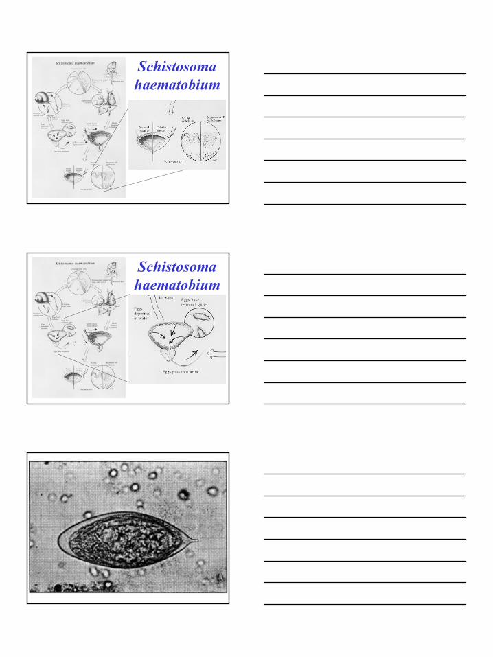
Order Strigiformesb. Uvulifer ambloplitis
1. causes "blackspot" on freshwater fisha. this is the neascus metacercaria
b. eggs shed, miracidium invades snails (Helisoma),
7
Uvulifer ambloplitisc. Two sporocyst generations in 6 weeks.
d. cercariae leave snail, float to surface and chase fishe. form metacercaria - melanin deposited by fish
makes black spots.
Uvulifer ambloplitis1. Not infective to humans, but usually
unacceptable to most fishermen.
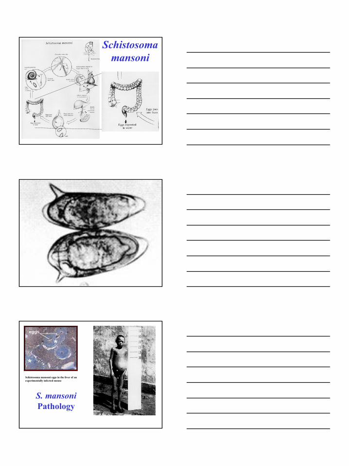
Family Schistosomatidae2. Three primary speciesa. Schistosoma mansoni:
Africa, Middle East, Central America and
Caribbeanb. S. japonicum: far east
c. S. haematobium: Africad. S. intercalatum -
Africa, similar to S.he. S. indicum - India

8
World Distributionof Schistosomiasis
Note that the disease is associated
with very old human
populations.

9
Schistosome Hosts3. Note different snail hosts in different schisto species:
a. S. haematobium: snail is Bulinusb. S. japonicum: snail is Oncomelania and some others
Schistosome Hostsc. S. indicum: snails are Indoplanorbis, Planorbis, Lymnea.
d. S. mansoni: snail is Biomphalaria
Schistosomiasis is usually contracted
at community water sources.
Control of schistosomiasis
often involves use of molluscicides

10
Schistosome Life CyclesEach have slightly
different pathology due to different locations in
definitive hosts.
Alaria canis·Fig. 1. Life cycle of Alariacanis (A. americana). 1 The adults (2.5-4.2 mm long) live in the anterior third of the small intestine of the final hosts (canids). 2 The operculate eggs are unembryonated when laid. 3 Larvae (miracidia) hatch in about 2 weeks after reaching water. 4 Miracidia swim actively and enter several species of helisomid snail (first inside which mother and daughter sporocyst are produced. The latter give rise to 5 The leave the snail during daylight hours and swim to the water surface, where they hang upside down. 6 If tadpoles (as intermediate hosts of the second type) pass by, the cercariae penetrate the skin. In about 2 weeks the cercariaebecome transformed into (6.1, 6.2 show surface view). 7-9 Two weeks after infection the mesocercariae are infectious for a series of paratenichosts, or directly for the final
host (canids) if this eats an infected tadpole (or an adult frog after its ). Inside the the mesocercariae are accumulated (8) in various tissues without further development. Large numbers of mesocercariae are very pathogenic for their hosts. If humans become accidentally infected, severe damage or death may occur. Mesocercariae which have reached the intestine of the final host penetrate into the body cavity and pass through the diaphragm into the lungs by the end of 2-3 weeks. Here, they transform into in about 5-6 weeks. The diplostomulae migrate up the trachea and are finally swallowed. Inside the intestine they mature in about 4 weeks and cause severe enteritis. FB, forebody; FT, forked tail; GP, genital pore; HB, hindbody; HF, holdfast organ; IN, intestine; OP, ; OS, oral sucker; OV, ovary; PH, pharynx; TE, UT, uterus with eggs; VI, VS, ventral sucker
SchistosomahaematobiumBulinus

11
Schistosomahaematobium
Schistosomahaematobium
Schistosomahaematobium
12
Schistosomahaematobium
Schistosomahaematobium

13
Schistosomahaematobium
Schistosomajaponicum
Oncomelania
Schistosomajaponicum

14
Schistosomajaponicum
Schistosomajaponicum

15
Schistosomajaponicum
S. japonicumPathology
Schistosomajaponicum

16
Schistosomajaponicum

17
Schistosomajaponicum
Schistosomamansoni
Biomphalaria
Schistosomamansoni

18
Schistosomamansoni
Schistosomamansoni
Schistosomamansoni
19
Schistosomamansoni
Schistosoma mansoni eggs in the liver of an experimentally infected mouse
S. mansoniPathology

20
Schistosoma Differences
a. Eggsb. Life cyclec. Pathology
Acquired immunity1. A possible context for
baptism?
Order Strigiformesd. Dioecy
1. high densities of males and females in same
host?2. Specialization as one
sex or the other can yield greater fitness than that obtained by individuals
with both sexes.
21
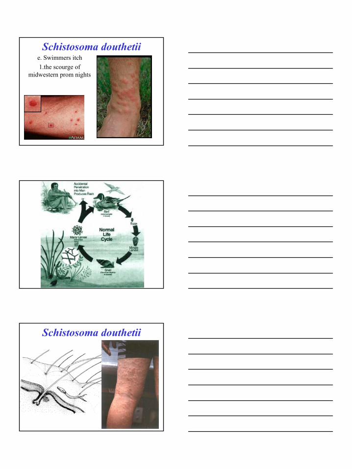
Schistosoma douthetiie. Swimmers itch 1.the scourge of
midwestern prom nights
Schistosoma douthetii