Embed Size (px)
Citation preview

BIO354: Cell Biology Laboratory 1
Laboratory 6
Analysis of the RNA Content of Yeast Cells,
Part A
I. Introduction
All cells contain a variety of biomolecules including proteins, carbohydrates, nucleic acids, and lipids. The
proportions of these molecules vary from cell to cell, as do the particular molecules within each general class. As
noted in the Introduction to Laboratory 4 (Determination of Protein Concentrations by Spectrophotometry), an
important step in the characterization of any cell is a precise biochemical description of its molecular content. The
purpose of this experiment is to analyze the RNA content of the yeast Saccharomyces cerevisiae. This unicellular
microorganism has served as a model of cellular processes in eukaryotes because it has a relatively small genome that
has been completely sequenced, it is susceptible to genetic analysis using both classical and molecular techniques, and
it can be easily grown in the laboratory.
In the first part of this two-week project, you will:
study yeast cells by light microscopy
prepare an extract of yeast cells which contains the RNAs for biochemical analysis
isolate a highly-purified sample of yeast RNA for molecular analysis by gel electrophoresis
determine the number of yeast cells in a liquid culture using three different methods.
Several different types of yeast will be available for this experiment: S. cerevisiae strain SEY6210, a haploid
laboratory strain; and Red Star baker’ yeast, another commercially-available aneuploids form used in baking. Each
group will use one of these samples but then compare the results.
In next week's lab, you will determine the concentration of RNA in the yeast extract using a spectrophotometric assay
and separate the different RNAs from yeast cells by horizontal agarose gel electrophoresis. This project is adapted
from an experiment described in a paper by Deutch, C. E. and Marshall, P. A. 2008. Analysis of the RNA Content of
the Yeast Saccharomyces cerevisiae. The American Biology Teacher 70: 537-545.
II. Pre-Lab Preparation
Read the Introduction, Background Information, and Experimental Procedures for this experiment. Also, please read
the sections of your Cell Biology book that deal with different biomolecules and with the roles of RNA in cell
biology. This experiment builds on the various techniques you learned in many of the earlier laboratory sessions.
Review those experiments as part of this project. After preparing for the lab, you should be able to answer the
following questions.
A. What are the general characteristics of baker's yeast?
B. How do yeast cells grow and reproduce?

BIO354: Cell Biology Laboratory 2
C. What types of RNA are found in yeast cells?
D. What are the functions of these different RNAs?
E. What is the purpose of treating yeast cells with trichloroacetic acid?
F. What is the purpose of treating yeast cells with perchloric acid?
G. What is left after treating yeast cells with trichloroacetic acid and perchloric acid?
H. Why is it better to use RNA prepared by the YeaStar method for electrophoresis than the RNA prepared by
acid extraction?
I. What is the purpose of treating yeast cells with Zymolase as part of the mini-prep RNA procedure?
J. Why is "RNAase free" water used in the preparation of the RNA?
K. What three methods will be used to determine the concentration of yeast cells in this experiment?
L. If 100 μl of a suspension of yeast cells that has been diluted to 10-4
gives 235 colonies on an agar plate, what
is the concentration of the original suspension?
M. Why does the turbidity of a culture increase as the cells grow?
III. Background Information
A. Saccharomyces cerevisiae
Saccharomyces cerevisiae (baker's yeast) is an important eukaryotic microorganism that is used industrially in the
production of breads and other baked goods. The cells are typically ovoid in shape and contain all of the standard
eukaryotic organelles (Figure 6.1).
Figure 6.1. Pictures showing a dark-field micrograph (A) and an electron micrograph (B) of yeast cells

BIO354: Cell Biology Laboratory 3
Unlike most other prokaryotic and eukaryotic cells that divide by binary fission, S. cerevisiae reproduces by a
process of budding. A small extension appears on the cell surface, and as chromosome duplication and mitosis
occur, one of the daughter nuclei is moved into the bud. The bud gradually increases in size and then separates
from the mother cell. In a culture of yeast cells, there will be some single cells, some cells with small buds, and
some cells with buds almost as large as the mother cell.
The yeast genome (that is, its total complement of genetic information) is only about three times the size of the
genome of the bacterium Escherichia coli and has been completely sequenced (Table 6.1). This genome contains
about 12 million base pairs, which is sufficient to code for about 6,000 proteins. It is thus significantly smaller
than the genomes of other eukaryotes such as Drosophila melanogaster (the common fruit fly) or Homo sapiens
(humans).
Table 1. Amounts of DNA in various genomes
The DNA molecules that make up the genome of S. cerevisiae are distributed among 16 chromosomes. These
DNA molecules can be separated from one another by gel electrophoresis (Figure 6.2).
Figure 6.2. Pulsed-field gel electrophoresis of budding yeast chromosomes. Intact cells embedded in a block of agarose are treated under very gentle conditions with proteases and detergents to free the chromosomal DNA from other cellular constituents. The DNA is then moved under the influence of an electrical field out of the agarose block and directly into an agarose gel. The technique uses a specialized gel apparatus in which the direction and strength of the electrophoretic field is varied periodically. This technique permits the separation of very long DNA molecules (of up to several million base pairs).
BIO354: Cell Biology Laboratory 4
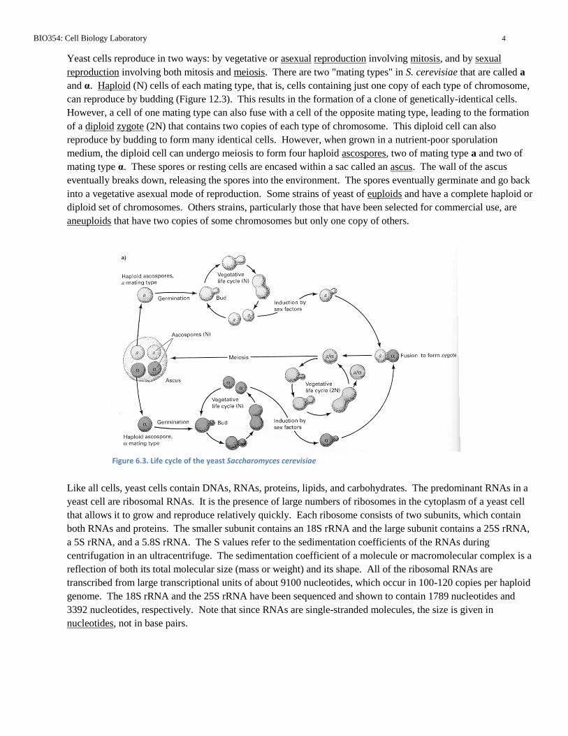
Yeast cells reproduce in two ways: by vegetative or asexual reproduction involving mitosis, and by sexual
reproduction involving both mitosis and meiosis. There are two "mating types" in S. cerevisiae that are called a
and α. Haploid (N) cells of each mating type, that is, cells containing just one copy of each type of chromosome,
can reproduce by budding (Figure 12.3). This results in the formation of a clone of genetically-identical cells.
However, a cell of one mating type can also fuse with a cell of the opposite mating type, leading to the formation
of a diploid zygote (2N) that contains two copies of each type of chromosome. This diploid cell can also
reproduce by budding to form many identical cells. However, when grown in a nutrient-poor sporulation
medium, the diploid cell can undergo meiosis to form four haploid ascospores, two of mating type a and two of
mating type α. These spores or resting cells are encased within a sac called an ascus. The wall of the ascus
eventually breaks down, releasing the spores into the environment. The spores eventually germinate and go back
into a vegetative asexual mode of reproduction. Some strains of yeast of euploids and have a complete haploid or
diploid set of chromosomes. Others strains, particularly those that have been selected for commercial use, are
aneuploids that have two copies of some chromosomes but only one copy of others.
Like all cells, yeast cells contain DNAs, RNAs, proteins, lipids, and carbohydrates. The predominant RNAs in a
yeast cell are ribosomal RNAs. It is the presence of large numbers of ribosomes in the cytoplasm of a yeast cell
that allows it to grow and reproduce relatively quickly. Each ribosome consists of two subunits, which contain
both RNAs and proteins. The smaller subunit contains an 18S rRNA and the large subunit contains a 25S rRNA,
a 5S rRNA, and a 5.8S rRNA. The S values refer to the sedimentation coefficients of the RNAs during
centrifugation in an ultracentrifuge. The sedimentation coefficient of a molecule or macromolecular complex is a
reflection of both its total molecular size (mass or weight) and its shape. All of the ribosomal RNAs are
transcribed from large transcriptional units of about 9100 nucleotides, which occur in 100-120 copies per haploid
genome. The 18S rRNA and the 25S rRNA have been sequenced and shown to contain 1789 nucleotides and
3392 nucleotides, respectively. Note that since RNAs are single-stranded molecules, the size is given in
nucleotides, not in base pairs.
Figure 6.3. Life cycle of the yeast Saccharomyces cerevisiae
BIO354: Cell Biology Laboratory 5
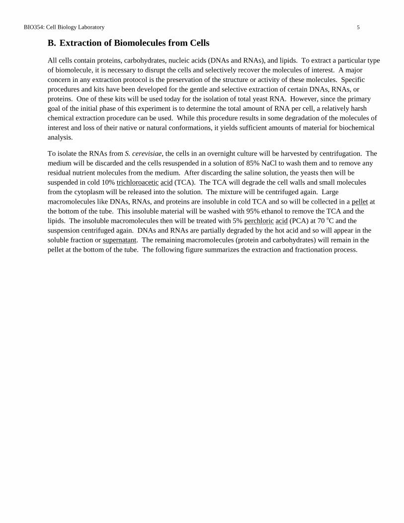
B. Extraction of Biomolecules from Cells
All cells contain proteins, carbohydrates, nucleic acids (DNAs and RNAs), and lipids. To extract a particular type
of biomolecule, it is necessary to disrupt the cells and selectively recover the molecules of interest. A major
concern in any extraction protocol is the preservation of the structure or activity of these molecules. Specific
procedures and kits have been developed for the gentle and selective extraction of certain DNAs, RNAs, or
proteins. One of these kits will be used today for the isolation of total yeast RNA. However, since the primary
goal of the initial phase of this experiment is to determine the total amount of RNA per cell, a relatively harsh
chemical extraction procedure can be used. While this procedure results in some degradation of the molecules of
interest and loss of their native or natural conformations, it yields sufficient amounts of material for biochemical
analysis.
To isolate the RNAs from S. cerevisiae, the cells in an overnight culture will be harvested by centrifugation. The
medium will be discarded and the cells resuspended in a solution of 85% NaCl to wash them and to remove any
residual nutrient molecules from the medium. After discarding the saline solution, the yeasts then will be
suspended in cold 10% trichloroacetic acid (TCA). The TCA will degrade the cell walls and small molecules
from the cytoplasm will be released into the solution. The mixture will be centrifuged again. Large
macromolecules like DNAs, RNAs, and proteins are insoluble in cold TCA and so will be collected in a pellet at
the bottom of the tube. This insoluble material will be washed with 95% ethanol to remove the TCA and the
lipids. The insoluble macromolecules then will be treated with 5% perchloric acid (PCA) at 70 oC and the
suspension centrifuged again. DNAs and RNAs are partially degraded by the hot acid and so will appear in the
soluble fraction or supernatant. The remaining macromolecules (protein and carbohydrates) will remain in the
pellet at the bottom of the tube. The following figure summarizes the extraction and fractionation process.

BIO354: Cell Biology Laboratory 6
Fractionation of Yeast Macromolecules
yeast cells in culture medium
wash with 0.85% NaCl
washed yeast cells
extract with cold 10% TCA
centrifuge
soluble fraction pellet of insoluble macromolecules
(small molecules)
wash with 95% ethanol
insoluble macromolecules
extract with 5% PCA at 70 oC
centrifuge
soluble fraction pellet of insoluble macromolecules
(RNAs, DNAs) (proteins, carbohydrates)
C. Isolation of Highly-Purified Yeast RNA
The acid extraction procedure described in Part B is designed to prepare sufficient amounts of RNA for
biochemical analysis. In this lab, you also will use a kit of reagents and spin columns to isolate a small amount
(10-25 g) of highly purified yeast RNA from 1.5 ml of a liquid culture. The kits to be used in this lab are made
by Zymo Research and sold under the name YeaStar RNA Kit. This type of kit is commonly used in molecular
biology, where most reactions are done in very small volumes (<50-100 l) and only small amounts of material
are required. The company claims that the RNA isolated with its kit can be used directly for Northern blot
analysis, poly-A mRNA isolation, and RT-PCR. The YeaStar RNA Kit costs $104 and includes the following
components, which are sufficient for 40 RNA preparations:
YeaStar RNA Kit Components
1000 units of Zymolase™
3.2 ml YR Digestion Buffer
6.4 ml YR Lysis Buffer
30 ml RNA Wash Buffer (with 100% ethanol)
4 ml RNAase-free Water
40 Zymo-spin III columns and collection tubes

BIO354: Cell Biology Laboratory 7
Zymolase™ is a mixture of enzymes that is specifically designed to degrade the polysaccharides that make up the
cell walls of S. cerevisiae and other fungi. Incubation of the yeast cells with the enzymes in YR Digestion Buffer
will convert the intact cells into osmotically sensitive protoplasts. Treatment of the protoplasts with YR Lysis
Buffer will lead to disruption of the plasma membrane and release of the cellular contents into solution. The cell
lysate will then be subjected to centrifugation in a microcentrifuge. This will result in the formation of a pellet of
insoluble molecules and a supernatant fraction containing the RNA. The supernatant will then be added to a
Zymo-spin III column and subjected to centrifugation. The column has a small membrane specifically designed
to retain RNA. The other macromolecules will pass through the membrane into a collection tube. The spin
column will be washed twice with RNA Wash Buffer to remove impurities. Finally, the column will be placed in
a new microcentrifuge tube and the RNA eluted from the spin column with RNase-free water. An essential factor
in the successful isolation of RNA from any cell is to avoid exposing the extract to any protein that contains
ribonuclease (RNase) activity. Therefore, the buffer solutions and water were all treated with a chemical to
inactivate this enzyme, and "virgin" plastic ware will be used in all of the steps. You must wear gloves
throughout this procedure to avoid the introduction of RNases from your hands into the preparation.
D. Counting Cells
To determine the macromolecular content of the individual yeast cells, it is necessary to count the exact number
of cells in a particular suspension. Because the yeasts are microscopic in size, there are three ways to do this: 1) a
direct microscopic count using a special slide called a hemocytometer; 2) a viable cell count, in which cells are
detected by their ability to form colonies on agar plates; and 3) an indirect count based on the turbidity of the cell
suspension. All three methods will be done as part of this experiment.
1. Direct Hemocytometer Counts
A hemocytometer is a special microscope slide that was first developed so that lab technicians in hospitals
could count red and white blood cells. However, it is commonly used to count other types of cells. The slide
consists of a central platform that is etched with a grid of lines a specific distance apart (Figure 6.4). There is
a moat on either side of the platform and supporting bridges on either side. A cover slip or glass is placed on
top of the bridges and a solution containing the cells to be counted (bacteria, yeasts, tissue culture cells, etc.)
is carefully introduced under the cover slip. When the slide is then examined microscopically, it is possible to
see the cells on the squares of the grid and to count them. By knowing the scale of the grid and the distance
between the grid and the coverslip, the volume of liquid over the squares can be determined. From this, the
number of cells/ml of solution can be calculated.

BIO354: Cell Biology Laboratory 8
In the hemocytometers we will use, the central portion of the grid is 1 mm x 1 mm, and is divided into 25
major squares. The cover slip sits 0.1 mm over the grid. Each of 25 major squares thus contains 0.2 mm x
0.2 mm x 0.1 mm = 0.004 mm3 or 0.004 l.
The 25 major squares are then divided into 16 smaller squares, each of which contains 0.05 mm x 0.05 mm x
0.1 mm or 0.00025 mm3 = 0.00025 l. The standard practice for using the hemocytometer is to count the
cells in at least 5 of the large squares and to determine the average. Most commonly, a person will count five
well separated squares, such as the four corner squares and one square in the middle of the grid.
For example, suppose you count 205 yeasts, 150 yeasts, 250 yeasts, 175 yeasts, and 200 yeasts in five of the
large squares. This gives an average of 196. The total cell count is thus:
Note that counts are usually expressed in scientific notation as yeast cells/ml. With yeast cells, a cell with a
bud is usually counted as one cell. Cells that fall on the lines at the edges of a square are usually counted if
more than half of the cell is inside the square.
Figure 6.4. Diagram of a hemocytometer, (a) side view, (b) top view, (c) view through a microscope on low power showing grid.
0.004 mm3 x 1 ml x 1 cm
3 x 1000 l = 0.004 l.
1 cm3 1000 mm
3 1 ml
196 yeasts x 1000 l = 4.90 x 10⁷ yeast cells/ml
0.004 l ml

BIO354: Cell Biology Laboratory 9
2. Viable Cell Counts
Another way to count microbial cells is to do a viable cell count. This involves making serial dilutions of a
cell suspension and spreading known volumes of each dilution on agar plates containing a nutrient medium.
As the plates are incubated, the cells grow and divide and eventually form colonies that can be seen by eye.
The colonies are then counted, and after correcting for the dilution factor, the count in viable cells/ml is
determined. (Figure 6.5)
Dilutions of the initial culture can be made in the nutrient medium, a stabilizing buffer, or a simple isotonic solution of
sodium chloride. You can make a 1/10 (10-1
) dilution by adding 1.0 ml to 9.0 ml of the diluents, or you can make a 1/10
(10-1
) dilution by adding 100 μl to 900 μl of the diluents. Refer back to Laboratory 1 (Scientific Calculations and Basic
Lab Techniques) for a review of serial dilutions. After incubating the agar plates, it is usually found that some of the
initial dilutions are still too concentrated and so give too many colonies to count. Other dilutions may be too dilute and
contain no organisms at all. In the middle, however, some dilutions give a reasonable number of colonies and these are
the plates that are used. Counts between 30 and 300 give the best statistical accuracy. If several dilutions give reasonable
counts, the viable counts/ml from these dilutions can be averaged. This method may not give the same result as a direct
hemocytometer count because some of the cells are dead or cannot form colonies on the nutrient medium that is used. In
the case of yeast cells, a cell with a bud will count as one cell since the mother cell and its bud will give rise to a single
colony.
Figure 6.5. Diagram showing procedure for creating a viable count of yeast cells.
BIO354: Cell Biology Laboratory 10
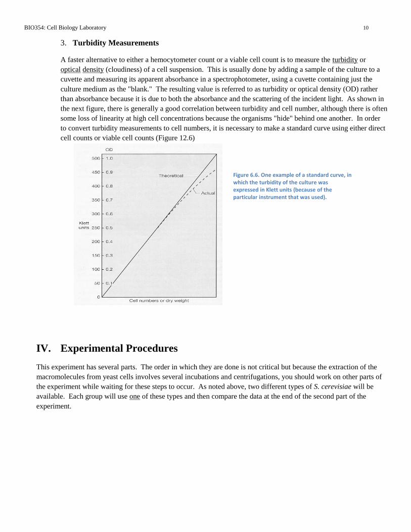
3. Turbidity Measurements
A faster alternative to either a hemocytometer count or a viable cell count is to measure the turbidity or
optical density (cloudiness) of a cell suspension. This is usually done by adding a sample of the culture to a
cuvette and measuring its apparent absorbance in a spectrophotometer, using a cuvette containing just the
culture medium as the "blank." The resulting value is referred to as turbidity or optical density (OD) rather
than absorbance because it is due to both the absorbance and the scattering of the incident light. As shown in
the next figure, there is generally a good correlation between turbidity and cell number, although there is often
some loss of linearity at high cell concentrations because the organisms "hide" behind one another. In order
to convert turbidity measurements to cell numbers, it is necessary to make a standard curve using either direct
cell counts or viable cell counts (Figure 12.6)
IV. Experimental Procedures
This experiment has several parts. The order in which they are done is not critical but because the extraction of the
macromolecules from yeast cells involves several incubations and centrifugations, you should work on other parts of
the experiment while waiting for these steps to occur. As noted above, two different types of S. cerevisiae will be
available. Each group will use one of these types and then compare the data at the end of the second part of the
experiment.
Figure 6.6. One example of a standard curve, in which the turbidity of the culture was expressed in Klett units (because of the particular instrument that was used).

BIO354: Cell Biology Laboratory 11
The following is a flow chart for this laboratory session:
Microscopic Observations of Yeast Cells (Section IVA)
Extraction of Macromolecules Counting Cells in an Overnight Culture of
for Biochemical Analysis S. cerevisiae with a Hemocytometer
(Section IVB) (Section IVD)
Isolation of Highly-Purified Yeast RNA Counting Cells in an Overnight Culture of
(Section IVC) S. cerevisiae by Plate Counts
(Section IVE)
Measuring the Turbidity of an Overnight
Culture of S. cerevisiae
(Section IVF)
A. Microscopic Observations of Yeast Cells
The objective of this part of the experiment is to observe the yeast S. cerevisiae with various types of light
microscopy. If necessary, go back and review the Background Information and Experimental Procedures in
Laboratory 2 (Introduction to Microscopy). This part of the lab will be done as a demonstration.
1. A Leica DME microscopes like you have used before will be set up with wet mounts of the two different
types of Saccharomyces cerevisiae. Pay particular attention to the type of yeast that you will be using but
look at the other two as well. Examine the cells first under bright-field (BF) optics. Because the yeasts
have no intrinsic color, it will be necessary to reduce the light intensity with the iris diaphragm or the
power source. Look at the cells with the 40X objective and make a drawing of what you see.
type of yeast cell used ___________________________________________________
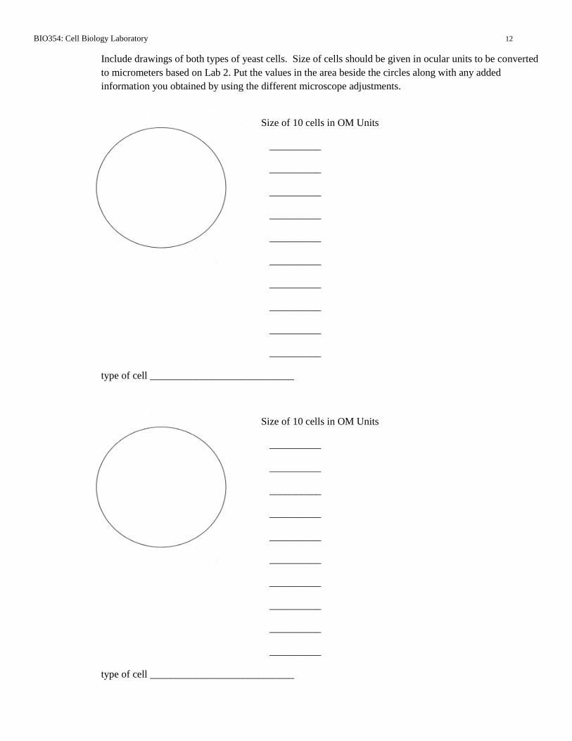
BIO354: Cell Biology Laboratory 12
Include drawings of both types of yeast cells. Size of cells should be given in ocular units to be converted
to micrometers based on Lab 2. Put the values in the area beside the circles along with any added
information you obtained by using the different microscope adjustments.
Size of 10 cells in OM Units
__________
__________
__________
__________
__________
__________
__________
__________
__________
__________
type of cell ____________________________
Size of 10 cells in OM Units
__________
__________
__________
__________
__________
__________
__________
__________
__________
__________
type of cell ____________________________
BIO354: Cell Biology Laboratory 13
2. Without moving the stage, rotate the condenser turret to the dark-field (DF) position. It will be necessary
to increase the illumination at this point because the yeasts will be visible only due to the reflected light.
Then rotate the condenser turret to the PH2 position for phase contrast observations. Also measure the
size of ten of each of the yeast cell types put the values in the area beside the circles along with any added
information you obtained by using the different microscope adjustments.
B. Extraction of Macromolecules for Biochemical Analysis
The objective of this part of the experiment is to prepare an extract of the yeast culture that can be used for a total
RNA determination. Because this procedure requires multiple centrifugation steps, you should start this part of
the experiment near the beginning of the class and do the other parts of the lab in between the various incubation
and centrifugation steps. Refer back to the flow chart as necessary. You should observe the extract carefully at
each step and record your observations.
1. Each group will be provided with a 25 mL culture of one form of S. cerevisiae. Be sure to note which
type of yeast you are going to use. The yeasts will have been grown overnight at 30oC in a rich nutrient
medium called YPD (1% yeast extract, 2% peptone, 2% dextrose). Swirl the flask to make an even
homogeneous suspension.
2. With a sterile 10 mL pipette and a green pipet-aid, remove 10.0 ml of the culture and add it to a 15 mL
plastic conical centrifuge tube.
3. Centrifuge the yeast suspension at 3,000 rpm for five minutes in a clinical centrifuge. Be sure use a
"balance tube" with the same volume. Carefully pour off (decant) the medium and discard it into the
designated container.
4. Add 5.0 mL of sterile saline (0.85% NaCl) to the tube and vortex the mixture carefully to resuspend the
yeast cells. Centrifuge the suspension at 3,000 rpm for five minutes and discard the wash solution into
the designated container.
5. Add 5.0 ml of cold 10% trichloroacetic acid (TCA) to the yeast cells in the tube and vortex the mixture
carefully. BE CAREFUL: TCA is a highly corrosive acid! Keep the suspension on ice for 20 minutes.
Centrifuge the suspension at 3,000 rpm for five minutes and discard the TCA solution into the designated
container.
6. Add 5.0 ml of 95% ethanol to the pellet of material in the tube and vortex the mixture carefully.
Centrifuge the suspension at 3,000 rpm for five minutes and discard the ethanol into the designated
container.
7. Add 5.0 ml of 5% perchloric acid (PCA) to the pellet of material in the tube and vortex the mixture
carefully. BE CAREFUL: PCA is also a highly corrosive acid! Heat the suspension in a 70oC water bath
for 20 minutes. Centrifuge the suspension at 3,000 rpm for five minutes and decant the PCA extract into
a new 15 ml plastic centrifuge tube. Label the tube with your initials and PCA extract. Place the tube in
the designated rack for storage in the refrigerator. Discard the original tube with the PCA-
insoluble material in the designated container.

BIO354: Cell Biology Laboratory 14
C. Isolation of Highly-Purified Yeast RNA
The objective of this part of the experiment is to use the YeaStar RNA Kit from Zymo Research to isolate highly-
purified RNA from an overnight culture of Saccharomyces cerevisiae. Because this protocol involves a 60
minute incubation period, you should start the procedure near the beginning of the lab and then work on the other
parts of the experiment.
1. Swirl the flask with the overnight culture of yeast cells to make a homogeneous suspension.
2. Put on a pair of gloves, and using a P-1000 micropipetter and a large sterile tip, add 1.5 ml (2 x 750 l) of
the culture to a sterile microcentrifuge tube. Centrifuge the sample in the microcentrifuge for 1 minute at
10,000 rpm. Carefully remove all of the supernatant with a micropipetter and discard it.
3. Add 80 l of YR Digestion Buffer and 5 l of Zymolase™ to the microcentrifuge tube and carefully
draw the liquid up and down to make an even suspension of the yeast cells. Cap the tube and place it in a
foam "float." Incubate the mixture for 60 minutes at 37oC.
4. After 60 minutes, remove the tube from the water bath and add 160 l of YR Lysis Buffer. Mix the
solution completely on a vortex mixer. Centrifuge the yeast lysate solution at 10,000 rpm for 2 minutes
in a microcentrifuge.
5. Place a Zymo-spin III column in a 2 ml plastic collection tube. Carefully remove the supernatant from
the yeast lysate with a micropipetter and add the liquid to the spin column.
6. Centrifuge the spin column/collection tube at 10,000 rpm for 1 minute in a microcentrifuge. Add 200 l
of RNA Wash Buffer to the spin column and centrifuge the spin column/collection tube at 10,000 rpm
for 1 minute. Add another 200 l of RNA Wash Buffer to the spin column/collection tube and
centrifuge again at 10,000 rpm for 1 minute.
7. Obtain a new sterile 1.5 ml microcentrifuge tube and place the spin column in it. Discard the 2 ml
collection tube with all of the other solutions. Add 60 l of RNase-free water to the membrane in the
spin column. Centrifuge the spin column/microcentrifuge tube for 10 seconds at 10,000 rpm.
8. Discard the spin column and cap the microcentrifuge tube. Label the tube with your initials and place
it in the designated rack for storage in the freezer at -20oC until next week's lab.

BIO354: Cell Biology Laboratory 15
D. Counting Cells in an Overnight Culture of S. cerevisiae with a Hemocytometer
The objective of this part of the lab is to determine the actual number of yeast cells per ml of the overnight YPD
culture using a hemocytometer. This number will be essential for the later calculations of the macromolecular
content. You should do this part of the experiment while you are doing the incubations required in Sections
B & C.
1. To determine the total cell count/ml, obtain a box containing a hemocytometer slide and the thick
coverslip that goes with it (BE CAREFUL: the hemocytometers and the special cover slips cost about
$125 each). Place the slide on the bench top in front of you and position the coverslip on top.
2. Make a 1/10 (10-1
) dilution of the overnight culture of S. cerevisiae in the following way. With a P-1000
micropipetter and a large sterile tip, add 900 L (0.9 mL) of sterile saline solution (0.85% NaCl) to a
sterile 1.5 mL microcentrifuge tube. Swirl the overnight culture of S. cerevisiae to make an even
homogeneous suspension. With a P-100 micropipetter and a small sterile tip, remove 100 L of the
suspension. Wipe the outside of the tip with a Kim-Wipe and add the liquid to the 10-1
dilution tube.
Draw the liquid up and down about 5 times to mix the solution. Then cap the tube and mix the solution
on a vortex mixer.
3. With a P-100 micropipetter and a sterile tip, remove 20 L of the diluted yeast suspension. Place the
pipet tip in the notch under the coverslip and slowly add the liquid to the slide. You will see the liquid
move over the surface of the slide. Add just enough liquid to cover the central part of the slide. The
coverslip should not float off of the bridges.
4. Obtain one of the Leica DME microscopes from the cabinet. Rotate the condenser turret on the Leica
DME microscope back to the bright field (BF) position. Place the hemocytometer on the stage of the
microscope and move it into position under the 10X objective. Bring the grid into focus using the coarse
focusing knob. It will be helpful to reduce the light intensity: the lines on the grid are designed to reflect
the light and the yeast cells have no natural color.
5. When you have found the grid, be sure that you understand its layout. Refer back to the figure in the
Background Information section if necessary. Then rotate the 40X objective into position. Adjust the
light intensity so that you can see both the grid and the yeast cells clearly. Depending on the density of
the overnight culture, the number of yeast cells/square will vary.
6. Count the yeast cells in five (5) of the 25 major squares. Note that each of these squares is divided into
16 smaller squares, so it may be easier to count the yeasts in the smaller squares first and then add up the
total. Record the data here
Square 1 Square 2 Square 3 Square 4 Square 5
# of Cells
BIO354: Cell Biology Laboratory 16
7. Calculate the average number of yeasts for the five large squares. Then multiply this value by 10 since
you made a 1/10 (10-1
) dilution of the original overnight culture. Finally, calculate the total cell count in
yeasts/ml. Remember that the volume of liquid over each large square is 0.004 L. (Specific details on
this calculation are included in the introductory information. Record your final value here)
8. When you are finished, carefully wash the hemocytometer and the cover slip with distilled water. Dry
them carefully with a Kim-Wipe and put them back in the box.
E. Counting Cells in an Overnight Culture of S. cerevisiae by Plate Counts
The objective of this part of the experiment is to determine the number of viable cells in the yeast culture by a
plate count. The plates that are set up in this week's lab will actually be counted in the next lab session.
1. To obtain a viable cell count for the yeast cell suspension, place seven (7) sterile 1.5 ml centrifuge tubes
in a rack in front of you. Label the tubes 10-1
, 10-2
, 10-3
, 10-4
, 10-5
, 10-6
, and 10-7
. With a P-1000
micropipetter and a large sterile tip, add 900 l (0.9 ml) of sterile saline solution (0.85% NaCl) to each
tube.
2. Swirl the overnight culture of S. cerevisiae again to make an even homogeneous suspension. With a
P-100 micropipetter and a small sterile tip, remove 100 l of the yeast culture. Wipe the outside of the tip
with a Kim-Wipe and add the liquid to the tube labeled 10-1
. Draw the liquid up and down about 5 times
to mix the solution. Discard the tip in the designated container. Push down the cap and vortex the
solution briefly. With a P-100 micropipetter and a small sterile tip, remove 100 l of the suspension in
the 10-1
dilution, wipe the outside of the tip, and add the liquid to the tube labeled 10-2
. Draw the liquid
up and down about 5 times to mix the solution. Discard the tip and vortex the solution briefly. Repeat
the process until all 7 serial dilutions have been made.
3. Obtain eight (8) plates of YPD agar medium. This is the same medium that the cells in the overnight
culture were grown in but solidified with 2% agar. Label the plates with your initials. Then label two
plates each with the numbers 10-4
, 10-5
, 10-6
, and 10-7
and your groups name or initials.
4. With a P-100 micropipetter and a small sterile tip, remove 100 l of the suspension in the tube labeled
10-4
. Because the yeasts tend to settle out quickly, mix the suspension first by vortexing it briefly first.
Carefully remove the lid of the petri dish and add the yeast suspension to the agar surface. Using a sterile
disposable spreader, distribute the liquid evenly over the agar surface. Place the spreader in the
designated container for disposal.
5. Repeat the process with the other plate labeled 10-4
using a new spreader.
6. In the same way, plate out two 100 l portions of each of the other dilutions of the yeast suspension (10-5
,
10-6
, and 10-7
). When all of the plates have been prepared, place them in the designated bin. The
instructor will incubate them for three days at 30 oC until the colonies become visible and then store them
in the refrigerator until next week's lab. You will count the colonies on the plates next week.
Hemocytometer Total Cell Count: _____________________________________________yeasts/mL

BIO354: Cell Biology Laboratory 17
F. Measuring the Turbidity of an Overnight Culture of S. cerevisiae
The objective of this part of the experiment is to determine the turbidity or optical density of the overnight yeast
culture. This turbidity is related to the total number of cells in the culture.
1. Turn on the Spectronic 20 Genesys spectrophotometer and allow it to warm up for 15 minutes. Set the
wavelength to 600 nm.
2. Using a sterile 5 ml pipet and a green pipet aid, transfer 3 ml of sterile liquid YPD medium to a plastic
disposable cuvette. Set the instrument to zero absorbance.
3. Swirl the overnight culture of S. cerevisiae to make an even homogeneous suspension. With another
sterile 5 ml pipet, transfer 3 ml of the overnight yeast culture to another plastic disposable cuvette.
Measure the absorbance (optical density, turbidity) of this suspension.
4. If the suspension is very dense and the spectrophotometer cannot read it or gives an absorbance greater
than 1.0, make a 1/10 (10-1
) dilution of the suspension in YPD medium. Record the result. Be sure the
multiply by 10 to get the turbidity of the original yeast suspension. Discard the cuvette with the yeast
culture in the designated container.
5. Using Figure 6.7 or 6.8, estimate the number of yeast cells/ml in your overnight culture.
Figure 6.7. Serial dilutions of a 24-hour culture of Red Star yeast cells were made. The absorbance of each diluted sample was then measured using a Gen 20 Spectrophotometer. Finally a sample of the solution was added to a hemocytometer and five squares were counted and the number of cells present was calculated for each dilution. Then a standard curve was created and a best-fit line determined.
Turbidity Total Cell Count: _____________________________________________yeasts/mL
BIO354: Cell Biology Laboratory 18
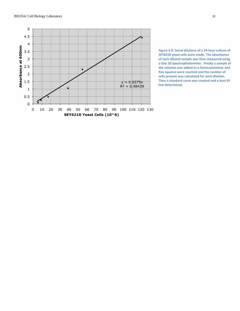
Figure 6.8. Serial dilutions of a 24-hour culture of SEY6210 yeast cells were made. The absorbance of each diluted sample was then measured using a Gen 20 Spectrophotometer. Finally a sample of the solution was added to a hemocytometer and five squares were counted and the number of cells present was calculated for each dilution. Then a standard curve was created and a best-fit line determined.