Embed Size (px)
Citation preview

ABSTRACT
Title of Thesis: BIOACTIVITY OF EPHEDRA: INTEGRATING
CYTOTOXICITY ASSESSMENT WITH REAL-TIME BIOSENSING
Kazuko Fukushima, Master of Science, 2004 Thesis Directed By: Dr. Y. Martin Lo, Department of Nutrition and Food
Science
Ephedrine-type alkaloids (ETA) are major active ingredients of Ephedra, a
traditional Chinese medicinal herb used to treat asthma and nasal congestion. Until
recently, large amounts of Ephedra were used in dietary supplements for weight loss
and athletic performance enhancement. However, indiscriminate consumption of
ETA-containing products has resulted in more than 1,000 reported cases of adverse
effects. The objective of this study is to evaluate bioactivity of ETA. The toxicities of
(-)-ephedrine and (+)-pseudoephedrine were measured using MTT assay on human
neuroblastoma (SH-SY5Y) and rat myoblastoma (H9c2 (2-1)), while the stress
responses of a panel of biosensing bioluminescent Escherichia coli strains were
analyzed. SH-SY5Y showed similar sensitivity to (-)-ephedrine and (+)-
pseudoephedrine, while H9c2 (2-1) could differentiate the cytotoxicity of (-)-
ephedrine and (+)-pseudoephedrine. The biosensing of the E. coli strains was highly
sensitive to the toxicity of ETA and could yield instantaneous response. The RLU
ratios dependent on the construct of strains gave unique fingerprinting pattern of
ETA.
BIOACTIVITY OF EPHEDRA: INTEGRATING CYTOTOXICITY ASSESSMENT
WITH REAL-TIME BIOSENSING
By
Kazuko Fukushima
Thesis submitted to the Faculty of the Graduate School of the University of Maryland, College Park, in partial fulfillment
of the requirements for the degree of Master of Science
2004 Advisory Committee: Dr. Y. Martin Lo, Chair Professor George A. Bean Dr. Arthur J. Miller

© Copyright by Kazuko Fukushima
2004

ii
Acknowledgements
My advisor Dr. Y. Martin Lo for his meticulous guidance, and for giving me
the chance to learn and challenge the new things.
Dr. Arthur J. Miller and Prof. George A. Bean for their helpful participation
and invaluable advice in my thesis committee.
Prof. David K. Y. Lei for welcoming me to his lab, and for providing
insightful suggestions. Dr. Libin Cui and Ms Tina for coaching me in cell culture
techniques from ABC.
My lab mate Ms Jing Wang for her great help in biosensing and for the
discussion helping me to understand the subject deeply.
All the lab mates: Ms Sanem Argin, Ms Pu Jing, Ms Julia Radinsky and Ms
Lisa Sadar for their friendship and warm encouragements.
My former lab mates Dr. April Hsu and Ms Brenda Fermin for their
continuing support since I first came here.
The National Personnel Authority, Japan for providing me with the
opportunity to learn in the United States for two years.
My supervisors and colleagues from the Ministry of Health, Labour and
Welfare, Japan, for their backup and for their hospitality when they visited D.C. area.
My sisters for cheering me up by their mindless chatter and e-mails, and my
parents for nurturing my independent spirit.

iii
Table of Contents Acknowledgements ...................................................................................................ii Table of Contents .................................................................................................... iii List of Tables............................................................................................................iv List of Figures ...........................................................................................................v CHAPTER 1: INTRODUCTION ..............................................................................1 CHAPTER 2: LITERATURE REVIEW....................................................................3
2.1 Ephedra ...........................................................................................................3 2.1.1 Botanical...................................................................................................3 2.1.2. Chemistry.................................................................................................4 2.1.3. Pharmacology ..........................................................................................5
2.2. Use of Ephedra ...............................................................................................6 2.2.1. Traditional Use.........................................................................................6 2.2.2. As Dietary Supplements ...........................................................................7
2.3. Assessment Tools ...........................................................................................8 2.3.1. Analytical Quantification .........................................................................8 2.3.2. In Vitro Cytotoxicity ................................................................................9 2.3.3. Biosensing Using Bioluminescent Reporter Bacteria ..............................10
CHAPTER 3: OBJECTIVES...................................................................................16 CHAPTER 4: MATERIALS AND METHODS.......................................................17
4.1. Cytotoxicity Assessment Using MTT Cell Proliferation Assay......................17 4.1.1. Materials ................................................................................................17 4.1.2. Cell Cultures ..........................................................................................17 4.1.3. MTT Assays...........................................................................................17 4.1.4. Data Analysis.........................................................................................18
4.2. Real-Time Biosensing Using Bioluminescent E. coli Strains.........................19 4.2.1. Bioluminescent E. coli strains ................................................................19 4.2.2. E. coli stress fingerprinting.....................................................................21 4.2.3. Data Analysis.........................................................................................22
CHAPTER 5: RESULTS AND DISCUSSION.......................................................23 5.1. Cytotoxicity Assessment Using MTT Cell Proliferation Assay......................23
5.1.1. Effects of Ephedrine and Pseudoephedrine on Cytoviability ...................23 5.1.2. Effects of Combined Treatments by Ephedrine and Pseudoephedrine .....26 5.1.3. Discussion..............................................................................................30
5.2. Real-Time Biosensing Using Bioluminescent E. coli Strains.........................34 CHAPTER 6: CONCLUSIONS..............................................................................45 Appendices..............................................................................................................47 References...............................................................................................................51
iv
List of Tables Table 4-1. Stress-responsive E. coli lux fusion strains ..............................................20 Table 5-1. The IC50 values of (-)-ephedrine and (+)-pseudoephedrine for two cell lines.........................................................................................................................27 Table A-1. RLU ratio obtained from six E. coli strains exposed to 0.03 mg/ml of (-)-ephedrine, (+)-pseudoephedrine or mixture of (-)-ephedrine and (+)-pseudoephedrine in different concentrations .......................................................................................47 Table A-2. RLU ratio obtained from six E. coli strains exposed to 0.04 mg/ml of (-)-ephedrine, (+)-pseudoephedrine or mixture of (-)-ephedrine and (+)-pseudoephedrine in different concentrations .......................................................................................48 Table A-3. RLU ratio obtained from six E. coli strains exposed to 0.05 mg/ml (-)-ephedrine, (+)-pseudoephedrine or mixture of (-)-ephedrine and (+)-pseudoephedrine in different concentrations .......................................................................................49 Table A-4. RLU ratio obtained from six E. coli strains exposed to 0.06 mg/ml (-)-ephedrine, (+)-pseudoephedrine or mixture of (-)-ephedrine and (+)-pseudoephedrine in different concentrations .......................................................................................50

v
List of Figures Figure 2-1. Chemical structures of (-)-ephedrine and (+)-pseudoephedrine ................5 Figure 2-2. Principle of biosensing using bioluminescent reporter bacteria ..............11 Figure 2-3. Bacterial bioluminescence pathway ......................................................13 Figure 5-1. Effects of ephedrine-type alkaloids on SH-SY5Y and H9c2 (2-1) cell viability ...................................................................................................................24 Figure 5-2. Sample dose-response curve. .................................................................25 Figure 5-3. H9c2 (2-1) cells treated with (-)-ephedrine ............................................28 Figure 5-4. Cumulative effects of two ephedrine-type alkaloids on the cytoviability of SH-SY5Y and H92c (2-1)........................................................................................29 Figure 5-5. RLU ratio obtained from six E. coli strains exposed to 0.03 mg/ml of (-)-ephedrine and (+)-pseudoephedrine .........................................................................40 Figure 5-6. RLU ratio obtained from six E. coli strains exposed to 0.04 mg/ml of (-)-ephedrine and (+)-pseudoephedrine. ........................................................................41 Figure 5-7. RLU ratio obtained from six E. coli strains exposed to 0.05 mg/ml of (-)-ephedrine and (+)-pseudoephedrine. ........................................................................42 Figure 5-8. RLU ratio obtained from six E. coli strains exposed to 0.06 mg/ml of (-)-ephedrine and (+)-pseudoephedrine. ........................................................................43 Figure 5-9. RLU ratio obtained from six E. coli strains exposed to 0.3 mg/ml of (-)-ephedrine and (+)-pseudoephedrine. . ......................................................................44
1
CHAPTER 1: INTRODUCTION
More than half of the U.S. adult population uses dietary supplement products,
and consumers spend approximately $12 billion annually on dietary supplements,
according to the 1998 Nutrition Business Journal Annual Industry Overview
(Nutrition Business Journal, 1998). In the last decade, large amounts of ephedrine-
type alkaloids (ETA) were used in numerous dietary supplements formulated for
weight reduction and athletic performance enhancement (Dulloo and Stock, 1993).
However, indiscriminate consumption of ETA-containing products has resulted in
more than 1,000 reported cases of poisoning and other serious effects since 1993,
some of which were fatal (FDA, 2000).
Used in the Far East to treat asthma, nose and lung congestion, and fever with
anhidrosis (Lee et al., 2000), Ephedra (ma huang), a traditional Chinese medicine
(TCM) derived from Ephedra sinica Stapf and other Ephedra species, is the major
sources of ETA. To date, the quality of Ephedra has been determined by the contents
of total ETA, with higher contents indicating better quality. However, although
individual ETA has similar pharmacological activity, they vary significantly in
potency (Cetaruk and Aaron, 1994). Both the contents and the profile of ETA in
Ephedra vary with plant species and varieties (Cui et al., 1991), plant parts (Liu et al.,
1993), seasons of harvest (Kasahara et al., 1986), and geographical origins (Zhang et
al., 1989). In addition to ETA, Ephedra also contains other phytoconstituents, which
may modify its pharmacological and toxicological activities. Therefore, the toxicity
of Ephedra cannot be totally accounted for by its ETA contents alone (FDA, 1997). A

2
bioassay is needed to determine the total toxicity of Ephedra due to the combined
effect of the alkaloids and other constituents.
The overall objective of this study is to evaluate bioactivity of ephedrine-type
alkaloids by measuring its cytotoxicity against selected cell lines and analyzing the
stress responses of a panel of six genetically engineered biosensing strains capable of
producing real-time responses to environmental stresses that cause specific cell
damage. To accomplish these objectives, the study will be divided into two parts. The
first part involves detecting and profiling the cytotoxicity of ephedrine-type alkaloids
against the two cell lines —human neuroblastoma and rat myoblastoma—using MTT
cell proliferation assay techniques. The second part involves a panel of six strains of
bioluminescent E. coli to identify the stress fingerprints induced by ephedrine-type
alkaloids.

3
CHAPTER 2: LITERATURE REVIEW
2.1 Ephedra
2.1.1 Botanical
Ephedra, also known as ma huang, belongs to the family Ephedraceae that is
an evolutionarily primitive plant family (Blumenthal and King, 1995). Ephedra
species favor dry, sandy or rocky environments, and are found in the temperate and
subtropical regions of China, Mongolia, India, parts of the Mediterranean and
Afghanistan as well as regions of North and Central America (Blumenthal and King,
1995). Although the genus Ephedra consists of more than 50 species (Schaneberg et
al., 2003), primary species of Ephedra are represented by Ephedra sinica, E. major
Host, E. gerardiana Wall, E. intermedia Schrenk & Meyer, and E. equisetina Bunge
(Morton, 1977).
Ephedra species are short, evergreen and almost leafless shrubs that grow
about 60 to 90 cm high (23.5 to 35.5 inches high). The stems are green in color,
slender, erect or reclining, small ribbed and channeled, about 1.5 mm in diameter and
usually terminating in a sharp point. Nodes are 4 to 6 cm apart, and small triangular
leaves appear at the stem nodes (Blumenthal and King, 1995). The nodes are
characteristically reddish brown. The stems usually branch form the base (Blumenthal
and King, 1995). They bear minute, yellow-green flowers and fruits, and emit a
strong pine-like odor and have an astringent taste (Blumenthal and King, 1995).
4
2.1.2. Chemistry
The major active ingredients of Ephedra are alkaloids that constitute 0.5 to 2.5
percent of the total mass, and are referred to as ephedrine-type alkaloids (Blumenthal
and King, 1995). The six optically active alkaloids that have been isolated from
Ephedra species are (-)-ephedrine, (+)-pseudoephedrine, (-)-N-methylephedrine, (+)-
N-methylpseudoephedrine, (-)-norephedrine, (+)-norpseudoephedrine. Usually, (-)-
ephedrine is the major isomer that comprises 30 to 90 percent of total alkaloid
fraction accompanied by (+)-pseudoephedrine, with trace amount of other ephedrine-
type alkaloids (Blumenthal and King, 1995). The total content of ephedrine-type
alkaloids depends on the species of Ephedra, time of year of harvest, weather
conditions, and altitude where the plant grows (Blumenthal and King, 1995), and can
exceed 2% (Bruneton, 1995). This variation according to environmental conditions
explains why some Ephedra containing dietary supplements of the same brand often
show alkaloid content markedly differing from label claims and also variation among
lots when analyzed chemically (Gurley et. al., 2000).
Preparation of ephedrine-type alkaloids from crude plant material involves an
acid/base extraction procedure (Reti, 1953). In addition to the extraction from plants,
ephedrine-type alkaloids can also be chemically synthesized, and most of the
ephedrine and pseudoephedrine used in western medicine has been manufactured
synthetically. However, these synthetic ephedrine-type alkaloids differ from the
natural forms in that they are racemic, i.e., optically inactive, because they are made
up of two enantiomorphic isomers (Abourashed et al., 2003).

5
In addition to the ephedrine-type alkaloids, other alkaloids and amino
compounds have been isolated from different species of Ephedra. The macrocyclic
spermine alkaloids, Ephedradines A-D, kynurenic acid derivatives, cyclopolyglycine,
methanoproline amino acids, flavones, flavanols, tannins, carboxylic acids, volatile
terpenes (Schaneberg et al., 2003).
Figure 2-1. Chemical structures of (-)-ephedrine and (+)-pseudoephedrine
(adapted from Abourashed et al., 2003)
2.1.3. Pharmacology
Ephedrine is a sympathomimetic substance that stimulates both - and -
adrenergic receptors (Abourashed et al., 2003). Stimulation of 1-adrenergic
receptors produces contraction of vascular smooth muscle, increased contractile force
of the heart and arrhythmias, glycogenolysis, gluconeogenesis, hyperpolarization, and
NHCH3
HO NHCH3
S S
CH3
CH3
S R
HO
HOCH3NHCH3RS SSNHCH3CH3HO
(-)-ephedrine (+)-pseudoephedrine

6
relaxation of intestinal smooth muscle. Stimulation of 2-adrenergic receptors
decreases insulin secretion, platelet aggregation and the release of norepinephrine
from the nerve terminals, and causes contraction of vascular smooth muscle.
Stimulation of 1-adrenergic receptors increases force and rate of contraction of the
heart, increased velocity of conduction through the atrioventricular node, and
increased rennin secretion. When used in therapeutic doses, stimulation of 2-
adrenergic receptors causes relaxation of the smooth muscle of the blood vessels and
bronchi (Mack, 1997). Stimulation of -adrenergic receptors including 3-subtype
involve in lypolysis and non-shivering thermogenesis (Abourashed et al., 2003). In
addition to its direct effects, ephedrine also displays indirect sympathetic activation
releasing norepinephrine from sympathetic neurons (Abourashed et al., 2003).
Ephedrine also has central nervous system (CNS) stimulant effects similar to those of
amphetamines, but less pronounced (McEvoy, 2000).
2.2. Use of Ephedra
2.2.1. Traditional Use
The Ephedra species have been dispensed in traditional Chinese medicines
(TCM) for at least 5,000 years (Morton, 1977). In traditional Chinese medicines,
dried stems of Ephedra species are used to alleviate symptoms caused by common
cold, influenza, asthma, bronchitis, nasal congestion and hay fever. They were also
used for a treatment of arthritis, fever, hives, lack of perspiration, headache, aching
joints and bones, wheezing, and low blood pressure (Leung and Foster, 1996). The
tissue used in TCM is the dried green stem of one of three Ephedra species (Ephedra

7
sinica, E. equisetina and E. intermedia), which are usually boiled in water and
administered as a hot tea. In contrast to the diaphoretic uses properties of ma-huang
(stem part), the root and rhizome of Ephedra species, called mahuanggen, have
antiperspirant property and are employed to treat spontaneous and night sweating
(Leung, 1990).
In Western medicine, ephedrine is used for the treatment of nasal congestion
due to hay fever, allergic rhinitis, asthma, and common cold (WHO, 1999). Also,
ephedrine salts are prescribed in the form of nasal sprays to relieve congestion and
swelling. When injected subcutaneously, ephedrine prevents hypotension during
anesthesia. Orally, it has been used in treating certain forms of epilepsy, nocturnal
enuresis, myasthenia gravis, and urticaria accompanying angioneurotic edema.
Pseudoephedrine, taken orally, is an effective nasal decongestant (Morton, 1977).
2.2.2. As Dietary Supplements
Approximately a decade ago, a new usage of Ephedra different from tradition
directions had been widespread in the United States. Focusing on the thermogenic
and lypolytic effects of Ephedra, dietary supplements containing Ephedra extracts
have been commercially promoted and used as a mean of weight reduction and
energy enhancement (Josefson, 1995). These dietary supplements are often combined
with other botanical ingredients such as St John's wort and stimulants including
guarana (caffeine source), carnitine, creatine (CANTOX, 2000).
However, since 1994, there has been increasing number of reports of adverse
reactions associated with the use of Ephedra containing products to the U.S. Food and
8
Drug Administration (FDA). Reported reactions varied from the milder adverse
effects such as nervousness, dizziness, tremor, headache, and gastrointestinal distress
to chest pain, myocardial infarction, hepatitis, stroke and death. Of 140 reports
submitted to the FDA between June 1997 and March 1999, 47% involved
cardiovascular symptoms and 18% neurological symptoms. Severe hypertension was
the single most frequent adverse effect followed by tachycardia, myocardial
infarction, stroke, seizure. Ten events resulted in death and 13 produced permanent
impairment (Haller and Benowitz, 2000).
2.3. Assessment Tools
2.3.1. Analytical Quantification
It has been reported that the type of alkaloid and the content of each alkaloid
in dietary supplements vary from product to product (Gurley et al., 2000). Several
analytical methods have been established to determine and quantify the ephedrine-
type alkaloids in Ephedra species and in dietary supplements containing Ephedra,
including capillary electrophoresis (Liu et al., 1992; Flurer et al., 1995; Chinaka et
al., 2000), chiral gas chromatography (Betz et al., 1997), gas chromatography with
mass spectrometry (GC-MS) (Hansen, 2001), high performance liquid
chromatography (HPLC) with ultraviolet (UV) detection (Gurley et al., 1998; Sheu
and Huang, 2001) and with mass spectrometry detection (LC-MS) (Gay et al., 2001)
and proton nuclear magnetic resonance (NMR) spectroscopy (Hanna, 1995).

9
Among the analytical methods developed to date, HPLC methods are
preferred because separation of all the six major bioactive components is achieved
using HPLC (Sheu and Huang, 2000). Improved HPLC methods are available
including different extraction methods (Hurlbut et al., 1998; Ichikawa et al., 2003),
types of column (Sheu and Huang, 2000), and mobile-phase compositions (Sheu and
Huang, 2000; Dong et al., 2002).
2.3.2. In Vitro Cytotoxicity
In vitro cytotoxicity methods are important tools to enhance our understanding
of hazardous effects caused by chemicals or bioactive components , which avoids
using animals (Broadhead and Combes, 2001). In vitro cytotoxicity tests provide
useful and necessary information in defining basal cytotoxicity, which is commonly
used as a starting point in an integral assessment of potential in vivo toxicity of
chemicals or active components in foods (Eisenbrand et al., 2002).
The endpoints frequently used in cytotoxicity testing are based on the
breakdown of the cellular permeability barrier, reduced mitochondrial function,
changes in cell morphology, and changes in cell replication (Eisenbrand et al., 2002).
Several methods have been developed for measurement of cell proliferation; counting
cells that exclude a dye (trypan blue), measuring released 51Cr-labeled protein after
cell lysis, measuring incorporation of radioactive nucleotides ([3H]thymidine or
[125I]iododeoxyuridine) during cell proliferation, and measuring colorimetric changes
of tetrazolium salts in active cells (Barile, 1994). Among these methods, the
colorimetric assay using tetrazolium salts are often employed as it does not involve

10
hazardous radio-active materials, and it is suitable for handling a large number of
samples. (Lee et al., 2000)
MTT (3-(4,5-dimetylthiazol-2-yl)-2,5-diphenyltetrazolium bromide) is a
tetrazolium salt that is reduced to yield a purple-colored water-insoluble formazan
product (Altman, 1976). Since MTT is cleaved only by active mitochondria in living
cells and not by dead cells or erythrocytes, MTT reduction is the most widely used
method for measuring cell proliferation and viability (Mosmann, 1983; Hansen et al.,
1989). The formazan salt is produced in proportion to the active cell number, and
accumulates within the cell since it is not membrane permeable. However when
dimethyl sulfoxide (DMSO), isopropanol or other suitable solvent is added, formazan
salt can be quantified calorimetrically. The MTT assay is simple and suitable for a
wide variety of cell lines (Barile, 1994). However, the MTT assay requires
monitoring as duration of MTT treatment, concentration of MTT used, and the
number of test cells. These experimental conditions need to be taken into
consideration when comparing inter-laboratory results (Lee et al., 2000).
2.3.3. Biosensing Using Bioluminescent Reporter Bacteria
Bioluminescent bacteria containing lux reporter fusions have been extensively
studied due to their potential as a sensing element in biosensors however they can
respond selectively to analytes and/or environmental stresses. Their responses could
then be converted into a signal (Daunert et al., 2000; Köhler et al., 2000).
11
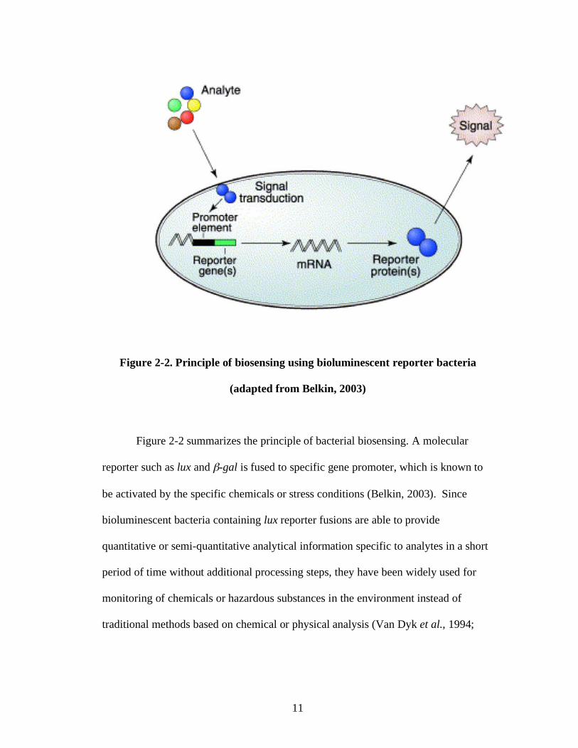
Figure 2-2. Principle of biosensing using bioluminescent reporter bacteria
(adapted from Belkin, 2003)
Figure 2-2 summarizes the principle of bacterial biosensing. A molecular
reporter such as lux and -gal is fused to specific gene promoter, which is known to
be activated by the specific chemicals or stress conditions (Belkin, 2003). Since
bioluminescent bacteria containing lux reporter fusions are able to provide
quantitative or semi-quantitative analytical information specific to analytes in a short
period of time without additional processing steps, they have been widely used for
monitoring of chemicals or hazardous substances in the environment instead of
traditional methods based on chemical or physical analysis (Van Dyk et al., 1994;

12
Belkin et al., 1996, 1997; Vollmer et al., 1997; Davidov et al., 2000; Rosen et al.,
2000).
Figure 2-3 summarizes the mechanism of light emitting reaction controlled by
each lux gene. The five structural genes encode luciferase (luxAB) and fatty acid
reductase complex (luxCDE) catalyzing the biosynthesis of fatty aldehyde substrate.
The bioluminescence reaction is a result of the oxidation of the fatty aldehyde and
reduced flavin mononucleotide (FMNH2) catalyzed by luciferase (luxAB) (Meighen,
1991).
In the genetically engineered bioluminescent bacteria, the lux reporter genes
controlling luciferase are placed under the control of a promoter that is activated by
the presence of specific chemicals and/or cellular activity. When bacteria are exposed
to hazardous chemicals or other environmental stresses, the genetic control
mechanism turns on the synthesis of luciferase, which produces a visible blue-green
light emission that is easy to monitor and quantify. The lux reporter genes have
advantages compared to lacZ genes and other reporter genes used in bacterial systems
because the activity can be monitored in real time without cell lysis (Rozen et al.,
2001). Moreover, if the five-gene luxCDABE reporter is used, the activity of the
reporter may be assayed directly without any additional substrate since all the
requirements for bioluminescence are readily available in bacteria.

13
Figure 2-3. Bacterial bioluminescence pathway
(adapted from Heitzer et al., 1998)
14
Various bioluminescent strains have been constructed to enable screening for
specific toxic mechanisms. However, it is impossible to cover all potential cellular
stress factors with a single reporter gene. Therefore, a panel of genetically engineered
strains should be used to increase their light production in response to a different type
of stress such as oxidative stress (Belkin et al., 1996), DNA damage (Vollmer et al.,
1997; Davidov et al., 2000) and protein damage (Van Dyk et al., 1994) caused by the
presence of organic chemical pollutants such as naphthalene, toluene and
isopropylbenzene. Choi and Gu (2002) developed a biosensor kit using four
recombinant bioluminescent E. coli strains. This biosensor enables one to detect
toxicity of different chemicals on-site by using freeze-dried E. coli cells which show
an increased bioluminescence under specific stressful conditions (e.g. DNA damage,
protein damage, membrane damage, and oxidative stress) (Choi and Gu, 2002).
The limitations of bioluminescent bacteria containing lux reporter fusions are
that they may need a longer time to return to the baseline signal after use, and hence
reversibility may be a problem. Also, the response tends to be slow relative to
enzyme-based sensors since the substrate must first diffuse through the cell wall
(D’Souza, 2001).
Bioluminescent bacteria containing lux reporter fusions have been mainly
used for environmental monitoring: they are also applied for food bacterial detection
in food, screening for bioactive component in food industry and food research, quality
control, and detection of naturally occurring hazardous components. Vansal and
Feller (1999) studied the direct effects of four different ephedrine isomers on human

15
1-, 2- and 3-adrenergic receptors in Chinese hamster ovary (CHO) cells transfected
with a 6 CRE-LUC plasmid by measuring the light production.
16
CHAPTER 3: OBJECTIVES
The objective of this study was to evaluate bioactivity of ephedrine-type
alkaloids by measuring its cytotoxicity against animal cell lines and analyzing the
stress responses of a panel of genetically engineered biosensing bacterial strains
capable of producing real-time responses to specific cell damages. This bioassay
integrating cytotoxicity assessment with real-time biosensing is a systematic approach
to screening Ephedra bioactivity, and the knowledge obtained from the damage
caused by active components of Ephedra on the living cells will help to establish in
vitro assessment tool, and to identify the damage mechanism of ephedrine-type
alkaloids.
To accomplish these objectives, the experiments was divided into two parts;
two cell lines, human neuroblastoma and rat myoblastoma, were used to detect and
profile the cytotoxicity of major ephedrine-type alkaloids, ephedrine and
pseudoephedrine, using a MTT cell proliferation assay. Secondly, six strains of
bioluminescent E. coli were used to identify the stress fingerprints induced by the
ephedrine-type alkaloids.

17
CHAPTER 4: MATERIALS AND METHODS
4.1. Cytotoxicity Assessment Using MTT Cell Proliferation Assay
4.1.1. Materials
Ephedrine-type alkaloids, (1R, 2S)-(-)-ephedrine (99%) and (1S, 2S)-(+)-
pseudoephedrine (98%), were purchased from Aldrich (Allentown, PA). And the
Minimum Essential Medium Alpha Medium and F-12 Nutrient Mixture (HAM) were
from Invitrogen life technologies (Carlsbad, CA). Dulbecco’s Modified Eagle’s
Medium (DMEM), MTT cell proliferation assay kit and other reagents for cell
cultures were from the American Type Culture Collection (ATCC, Manassas, VA).
4.1.2. Cell Cultures
Human neuroblastoma cell line (SH-SY5Y) (ATCC #CRL-2266) and rat
myoblast cell line (H9c2 (2-1)) (ATCC #CRL-1446) were purchased from ATCC.
SH-SY5Y and H9c2 (2-1) were routinely maintained in 1:1 Minimum Essential
Medium/F-12 Nutrient Mixture (HAM) and in Dulbecco’s Modified Eagle’s Medium
(DMEM) respectively. Both culture media are supplemented with 10% fetal bovine
serum. Cells were incubated in a humidified atmosphere of 5% CO2 at 37°C.
4.1.3. MTT Assays
MTT cell proliferation assay kit was used to determine the cytotoxicity of
ephedrine-type alkaloids. Cells were harvested by centrifugation from the
18
maintenance cultures in the exponential phase, stained with trypan blue and counted
by a hemocytometer. Cell suspensions (200 l) were dispensed into 96-well flat-
bottomed tissue culture plates at concentrations of 2 x 10⁵ cells/ml for SH-SY5Y and
3 x 10⁴ cells/ml for H9c2 (2-1). After a recovery period (72-hour for SH-SY5Y and
48-hour for H9c2 (2-1)), the media were removed from the wells with a hypodermic
needle attached to a suction line, and then various concentrations of ephedrine-type
alkaloids ranging from 0.1 to 1.0 mg/ml diluted in 200 l growth media were added
to each well. Control wells received only 200 l growth media. Plates were incubated
at 37°C in 5% CO2 for 72 hours. The media was then aspirated from the wells with a
hypodermic needle attached to a suction line, and 100 l of fresh media added. Ten l
of MTT Reagent was added to each well, and the plates were incubated for 4 hours.
One hundred l of Detergent Reagent (sodium dodecyl sulfate) was added to each
well, and the plates left at room temperature in the dark overnight. The absorbance of
each well was read by the Opsys MR Microplate Reader (Thermo Labsystems,
Chantilly, VA) at 570 nm with 690 nm as the reference wavelength.
4.1.4. Data Analysis
The relative viability of the treated cells as compared to the control cells was
expressed as the % cytoviability, using the following formula:
% cytoviability = [A570 of treated cells] 100% / [A570 of control cells].
The IC50 (median inhibition concentration) was determined by nonlinear
regression analysis of the corresponding dose response curve utilizing the analytical
software package GraphPad Prism Version 4 (GraphPad Software, San Diego, CA).
19
The results were presented as the mean ± standard error of the mean (S.E.M.) of four
experiments, and Student’s t-test was used to assess the significance of the data.
4.2. Real-Time Biosensing Using Bioluminescent E. coli Strains
4.2.1. Bioluminescent E. coli strains
Six bioluminescent E. coli strains obtained from DuPont Genetics Lab (DuPont
Company, Wilmington, DE) was used in this study. Each strain contained different
selected stress-responsive promoter fused to the Photorhabdus luminescence
luxCDABE reporter. The panel strains were chosen to represent a range of stress
responses to result in different patterns of induced gene expression. Each strain
responds respectively to oxidative damage, internal acidification, DNA damage,
protein damage, “super-stationary phase” and sigma S stress (Table 4-1) (Van Dyk,
1998). There are two sets of the stress-responsive E. coli strains, and one set
introduces an outer membrane mutation, tolC, which enables highly sensitive
detection of a variety of organic molecules because their ability to pump out
undesired molecules is limited (Davidov et al., 2000). In this experiment, the set of
six tolC- strains was used.

20
Table 4-1. Stress-responsive E. coli lux fusion strains (adapted from Van Dyk, 1998)
Stress Response
Regulatory Circuit
Promoter Fused to lux
Strain Name
tolC
allele Plasmid-
containing
DNA damage SOS recA DPD1710 DPD2222
+ -
No No
“super-stationary phase”
? o513 DPD2173 DPD2232
+ -
Yes Yes
sigma S stress response
Stationary phase ( s)
yciG DPD2161 DPD2233
+ -
Yes Yes
protein damage Heatshock
( 32) grpE DPD3084
DPD2234 + -
No No
oxidative damage
OxyR & s katG DPD2227 DPD2238
+ -
Yes Yes
internal acidification
Mar/sox/Rob inaA DPD2226 DPD2240
+ -
Yes Yes
21
The E. coli strains were maintained in a 70% glycerol suspension at -78°C.
Prior to the assay, the stock cultures were transferred to 250 ml flasks with 50 ml
sterilized Luria Bertani (LB) medium and incubated for 12 hours at 37°C on a model
1575 orbital shaking incubator (VWR Scientific, Cornelius, OR) at 300 rpm. To
ensure stability of the plasmids containing lux fusion genes, ampicillin (100 µg/ml)
was added in the growth media of all the strains except for strains DPD2222 and
DPD2234, which have the lux gene fusion in their chromosomes.
4.2.2. E. coli stress fingerprinting
Cultures of each E. coli strain were diluted with sterile distilled water at a
ratio of 1:10. Nine hundred µl of culture solution was placed in a transparent glass
cuvette. Luminescence from E. coli of each cuvette was measured by a TD-20e
luminometer (Turner Designs, Sunnyvale, CA) and the luminescence values were
presented as instrument’s arbitrary relative light unit (RLU). The RLU value was
recorded again after 100 µl of ephedrine-type alkaloids. The difference and ratio of
the two RLU values (“before RLU” and “after RLU”) were both calculated to
indicate the stress responses from the bioluminescent E. coli strains.
RLU = “after RLU” – “before RLU”
RLU ratio = “after RLU”/”before RLU”
If the RLU rations are greater than 1.0 (“lights-on”), it indicates that the lux
gene fusion is expressed because of the ephedrine-type alkaloids. If the RLU rations
22
are less than 1.0 (“lights-off”), it suggests a dampening of bioluminescent E. coli
strains in the presence of ephedrine-type alkaloids.
4.2.3. Data Analysis
Data are presented as mean ± the standard errors of the mean (S. E. M.).
Statistical analysis was performed using either Student’s t-test or analysis of variance
(ANOVA) utilizing the analytical software package GraphPad Prism Version 4
(GraphPad Software, San Diego, CA). For ANOVA, pairwise comparisons between
treatments were made using Tukey’s Multiple Test Comparison.
23
CHAPTER 5: RESULTS AND DISCUSSION
5.1. Cytotoxicity Assessment Using MTT Cell Proliferation Assay
5.1.1. Effects of Ephedrine and Pseudoephedrine on Cytoviability
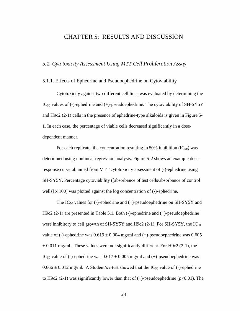
Cytotoxicity against two different cell lines was evaluated by determining the
IC50 values of (-)-ephedrine and (+)-pseudoephedrine. The cytoviability of SH-SY5Y
and H9c2 (2-1) cells in the presence of ephedrine-type alkaloids is given in Figure 5-
1. In each case, the percentage of viable cells decreased significantly in a dose-
dependent manner.
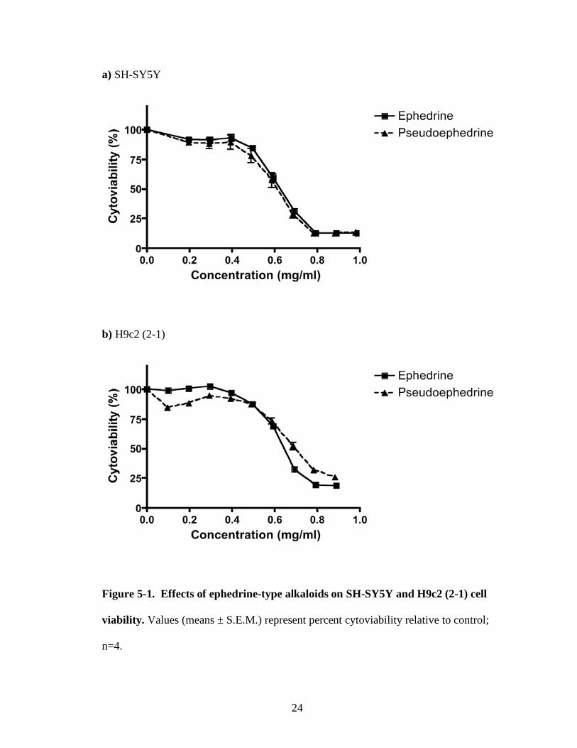
For each replicate, the concentration resulting in 50% inhibition (IC50) was
determined using nonlinear regression analysis. Figure 5-2 shows an example dose-
response curve obtained from MTT cytotoxicity assessment of (-)-ephedrine using
SH-SY5Y. Percentage cytoviability ([absorbance of test cells/absorbance of control
wells] 100) was plotted against the log concentration of (-)-ephedrine.
The IC50 values for (-)-ephedrine and (+)-pseudoephedrine on SH-SY5Y and
H9c2 (2-1) are presented in Table 5.1. Both (-)-ephedrine and (+)-pseudoephedrine
were inhibitory to cell growth of SH-SY5Y and H9c2 (2-1). For SH-SY5Y, the IC50
value of (-)-ephedrine was 0.619 ± 0.004 mg/ml and (+)-pseudoephedrine was 0.605
± 0.011 mg/ml. These values were not significantly different. For H9c2 (2-1), the
IC50 value of (-)-ephedrine was 0.617 ± 0.005 mg/ml and (+)-pseudoephedrine was
0.666 ± 0.012 mg/ml. A Student’s t-test showed that the IC50 value of (-)-ephedrine
to H9c2 (2-1) was significantly lower than that of (+)-pseudoephedrine (p<0.01). The
24
a) SH-SY5Y
b) H9c2 (2-1)
Figure 5-1. Effects of ephedrine-type alkaloids on SH-SY5Y and H9c2 (2-1) cell
viability. Values (means ± S.E.M.) represent percent cytoviability relative to control;
n=4.
25
Figure 5-2. Sample dose-response curve. Percentage cytoviability ([absorbance of
treated cells]/[absorbance of control cells] 100) plotted against the concentration of
ephedrine-type alkaloids.
IC50
26
results indicate that H9c2 (2-1) could differentiate the cytotoxicity of (-)-ephedrine
and (+)-pseudoephedrine while SH-SY5Y showed similar sensitivity to both
ephedrine-type alkaloids. Figure 5-3 shows morphological changes of H9c2 (2-1) cell
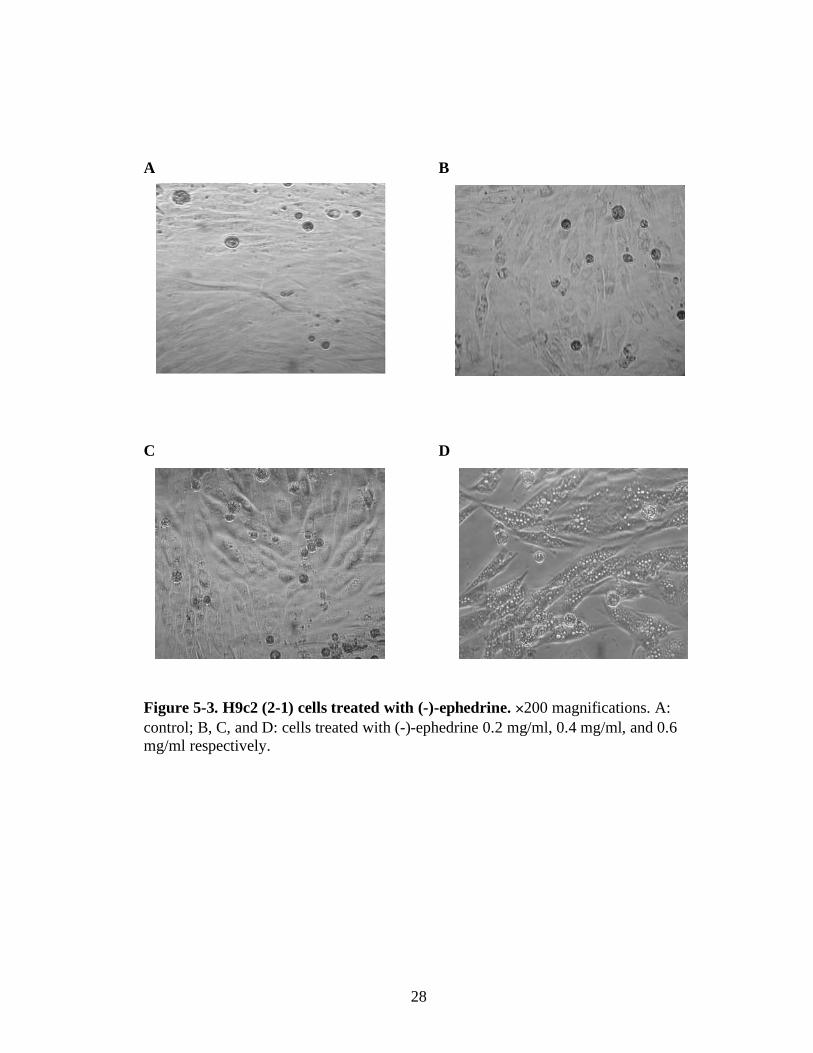
line when it was treated with (-)-ephedrine for 72 hours. As the concentration of (-)-
ephedrine increases, swelling of the cells became prominent and vacuolar
degeneration was observed microscopically.
5.1.2. Effects of Combined Treatments by Ephedrine and Pseudoephedrine
The cumulative effects of (-)-ephedrine and (+)-pseudoephedrine on the
cytoviability of SH-SY5Y and H9c2 (2-1) cells were illustrated in Figure 5-4. At low
concentrations of (-)-ephedrine (0.2 mg/ml) and (+)-pseudoephedrine (0.2 mg/ml), the
cytoviability of SH-SY5Y cells were suppressed 11 and 12%, respectively. The
combined treatment suppressed cytoviability 18%, slightly lowering the 23%
predicted by the sum of the individual suppression. Other combination of (-)-
ephedrine (0.4 mg/ml) and (+)-pseudoephedrine (0.2 mg/ml) suppressed the
cytoviability to the extent predicted by the individual effects while the combination of
(-)-ephedrine (0.2 mg/ml) and (+)-pseudoephedrine (0.4 mg/ml) suppressed the
cytoviability to the larger extent than additive effect. For H9c2 (2-1), on the other
hand, all the combinations of (-)-ephedrine and (+)-pseudoephedrine showed lesser
suppression of cytoviability than predicted value from the individual effects.

27
Table 5-1. The IC50 values of (-)-ephedrine and (+)-pseudoephedrine for two cell
lines.
IC50 (mg/ml)
Cell Lines ephedrine pseudoephedrine
SH-SY5Y 0.619 ± 0.004a 0.605 ± 0.011a
H9c2 (2-1) 0.617 ± 0.005a 0.666 ± 0.012b
Each value is the mean ± S.E.M., n=4.
IC50: concentration of test sample to inhibit cytoviability by 50%
Values followed with an identical letter are not significantly different (p<0.01).
28
A B
C D
Figure 5-3. H9c2 (2-1) cells treated with (-)-ephedrine. 200 magnifications. A:
control; B, C, and D: cells treated with (-)-ephedrine 0.2 mg/ml, 0.4 mg/ml, and 0.6 mg/ml respectively.

29
a) SH-SY5Y
b) H9c2 (2-1)
Figure 5-4. Cumulative effects of two ephedrine-type alkaloids on the
cytoviability of SH-SY5Y and H92c (2-1). Shown are mean ± S. E. M. for n=6.
E: (-)-ephedrine, P: (+)-pseudoephedrine.
30
5.1.3. Discussion
Among the adverse events potentially associated with dietary supplements
containing ephedrine-type alkaloids reported to FDA from 1997 through 1999,
approximately 60% of the total reports were characterized as clinically serious such
as deaths, cardiovascular events, and serious nervous system effects. Serious nervous
system effects including seizures, psychosis and depression accounted for 16% of the
total adverse events (FDA, 2000). Lee et al. (2000) assayed the cytotoxicity of pure
ephedrine and extracts of Ephedra using a mouse neuroblastoma cell line (Neuro-2a)
and reported that Neuro-2a was more sensitive to Ephedra extracts compared to other
cell lines including human hepatoblastoma (HepG2). In this study, a human
neuroblastoma cell line SH-SY5Y, which is frequently used in the assessments of
cytotoxicity or protective effects for other chemicals (Slaughter et al., 2002; Legros et
al., 2004; Miglio et al., 2004), was employed instead of mouse neuroblastoma cell
line, to investigate whether it would show similar sensitivity to ephedrine-type
alkaloids.
Serious cardiovascular events that included myocardial infraction (MI),
unstable angina, dysrhythmias and transient ischemic attacks, accounted for 31% of
reported adverse events (FDA, 2000). Therefore, rat myocardium cell line (H9c2 (2-
1)) was used in this MTT cytotoxicity assessment in addition to SH-SY5Y. The H9c2
(2-1) cell line is a permanent cell line derived from rat heart tissue, that shows
morphological characteristics similar to those of immature embryonic cardiocytes and
has preserved several elements of the electrical and hormonal signaling pathways
31
found in adult rat cardiac myocytes (Hescheler et al., 1991). Also, it has been
reported that H9c2 (2-1) expresses both 1- and 2-adrenergic receptors on which
ephedrine acts (Dangel et al., 1996). Therefore, H9c2 (2-1) cell line was expected to
be a useful model for cytotoxicity assessment of ephedrine-type alkaloids.
With regard to the cytotoxicity of (-)-ephedrine, the IC50 values achieved from
the current study (0.615 ~ 0.616 mg/ml) were close to the results previously reported
(Lee et al., 2000). However, Lee et al. also reported that the cytotoxicity of (-)-
ephedrine was significantly higher than that of (+)-pseudoephedrine when they
determined the % cytoviability of equimolar concentrations (3.0 mM = approximately
0.50 mg/ml) of (-)-ephedrine and (+)-pseudoephedrine for HepG2 cell line (30.8% for
(-)-ephedrine and 89.4% for (+)-pseudoephedrine). In the current study, the %
cytoviability of 0.50 mg/ml of (-)-ephedrine and (+)-pseudoephedrine was 83.7% and
76.5% for SH-SY5Y and 91.4% and 86.1% for H9c2 (2-1) respectively, and
Student’s t-test showed no significant difference in either cell line at this level of
concentration. The same can be said for the IC50 values. There was no significant
difference between (-)-ephedrine and (+)-pseudoephedrine for SH-SY5Y. Although (-
)-ephedrine is considered to be more potent and toxic than (+)-pseudoephedrine
clinically (Tang, 1996), it is possible that the sensitivity to (-)-ephedrine and (+)-
pseudoephedrine is different from cell line to cell line, and it may not be necessarily
appropriate to suggest that (-)-ephedrine is more cytotoxic than (+)-pseudoephedrine.
For H9c2 (2-1), (-)-ephedrine was proved to be more cytotoxic but the difference was
less prominent.
32
Although there is a little data on blood and tissue levels of (-)-ephedrine and
other ephedrine-type alkaloids in humans, Haller et al. (2002) reported that maximum
plasma concentrations were 63.5 ng/ml for ephedrine and 24.1 ng/ml for
pseudoephedrine 2.4 hours after the ingestion of dietary supplements containing
ephedrine-type alkaloids (17.3 mg ephedrine, 5.3 mg pseudoephedrine and
insignificant amounts of the other alkaloids on an average). Other studies also
showed that the therapeutic plasma levels of ephedrine in humans following ingestion
of ephedrine in the form Ephedra sinica capsules (19 mg ephedrine), an ephedrine
tablet (20 mg ephedrine), or an ephedrine solution (22 mg) are 81 ng/ml, 74 ng/ml,
and 79 ng/ml respectively (Vansal and Feller, 1999). The IC50 values achieved from
the present study were 0.605 ~ 0.666 mg/ml, which were several orders of magnitude
higher than the therapeutic plasma levels. Therefore, it is difficult to directly associate
the in vitro cytotoxic effects with the actual adverse responses of human.
Ephedrine is both a direct and indirect adrenergic agonist (Abourashed et al.,
2003). That is, ephedrine activates adrenergic receptors both by direct agonist activity
as well as by releasing norepinephrine via carrier-mediated exchange mechanism.
Ephedrine possesses two asymmetrical carbon atoms and exists as four isomers.
Among these four ephedrine isomers, (-)-ephedrine and (+)- pseudoephedrine are
naturally contained in some of the Ephedra species (Vansal and Feller, 1999). Vansal
and Feller (1999) reported (-)-ephedrine was the most potent of the four ephedrine
isomers on human -adrenergic receptors expressed in Chinese hamster ovary cells.
However, Rothman et al. (2003) reported that ephedrine-type alkaloids showed no
agonist effects at 1- and -adrenergic receptors and they suggested that

33
pharmacological actions of ephedrine and its derivatives resulted primarily from
release of norepinephrine rather than direct activation of adrenergic receptors.
Although the cell lines employed in this study are reported to express several
subtypes of the adrenergic receptors (Dangel et al., 1996), it is not known if their
response resulted from the direct effects on a specific adrenergic receptor or from
indirect effects. However, the MTT assay using these cell lines may serve as an
efficient model to detect general cytotoxicity of compounds included in dietary
supplements.
Although little is known about the cytotoxicity of other ephedrine-type
alkaloids and compounds in Ephedra, norpseudoephedrine is known to be more
potent than (-)-ephedrine with regard to CNS stimulation (Kalix, 1991). Moreover,
the presence of toxins in Ephedra other than (-)-ephedrine is suggested since IC50
values of Ephedra extracts normalized by their (-)-ephedrine contents were
significantly lower than pure (-)-ephedrine (Lee et al., 2000). Dietary supplements
containing ephedrine-type alkaloids often include other agents including stimulants,
diuretics and cathartics (CANTOX, 2000). The synergistic interaction between
ephedrine-type alkaloids and caffeine is well known (Haller et al., 2004). Therefore,
it may also be useful to evaluate the interaction between ephedrine-type alkaloids and
other functional compounds using this MTT cytotoxicity assessment.
In response to the FDA’s decision to ban the sales of Ephedra-containing
dietary supplements, manufacturers and companies have developed new “Ephedra-
free” dietary supplements (Marcus and Grollman, 2003). One of the most popular
substitutes for Ephedra is Citrus aurantium (bitter orange). Its major active

34
component “synephrine” is an ephedrine-type alkaloid and the combination of
synephrine and caffeine has the similar potential to induce cardiac arrhythmias,
hypertension, heart attacks, and strokes as the combination of Ephedra and caffeine
(Marcus and Grollman, 2003). The association of dietary supplements containing
bitter oranges with myocardial infarction has been reported (Nykamp et al., 2004).
Therefore, there is a concern that the misuse of “emerging” dietary supplements
containing botanical substances might cause other health problems. The MTT
cytotoxicity assay used in the present study could serve as a useful assessment tool to
analyze the cytotoxic pattern of other botanical substances contained in dietary
supplements and to predict potential adverse effects.
5.2. Real-Time Biosensing Using Bioluminescent E. coli Strains
The RLU ratio of the E. coli strains exposed to (-)-ephedrine and (+)-
pseudoephedrine at differing concentrations (0.03 mg/ml to 0.06mg/ml) is
summarized in Figures 5-5 - 5-8. All strains showed increased bioluminescence in
response to the stress caused by (-)-ephedrine and (+)-pseudoephedrine at the
concentrations as low as 0.03 mg/ml. This concentration was much lower than the
ID50 (0.605 ~ 0.666 mg/ml) obtained from the cytotoxicity assessment using human
and rat cell lines.
The RLU ratio of a panel of bioluminescent E. coli strains exposed to (-)-ephedrine
and (+)-pseudoephedrine at a concentration of 0.3 mg/ml is presented in Figure 5-9.
Both (-)-ephedrine and (+)-pseudoephedrine decreased the RLU ratios and was toxic
to all strains. The RLU ratios in four strains (DPD2232, DPD2233, DPD2238 and

35
DPD2240) for (-)-ephedrine were less than 1.0 (“Lights-off” response) which could
be attributed to inhibition of cellular metabolism required for production of energy or
reduction power (Chatterjee and Meighen, 1993), and the RLU ratios were
significantly lower than those of (+)-pseudoephedrine, whereas there was no
significant difference in strains DPD2222 and DPD2234. These results indicate that
this biosensing panel was sensitive to the bioactive effects caused by ephedrine-type
alkaloids and it distinguished (-)-ephedrine and (+)-pseudoephedrine. The strains
DPD2222 and DPD2234 showed less intense response to (-)-ephedrine compared to
other 4 strains. A possible reason for this is that the host strain of DPD2222 and
DPD2234 is MM28, which is different from the parental strain of other 4 strains
(GC4468). Moreover, DPD2222 and DPD2234 have their lux-fusions in their
chromosome while the other strains have plasmids containing lux-fusions. These
might affect the sensitivity to detect the toxicity of ephedrine-type alkaloids in short
time.
At a concentration of 0.03 mg/ml, (-)-ephedrine induced increased
bioluminescence in DPD2222, DPD2232 and DPD2234. In contrast, (+)-
pseudoephedrine induced bioluminescence in DPD2222, DPD2232 and DPD2233.
Strains DPD2238 and DPD2240 showed no significant response to either (-)-
ephedrine or (+)-pseudoephedrine. Also, RLU ratios of (-)-ephedrine and (+)-
pseudoephedrine were not significantly different in all strains.
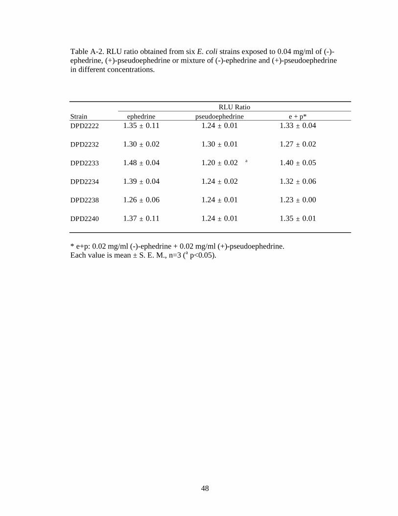
At a concentration of 0.04 mg/ml, (-)-ephedrine induced increased
bioluminescence in DPD2232, DPD2233 and DPD2234, and the RLU ratio of
DPD2233 was significantly higher than (+)-pseudoephedrine. In contrast, (+)-
36
pseudoephedrine only increased the RLU ratio in strain DPD2232. Strains DPD2222,
DPD2238 and DPD2240 showed no significant responses either to (-)-ephedrine or
(+)-pseudoephedrine.
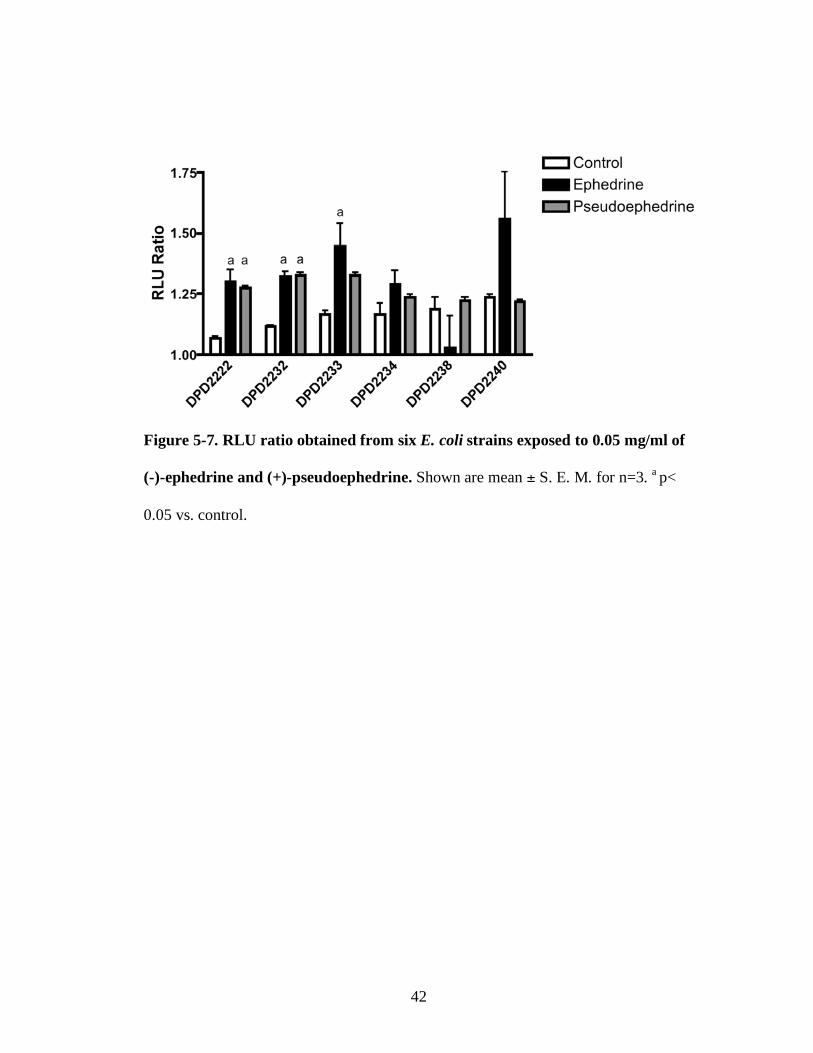
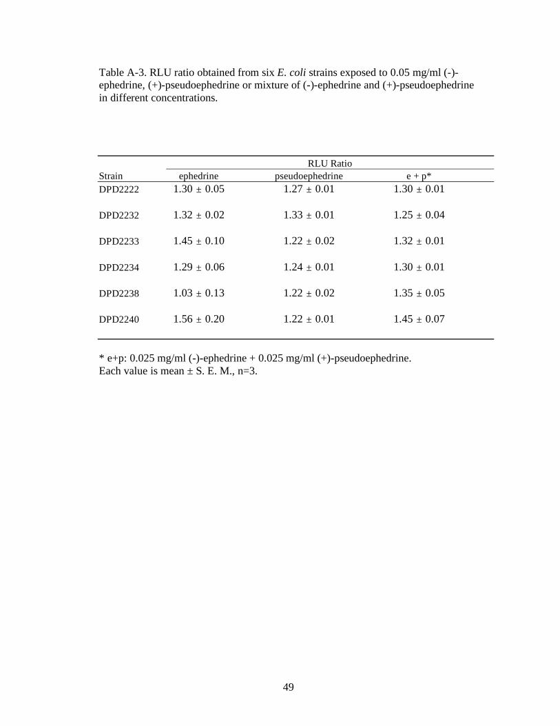
At the concentration of 0.05 mg/ml, (-)-ephedrine induced increased
bioluminescence in DPD2222, DPD2232 and DPD2233 compared to the control
although they were not significantly different from those of (+)-pseudoephedrine. On
the other hand, (+)-pseudoephedrine also increased the RLU ratio of strains DPD2222
and DPD2232. Strains DPD2234, DPD2238 and DPD2240 showed no significant
responses either to (-)-ephedrine or (+)-pseudoephedrine.
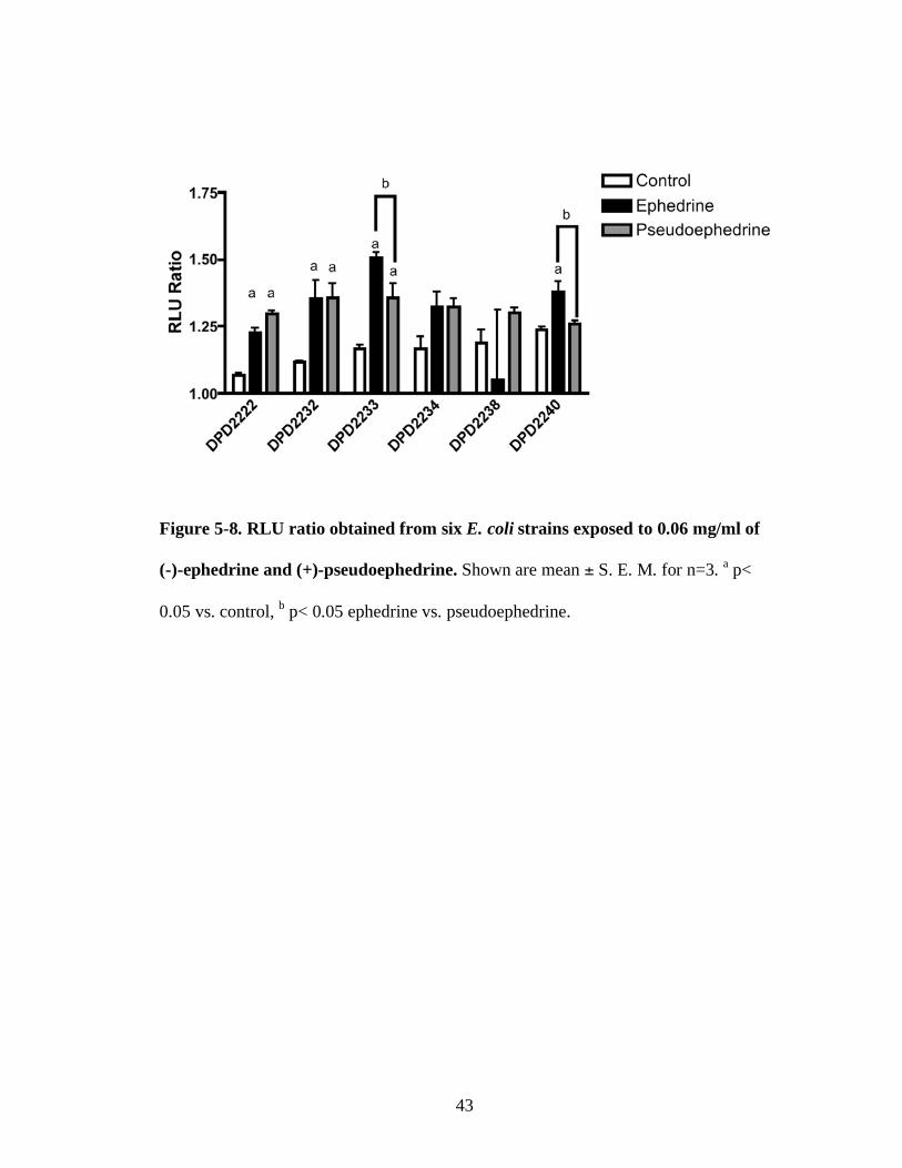
At a concentration of 0.06 mg/ml, (-)-ephedrine induced bioluminescence in
DPD2222, DPD2232, DPD2233 and DPD2240 compared to the control although they
were not significantly different from those of (+)-pseudoephedrine. (+)-
Pseudoephedrine also increased the RLU ratio in strains DPD2222 and DPD2232.
Strains DPD2234 and DPD2238 showed no significant responses either to (-)-
ephedrine or (+)-pseudoephedrine.
Strain DPD2233 showed increased response to (-)-ephedrine in the
concentration range of 0.04 mg/ml ~ 0.06 mg/ml. Strain DPD2233 is constructed to
contain the plasmid in which the E. coli yciG promoter is fused to luxCDABE genes.
As expression of yciG gene is under control of the stationary phase sigma factor s,
the yciG-lux fusion is expected to report on the activation of the s-dependent stress
response (Van Dyk, 1998).
In the present study, (-)-ephedrine slightly increased the bioluminescence of
strain DPD2234 at the lower concentration (0.03 mg/ml and 0.04 mg/ml), but showed

37
no significant increase of RLU ratio when exposed to (+)-pseudoephedrine.
Escherichia coli strain DPD2234 contains chromosomal insertion of a grpE promoter
fused to the P. luminescens luxCDABE. Since grpE gene is in the heat shock regulon
controlled by 32, the grpE-lux fusion responds to stresses that induce this protein-
damage responsive regulon (Van Dyk, 1998), and it is also known to respond to a
variety of stresses and chemicals (Van Dyk et al., 1995). Increased RLU ratio of the
strain DPD2234 indicated the induction of the general stress-response by (-)-
ephedrine.
Both (-)-ephedrine and (+)-pseudoephedrine appeared to increase the RLU
ratio of strain DPD2222, which is designed to respond to DNA damage through SOS
regulatory circuit. The increased RLU ratio of DPD2222 indicated the possibility that
DNA damage-sensing SOS response was induced by both (-)-ephedrine and (+)-
pseudoephedrine. Strain DPD2222 is the one that contains chromosomal insertion of
E. coli recA promoter fused to the P. luminescens luxCDABE. When DNA damage is
present, a resultant single stranded DNA acts as a signal for induction of the SOS
response (Daunert et al., 2000). According to the studies on the genotoxicity of
ephedrine and Ephedra extract, both in vitro and in vivo, ephedrine sulfate had no
genotoxicity in three strains of Salmonella typhimurium (TA97, TA98, TA100 and
TA1535), nor in cultured Chinese hamster ovary (CHO) cells (NTP, 1986). Hillard et
al. (1998) reported that ephedrine sulfate was negative in two chromosome aberration
tests using CHO cells. These studies demonstrated that ephedrine and Ephedra are not
genotoxic. Although SOS activation in itself does not imply genotoxicity, the two
activities are reported to be correlated (Davidov et al., 2000). Further information is
38
thus needed to determine whether mechanisms other than DNA damages are involved
in the stress response obtained in the present study, or ephedrine-type alkaloids cause
DNA damage but not mutation.
Strain DPD2238 showed no significant increase of RLU ratio when exposed
to either (-)-ephedrine or (+)-pseudoephedrine in different concentrations. The strain
DPD2238 is constructed to contain the plasmid in which the E. coli katG (catalase)
promoter is fused to luxCDABE genes. An E. coli strain harboring this plasmid is
known to exhibit low basal levels of luminescence, which increased up to 1,000-fold
in the presence of oxidative stress such as hydrogen peroxide, organic peroxides,
alcohols and cigarette smoke (Belkin et al., 1996). Kang et al. (1998) reported that
ephedrine exerted mild antioxidant activity in vitro. The result from the present study
that oxidative stress response was absent supports their observations. If direct
correlations could be established between antioxidant activity and oxidative stress
response, strain DPD2238 could be used as a tool to assay antioxidant property of
ephedrine-type alkaloids.
Strain DPD2232 is constructed to contain the plasmid in which the E. coli
o513 promoter is fused to luxCDABE genes. Although the regulation of o513 has not
been well characterized, it was reported that expression of a lux fusion to an open
reading frame o513 is highly induced as the culture ages, suggesting that stationary
phase induces the expression of o513; however such expression is not controlled by
s (Van Dyk, 1998). Van Dyk (1998) also observed that o513-lux fusion did not yield
increased bioluminescence to the wide range of chemicals (e.g., hydrogen peroxide,
nalidixic acid, ethanol, sodium salicylate, and paraquat). Rather, it gave a response
39
ratio of less than 1.0 (“lights off”). Therefore, Van Dyk (1998) proposed to use the
strain containing o513-lux fusion as a general indicator of toxicity. Interestingly, both
(-)-ephedrine and (+)-pseudoephedrine significantly induced bioluminescence of the
o513-lux fusion strain (DPD2232) compared to the control, and the “lights off”
response was only observed for (-)-ephedrine at the high concentration of 0.3 mg/ml.
This result suggests the possibility that o513 promoter is activated in response to the
stress caused by ephedrine-type alkaloids.

40
Figure 5-5. RLU ratio obtained from six E. coli strains exposed to 0.03 mg/ml of
(-)-ephedrine and (+)-pseudoephedrine. Shown are mean ± S. E. M. for n=3. a p<
0.05 vs. control.

41
Figure 5-6. RLU ratio obtained from six E. coli strains exposed to 0.04 mg/ml of
(-)-ephedrine and (+)-pseudoephedrine. Shown are mean ± S. E. M. for n=3. a p<
0.05 vs. control, b p< 0.05 ephedrine vs. pseudoephedrine.
42
Figure 5-7. RLU ratio obtained from six E. coli strains exposed to 0.05 mg/ml of
(-)-ephedrine and (+)-pseudoephedrine. Shown are mean ± S. E. M. for n=3. a p<
0.05 vs. control.
43
Figure 5-8. RLU ratio obtained from six E. coli strains exposed to 0.06 mg/ml of
(-)-ephedrine and (+)-pseudoephedrine. Shown are mean ± S. E. M. for n=3. a p<
0.05 vs. control, b p< 0.05 ephedrine vs. pseudoephedrine.

44
Figure 5-9. RLU ratio obtained from six E. coli strains exposed to 0.3 mg/ml of (-
)-ephedrine and (+)-pseudoephedrine. Shown are mean ± S. E. M. for n=3.
45
CHAPTER 6: CONCLUSIONS
In the present study, the bioactivity of two major ephedrine-type alkaloids
were evaluated by measuring its cytotoxicity against the cell lines and analyzing the
stress response of a panel of genetically engineered biosensing E. coli strains capable
of producing real-time responses to specific cell damages.
The two ephedrine-type alkaloids, (-)-ephedrine and (+)-pseudoephedrine,
showed cytotoxicity to the human neuroblastoma SH-SY5Y and rat myoblastoma
H9c2 (2-1) cell lines. SH-SY5Y cell lines showed similar sensitivity to (-)-ephedrine
and (+)-pseudoephedrine while H9c2 (2-1) cell line was able to differentiate the
cytotoxicity of (-)-ephedrine and (+)-pseudoephedrine. MTT assay using established
cell lines could provide information on general information on cytotoxicity of
ephedrine-type alkaloids, but a variety of cell lines should be used to assess
organ/tissue toxicity.
Biosensing using a panel of bioluminescent E. coli strains was highly sensitive
to ephedrine-type alkaloids and could produce a rapid response at the concentration as
low as 0.03 mg/ml. Moreover, these E. coli strains distinguished the toxicity of (-)-
ephedrine and (+)-pseudoephedrine; the bioluminescence from the E. coli strains was
significantly suppressed by (-)-ephedrine compared to (+)-pseudoephedrine at the
high concentration 0.3 mg/ml. At the lower concentrations, (-)-ephedrine generally
induced higher bioluminescence than (+)-pseudoephedrine did throughout the
experiments. As the RLU ratios dependent on the construct of strains gave unique

46
fingerprinting pattern of ephedrine-type alkaloids, this biosensing panel has a
potential to be a very effective clue for clarifying the toxicity mechanism of
ephedrine-type alkaloids. However, this biosensing panel did not give clear dose-
dependent response in the range of concentrations used in the present study. In order
to obtain quantitative information, it might be needed to assess the stress response
using wider range of concentration.

47
Appendices
Table A-1. RLU ratio obtained from six E. coli strains exposed to 0.03 mg/ml of (-)-ephedrine, (+)-pseudoephedrine or mixture of (-)-ephedrine and (+)-pseudoephedrine in different concentrations.
RLU Ratio
Strain ephedrine pseudoephedrine e + p*
DPD2222 1.31 ± 0.01 1.28 ± 0.02 1.18 ± 0.02 a
DPD2232 1.32 ± 0.05 1.31 ± 0.02 1.20 ± 0.03
DPD2233 1.29 ± 0.06 1.33 ± 0.03 1.31 ± 0.02
DPD2234 1.36 ± 0.01 1.27 ± 0.03 1.31 ± 0.07
DPD2238 1.26 ± 0.04 1.26 ± 0.02 1.29 ± 0.01
DPD2240 1.24 ± 0.00 1.34 ± 0.06 1.37 ± 0.06
* e+p: 0.015 mg/ml (-)-ephedrine + 0.015 mg/ml (+)-pseudoephedrine. Each value is mean ± S. E. M., n=3 (a p<0.05).
48
Table A-2. RLU ratio obtained from six E. coli strains exposed to 0.04 mg/ml of (-)-ephedrine, (+)-pseudoephedrine or mixture of (-)-ephedrine and (+)-pseudoephedrine in different concentrations.
RLU Ratio
Strain ephedrine pseudoephedrine e + p*
DPD2222 1.35 ± 0.11 1.24 ± 0.01 1.33 ± 0.04
DPD2232 1.30 ± 0.02 1.30 ± 0.01 1.27 ± 0.02
DPD2233 1.48 ± 0.04 1.20 ± 0.02 a 1.40 ± 0.05
DPD2234 1.39 ± 0.04 1.24 ± 0.02 1.32 ± 0.06
DPD2238 1.26 ± 0.06 1.24 ± 0.01 1.23 ± 0.00
DPD2240 1.37 ± 0.11 1.24 ± 0.01 1.35 ± 0.01
* e+p: 0.02 mg/ml (-)-ephedrine + 0.02 mg/ml (+)-pseudoephedrine. Each value is mean ± S. E. M., n=3 (a p<0.05).
49
Table A-3. RLU ratio obtained from six E. coli strains exposed to 0.05 mg/ml (-)-ephedrine, (+)-pseudoephedrine or mixture of (-)-ephedrine and (+)-pseudoephedrine in different concentrations.
RLU Ratio
Strain ephedrine pseudoephedrine e + p*
DPD2222 1.30 ± 0.05 1.27 ± 0.01 1.30 ± 0.01
DPD2232 1.32 ± 0.02 1.33 ± 0.01 1.25 ± 0.04
DPD2233 1.45 ± 0.10 1.22 ± 0.02 1.32 ± 0.01
DPD2234 1.29 ± 0.06 1.24 ± 0.01 1.30 ± 0.01
DPD2238 1.03 ± 0.13 1.22 ± 0.02 1.35 ± 0.05
DPD2240 1.56 ± 0.20 1.22 ± 0.01 1.45 ± 0.07
* e+p: 0.025 mg/ml (-)-ephedrine + 0.025 mg/ml (+)-pseudoephedrine. Each value is mean ± S. E. M., n=3.

50
Table A-4. RLU ratio obtained from six E. coli strains exposed to 0.06 mg/ml (-)-ephedrine, (+)-pseudoephedrine or mixture of (-)-ephedrine and (+)-pseudoephedrine in different concentrations.
RLU Ratio
Strain ephedrine pseudoephedrine e + p
DPD2222 1.23 ± 0.02 a 1.30 ± 0.01 b 1.29 ± 0.01 ab
DPD2232 1.36 ± 0.07 1.36 ± 0.05 1.15 ± 0.01
DPD2233 1.51 ± 0.02 a 1.30 ± 0.04 ab 1.24 ± 0.04 b
DPD2234 1.32 ± 0.06 1.32 ± 0.03 1.20 ± 0.01
DPD2238 1.05 ± 0.26 1.30 ± 0.02 1.17 ± 0.02
DPD2240 1.38 ± 0.04 1.26 ± 0.01 1.32 ± 0.03
* e+p: 0.03 mg/ml (-)-ephedrine + 0.03 mg/ml (+)-pseudoephedrine. Each value is mean ± S. E. M., n=3. Values followed with an identical letter are not significantly different (p<0.05).

51
References
Abourashed, E. A., El-Alfy, A. T., Khan, I. A., and Walker, L. (2003). Ephedra in
perspective--a current review. Phytother Res 17, 703-712.
Altman, F. P. (1976). Tetrazolium salts and formazans. Prog Histochem Cytochem 9,
1-56.
Anon. (1998). Nutrition Business Journal Annual Industry Overview. Nutrition
Business Journal.
Barile, F. A. (1994). Introduction to in vitro cytotoxicology: mechanisms and
methods (Boca Raton, FL, CRC Press).
Belkin, S. (2003). Microbial whole-cell sensing systems of environmental pollutants.
Current Opinion in Microbiology 6, 206-212.
Belkin, S., Smulski, D., Vollmer, A., Van Dyk, T., and LaRossa, R. (1996). Oxidative
stress detection with Escherichia coli harboring a katG'::lux fusion. Appl
Environ Microbiol 62, 2252-2256.
Belkin, S., Smulski, D. R., Dadon, S., Vollmer, A. C., Van Dyk, T. K., and LaRossa,
R. A. (1997). A panel of stress-responsive luminous bacteria for the
detection of selected classes of toxicants. Wat Res 31, 3009-3016.
Betz, J. M., Gay, M. L., Mossoba, M. M., Adams, S., and Portz, B. S. (1997). Chiral
gas chromatographic determination of ephedrine-type alkaloids in dietary
supplements containing ma huang. J AOAC Int 80, 303-315.
Blumenthal, M., and King, P. (1995). Ma Huang: Ancient herb, modern medicine,
regulatory dilemma; A review of the botany, chemistry, medicinal uses,

52
safety concerns, and legal status of Ephedra and its alkaloids. Herbal Gram,
22+.
Broadhead, C. L., and Combes, R. D. (2001). The current status of food additives
toxicity testing and the potential for application of the three Rs. Altern Lab
Anim 29, 471-485.
Bruneton, J. (1995). Pharmacognosy, phytochemistry, medicinal plants (Paris,
Lavoisier).
CANTOX (2000). Safety Assessment and Determination of a Tolerable Upper Limit
for Ephedra (Ontario, Canada, CANTOX Health Sciences International).
Cetaruk, E. W., and Aaron, C. K. (1994). Emergency medical clinics of north
america. Hazards of nonprescription medications 12, 483-510.
Chatterjee, J., and Meighen, E. A. (1995). Biotechnological Applications of Bacterial
Bioluminescence (lux) Genes. Photochemistry and Photobiology 62, 641-
650.
Chinaka, S., Tanaka, S., Takayama, N., Komai, K., Ohshima, T., and Ueda, K.
(2000). Simultaneous chiral analysis of methamphetamine and related
compounds by capillary electrophoresis. J Chromatogr B Biomed Sci Appl
749, 111-118.
Choi, S. H., and Gu, M. B. (2002). A portable toxicity biosensor using freeze-dried
recombinant bioluminescent bacteria. Biosens Bioelectron 17, 433-440.
Cui, J. F., Niu, C. Q., and Zhang, J. S. (1991). [Determination of six Ephedra
alkaloids in Chinese Ephedra (ma huang) by gas chromatography]. Yao Xue
Xue Bao 26, 852-857. [Article in Chinese]
53
Dangel, V., Giray, J., Ratge, D., and Wisser, H. (1996). Regulation of beta-
adrenoceptor density and mRNA levels in the rat heart cell-line H9c2.
Biochem J 317, 925-931.
Daunert, S., Barrett, G., Feliciano, J. S., Shetty, R. S., Shrestha, S., and Smith-
Spencer, W. (2000). Genetically engineered whole-cell sensing systems:
coupling biological recognition with reporter genes. Chem Rev 100, 2705-
2738.
Davidov, Y., Rozen, R., Smulski, D. R., Van Dyk, T. K., Vollmer, A. C., Elsemore,
D. A., LaRossa, R. A., and Belkin, S. (2000). Improved bacterial SOS
promoter:: lux fusions for genotoxicity detection. Mutat Res 466, 97-107.
Dong, X., Sun, H., Lu, X., Wang, H., Liua, S., and Wang, N. (2002). Separation of
ephedrine stereoisomers by molecularly imprinted polymers--influence of
synthetic conditions and mobile phase compositions on the chromatographic
performance. Analyst 127, 1427-1432.
D’Souza, S. F. (2001). Microbial biosensors. Biosens Bioelectron 16, 337-353.
Dulloo, A. G., and Stock, M. J. (1993). Ephedrine as a thermogenic drug.
International J Obes Relat Metab Disord 17, S1-S2.
Eisenbrand, G., Pool-Zobel, B., Baker, V., Balls, M., Blaauboer, B. J., Boobis, A.,
Carere, A., Kevekordes, S., Lhuguenot, J. C., Pieters, R., and Kleiner, J.
(2002). Methods of in vitro toxicology. Food Chem Toxicol 40, 193-236.
Flurer, C. L., Lin, L. A., Satzger, R. D., and Wolnik, K. A. (1995). Determination of
ephedrine compounds in nutritional supplements by cyclodextrin-modified
capillary electrophoresis. J Chromatogr B Biomed Appl 669, 133-139.
54
Food and Drug Administration (1997). Dietary supplements containing ephedrine
alkaloids; proposed rule. Federal Register 62, 30677-30724.
Food and Drug Administration (2000). Assessment of Public Health Risks Associated
with the Use of Ephedrine Alkaloid-contaioning Dietary Suplements.
Gay, M. L., White, K. D., Obermeyer, W. R., Betz, J. M., and Musser, S. M. (2001).
Determination of ephedrine-type alkaloids in dietary supplements by
LC/MS using a stable-isotope labeled internal standard. J AOAC Int 84,
761-769.
Gurley, B. J., Gardner, S. F., and Hubbard, M. A. (2000). Content versus label claims
in ephedra-containing dietary supplements. Am J Health Syst Pharm 57,
963-969.
Gurley, B. J., Wang, P., and Gardner, S. F. (1998). Ephedrine-type alkaloid content of
nutritional supplements containing Ephedra sinica (ma-huang) as
determined by high performance liquid chromatography. J Pharm Sci 87,
1547-1553.
Haller, C. A., and Benowitz, N. L. (2000). Adverse cardiovascular and central
nervous system events associated with dietary supplements containing
ephedra alkaloids. N Engl J Med 343, 1833-1838.
Haller, C. A., Jacob, I., Peyton, and Benowitz, N. L. (2002). Pharmacology of
ephedra alkaloids and caffeine after single-dose dietary supplement use.
Clinical Pharmacology & Therapeutics 71, 421-432.

55
Haller, C. A., Jacob, I., Peyton, and Benowitz, N. L. (2004). Enhanced stimulant and
metabolic effects of combined ephedrine and caffeine. Clinical
Pharmacology & Therapeutics 75, 259-273.
Hanna, G. M. (1995). Determination of ephedrine, pseudoephedrine, and
norephedrine in mixtures (bulk and dosage forms) by proton nuclear
magnetic resonance spectroscopy. J AOAC Int 78, 946-954.
Hansen, L. B. (2001). A stable gas chromatography-mass spectrometry analysis
system to characterize ma huang products found in health foods and
supplements. J Pharm Sci 90, 943-948.
Hansen, M. B., Nielsen, S. E., and Berg, K. (1989). Re-examination and further
development of a precise and rapid dye method for measuring cell
growth/cell kill. J Immunol Methods 119, 203-210.
Heitzer, A., Applegate, B., Kehrmeyer, S., Pinkart, H., Webb, O. F., Phelps, T. J.,
White, D. C., and Sayler, G. S. (1998). Physiological considerations of
environmental applications of lux reporter fusions. Journal of
Microbiological Methods 33, 45-57.
Hescheler, J., Meyer, R., Plant, S., Krautwurst, D., Rosenthal, W., and Schultz, G.
(1991). Morphological, biochemical, and electrophysiological
characterization of a clonal cell (H9c2) line from rat heart. Circ Res 69,
1476-1486.
Hilliard, C. A., Armstrong, M. J., Bradt, C. I., Hill, R. B., Greenwood, S. K., and
Galloway, S. M. (1998). Chromosome aberrations in vitro related to
56
cytotoxicity of nonmutagenic chemicals and metabolic poisons. Environ
Mol Mutagen 31, 316-326.
Hurlbut, J. A., Carr, J. R., Singleton, E. R., Faul, K. C., Madson, M. R., Storey, J. M.,
and Thomas, T. L. (1998). Solid-phase extraction cleanup and liquid
chromatography with ultraviolet detection of ephedrine alkaloids in herbal
products. J AOAC Int 81, 1121-1127.
Ichikawa, M., Udayama, M., Imamura, K., Shiraishi, S., and Matsuura, H. (2003).
HPLC determination of (+)-pseudoephedrine and (-)-ephedrine in Japanese
herbal medicines containing Ephedra herb using solid-phase extraction.
Chem Pharm Bull (Tokyo) 51, 635-639.
Josefson, D. (1996). Herbal stimulant causes US deaths. Bmj 312, 1378-1379.
Kalix, P. (1991). The pharmacology of psychoactive alkaloids from ephedra and
catha. J Ethnopharmacol 32, 201-208.
Kang, M. Y., Tsuchiya, M., Packer, L., and Manabe, M. (1998). In vitro study on
antioxidant potential of various drugs used in the perioperative period. Acta
Anaesthesiol Scand 42, 4-12.
Kasahara, Y., Hayasaka, H., Oba, K., and Hikino, H. (1986). Seasonal dynamics of
the accumulation of ephedrine alkaloids in Ephedra distachya herbs.
Shoyakugaku Zasshi 40, 390-392.
Kohler, S., Belkin, S., and Schmid, R. D. (2000). Reporter gene bioassays in
environmental analysis. Fresenius J Anal Chem 366, 769-779.
57
Lee, M. K., Cheng, B. W. H., Che, C. T., and Hsieh, D. P. H. (2000). Cytotoxicity
Assessment of Ma-huang (Ephedra) under Different Conditions of
Preparation. Toxicol Sci 56, 424-430.
Legros, H., Dingeval, M.-G., Janin, F., Costentin, J., and Bonnet, J.-J. (2004).
Toxicity of a Treatment Associating Dopamine and Disulfiram for
Catecholaminergic Neuroblastoma SH-SY5Y Cells: Relationships with 3,4-
Dihydroxyphenylacetaldehyde Formation. NeuroToxicology 25, 365-375.
Leung, A. Y. (1990). Chinese medicinals. In Advances in new crops, J. Janick, and J.
E. Simon, eds. (Portland, OR., Timber Press), pp. 499-510.
Leung, A. Y., and Foster, S. (1996). Encyclopedia of common natural ingredients
used in food, drugs, and cosmetics, 2nd ed. edn (New York, Wiley).
Liu, Y. M., and Sheu, S. J. (1992). Determination of ephedrine alkaloids by capillary
electrophoresis. J Chromatogr 600, 370-372.
Liu, Y. M., Sheu, S. J., Chiou, S. H., Chang, H. C., and Chen, Y. P. (1993). A
comparative study on commercial samples of Ephedrae herba. Planta
Medica 59, 376-378.
Mack, R. B. (1997). "All but death, can be adjusted". Ma Huang (ephedrine)
adversities. N C Med J 58, 68-70.
Marcus, D. M., and Grollman, A. P. (2003). Ephedra-free is not danger-free. Science
301, 1669-1671
McEvoy, G. K., ed. (2000). AHFS drug information (Bethesda, MD, authority of the
Board of Directors of the American Society of Hospital Pharmacists).
58
Meighen, E. A. (1991). Molecular biology of bacterial bioluminescence. Microbiol
Rev 55, 123-142.
Miglio, G., Varsaldi, F., Francioli, E., Battaglia, A., Canonico, P. L., and Lombardi,
G. (2004). Cabergoline protects SH-SY5Y neuronal cells in an in vitro
model of ischemia. European Journal of Pharmacology 489, 157-165.
Morton, J. F. (1977). Major medicinal plants: botany, culture, and uses (Springfield,
Ill, Charles C Thomas Publishers).
Mosmann, T. (1983). Rapid colorimetric assay for cellular growth and survival:
Application to proliferation and cytotoxicity assays. Journal of
Immunological Methods 65, 55-63.
Nykamp, D. L., Fackih, M. N., and Compton, A. L. (2004). Possible association of
acute lateral-wall myocardial infarction and bitter orange supplement. Ann
Pharmacother 38, 812-816.
Program, N. T. (1986). NTP Toxicology and Carcinogenesis Studies of Ephedrine
Sulfate (CAS No. 134-72-5) in F344/N Rats and B6C3F1 Mice (Feed
Studies). Natl Toxicol Program Tech Rep Ser 307, 1-186.
Reti, L. (1953). Ephedra Bases. In The Alkaloids, R. H. F. Manske, and H. L.
Holmes, eds. (New York, NY, Academic Press).
Rosen, R., Davidov, Y., LaRossa, R. A., and Belkin, S. (2000). Microbial sensors of
ultraviolet radiation based on recA'::lux fusions. Appl Biochem Biotechnol
89, 151-160.
Rothman, R. B., Vu, N., Partilla, J. S., Roth, B. L., Hufeisen, S. J., Compton-Toth, B.
A., Birkes, J., Young, R., and Glennon, R. A. (2003). In vitro

59
characterization of ephedrine-related stereoisomers at biogenic amine
transporters and the receptorome reveals selective actions as norepinephrine
transporter substrates. J Pharmacol Exp Ther 307, 138-145.
Rozen, Y., Dyk, T. K., LaRossa, R. A., and Belkin, S. (2001). Seawater activation of
Escherichia coli gene promoter elements: dominance of rpoS control.
Microb Ecol 42, 635-643.
Schaneberg, B. T., Crockett, S., Bedir, E., and Khan, I. A. (2003). The role of
chemical fingerprinting: application to Ephedra. Phytochemistry 62, 911-
918.
Sheu, S. J., and Huang, M. H. (2000). Separation of the Ephedra Alkaloids by RPLC.
Journal of Food and Drug Analysis 8, 337-341.
Sheu, S. J., and Huang, M. H. (2001). Determination of Ephedra alkaloids by high-
performance liquid chromatography. Chromatographia 54, 117-119.
Slaughter, M. R., Thakkar, H., and O'Brien, P. J. (2002). Effect of diquat on the
antioxidant system and cell growth in human neuroblastoma cells.
Toxicology and Applied Pharmacology 178, 63-70.
Tang, D. H. (1996). Ephedra. Clinical Toxicology Review 18.
Van Dyk, T., Majarian, W., Konstantinov, K., Young, R., Dhurjati, P., and LaRossa,
R. (1994). Rapid and sensitive pollutant detection by induction of heat shock
gene- bioluminescence gene fusions. Appl Environ Microbiol 60, 1414-
1420.
60
Van Dyk, T. K. (1998) Bioluminescent gene fusions for characterization of
xenobiotic-induced gene expression changes in Escherichia coli, University
of Delaware.
Van Dyk, T. K., Reed, T. R., Vollmer, A. C., and LaRossa, R. A. (1995). Synergistic
induction of the heat shock response in Escherichia coli by simultaneous
treatment with chemical inducers. J Bacteriol 177, 6001-6004.
Vansal, S. S., and Feller, D. R. (1999). Direct effects of ephedrine isomers on human
beta-adrenergic receptor subtypes. Biochem Pharmacol 58, 807-810.
Vollmer, A., Belkin, S., Smulski, D., Van Dyk, T., and LaRossa, R. (1997). Detection
of DNA damage by use of Escherichia coli carrying recA'::lux, uvrA'::lux,
or alkA'::lux reporter plasmids. Appl Environ Microbiol 63, 2566-2571.
World Health Organization (1999). Herba Ephedrae. In WHO monographs on
selected medicinal plants (Geneva, Switzerland, World Health
Organization), pp. 145-153.
Zhang, J. S., Tian, Z., and Lou, Z. C. (1989). [Quality evaluation of twelve species of
Chinese Ephedra (ma huang)]. Yao Xue Xue Bao 24, 865-871. [Article in
Chinese]