Embed Size (px)
Citation preview
Biochemical Characterization of Rat P450 2C11 Fused to Rat or BacterialNADPH-P450 Reductase Domains†
Christian Helvig‡ and Jorge H. Capdevila*,‡,§
Departments of Medicine and Biochemistry, Vanderbilt UniVersity Medical School, NashVille, Tennessee 37232
ReceiVed NoVember 8, 1999; ReVised Manuscript ReceiVed January 13, 2000
ABSTRACT: cDNAs coding for rat P450 2C11 fused to either a bacterial (the NADPH-cytochrome P450BM3 reductase domain of P450 BM3) or a truncated form of rat NADPH-P450 reductases were expressedin Escherichia coliand characterized enzymatically. Measurements of NADPH cytochromec reductaseactivity showed fusion-dependent increases in the rates of cytochromec reduction by the bacterial or themammalian flavoprotein (21 and 48%, respectively, of the rates observed with nonfused enzymes). Neitherthe bacterial flavoprotein nor the truncated rat reductase supported arachidonic acid metabolism by P4502C11. In contrast, fusion of P450 2C11 to either reductase yielded proteins that metabolized arachidonicacid to products similar to those obtained with reconstituted systems containing P450 2C11 and native ratP450 reductase. Addition of a 10-fold molar excess of rat P450 reductase markedly increased the rates ofmetabolism by both fused and nonfused P450s 2C11. These increases occurred with preservation of theregioselectivity of arachidonic acid metabolism. The fusion-independent reduction of P450 2C11 by bacterialP450 BM3 reductase was shown by measurements of NADPH-dependent H2O2 formation [73( 10 and10 ( 1 nmol of H2O2 formed min-1 (nmol of P450)-1 for the reconstituted and fused protein systems,respectively]. These studies demonstrate that (a) a self-sufficient, catalytically active arachidonateepoxygenase can be constructed by fusing P450 2C11 to mammalian or bacterial P450 reductases and (b)the P450 BM3 reductase interacts efficiently with mammalian P450 2C11 and catalyzes the reduction ofthe heme iron. However, fusion is required for metabolism and product formation.
Cytochrome P450 enzymes (P450s)1 are involved in themetabolism of a variety of exogenous and endogenous(steroids, vitamins, fatty acids, plant hormones, etc.) com-pounds (1-4). Molecular biology has contributed extensivelyto our understanding of the genetics, molecular, and enzy-matic properties of these enzymes. Among these advances,the design and the expression of recombinant proteinscontaining fused P450 heme and NADPH-P450 reductasedomains (5-7) have provided useful tools for enzymatic and
mechanistic studies of P450 heme/flavin electron transport,catalytic turnover, and product formation (8, 9). Fused P450/reductase constructs have been modeled after P450 BM-3(Cyp102), a soluble enzyme isolated fromBacillus mega-terium (10) that contains, in a single 119 kDa polypeptide,a P450 heme domain with its carboxy terminus fused to theamino terminus of a P450 reductase domain (10, 11). P450BM3 catalyzes the epoxidation andω-3 hydroxylation ofarachidonic acid (AA) (12) and theω-1, ω-2, and ω-3hydroxylation of medium and long chain saturated fatty acids(13-15). During catalytic turnover by P450 BM3, substrate,oxygen, and NADPH are utilized in an approximately 1:1:1molar ratio, demonstrating an efficient coupling of electrontransport to oxygen reduction and fatty acid oxidation bythis self-contained, high-turnover, catalytic unit. More recentexamples of eukaryotic fused P450 monooxygenases are thenitric oxide synthases (16, 17) and a fungal fatty acidω-1/ω-3 hydroxylase (18).
Microsomal P450 catalyzes the in vivo metabolism of AAto a series of bioactive eicosanoids, including EETs, DHETs,and 19- and 20-(OH)-AA (1, 19-22). These metabolites havebeen shown to influence vascular physiology (1, 19-22) andto be implicated in the pathophysiology of experimentalspontaneous and salt-sensitive hypertension (19-23). How-ever, a lack of suitable experimental models, i.e., culturedcells expressing significant levels of relevant P450 isoforms,has limited the application of mechanistic approaches to thestudy of the functional significance of this branch of the AAcascade. To overcome some of these limitations, cell
† This work was supported by USPHS Grant NIHGM 37922.N-terminal amino acid sequences were done at the Vanderbilt MedicalCenter Protein Sequencing Core Facility, a Vanderbilt Cancer CenterShared Resource, supported by an NCI Center Grant (CA 68485).
* To whom correspondence should be addressed at Department ofMedicine, Vanderbilt University Medical Center, Medical Center NorthS-3223, Nashville, TN 37232. Telephone: (615) 322-4968.
‡ Department of Medicine.§ Department of Biochemistry.1 Abbreviations: P450, cytochrome P450; OR, rat liver NADPH-
P450 reductase; ORtr, a truncated form of OR that lacks its membraneinsertion peptide; BM3OR, bacterial NADPH-P450 reductase; [2C11-OR], a protein containing fused P450 2C11 and OR domains; [2C11-ORtr], a protein containing fused P450 2C11 and ORtr domains;[2C11-BM3OR], a protein containing fused P450 2C11 and BM3ORdomains; [2C11-FMN], a protein containing a P450 2C11 fused tothe FMN domain of the OR; AA, arachidonic acid; EET,cis-epoxyeicosatrienoic acid; HETE, hydroxyeicosatetraenoic acid; 20-OH-AA, 20-hydroxyeicosatetraenoic acid; DLPC, dilauroylphosphatidyl-choline; SDS, sodium dodecyl sulfate; PAGE, polyacrylamide gelelectrophoresis; HPLC, high-pressure liquid chromatography; RP,reverse phase; PVDF, poly(vinylidene difluoride); SOD, superoxidedismutase; aa, amino acid; CHAPS, 3-[(3-cholamidopropyl)dimethyl-ammonio]-1-propanesulfonate.
5196 Biochemistry2000,39, 5196-5205
10.1021/bi992578v CCC: $19.00 © 2000 American Chemical SocietyPublished on Web 04/07/2000

transfection techniques are being extensively applied to thecharacterization of cDNA-dependent cellular phenotypes (24,25). As part of our studies of the cellular roles of the P450-derived eicosanoids, our goal is to develop self-sufficientcatalytic units consisting of eukaryotic P450 epoxygenasesfused to P450 reductase, to utilize these constructs for celland/or organ transfection, and to characterize cDNA-de-pendent, AA monooxygenase-associated phenotypes. Wereport here the design, construction, and biochemical char-acterization of self-contained catalytic units consisting ofP450 2C11, the predominant 2C isoform in the rat liver (26),fused to either mammalian or bacterial P450 reductases andcapable of AA metabolism in the presence of only NADPHand oxygen.
MATERIALS AND METHODS
Plasmid and Enzymes Used.pCWori+ containing the P4503A4 ORF fused to a truncated form of P450 reductase (ORtr)by means of a Ser-Thr linker (3A4/ORtr) was a gift of Dr.F. P. Guengerich (Department of Biochemistry, VanderbiltUniversity Medical School, Nashville, TN) (27, 28). ThepBluescript and pCR2.1 cloning vectors were from Strat-agene (LaJolla, CA) and InVitrogene (Carlsbad, CA),respectively. The rat liver NADPH-P450 reductase (OR)cDNA was a gift of Dr. C. Kasper (McArdle Laboratory,University of Wisconsin). The P450 BM3 (BM3) cDNA wasa gift of Dr. Julian Peterson (Department of Biochemistry,Southwestern Medical Center, Dallas, TX). The P450 BM3reductase domain (BM3OR) was obtained byRsaI/XhoIdigestion of the BM3 cDNA (in pIBI) (27), cloned into theEcoRV andXhoI sites of the pET-30b vector (Novagen, WI),and expressed in BL 21E. coli. Recombinant BM3OR andOR were affinity-purified using a His-Tag system (Novagen,WI). The FAD domain of the BM3OR was a gift of Dr.Julian Peterson.
Construction of Plasmids Coding for P450 2C11 Fusedto either Rat or Bacterial P450 Reductase.P450 3A4 wasremoved from a fused P450 3A4-P450 reductase constructin pCWori+ by NdeI and SalI digestion. After gel purifica-tion, the linearized plasmid was ligated to aNdeI-SalI 2C11fragment obtained by PCR amplification of the rat P4502C11 cDNA (in pBluescript) using the following primers:(a) a 30-mer sense oligonucleotide containing the italicizedNdeI site as part of the P450 2C11 ATG initiation codon(5′-GGAATTC CATATGGATCCAGTCCTAGTCCT-3′) and(b) a 31-mer antisense primer containing the italicizedSalIsite upstream of a TAA stop codon (5′-ACGCGTCGACG-GCAGATGAGAGCT TAGAGAG-3′). Prior to ligation, thePCR product was sequenced to document the fidelity of PCRamplification. The resulting PCWori+, in which the 3′-endof P450 2C11 was fused to the 5′-end of the ORtr by meansof a Ser-Thr linker [2C11-ORtr], was sequenced andexpressed. A modified version of this plasmid, in which theFMN domain of the ORtr was removed, was prepared bySalI-BglI digestion of [2C11-ORtr] in PCWori+, followedby purification, filling with Kleenow polymerase, andcircularization. The FMN domain of ORtr was defined asthe polypeptide positioned between residues 57 and 245 ofthe published protein sequence (30).
To generate a cDNA coding for a P450 2C11 fused to thefull-length P450 reductase [2C11-OR], the 5′-end of the
OR cDNA was modified by PCR to introduce an oligo-nucleotide coding for a Pro-Ser-Thr linker and to replacethe cDNA’s ATG initiation codon with aSalI site. A SalI-AVrII fragment, containing an ATG-minus OR reading frame,was generated by PCR amplification of the OR clone usingthe following primers: (a) a 26-mer sense oligonucleotidewith the italicizedSalI site (5′-CCGTCGACTGGGGACTCT-CACGAAGA-3′) and (b) a 22-mer antisense primer withthe italicizedAVrII site and the TAA stop codon (bold) (5′-CCTAGGTGATTA CAGGGAG CGA-3′). The purified PCRproduct was sequenced and ligated to aSalI-AVrll digestof the [2C11-ORtr] in PCWori+. After confirming ligationand cloning integrity, the resulting [2C11-OR] plasmid wasused for expression.
Similar approaches were utilized for the construction ofPCWori+ expression plasmids containing P450 2C11 fusedto the N-amino terminal of the P450 BM3 reductase [2C11-BM3OR] (31). The 5′-end of the BM3OR cDNA wasmodified by PCR to incorporate an oligonucleotide codingfor a Pro-Ser-Arg linker and anAVaI site and using thefollowing primers: (a) a 23-mer sense oligonucleotidecontaining the italicizedAVaI site (5′-CCCTCGAGCAAA-AAGGCAGAAAA-3 ′) and (b) a 20-mer antisense primercontaining the italicizedXbaI site (5′-TCTAGAGGATCT-GCTGTCAC-3′). After purification and sequence analysis,the fragment was ligated into aSalI-XbaI digest of the[2C11-OR] construct in PCWori+.
To improve bacterial translation and protein expression,we modified the 5′-end of the P450 2C11 cDNA by replacingthe cDNA’s ATG initiation codon with the first 41 nucle-otides of the published sequence for P450 BM3 (32). Theadded sequence contained the Shine-Dalgarno sequence, thecDNA initiation codon, and the codons corresponding to thefirst eight amino acid residues in P450 BM3 (32). For thesepurposes, we synthesized complementary 47-mer oligonucle-otides containing the italicized start codon and Shine-Dalgarno sequences: (A) (5′-GATCCAAGTGAAAGAGG-GATAACATG ACAATTAAAGAAATGCCTCA G-3′) (B)(5′-GTTCACTTTCTCCCTATTGTACTGTTAAT TTCTT-TACGGAGTCCTAG -3′). After phosphorylation, the oli-gonucleotides were annealed by heating the mixture at 90°C and slowly cooling to room temperature in the presenceof 20 mM Tris-HCl pH 7.5, 2 mM MgCl2, and 50 mM NaCl.Annealing generated aBamHI cohesive end (bold). Aftergel purification, the double-stranded DNA fragment wasligated into pCW:2C11/RatORtr previously digested withBamHI and dephosphorylated. In view of low ligation yields,after 12 h at 16°C the ligation mixture was PCR-amplifiedusing two primers: (a) a 22-mer sense oligonucleotide (5′-GATATCAAGTGAAAGAGGGATA-3′) containing anEcoRVsite (italicized) replacing theBamHI site of primer (A) and(b) a 20-mer antisense oligonucleotide reverse complementto P450 2C11 sequence (5′-AAATT CTTAAGTACTTGGTT-3′) containing anAflII site (italicized). Gel-purified PCRproducts were ligated into the pCR2.1 vector and sequenced.The EcoRV-AflII fragment was released from pCR2.1plasmid and ligated into either [2C11-ORtr], [2C11-OR],or [2C11-BM3OR] constructs opened byBamHI (blunted)andAflII digestion.
Expression and Purification of Fusion Proteins.DH5R E.coli were transformed with the pCWori+ vector containingthe [2C11-ORtr], [2C11-OR], or [2C11-BM3OR] inserts
Arachidonate Oxidation by Fused P450-Reductase Proteins Biochemistry, Vol. 39, No. 17, 20005197
in the presence of ampicillin. Cells were grown in modifiedTB containing thiamin (1 mM), potassium phosphate buffer,pH 7.7 (100 mM), IPTG (1 mM),δ-aminolevulinic acid (1mM), and a rare salt solution (33). After 48 h at 28°C, cellswere harvested by centrifugation (2800g, 12 min, 4°C); lysedin 0.1 M Tris-HCl, pH 7.4, containing 10 mM CHAPS(Sigma Chemical Co., MO), 20% glycerol, and 1 mMEDTA; and centrifuged for 10 min at 2800g. The resultingpellets were resuspended in the lysis buffer and utilized todetermine expression levels by CO-difference spectroscopy(34).
For the isolation of the membrane fraction, cells expressingthe different P450 2C11 constructs were collected bycentrifugation, and the cell pellets were suspended in TSEbuffer (50 mM Tris-acetate, pH 7.6, containing 250 mMsucrose and 0.25 mM EDTA). A solution of freshly preparedlysozyme was added (0.25 mg/mL, final concentration), andthe mixture was then gently shaked at 4°C for 30-45 min.After centrifugation (2800g for 12 min at 4°C), the resultingspheroplasts were suspended in 0.1 M NaPi buffer, pH 7.4,containing 20% glycerol, 0.1 mM EDTA, 0.1 mM DTT, 1mM PMSF, 0.1µg/mL leupeptin, and 0.04 U/mL aprotinin,and disrupted by sonication at 4°C. After a 10-mincentrifugation at 4000g, the supernatants were centrifugedat 100000g for 1 h. The resulting membrane pellets weresuspended in 10 mM Tris-HCl buffer (pH 7.5) containing0.25 M sucrose and used for either protein purification orenzymatic characterization.
For protein purification, the membrane fractions weresuspended in 10 mM Tris-HCl buffer (pH 7.5) containing0.25 M sucrose and solubilized by the dropwise addition of10% sodium cholate (w/v) (Sigma Chemical Co., MO) toobtain a final concentration of 1%. After a 12-16 hincubation at 4°C, insoluble proteins were removed bycentrifugation (1 h, 140000g), the P450 and sodium cholateconcentrations of the high-speed supernatant were adjustedto 1µM and 0.4%, respectively, and the resulting suspensionwas immediately loaded onto an octyl-sepharose 4B (Phar-macia Biotech AB, Uppsala, Sweden) column (5× 10 cm).Extensive column washing was followed by elution of boundrecombinant P450s in the presence of 0.4% (v/v) Emulgen911 (Kao Chemical Co., Tokyo, Japan). After dialysis versus10 mM sodium phosphate buffer (pH 7.5) containing 20%(v/v) glycerol, 0.2% (w/v) sodium cholate, 0.1 mM DTT,and EDTA (buffer A) (16 h, 100 vol), recombinant fusionproteins were loaded onto a hydroxylapatite (Bio-RadLaboratories, Richmond, CA) column (2× 12 cm) equili-brated with buffer A. The column was washed with 10 volof the equilibrating buffer, and the bound P450s were theneluted in the presence of 0.3 M sodium phosphate (pH 7.4)in buffer A and dialyzed [vs 100 vol of 10 mM sodiumphosphate buffer (pH 7.4) containing 20% glycerol and 0.2%(v/v) sodium cholate]. The specific contents of the purifiedproteins were 10, 4, 6, and 5 nmol of P450s 2C11 per mg ofprotein, respectively, for P450s 2C11, [2C11-ORtr], [2C11-BM3OR], and [2C11-FMN].
Enzymatic ActiVities, Product Characterization.Incuba-tions were performed exactly as previously described (35).Briefly, purified recombinant P450 2C11, [2C11-ORtr],[2C11-OR], and [2C11-BM3OR] (10-40 pmol) weremixed with a solution of dilauroylphosphatidylcholine (2 mg/mL; 5-25 µL) and, when needed, 0.5-10 µL of a 20 µM
solution of NADPH-P450 reductase (to obtain P450/NADPH-P450 reductase molar ratios between 1:1 and 1:20). After10 min at room temperature, the mixtures were diluted 10-fold by the addition of 50 mM Tris-HCl buffer (pH 7.5)containing 0.15 M KCl, 10 mM MgCl2, 8 mM sodiumisocitrate, and isocitrate dehydrogenase (0.5 IU/mL) andpreincubated 1 min at 35°C prior to the addition of a 20mM solution of [1-14C]sodium arachidonate in 0.1 M Tris-HCl buffer (pH 8.0) (20-50 µCi/µmol; 50-100 µM, finalconcentration) (35). Reactions were initiated with NADPH(1 mM, final concentration).
E. coli membrane fractions, containing expressed recom-binant proteins, were suspended in 50 mM Tris-HCl buffer(pH 7.5) containing 0.15 M KCl, 10 mM MgCl2, 8 mMsodium isocitrate, and isocitrate dehydrogenase (0.5 IU/mL)to a final concentration of 1 mg of protein/mL. After 1 minat 35 °C, [1-14C]arachidonic acid (20-50 µCi/µmol; 50-100 µM final concentration) was added, and the reactionswere initiated by the addition of NADPH (1 mM, finalconcentration) and continued at 35°C under constant mixing.Aliquots of the reaction mixture were withdrawn at differenttime points, and the organic soluble products were extractedinto acidified ethyl ether and resolved and quantified by RP-HPLC with on-line liquid scintillation counting (Flo-oneâCounter; Radiomatic Instruments, Tampa, FL) (35).
Other Methods.Polyclonal antibodies against purifiedrecombinant P450 2C11 were raised in female White NewZealand rabbits (36) and purified by affinity chromatographyon a Protein G Superose HR 10/2 column (PharmaciaBiotech AB, Uppsala, Sweden). After (NH4)2SO4 precipita-tion and dialysis against 20 vol of 20 mM sodium phosphatebuffer (pH 7.0) (36), samples of anti-2C11 rabbit IgG wereloaded (at 1 mL/min) onto the affinity column, and thecolumn then was washed for 10 min with 20 mM sodiumphosphate buffer (pH 7.0). The anti-2C11 antibodies wereeluted with a linear buffer gradient from initially 100% 20mM sodium phosphate buffer (pH 7.0) to 100% 100 mMglycine (pH 2.7) over 20 min at 1 mL/min. Antigen-antibody reactions were detected using a SuperSignalSubstrate Western Blotting kit (Pierce, Rockford, IL) andthe manufacturer’s instructions. Densitometric analyses weredone by using a Raiser camera and Alpha Imager 950software package. Protein concentrations were determinedusing the Bio-Rad Protein Assay reagent (Bio-Rad Labora-tories). Cytochrome P450 concentrations were determinedaccording to Omura and Sato (34). NADPH-cytochromecreductase activities were measured at 25°C in 50 mMpotassium phosphate buffer (pH 7.7) containing 40µMbovine heart cytochromec and 0.1 mM EDTA. Reactionswere initiated by the addition of NADPH (50 mM, finalconcentration), and the reduction of cytochromec wasmonitored at 550 nm. Hydrogen peroxide was determinedas described (37). Briefly, recombinant enzymes (0.1µM),suspended in 50 mM Tris-HCl buffer (pH 7.5) containing0.15 M KCl, 10 mM MgCl2, 8 mM sodium isocitrate, andisocitrate dehydrogenase (0.5 IU/mL) were incubated at 35°C with constant mixing and in the absence or the presenceof AA (100 µM, final concentration) and NADPH. Aliquotsof the incubation mixtures were withdrawn at different pointsin time and, after the addition of TCA, Fe(NH4)2(SO4)2, andKSCN, the H2O2-dependent formation of Fe(SCN)3 wasmeasured at 480 nm (37).
5198 Biochemistry, Vol. 39, No. 17, 2000 Helvig and Capdevila
RESULTS AND DISCUSSION
Expression of 2C11 Fusion Proteins in E. coli.Thecharacterization of wild type and modified P450 isoformsrequires access to quantities of recombinant protein that aresufficient for purification and enzymatic studies. Becauseof their inherent simplicity, low cost, and short time requiredfrom cloning to expression, bacterial expression systems arenow widely utilized for the expression of mammalian P450(38-40). However, as recognized early on, the expressionof most mammalian P450 requires extensive modificationsto the protein N-terminal amino acid sequence (38-40). Inmost cases, these modifications are designed to replace theprotein’s first 8-25 amino acids and to add a bacterialribosome recognition site to the cDNA (40). Nevertheless,the nature and the extent of these modifications remainmostly empirical and nearly P450 isoform-specific. For theE. coli expression of P450 2C11 cDNA constructs, weemployed a strategy similar to that described in ref40.Briefly, the cDNA ATG initiation codon was removed andreplaced by a sequence coding for the P450 BM3 translationinitiator consensus (Shine-Dalgarno), its ATG initiationcodon, and the sequence coding for the protein’s first eightamino acids. The thus modified P450 2C11 cDNA was thencloned into the pCWori+ vector and expressed in DH5R E.coli. As shown in Table 1, and with the exception of the[2C11-OR] construct, all cDNAs were expressed at a highlevel (between 163 and 211 nmol of P450 protein/L ofculture). For the [2C11-OR] protein, the presence of theOR membrane insertion peptide markedly reduced thecapacity of theE. coli to biosynthesize the correspondingfused protein (Table 1). Thus, for example, as compared tothe construct containing a truncated OR sequence ([2C11-ORtr]), this hydrophobic peptide, positioned between thehemoprotein and the flavoprotein domains, reduced overallexpression levels by∼8-fold (Table 1).
Lysozyme digestion of cDNA expressingE. coli, followedby sonication and high-speed centrifugation (105000g; 60min), showed that, with the exception of the [2C11-OR]where low expression levels precluded accurate spectropho-tometric detection, all recombinant proteins were recoveredassociated with the membrane fractions isolated by high-speed centrifugation (Table 1). These results differ from thoseobtained with P450s 2E1 and 2B4, in which approximately73 and 80% of the corresponding recombinant proteins wererecovered in the cell cytosol (40). The reported highersolubility of the recombinant P450s 2E1 and 2B4 is likelydue to differences in the extent of modifications introducedinto their N-amino terminals, i.e., the replacement of theproteins first 20 N-terminal amino acids with the P450 BM3
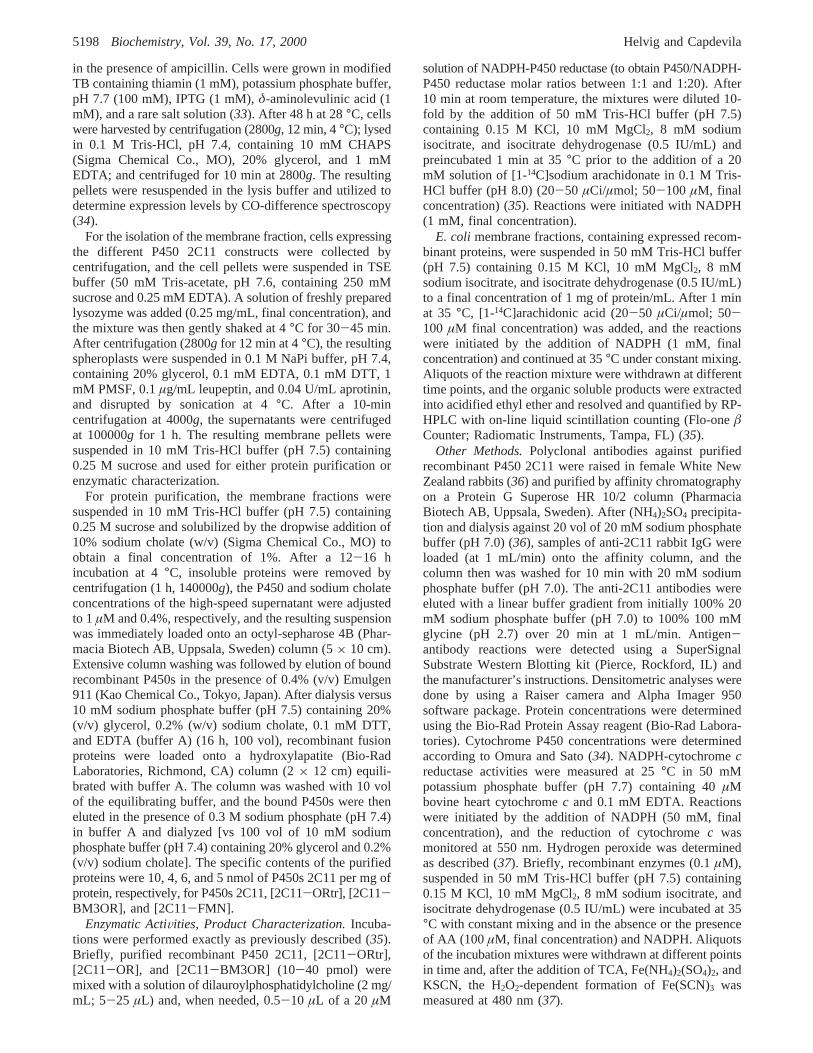
first 16 amino acids (40), instead of the 8 residues utilizedin this work.
To obviate alternate, non-P450 OR-catalyzed pathways ofP450 2C11 reduction (41), the expressed proteins weresolubilized and purified as described in Materials andMethods. SDS-PAGE analysis of the purified enzymesindicated that they were obtained with overall purities of 80,60, 80, and 40% for P450s 2C11, [2C11-ORtr], [2C11-BM3OR], and [2C11-FMN], respectively (not shown).Immunoelectrophoresis using polyclonal anti-2C11 antibod-ies (36) showed that all purified enzymes contained an anti-2C11 inmunoreactive protein band with average MWscorresponding to those predicted from the individual cDNA-translated protein sequences (58, 128, 135, 122, and 80 kDafor P450s 2C11, [2C11-ORtr], [2C11-OR], [2C11-BM3OR], and [2C11-FMN], respectively) (Figure 1A).Included in Figure 1A (lane 5) are the inmunoreactiveproperties of a modified [2C11-BM3OR] protein with aP450-BM3OR linker containing six additional residues(Ile-Pro-Leu-Gly-Gly-Ile) (29). Low expression levelsand the unstable nature of the [2C11-OR] protein made itsfurther purification and spectral quantification impractical.As an alternative, we compared by Western blot analysisthe anti 2C11-immunoreactivity of this protein with that ofthe [2C11-ORtr] enzyme and estimated its P450 2C11specific content by densitometric analysis (Figure 1B). It wascalculated that the P450 2C11 content in the [2C11-OR]membrane fractions isolated after lysozyme digestion, soni-cation, and high-speed centrifugation corresponded to ap-proximately one-fifth of that obtained with the [2C11-ORtr]cDNA expression (Figure 1B).
The absolute spectrum of recombinant [2C11-ORtr] andof [2C11-BM3OR] were similar with Soret,R-, andâ-bandsat 414, 570, and 537 nm, respectively. A broad absorptionband centered at around 480 nm was indicative of the spectralcontributions of the fused OR FMN and FAD prostheticgroups. Reduction with sodium dithionite resulted in theattenuation and displacement of the Soret band to 416 nm
Table 1: Expression Levels of Recombinant P450 2C11 Constructsin E. coli
recombinant enzymescell suspensions
(nmol/L)amembrane fraction
(nmol/mg)
2C11 206( 52 6.9[2C11-ORtr] 182( 8 2.1[2C11-OR] 23( 11 undetectable[2C11-BM3OR] 163( 27 2.8[2C11-FMN] 211 ( 25 1.8
a Values are averages calculated from at least three differentexperiments( SE.
FIGURE 1: Immunoelectrophoresis characterization of the purifiedrecombinant forms of P450 2C11. Purified P450s 2C11, [2C11-ORtr], [2C11-OR], [2C11-BM3OR], a modified [2C11-BM3OR]protein containing six additional aa residues in the P450-BM3ORlinker (29), and [2C11-FMN] (lanes 1-6, respectively; 1 pmol ofP450 each) (A) or samples of purified [2C11-OR] (lanes 1-4,respectively; 5, 10, 20, and 40µg of protein each) and of [2C11-ORtr] (lanes 5-8, respectively; 5, 10, 20, and 40µg of proteincontaining 1.2, 2.4, 4.8, and 9.6 pmol of P450 2C11 each) (B) weresubmitted to SDS-PAGE (100× 60 × 1 mm, 10% acrylamidegel slabs). After transfer to PVDF membranes, the blots wereincubated, first, with a polyclonal antibody raised against purifiedrecombinant P450 2C11, and second, with a peroxidase-coupledanti-rabbit IgG. The sample’s chemioluminescense was detectedusing a Super Signal kit and Kodak X-ray film. P450 2C11 contentswere estimated by densitometric analysis. See Materials andMethods for further details. Shown are the relative electrophoreticmobilities of protein molecular weight standards.
Arachidonate Oxidation by Fused P450-Reductase Proteins Biochemistry, Vol. 39, No. 17, 20005199
and the replacement of theR- and â-bands for a broadabsorption band centered at around 542 nm. The addition ofCO to a solution of reduced [2C11-ORtr] or [2C11-BM3OR] leads to the rapid development of a strongabsorption band at 450 nm, with shoulders at 422 and 563nm, indicative of the formation of the CO-bound complexof reduced [2C11-ORtr] or [2C11-BM3OR]. Experimentsin which CO was added to the cuvette prior to reductionshowed that, for both proteins, heme reduction was rapidand completed within the first few seconds of dithioniteaddition (34).
Cytochrome c Reduction.To characterize the integrity ofNADPH/FAD/FMN electron transfer and to investigatewhether protein steric factors affected cytochromec/reductaseinteractions, we determined the cytochromec reductaseactivity of the fused proteins for comparison to that of ORand BM3OR. As shown in Table 2, the addition of purifiedP450 2C11 had only a minor effect on the rates ofcytochromec reduction catalyzed by the mammalian andbacterial oxido reductases. These results show that thepresence of purified P450 2C11 does not alter the rates ofFAD/FMN/cytochromec electron transfer. On the otherhand, and as compared with the corresponding purifiedenzymes, fusion of the flavoproteins’ amino-end to P4502C11 carboxyl-end increased the cytochromec reductaseactivity of both the mammalian and the bacterial oxidoreductases (40 and 21% increase in activity for the [2C11-ORtr] and [2C11-BM3OR] fusion proteins, respectively)(Table 2). With the exception of the [2C11-BM3OR]protein, the P450 2C11 rate effects were, for the most part,SOD-insensitive (Table 2) suggesting direct donor-acceptor,protein/protein, electron transfer and not a bulk solvent O2
•-
mediated process. While the mechanisms and/or the pathsof electron transfer from the OR prosthetic groups to theheme of cytochromec are not understood, the observed P4502C11 fusion-dependent increases in flavoprotein turnoversuggest that fused P450 heme/cytochromec heme interac-tions may be responsible for these augmented rates ofNADPH/cytochrome electron transport. However, clotrima-zole (between 1 and 5µM), a strong cytochrome P450 hemeligand and potent inhibitor, had no effect on the rates ofcytochromec reduction by the fused proteins (not shown).As with P450 2C11, the presence of the bacterial FMNdomain fused to the carboxyl-end of P450 2C11 had noeffects on the rate of cytochromec reduction by the OR(Table 2). Finally, highly variable, preparation-dependentrates of cytochromec reduction were observed withE. colimembranes containing recombinant [2C11-OR] proteins.
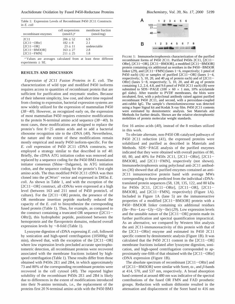
Metabolism of Arachidonic Acid.To evaluate and comparethe enzymatic activities of the fused proteins to that ofnonfused P450 2C11, we first reconstituted the AA mo-nooxygenase activity of purified P450 2C11 in the presenceof DLPC, NADPH, and variable P450/reductase molar ratios(42). Incubation of recombinant P450 2C11 with an equimo-lar concentration of OR and AA (70-100 µM, finalconcentration) resulted in the NADPH- and time-dependentformation of metabolites with the chromatographic propertiesof mixtures of authentic 14,15-, 11,12-, 8,9-, and 5,6-EETs(35, 36) (Figure 2). Approximately 27% of the total P4502C11 AA oxidation products eluted with HPLC retentiontimes similar to those of mixtures of HETEs, including 12-and 15-HETE, and are yet to be fully characterized (Figure2). Catalytic activity was P450- and OR-dependent andrequired the presence of 150 mM KCl for optimal turnover.As with other P450 AA monooxygenases (8, 43), turnoverrates increased almost linearly with increasing OR andreached a maximum at a molar ratio of OR to P450 2C11 ofapproximately 10 (Table 3). Those OR-dependent rate effectsindicate that during the catalysis of AA oxidation, electrontransfer and heme reduction are rate limiting. While thetruncated form of the OR, the ORtr, was unable to supportmetabolism by purified recombinant P450 2C11 (44, 45),its fusion to the carboxyl-terminal of P450 2C11 yielded aself-sufficient catalyst that metabolized AA at approximatelyone-fourth the rate of the reconstituted system but requiringonly air and NADPH for full activity (Table 3). As withpurified P450 2C11, metabolism by the [2C11-ORtr] proteinwas also stimulated by the addition of a 10-fold molar excessof purified P450 OR (22-fold over the rates in the absenceof exogenously added OR; Table 3). These results are similarto data obtained with several nonfused P450 isoforms (42)and indicate that (a) the presence of an ORtr moleculeconnected to the P450 2C11 carboxy-terminal does notpreclude catalytically productive interactions with additional
Table 2: Rates of Cytochromec Reduction by RecombinantEnzymes
cytochromec reduceda
enzyme system - SOD + SOD
OR 2925( 120 2878( 642C11+ OR 2270( 100 2147( 94[2C11-ORtr] 4316( 102 4091( 143BM3OR 3140( 102 3354( 1852C11+ BM3OR 1879( 49 1623( 98[2C11-BM3OR] 3808( 120 2979( 162[2C11-FMN] + OR 2027( 149 1925( 132
a In nmol of cytochromec reduced min-1 (nmol of flavoprotein)-1
at 25°C. Values are the averages calculated from at least three differentexperiments( SE.
FIGURE 2: Chromatographic resolution of the AA metabolitesgenerated by P450 2C11 reconstituted with rat P450 reductase orfused to a bacterial P450 reductase. Reaction mixtures containingeither a combination of P450 2C11 and OR (0.02µM each, finalconcentration) (bottom frame) or, alternatively, the [2C11-BM3OR] fusion protein (0.1µM, final concentration) (top frame)were incubated at 35°C for 15 min and in the presence of DLPC(50 µg/mL) and a buffer system containing NADPH, an NADPH-regenerating system, and [1-14C]AA (20 µCi/µmol; 100 µM).Reactions were terminated by the addition of acidified ethyl ether,and the organic soluble products were extracted and resolved byRP-HPLC as described in Materials and Methods. Shown are theradiochromatograms derived from incubation mixtures containing4 and 20 nmol of P450 2C11 and [2C11-BM3OR], respectively.
5200 Biochemistry, Vol. 39, No. 17, 2000 Helvig and Capdevila
OR molecules and (b) the existence of multiple electronpathways and/or P450 [2C11-ORtr]-OR molecular interac-tions leading to productive electron flow from the flavopro-tein to the P450 heme iron.
As shown in Table 3, the P450 BM3 flavoprotein domain(BM3OR) (29), even when added in a 10-fold molar excess,was unable to support AA oxidation by purified P450 2C11(Table 3). On the other hand, fusion of the BM3OR N-aminoterminal to the carboxyl-end of P450 2C11 yielded a proteinthat metabolized AA at a rate approximately one-fifth of thatof the [2C11-ORtr] enzyme (Figure 2, Table 3). As with2C11 and [2C11-ORtr], the presence of a 10-fold molarexcess of purified OR, but not BM3OR, markedly increasedthe catalytic turnover of the [2C11-BM3OR] protein (Table3). To the best of our knowledge, this is the first demonstra-tion of catalytically productive electron transfer between theheme iron of a mammalian P450 and a soluble flavoproteinof bacterial origin such as the BM3OR. Furthermore, thedemonstrated inability of the purified BM3OR to supportcatalytic turnover by P450 2C11 suggests that this lack ofactivity is either caused by limited productive interactionsbetween these proteins or electronic and/or mechanisticreasons. In this regard, P450 2C11 fused to the FMN domainof the BM3OR was also unable to support AA metabolismin the presence of OR or BM3OR (not shown). Importantly,regardless of the nature and/or concentration of the proximalelectron donor to the heme iron, for example, the fused ORtror BM3OR flavoproteins or, alternatively, the purified OR,the regiochemistry of AA oxidation by these enzymes wassimilar and under the control of P450 2C11 (36) (Table 4).Published results have also demonstrated that the fusion ofP450 and OR does not result in significant changes incatalytic efficiency or product chemistry (46, 47).
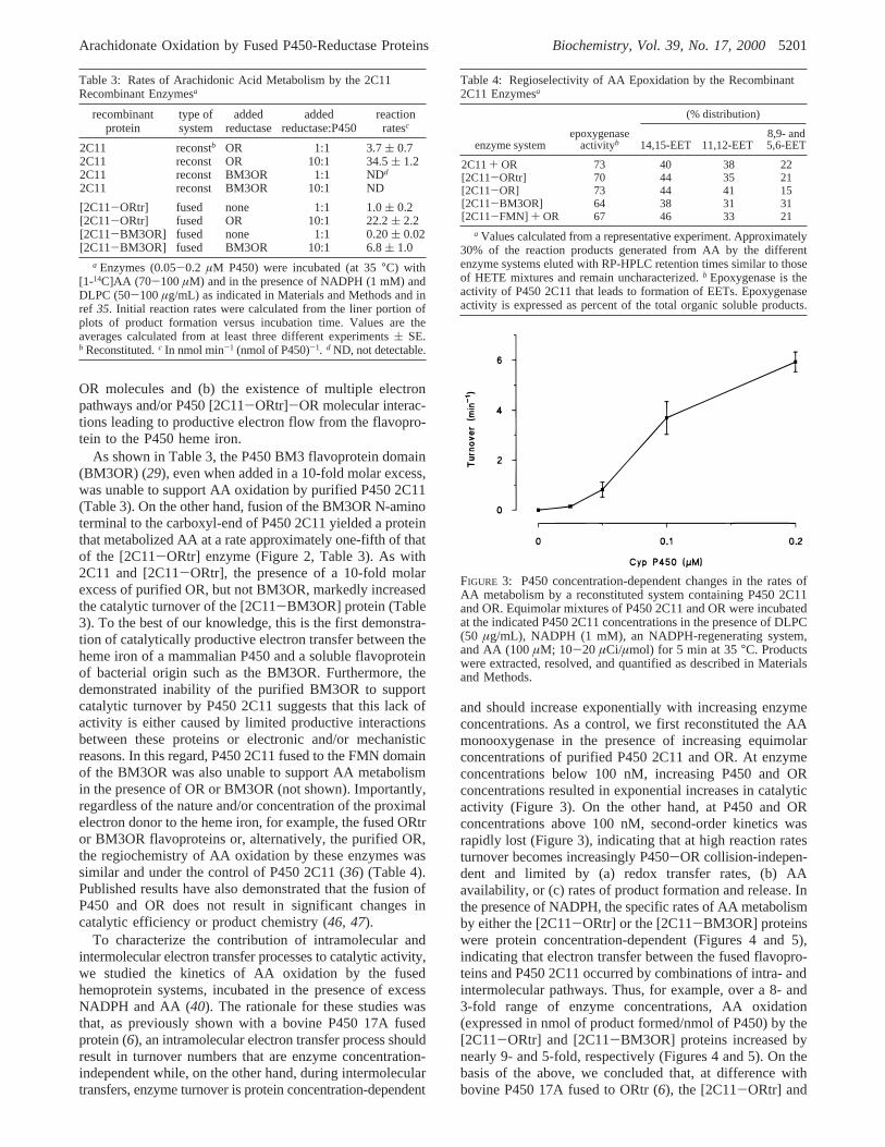
To characterize the contribution of intramolecular andintermolecular electron transfer processes to catalytic activity,we studied the kinetics of AA oxidation by the fusedhemoprotein systems, incubated in the presence of excessNADPH and AA (40). The rationale for these studies wasthat, as previously shown with a bovine P450 17A fusedprotein (6), an intramolecular electron transfer process shouldresult in turnover numbers that are enzyme concentration-independent while, on the other hand, during intermoleculartransfers, enzyme turnover is protein concentration-dependent
and should increase exponentially with increasing enzymeconcentrations. As a control, we first reconstituted the AAmonooxygenase in the presence of increasing equimolarconcentrations of purified P450 2C11 and OR. At enzymeconcentrations below 100 nM, increasing P450 and ORconcentrations resulted in exponential increases in catalyticactivity (Figure 3). On the other hand, at P450 and ORconcentrations above 100 nM, second-order kinetics wasrapidly lost (Figure 3), indicating that at high reaction ratesturnover becomes increasingly P450-OR collision-indepen-dent and limited by (a) redox transfer rates, (b) AAavailability, or (c) rates of product formation and release. Inthe presence of NADPH, the specific rates of AA metabolismby either the [2C11-ORtr] or the [2C11-BM3OR] proteinswere protein concentration-dependent (Figures 4 and 5),indicating that electron transfer between the fused flavopro-teins and P450 2C11 occurred by combinations of intra- andintermolecular pathways. Thus, for example, over a 8- and3-fold range of enzyme concentrations, AA oxidation(expressed in nmol of product formed/nmol of P450) by the[2C11-ORtr] and [2C11-BM3OR] proteins increased bynearly 9- and 5-fold, respectively (Figures 4 and 5). On thebasis of the above, we concluded that, at difference withbovine P450 17A fused to ORtr (6), the [2C11-ORtr] and
Table 3: Rates of Arachidonic Acid Metabolism by the 2C11Recombinant Enzymesa
recombinantprotein
type ofsystem
addedreductase
addedreductase:P450
reactionratesc
2C11 reconstb OR 1:1 3.7( 0.72C11 reconst OR 10:1 34.5( 1.22C11 reconst BM3OR 1:1 NDd
2C11 reconst BM3OR 10:1 ND
[2C11-ORtr] fused none 1:1 1.0( 0.2[2C11-ORtr] fused OR 10:1 22.2( 2.2[2C11-BM3OR] fused none 1:1 0.20( 0.02[2C11-BM3OR] fused BM3OR 10:1 6.8( 1.0
a Enzymes (0.05-0.2 µM P450) were incubated (at 35°C) with[1-14C]AA (70-100µM) and in the presence of NADPH (1 mM) andDLPC (50-100µg/mL) as indicated in Materials and Methods and inref 35. Initial reaction rates were calculated from the liner portion ofplots of product formation versus incubation time. Values are theaverages calculated from at least three different experiments( SE.b Reconstituted.c In nmol min-1 (nmol of P450)-1. d ND, not detectable.
Table 4: Regioselectivity of AA Epoxidation by the Recombinant2C11 Enzymesa
(% distribution)
enzyme systemepoxygenase
activityb 14,15-EET 11,12-EET8,9- and5,6-EET
2C11+ OR 73 40 38 22[2C11-ORtr] 70 44 35 21[2C11-OR] 73 44 41 15[2C11-BM3OR] 64 38 31 31[2C11-FMN] + OR 67 46 33 21
a Values calculated from a representative experiment. Approximately30% of the reaction products generated from AA by the differentenzyme systems eluted with RP-HPLC retention times similar to thoseof HETE mixtures and remain uncharacterized.b Epoxygenase is theactivity of P450 2C11 that leads to formation of EETs. Epoxygenaseactivity is expressed as percent of the total organic soluble products.
FIGURE 3: P450 concentration-dependent changes in the rates ofAA metabolism by a reconstituted system containing P450 2C11and OR. Equimolar mixtures of P450 2C11 and OR were incubatedat the indicated P450 2C11 concentrations in the presence of DLPC(50 µg/mL), NADPH (1 mM), an NADPH-regenerating system,and AA (100µM; 10-20 µCi/µmol) for 5 min at 35°C. Productswere extracted, resolved, and quantified as described in Materialsand Methods.
Arachidonate Oxidation by Fused P450-Reductase Proteins Biochemistry, Vol. 39, No. 17, 20005201
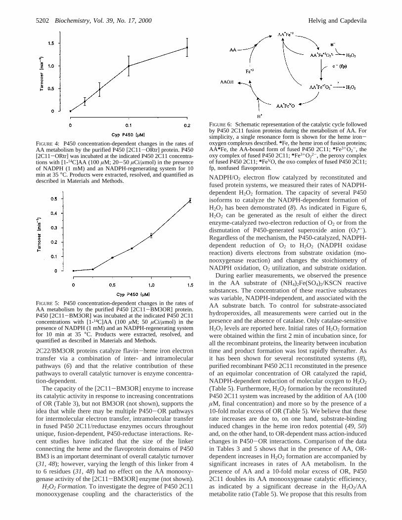
2C22/BM3OR proteins catalyze flavin-heme iron electrontransfer via a combination of inter- and intramolecularpathways (6) and that the relative contribution of thesepathways to overall catalytic turnover is enzyme concentra-tion-dependent.
The capacity of the [2C11-BM3OR] enzyme to increaseits catalytic activity in response to increasing concentrationsof OR (Table 3), but not BM3OR (not shown), supports theidea that while there may be multiple P450-OR pathwaysfor intermolecular electron transfer, intramolecular transferin fused P450 2C11/reductase enzymes occurs throughoutunique, fusion-dependent, P450-reductase interactions. Re-cent studies have indicated that the size of the linkerconnecting the heme and the flavoprotein domains of P450BM3 is an important determinant of overall catalytic turnover(31, 48); however, varying the length of this linker from 4to 6 residues (31, 48) had no effect on the AA monooxy-genase activity of the [2C11-BM3OR] enzyme (not shown).
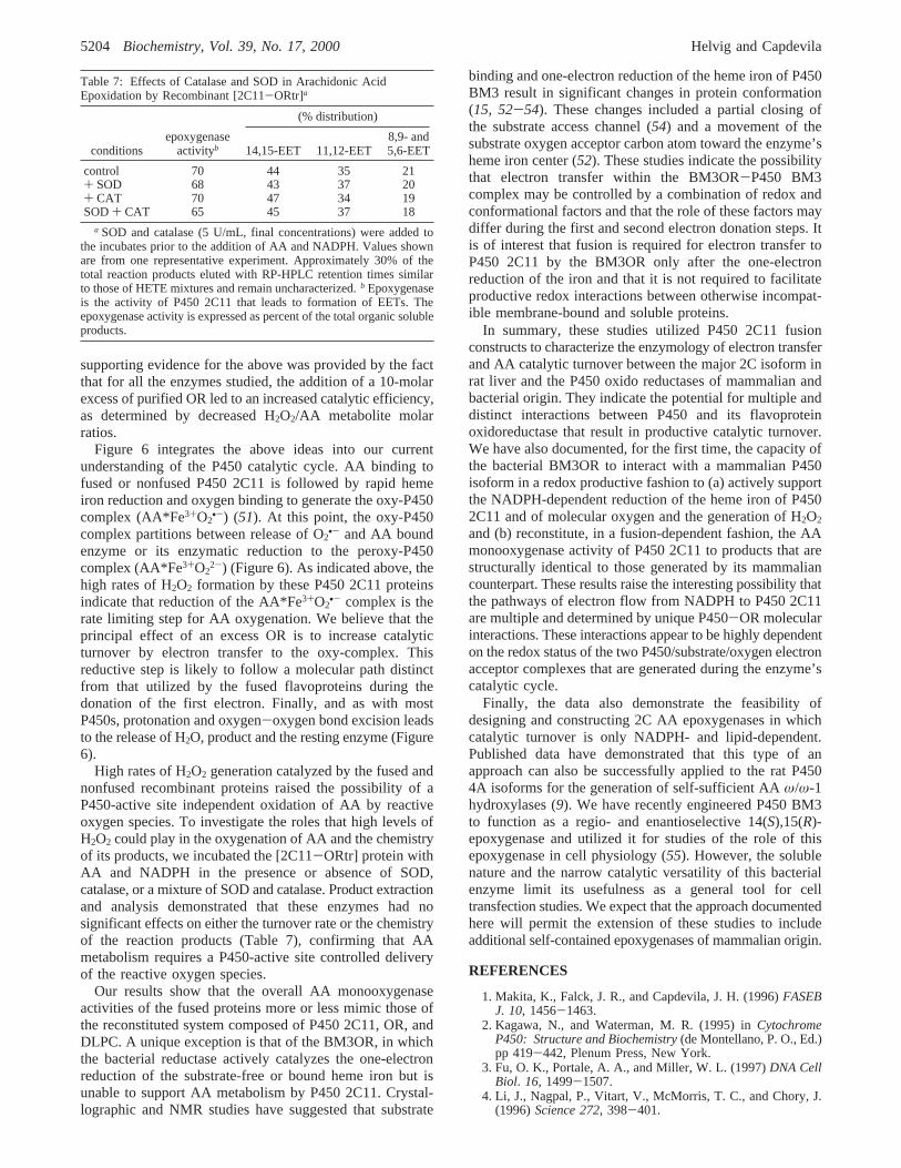
H2O2 Formation.To investigate the degree of P450 2C11monooxygenase coupling and the characteristics of the
NADPH/O2 electron flow catalyzed by reconstituted andfused protein systems, we measured their rates of NADPH-dependent H2O2 formation. The capacity of several P450isoforms to catalyze the NADPH-dependent formation ofH2O2 has been demonstrated (8). As indicated in Figure 6,H2O2 can be generated as the result of either the directenzyme-catalyzed two-electron reduction of O2 or from thedismutation of P450-generated superoxide anion (O2
•-).Regardless of the mechanism, the P450-catalyzed, NADPH-dependent reduction of O2 to H2O2 (NADPH oxidasereaction) diverts electrons from substrate oxidation (mo-nooxygenase reaction) and changes the stoichiometry ofNADPH oxidation, O2 utilization, and substrate oxidation.
During earlier measurements, we observed the presencein the AA substrate of (NH4)2Fe(SO4)2/KSCN reactivesubstances. The concentration of these reactive substanceswas variable, NADPH-independent, and associated with theAA substrate batch. To control for substrate-associatedhydroperoxides, all measurements were carried out in thepresence and the absence of catalase. Only catalase-sensitiveH2O2 levels are reported here. Initial rates of H2O2 formationwere obtained within the first 2 min of incubation since, forall the recombinant proteins, the linearity between incubationtime and product formation was lost rapidly thereafter. Asit has been shown for several reconstituted systems (8),purified recombinant P450 2C11 reconstituted in the presenceof an equimolar concentration of OR catalyzed the rapid,NADPH-dependent reduction of molecular oxygen to H2O2
(Table 5). Furthermore, H2O2 formation by the reconstitutedP450 2C11 system was increased by the addition of AA (100µM, final concentration) and more so by the presence of a10-fold molar excess of OR (Table 5). We believe that theserate increases are due to, on one hand, substrate-bindinginduced changes in the heme iron redox potential (49, 50)and, on the other hand, to OR-dependent mass action-inducedchanges in P450-OR interactions. Comparison of the datain Tables 3 and 5 shows that in the presence of AA, OR-dependent increases in H2O2 formation are accompanied bysignificant increases in rates of AA metabolism. In thepresence of AA and a 10-fold molar excess of OR, P4502C11 doubles its AA monooxygenase catalytic efficiency,as indicated by a significant decrease in the H2O2/AAmetabolite ratio (Table 5). We propose that this results from
FIGURE 4: P450 concentration-dependent changes in the rates ofAA metabolism by the purified P450 [2C11-ORtr] protein. P450[2C11-ORtr] was incubated at the indicated P450 2C11 concentra-tions with [1-14C]AA (100 µM; 20-50 µCi/µmol) in the presenceof NADPH (1 mM) and an NADPH-regenerating system for 10min at 35°C. Products were extracted, resolved, and quantified asdescribed in Materials and Methods.
FIGURE 5: P450 concentration-dependent changes in the rates ofAA metabolism by the purified P450 [2C11-BM3OR] protein.P450 [2C11-BM3OR] was incubated at the indicated P450 2C11concentrations with [1-14C]AA (100 µM; 50 µCi/µmol) in thepresence of NADPH (1 mM) and an NADPH-regenerating systemfor 10 min at 35 °C. Products were extracted, resolved, andquantified as described in Materials and Methods.
FIGURE 6: Schematic representation of the catalytic cycle followedby P450 2C11 fusion proteins during the metabolism of AA. Forsimplicity, a single resonance form is shown for the heme iron-oxygen complexes described.*Fe, the heme iron of fusion proteins;AA*Fe, the AA-bound form of fused P450 2C11;*Fe3+O2
-, theoxy complex of fused P450 2C11;*Fe3+O2
2-, the peroxy complexof fused P450 2C11;*FeVO, the oxo complex of fused P450 2C11;fp, nonfused flavoprotein.
5202 Biochemistry, Vol. 39, No. 17, 2000 Helvig and Capdevila

an OR concentration-dependent increase in electron transferto the one-electron reduced form of substrate and oxygen-bound P450 2C11 (the AA*Fe3+O2
•-complex; Figure 6).As compared with the reconstituted system, fusion to the
ORtr did not significantly change the rate of H2O2 generationby P450 2C11 (Table 5). Furthermore, NADPH-dependentperoxide generation by the [2C11-ORtr] enzyme wassubstrate-independent and substantially less affected by thepresence of exogenously added OR (Table 5). Again,comparison of the data in Tables 3 and 5 indicated that (a)fusion does not improve the catalytic efficiency of P450 2C11for AA metabolism, that is, the [2C11-ORtr] protein divertsan important part of the NADPH-supplied electron to H2O2,as revealed by high H2O2 molar ratios (Table 5); (b) the lackof a substrate effect on the rates of H2O2 formation suggeststhat most of the peroxide formed is derived from the one-electron reduction of P450 and the dismutation of the O2
•-
released from the P450 2C11 oxy complex (51) (Fe3+O2•-,
Figure 6); and (c) the marked increase in the efficiency ofthe AA monooxygenase observed after the addition of a 10-fold molar excess of OR to the [2C11-ORtr] protein (Table5) suggests that the exogenously added OR facilitates thedonation of the second electron to the substrate-bound oxy-P450 complex, (AA*Fe3+O2
•-; Figure 6), the required stepfor the subsequent generation of the iron-bound oxidantspecies (AA*FeVO, Figure 6).
As discussed, the P450 2C11 AA monooxygenase couldnot be reconstituted in the presence of a 1:1 or a 10:1 molarexcess of the bacterial flavoprotein reductase, the BM3OR.However, under similar conditions, the BM3OR was aneffective P450 2C11 reductase and actively supported H2O2
formation by this enzyme in an AA and BM3OR concentra-tion-dependent fashion (Table 6). When reconstituted withthe BM3OR in a 1:1 molar ratio, P450 2C11 catalyzed H2O2
generation at nearly double the rate of the mammalianflavoprotein (∼1.8-fold faster) (Tables 5 and 6). As with theOR, the rates of H2O2 formation were increased by theaddition of AA and a 10-molar excess of BM3OR (Table6), reflecting AA-dependent changes in P450 redox potentialand BM3OR mass action kinetic effects. The observationthat a reconstituted system composed of P450 2C11 and P450BM3OR actively catalyzes the NADPH-dependent reductionof O2 to H2O2, but not AA oxidation, indicates that thebacterial flavoprotein supports the one-electron reduction ofsubstrate-free or bound P450 2C11 but not that of the oxycomplex of ferrous P450 2C11 (Figure 6) (51). The aboveinterpretation implies that the BM3OR-supported P450 2C11-dependent formation of H2O2 results from the one-electronreduction of O2 and O2
•- dismutation (51).Fusion of the BM3OR to P450 2C11 markedly reduced
the rate of P450 2C11-dependent H2O2 formation by the[2C11-BM3OR] protein incubated in either the presenceor the absence of AA (Table 6). As illustrated in Table 3,only after fusion was the BM3OR capable of supporting AAmetabolism, albeit at rates lower that those of the [2C11-ORtr] enzyme. A comparison of the molar ratio between theH2O2 formed and the extent of AA metabolism (the H2O2/AA ratio) shows that the [2C11-ORtr] and [2C11-BM3OR]proteins metabolize AA with similar catalytic efficiencies(Tables 5 and 6). The addition of a 10-fold molar excess ofBM3OR to the [2C11-BM3OR] protein resulted in a greatlystimulated rate of H2O2 formation (10-fold increase; Table6), an effect that was further augmented by the inclusion ofAA (100 µM, final concentration) (Table 6). Finally, and atdifference with the OR (Table 3), the addition of exogenousBM3OR to incubates containing [2C11-BM3OR] and AAdid not alter the rates of AA metabolism by the [2C11-BM3OR] protein but led to high rates of H2O2 formationand a low catalytic efficiency (Table 6). Taken together, theseexperiments showed that (a) fusion reduces the rate at whichthe BM3OR transfers the first electron to the heme iron ofsubstrate-bound or substrate-free P450 2C11 (Figure 6).However, fusion is required for the BM3OR-catalyzedtransfer of the second electron to the AA-bound oxy-P450complex, oxygen activation, and metabolism; (b) as with thereconstituted system, exogenously added (nonfused) BM3ORactively catalyzes the one-electron reduction of the [2C11-BM3OR] heme iron and H2O2 formation via superoxidedismutation; and (c) through unique conformational and/orchanges in redox properties, fusion of the N-terminal ofBM3OR to the carboxyl-end of P450 2C11 opens a catalyti-cally productive pathway of electron flow to the P450. Thispresumably intramolecular pathway of electron transfer isnot functional when the purified BM3OR is incubated witheither the P450 2C11 or the [2C11-BM3OR] proteins.
The mechanisms of H2O2 formation by these proteins, thatis, P450 2C11-dependent one- or two-electron reduction ofO2 (to either O2
•- or O22- ) remain to be established;
however, a one-electron reduction, O2•--mediated pathway
for the P450-catalyzed formation of H2O2 has been proposed(51). Nevertheless, regardless of the nature of the immediateH2O2 precursor, the observed high levels of NADPH oxidaseactivity support the idea that, for all the three enzyme systemsstudied, reduction of the oxy-P450 complex (51) and/oroxygen insertion into the substrate carbon template are therate-limiting steps during AA monooxygenation. Additional
Table 5: Rates of OR-Dependent H2O2 Formation by RecombinantP450 Proteinsa
enzyme system 2C11+ OR [2C11-ORtr]
OR/P450 molar ratio 1:1 10:1 1:1 10:1
minus AA 40( 9 142( 6 46( 2 64( 4plus AA 55( 9 197( 8 45( 6 77( 2
H2O2/AA metabolites 15 6 45 3a Enzymes were incubated with NADPH in the presence or in the
absence of AA exactly as described in Table 2 and Materials andMethods. Catalase-sensitive H2O2 concentrations, determined from atleast three different experiments, are given as nmol of H2O2 formed(nmol of P450)-1 min-1 at 35°C and are the averages( SE.
Table 6: Rates of BM3OR-Dependent H2O2 Formation byRecombinant P450 Proteinsa
enzyme system 2C11+ BM3OR [2C11-BM3OR]
BM3OR/P450 molar ratio: 1:1 10:1 1:1 10:1
minus AA 73( 10 131( 2 10( 1 100( 5plus AA 142( 13 147( 6 13( 1 183( 9
H2O2/AA metabolites NAb NA 65 915a Enzymes were incubated with NADPH in the presence or in the
absence of AA exactly as described in Tables 2 and 5 and in Materialsand Methods. Catalase-sensitive H2O2 concentrations, determined fromat least three different experiments, are given as nmol of H2O2 formed(nmol of P450)-1 min-1 at 35 °C and are the averages( SE. b NA,not applicable.
Arachidonate Oxidation by Fused P450-Reductase Proteins Biochemistry, Vol. 39, No. 17, 20005203
supporting evidence for the above was provided by the factthat for all the enzymes studied, the addition of a 10-molarexcess of purified OR led to an increased catalytic efficiency,as determined by decreased H2O2/AA metabolite molarratios.
Figure 6 integrates the above ideas into our currentunderstanding of the P450 catalytic cycle. AA binding tofused or nonfused P450 2C11 is followed by rapid hemeiron reduction and oxygen binding to generate the oxy-P450complex (AA*Fe3+O2
•-) (51). At this point, the oxy-P450complex partitions between release of O2
•- and AA boundenzyme or its enzymatic reduction to the peroxy-P450complex (AA*Fe3+O2
2-) (Figure 6). As indicated above, thehigh rates of H2O2 formation by these P450 2C11 proteinsindicate that reduction of the AA*Fe3+O2
•- complex is therate limiting step for AA oxygenation. We believe that theprincipal effect of an excess OR is to increase catalyticturnover by electron transfer to the oxy-complex. Thisreductive step is likely to follow a molecular path distinctfrom that utilized by the fused flavoproteins during thedonation of the first electron. Finally, and as with mostP450s, protonation and oxygen-oxygen bond excision leadsto the release of H2O, product and the resting enzyme (Figure6).
High rates of H2O2 generation catalyzed by the fused andnonfused recombinant proteins raised the possibility of aP450-active site independent oxidation of AA by reactiveoxygen species. To investigate the roles that high levels ofH2O2 could play in the oxygenation of AA and the chemistryof its products, we incubated the [2C11-ORtr] protein withAA and NADPH in the presence or absence of SOD,catalase, or a mixture of SOD and catalase. Product extractionand analysis demonstrated that these enzymes had nosignificant effects on either the turnover rate or the chemistryof the reaction products (Table 7), confirming that AAmetabolism requires a P450-active site controlled deliveryof the reactive oxygen species.
Our results show that the overall AA monooxygenaseactivities of the fused proteins more or less mimic those ofthe reconstituted system composed of P450 2C11, OR, andDLPC. A unique exception is that of the BM3OR, in whichthe bacterial reductase actively catalyzes the one-electronreduction of the substrate-free or bound heme iron but isunable to support AA metabolism by P450 2C11. Crystal-lographic and NMR studies have suggested that substrate
binding and one-electron reduction of the heme iron of P450BM3 result in significant changes in protein conformation(15, 52-54). These changes included a partial closing ofthe substrate access channel (54) and a movement of thesubstrate oxygen acceptor carbon atom toward the enzyme’sheme iron center (52). These studies indicate the possibilitythat electron transfer within the BM3OR-P450 BM3complex may be controlled by a combination of redox andconformational factors and that the role of these factors maydiffer during the first and second electron donation steps. Itis of interest that fusion is required for electron transfer toP450 2C11 by the BM3OR only after the one-electronreduction of the iron and that it is not required to facilitateproductive redox interactions between otherwise incompat-ible membrane-bound and soluble proteins.
In summary, these studies utilized P450 2C11 fusionconstructs to characterize the enzymology of electron transferand AA catalytic turnover between the major 2C isoform inrat liver and the P450 oxido reductases of mammalian andbacterial origin. They indicate the potential for multiple anddistinct interactions between P450 and its flavoproteinoxidoreductase that result in productive catalytic turnover.We have also documented, for the first time, the capacity ofthe bacterial BM3OR to interact with a mammalian P450isoform in a redox productive fashion to (a) actively supportthe NADPH-dependent reduction of the heme iron of P4502C11 and of molecular oxygen and the generation of H2O2
and (b) reconstitute, in a fusion-dependent fashion, the AAmonooxygenase activity of P450 2C11 to products that arestructurally identical to those generated by its mammaliancounterpart. These results raise the interesting possibility thatthe pathways of electron flow from NADPH to P450 2C11are multiple and determined by unique P450-OR molecularinteractions. These interactions appear to be highly dependenton the redox status of the two P450/substrate/oxygen electronacceptor complexes that are generated during the enzyme’scatalytic cycle.
Finally, the data also demonstrate the feasibility ofdesigning and constructing 2C AA epoxygenases in whichcatalytic turnover is only NADPH- and lipid-dependent.Published data have demonstrated that this type of anapproach can also be successfully applied to the rat P4504A isoforms for the generation of self-sufficient AAω/ω-1hydroxylases (9). We have recently engineered P450 BM3to function as a regio- and enantioselective 14(S),15(R)-epoxygenase and utilized it for studies of the role of thisepoxygenase in cell physiology (55). However, the solublenature and the narrow catalytic versatility of this bacterialenzyme limit its usefulness as a general tool for celltransfection studies. We expect that the approach documentedhere will permit the extension of these studies to includeadditional self-contained epoxygenases of mammalian origin.
REFERENCES
1. Makita, K., Falck, J. R., and Capdevila, J. H. (1996)FASEBJ. 10, 1456-1463.
2. Kagawa, N., and Waterman, M. R. (1995) inCytochromeP450: Structure and Biochemistry(de Montellano, P. O., Ed.)pp 419-442, Plenum Press, New York.
3. Fu, O. K., Portale, A. A., and Miller, W. L. (1997)DNA CellBiol. 16, 1499-1507.
4. Li, J., Nagpal, P., Vitart, V., McMorris, T. C., and Chory, J.(1996)Science 272, 398-401.
Table 7: Effects of Catalase and SOD in Arachidonic AcidEpoxidation by Recombinant [2C11-ORtr]a
(% distribution)
conditionsepoxygenase
activityb 14,15-EET 11,12-EET8,9- and5,6-EET
control 70 44 35 21+ SOD 68 43 37 20+ CAT 70 47 34 19SOD+ CAT 65 45 37 18
a SOD and catalase (5 U/mL, final concentrations) were added tothe incubates prior to the addition of AA and NADPH. Values shownare from one representative experiment. Approximately 30% of thetotal reaction products eluted with RP-HPLC retention times similarto those of HETE mixtures and remain uncharacterized.b Epoxygenaseis the activity of P450 2C11 that leads to formation of EETs. Theepoxygenase activity is expressed as percent of the total organic solubleproducts.
5204 Biochemistry, Vol. 39, No. 17, 2000 Helvig and Capdevila

5. Murakami, H., Yabusaki, Y., Sakaki, M., and Ohkawa, H.(1987)DNA 6, 189-197.
6. Shet, M. S., Fisher, C. W., Arlotto, M. P., Shackleton, C. H.,Holmans, P. L., Martin-Wixtrom, C. A., Saeki, Y., andEstabrook R. W. (1994)Arch. Biochem. Biophys. 311, 402-417.
7. Fisher, C. W., Shet, M. S., and Estabrook, R. W. (1996)Methods Enzymol. 272, 15-25.
8. Shet, M. S., Faulkner, K. M., Holmans, P. L., Fisher, C. W.,and Estabrook R. W. (1995)Arch. Biochem. Biophys. 318,314-321.
9. Fisher, C. W., Shet, M. S., Claudle, D. L., Martin-Wixtrom,C. A., and Estabrook (1992)Proc. Natl. Acad. Sci. U.S.A. 89,10817-10821.
10. Narhi, L. O., and Fulco, A. J. (1986)J. Biol. Chem. 261,7160-7169.
11. Wen, L. P., and Fulco A. J. (1987)J. Biol. Chem. 262, 6676-6682.
12. Graham-Lorence, S., Truan, G., Peterson, J. A., Falck, J. R.,Wei, S., Helvig, C., and Capdevila, J. H. (1997)J. Biol. Chem.272, 1127-1135.
13. Miura, Y., and Fulco, A. J. (1975)Biochim. Biophys. Acta388, 305-317.
14. Ho, P. P., and Fulco A. J. (1976)Biochim. Biophys. Acta 431,249-256.
15. Ravichandran, K. G., Boddupalli, S. S., Hasermann, C. A.,Peterson, J. A., and Deisenhofer, J. (1993)Science 261, 731-736.
16. McMillan, K., Bredt, D. S., Hirsch, D. J., Snyder, S. H., Clarck,J. E., and Master, B. S. I. (1992)Proc. Natl. Acad. Sci. U.S.A.89, 11141-11145.
17. Geller, D. A., Lowenstein, C. J., Shapiro, R. A., Nussler, A.K., DiSilvio, M., Wang S. C., Nakayama, O. K., Simmons,R. L, Snyder, S. H., and Billiar, T. R. (1993)Proc. Natl. Acad.Sci. U.S.A. 90, 3491-3495.
18. Nakayama, N., Takemae, A., and Shoun H. (1996)J. Biochem.119, 435-440.
19. Oliw, E. H., Bylund, J., and Herman, C. (1996)Lipids 31,1003-1021.
20. MacGiff, J. C., Steinberg, M., and Quilley, J. (1996)TrendsCardioVasc. Med. 6, 4-10.
21. Harder, D. R., Lange, A. R., Gebremedhin, D., Birks, E. K.,and Roman, R. J. (1997)J. Vasc. Res. 32, 79-92.
22. McGiff, J. C., and Quilley, J. (1999)Am. J. Physiol. 277, R607-R623.
23. Stec, D. E., Deng, A. Y., Rapp, J. P., and Roman, R. J. (1996)Hypertension 27, 564-568.
24. Karara, A., Makita, K., Jacobson, H. R., Falck, J. R.,Guengerich, F. P., DuBois, R. N., and Capdevila, J. H. (1993)J. Biol. Chem. 268, 13565-13570.
25. Cederbaum, A. I. (1998)Biofactors 8, 93-96.26. Guengerich, F. P., Dannan, G. A., Wright, S. T., Martin, M.
V., and Kaminsky, L. S. (1982)Biochemistry 21, 6019-6030.27. Gillam, E. M., Baba, T., Kim, B. R., Ohmori, S., and
Guengerich, F. P. (1993)Arch. Biochem. Biophys. 305, 123-131.
28. Gillam, E. M., Guo, Z., Ueng, Y. F., Yamazaki, H., Cock, I.,Reilly, P. E., Hooper, W. D., and Guengerich, F. P. (1995)Arch. Biochem. Biophys. 317, 374-384.
29. Boddupali, S. S., Oster, T., Estabroock, R. W., and Peterson,J. A. (1992)J. Biol. Chem.267, 10375-10380.
30. Porter, T. D., and Kasper, C. B. (1985)Proc. Natl. Acad. Sci.U.S.A. 82, 973-977.
31. Govindaraj, S., and Poulos, T. L. (1995)Biochemistry 34,11221-11226.
32. Ruettinger, R. T., Wen, L. P., and Fulco, A. J. (1989)J. Biol.Chem. 264, 10987-10995.
33. Bauer, S., and Shiloach, J. (1974)Biotechnol. Bioeng. 16,933-941.
34. Omura, T., and Sato, R. (1964)J. Biol. Chem. 239, 2370-2378.
35. Capdevila, J. H., Falck, J. R., Dishman, E., and Karara, A.(1990)Methods Enzymol. 187, 385-394.
36. Capdevila, J. H., Wei, S., Yan, Y., Karara, A., Jacobson, H.R., Falk, J. R., Guengerich, F. P., and Dubois, R. N. (1992)J.Biol. Chem. 267, 21720-21726.
37. Hildebrandt, A. G., and Roots, I. (1975)Arch. Biochem.Biophys. 171, 385-397.
38. Barnes, H. J., Arlotto, M. P., and Waterman, M. R. (1991)Proc. Natl. Acad. Sci. U.S.A. 88, 5597-5601.
39. Barnes, H. (1996)Methods Enzymol. 272, 3-14.40. Pernecky, S. J., and Coon M. J. (1996)Methods Enzymol. 272,
25-34.41. Jenkins, C. M., and Waterman, M. R. (1998)Biochemistry
37, 6106-6113.42. Helvig, C., Dishman, E., and Capdevila, J. H. (1998)Bio-
chemistry 37, 12546-12558.43. Shet, M. S., Fisher, C. W., Holmans, P. L, and Estabrook R.
W. (1996)Arch. Biochem. Biophys. 330, 199-208.44. Lu, A. Y. H., Junk, K. W., and Coon, M. J. (1969)J. Biol.
Chem. 244, 3714-3721.45. Black, S. D., and Coon, M. J. (1982)J. Biol. Chem. 257,
5929-5938.46. Parikh, A., and Guengerich, F. P. (1997)Protein Expression
Purif. 3, 346-354.47. Chaurasia, C. S., Alterman, M. A., Lu, P., and Hanziik, P. P.
(1995)Arch. Biochem. Biophys. 317, 161-169.48. Govindaraj, S., and Poulos, T. L. (1996)Protein Sci. 5, 1389-
1393.49. Gunsalus, I. C., Meeks, J. R., Lipscomb, J. D., Debrunner, P.,
and Munck, E. (1974) inMolecular Mechanisms of OxygenActiVation (Hayaishi, O., Ed.) Chapter 14, pp 559-613,Academic Press, New York.
50. Makino, R., Iizuka, T., Sakaguchi, K., and Ishimura, Y. (1982)in Oxygenase and Oxygen Metabolism(Nozaki, M., Yama-moto, S., Ishimura, Y., Coon, M. J., Ernster, L., and Estabrook,R. W., Eds.) pp 467-477, Academic Press, New York.
51. Loida, P. J., and Sligar, S. G. (1993)Biochemistry 32, 11530-11538.
52. Li, H., and Poulos, T. L. (1997)Nat. Struct. Biol. 4, 140-146.
53. Li, H. Y., and Poulos, T. L. (1995)Acta Crystallogr. D51,21-32.
54. Modi, S., Primrose, W. U., Boyle, J. M. B., Gibson, C. F.,Lian, L.-Y., and Roberts, G. C. K. (1995)Biochemistry 4,8982-8988.
55. Chen, J. K., Wang, D. W., Falck, J. R., Capdevila, J., andHarris, R. C. (1999)J. Biol. Chem. 274, 4764-4769.
BI992578V
Arachidonate Oxidation by Fused P450-Reductase Proteins Biochemistry, Vol. 39, No. 17, 20005205