Embed Size (px)
Citation preview
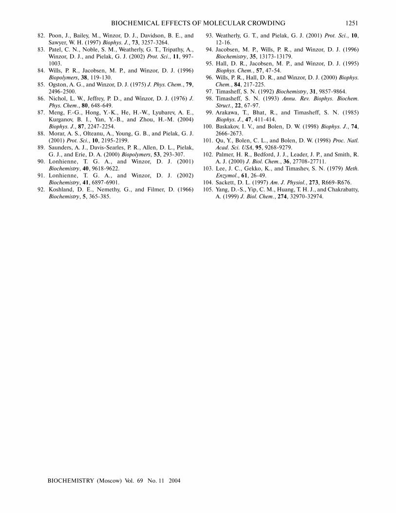
A distinctive property of living systems is that bio-chemical processes proceed in a medium containing highconcentrations of macromolecules (50-400 mg/ml) [1-3], the concentrations of single macrosolute species beingnot so high. However, overall the macromolecules presentin the cell occupy a significant part of the total volume ofthe medium (about 40%). Therefore, the accessible vol-ume in the cell is reduced. In the literature, such condi-tions in the living cell have been termed “macromolecu-lar crowding”. Figure 1 (see color insert) demonstratesthe crowding conditions in the cytoplasm of an intact cell[4].
In multicellular organisms, crowding is not confinedto cellular interiors, but also occurs in the extracellularmatrix of tissues. For instance, cartilage is a biologicalsubstance with well pronounced crowding. In blood plas-ma, the total concentration of macromolecules reaches80 g/liter, this concentration being sufficient to cause sig-nificant crowding effects. A type of crowding designatedas a “macromolecular confinement” exists in situationswhen macromolecules are included in small cellular com-partments or pores with dimensions comparable to thesize of large macromolecules. Such compartments arecreated, in particular, in the cytoskeleton lattice or in thecentral cavity of the chaperonine GroEL [5-7]. Whendescribing the influence of crowding on biological reac-
tions, the following terms are used in the literature:macromolecular crowding, molecular crowding, and alsomacromolecular confinement, but for all these terms theeffect of the excluded volume is implied. The term“molecular crowding” is used when crowded conditionsis caused by high concentrations of low-molecular-weightcosolutes (for example, osmolytes).
The technique of cryoelectron tomography [4, 8]and investigation of the movement of the fluorescent pro-teins in animal cells [9] are currently used to study crowd-ing in vivo. Direct evidence for the crowded state of cellinteriors is provided by the technique of cryoelectrontomography when thin intact cells are frozen rapidly inliquid ethane. This method has unique possibilities forreconstruction of three-dimensional images of themacromolecular complexes in their natural environment.At the present time, a resolution of 5-8 nm has beenreached, which is enough for recognition of complexeswith dimensions 20-50 nm and for their study in crowdedconditions in different cell compartments [8]. The highdensity of actin filaments (part of the cellular “skeleton”)and ribosomes (Fig. 1) supports the view that cytoplasm isfilled with large ensembles of macromolecules, whichform functional complexes, rather than with freely diffus-ing and colliding macromolecules [3, 10]. Direct observa-tion of fluorescent proteins both within the cytosol andinside the cellular compartments shows that their diffu-sion rate is reduced by factors in the range 3-8 comparedto the diffusion rate in solution, which is consistent withpredictions of macromolecular crowding theory [9].
Any reaction, which increases the available cellularvolume, will be stimulated by crowded conditions.
Biochemistry (Moscow), Vol. 69, No. 11, 2004, pp. 1239-1251. Translated from Biokhimiya, Vol. 69, No. 11, 2004, pp. 1522-1536.Original Russian Text Copyright © 2004 by Chebotareva, Kurganov, Livanova.
REVIEW
0006-2979/04/6911-1239 ©2004 MAIK “Nauka/Interperiodica”
Abbreviations: GAPD) D-glyceraldehyde 3-phosphate dehydro-genase; PDI) protein disulfide isomerase; PEG) polyethyleneglycol; TMAO) trimethylamine N-oxide; HbCN) cyanomethe-moglobin.* To whom correspondence should be addressed.
Biochemical Effects of Molecular Crowding
N. A. Chebotareva*, B. I. Kurganov, and N. B. Livanova
Bach Institute of Biochemistry, Russian Academy of Sciences, Leninsky pr. 33, Moscow 119071, Russia;fax: (7-095) 954-2732; E-mail: [email protected]
Received July 21, 2004
Abstract—Cell cytoplasm contains high concentrations of high-molecular-weight components that occupy a substantial partof the volume of the medium (crowding conditions). The effect of crowding on biochemical processes proceeding in the cell(conformational transitions of biomacromolecules, assembling of macromolecular structures, protein folding, protein aggre-gation, etc.) is discussed in this review. The excluded volume concept, which allows the effects of crowding on biochemicalreactions to be quantitatively described, is considered. Experimental data demonstrating the biochemical effects of crowdingimitated by both low-molecular-weight and high-molecular-weight crowding agents are summarized.
Key words: excluded volume, crowding, protein folding, association, denaturation, aggregation
1240
BIOCHEMISTRY (Moscow) Vol. 69 No. 11 2004
CHEBOTAREVA et al.
Macromolecular self-association with participation ofdifferent macromolecules, protein and nucleic acid fold-ing with formation of the compact structures, the forma-tion of the aggregates and amyloid inclusion bodies insome diseases (Parkinson’s disease, Alzheimer’s disease,etc.) are such types of reaction.
The goal of the present review is to discuss the influ-ence of crowding on the biochemical processes in the cell.
EFFECTS OF CROWDINGON REACTION EQUILIBRIA
AND REACTION RATESWITH PARTICIPATION
OF MACROMOLECULES
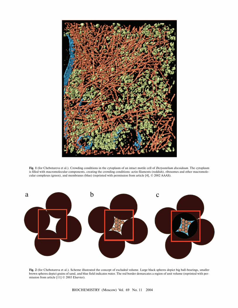
“Macromolecular crowding” is more accuratelytermed “the excluded volume effect” because its mostbasic characteristic is the mutual impenetrability of allsolute molecules. This nonspecific steric repulsion isalways present, regardless of any other attractive or repul-sive interactions that might occur between the solute mol-ecules. Thus crowding, like gravity, cannot be excludedand organisms have to cope with its consequences [6, 7].The influence of the excluded volume on the hydrody-namic and thermodynamic properties of compact globu-lar proteins can be explained using models, which consid-er protein molecules as hard particles of spherical shape[5]. In order to illustrate the concept of excluded volume,it is possible to employ a macroscopic analogy [11].Consider a beaker filled to the brim with metallic ballshaving 5 mm diameter (Fig. 2, reprinted from [11] withpermission from Elsevier Publisher). The randomlyclose-packed ball bearings occupy about 65% of the vol-ume of the beaker, leaving about 35% in the intersticesbetween the ball bearings. Even though the interstitialvolume is “empty”, it is impossible to add even a singleadditional ball bearing of the same size to the beaker withclose-packed balls (Fig. 2a, see color insert). In otherwords, the volume available to ball bearings (i.e., the totalvolume minus the excluded volume) has become zero. Atthe same time, the interstitial volume is available to parti-cles that are smaller, for instance, to grains of sand. If wepour sand into the beaker, it will “fill” the intersticesbetween the ball bearings. But in reality, grains of sandcould occupy only about 65% of the volume occupied bybig balls (Fig. 2b). If the beaker is filled with sand in thisfashion, the volume remaining between bearing balls andgrains of sand will correspond to about 10% of the totalvolume of the beaker. This part of the volume is excludedto both ball bearings and grains of sand, but may be occu-py by the particles of the smaller size, for example, bymolecules of water (Fig. 2c).
Volume exclusion and thermodynamic activity of asubstance. Consider the effect of macromolecular crowd-ing on the thermodynamic activity of a substance. The
chemical potential of solute species i can be partitionedinto ideal and nonideal contributions:
µi = µiideal + µi
nonideal . (1)
The ideal contribution is the free energy changeexpected in the absence of solute–solute interactions:
µiideal = µ i
0 + kT lnci , (2)
where µi0 is the standard state chemical potential of
species i, k is Boltzmann’s constant, T the absolute tem-perature, and ci the concentration (in molar or w/v units).The nonideal contribution is the free energy change asso-ciated with the equilibrium free energy of nonspecificinteraction between a molecule of solute species i and allother solute molecules in the solution:
µinonideal = kT lnγi , (3)
where γi is the activity coefficient. Equations (1)-(3) canbe transformed into the following:
µi = µi0 + RT lnai , (4)
where ai = γici is an effective concentration called thethermodynamic activity.
It is essential to keep in mind that the theory ofchemical equilibrium is based upon the use of activitiesrather than concentrations. In a very dilute solution, thenon-ideal contribution of the chemical potential can beneglected, and concentrations can be used instead ofactivities. In a crowded biological medium, the activity ofsome species could significantly exceed their respectiveconcentration [6, 7].
The theoretical aspects of the effect of excluded vol-ume on chemical reaction were discussed by Minton andcoworkers, Ellis and coworkers, Winzor and coworkers,and many others [11-19]. Consider a generalizedreversible reaction in solution:
r1R1 + r2R2 + ... →← p1P1 + p2P2 + ..., (5)
where ri is the stoichiometric coefficient of reactantspecies Ri and pi is the stoichiometric coefficient of prod-uct species Pi. The equilibrium molar concentrations ofreactants are related by following relationship:
. (6)
Here K denotes the equilibrium constant, whichunder crowding conditions should be considered as anapparent constant related to the true equilibrium constantK 0 by the equation:
BIOCHEMICAL EFFECTS OF MOLECULAR CROWDING 1241
BIOCHEMISTRY (Moscow) Vol. 69 No. 11 2004
K = K 0Γ, (7)
where Γ is the “nonideality factor”. The value of Γ couldbe expressed via the thermodynamic activity coefficientsγi for each i-th component:
(8)
To explain the thermodynamic nonideality initiatedby crowding on the rates of the forward and backwardreaction the theory of absolute reaction rates could beused [20]. For the values of the effective forward andbackward reaction rate constants (kf and kb, respectively)the following expressions should be written:
,
(9)
,
where kf0 and kb
0 denote, respectively, the forward andbackward rate constants in the infinitely dilute solution ofall macrosolutes, and γT denotes the thermodynamicactivity coefficient of the transition-state complex.
In a solution containing macromolecules that inter-act exclusively via steric repulsion, a very simple relation-ship exists between the effective and the real concentra-tion of each solute:
γi = ai/ci = νtot/νa,i , (10)
where ai and ci are the thermodynamic activity and con-centration of the solute i, respectively; νtot is the total vol-ume; and νa,i is the volume available to the solute i. Fromthe practical point of view it is very important that usual-ly the activity coefficients of the proteins could be calcu-lated rather exactly using simple structural models, wherethe hard globular proteins are represented by hard spher-ical or spherocylindrical particles [13, 21-23]. Evaluatingin such a way the activity coefficients of proteins andother macromolecules and the value of nonideality factorΓ under crowding conditions, it is possible to predict theeffect of crowding on the equilibrium position and therates of the reactions proceeding with the participation ofthe biomacromolecules [11].
To imagine how strong could be deviations from ide-ality under crowding conditions, let us evaluate the activ-ity coefficient of a globular test-protein T in a solutioncontaining a second globular protein C, which could becalled a crowding agent. Let us denote as φ the fraction ofthe total volume that is occupied by crowder. Both test-
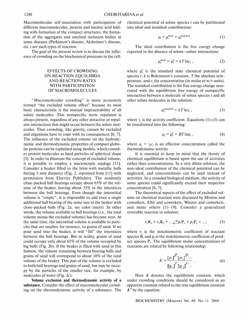
protein and protein crowding agent are considered ashard spherical particles with molecular masses strictlyproportional to the particle radius in third degree. Figure3 presents the theoretical dependence of the activity coef-ficient of the test-protein T (γT) on φ value, increasingwith the filling of volume by the crowder. The dependenceof logγT on φ is calculated under the different relation-ships of the particle masses MT/MC (MT and MC are themasses of test-protein and crowding agent, respectively).As shown in the figure, the activity coefficient of test-pro-tein T could be several orders of magnitude higher thanthe value in very dilute solutions (γT = 1) at the level ofvolume occupancy by crowding agent corresponding tophysiological conditions. For example, the dark circle inthe Fig. 3 indicates that at φ = 0.3 the value of the activi-ty coefficient of the test-protein T exceeds 100. This valueof γT is close to the experimentally measured activitycoefficient of hemoglobin in a 350 g/liter solution com-parable to its content in a red blood cell.
Let us discuss now the influence of crowding onreversible protein isomerization:
R →← P , (11)
where R and P are conformational states of the proteinmolecule that are distinguished by the volume (the com-pactness degree). Allosteric transitions of proteinsinduced by the binding of allosteric ligands and reversibleprotein denaturation are, in particular, such kinds of
Fig. 3. Dependence of logarithm of activity coefficient (γT) ofspherical particles of species T with mass of MT plotted as a func-tion of φ, the fractional occupancy of volume of the crowdingagent C with mass of MC [11]. The dependence of logγT on φ iscalculated at different values of MT/MC: 0.1 (1), 0.3 (2), 1 (3), 3(4), and 10 (5).
1
4
3
2
2
1
0
3
4
0.1 0.2 0.3 0.4
φ
log
γ T
0
5
1242 CHEBOTAREVA et al.
BIOCHEMISTRY (Moscow) Vol. 69 No. 11 2004
processes. Processes of this type are accompanied by achange in the compactness of the protein molecule. In avery well known model of allosteric proteins proposed byMonod, Wyman, and Changeux [24], the equilibriumbetween “relaxed” protein conformation R, stabilized bysubstrates and allosteric activators, and “tense”, morecompact conformation T, stabilized by allostericinhibitors, is considered.
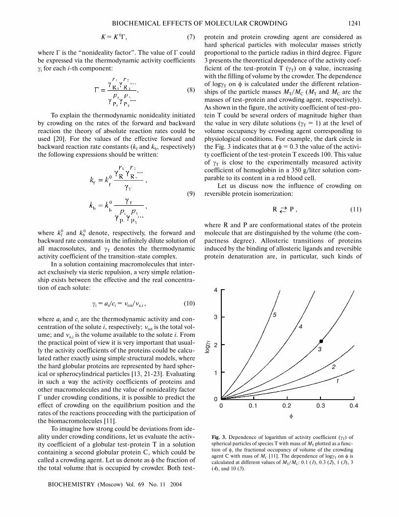
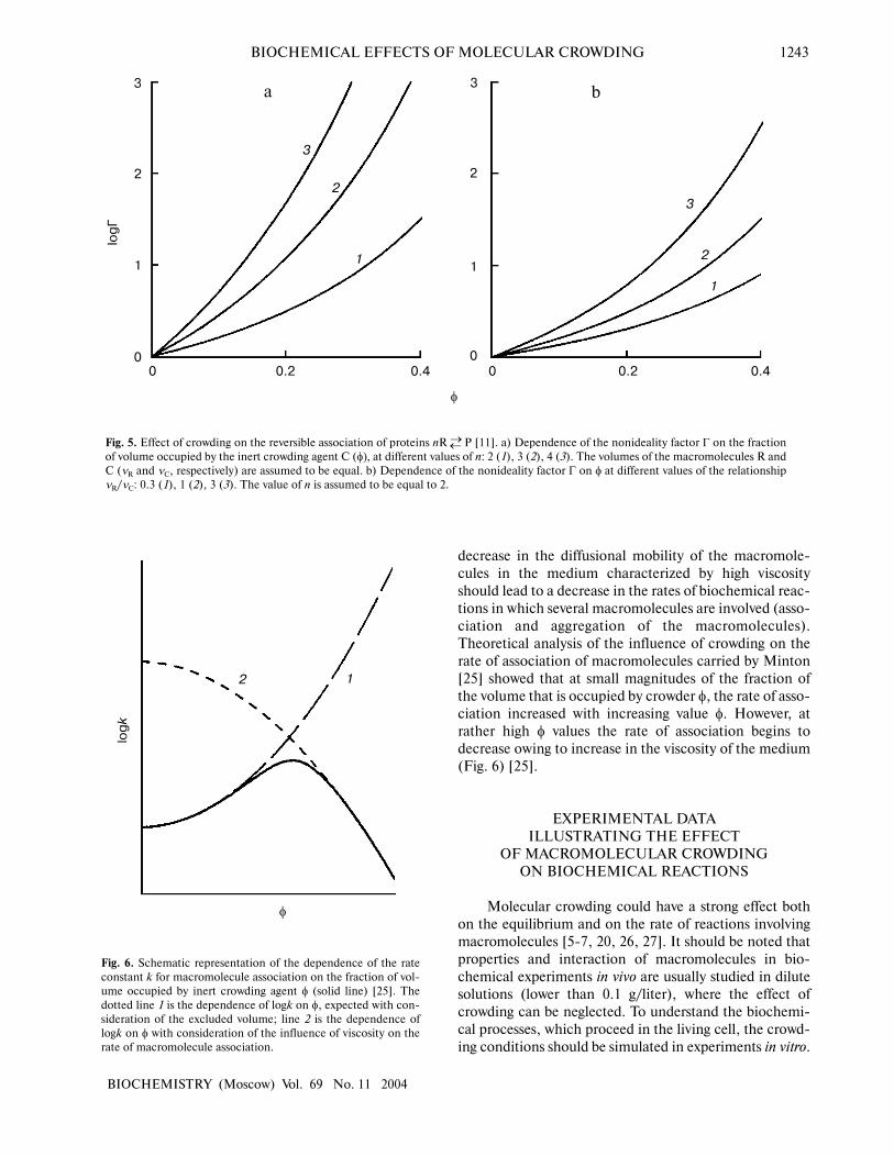
Figure 4 demonstrates the shift of equilibrium (11) inthe presence of macromolecular crowding agent (C). Allmacromolecular substances (R, P, and C) are consideredas hard spherical particles. The situation is discussedwhen the conformational transition in the model proteinis accompanied by an increase in the effective volume ofthe molecule, i.e., the radius of the particle P (rP) exceedsthe radius of the particle R (rR). Such a situation occurs,for instance, under the unfolding (denaturation) of a pro-tein molecule or under the allosteric transition of theform T into the form R. The nonideality factor Γ =γR/γP < 1. The higher is the part of excluded volumeoccupied by the crowding agent φ, the more the nonide-ality factor Γ deviates from unity. Figure 4a illustrates theenhancement of the crowding effect on the increase in theratio of the protein molecular radii rP/rR. Figure 4b showsthat the crowding effect is more pronounced withincrease in the relative size of the model protein, i.e., atthe increase in the ratio of the macromolecules volumesof the model proteins and crowding agent vR/vC [11].
Consider the influence of crowding on the reversibleassociation of protein molecules
nR →← P , (12)
where R is monomer and P is oligomer composed from nmonomers. The behavior of the associating enzyme sys-tem under crowding conditions is presented in Fig. 5. It issupposed that all macrosolute species (R, P, and C) canbe represented as hard spherical particles, and that thevolume of the oligomer P is equal to n times the volumeof monomer R. The nonideality factor Γ = γR
n/γP. Theactivity coefficient for the monomer γR is less than theactivity coefficient for the oligomer γP. However, becausen > 1, the magnitude of the nonideality factor Γ is higherthan unity. The higher is the part of the excluded volumeoccupied by crowder, φ, the stronger is the deviation ofthe nonideality factor Γ from unity. Figure 5a illustratesthe enhancement of the crowding effect on the increase inthe number of monomers involved into association (i.e.,increase in the value of n). Figure 5b shows that thecrowding effect is more pronounced on the increase inthe relative dimensions of the model protein, i.e., on theincrease in the ratio of the volume of the model proteinmacromolecules to the volume of the crowding agentνR/νC [11].
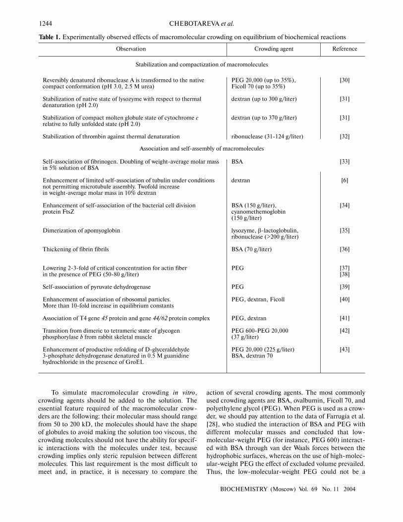
Crowding has a complex effect on the rate of bio-chemical reactions. On one hand, under crowding condi-tions the thermodynamic activities of the reactantsincrease, and, on the other hand, crowding reduces diffu-sion, and, therefore, the possibility of the meeting of tworeactants decreases. The overall result of these opposingfactors depends on the nature of each reaction. The
Fig. 4. Influence of crowding on the reversible conformational transition of proteins R →← P [11]. a) Dependence of the nonideality factorΓ on the fraction of volume occupied by the inert crowding agent C (φ), at different values of the relationship rP/rR (rP and rR are radii ofprotein molecules P and R, respectively): 1.1 (1), 1.2 (2), 1.3 (3). The volumes of the macromolecules R and C (vR and vC, respectively) areassumed to be equal. b) Dependence of the nonideality factor Γ on φ at different values of relationship νR/νC. The relationship rP/rR isassumed to be equal to 1.2.
1
0
–0.5
2
–1.0
–1.5
0
3
0.2 0.4
φ
log
Г
–2.0
3
2
1
0
–0.5
–1.0
–1.5
–2.00 0.2 0.4
a b
BIOCHEMICAL EFFECTS OF MOLECULAR CROWDING 1243
BIOCHEMISTRY (Moscow) Vol. 69 No. 11 2004
decrease in the diffusional mobility of the macromole-cules in the medium characterized by high viscosityshould lead to a decrease in the rates of biochemical reac-tions in which several macromolecules are involved (asso-ciation and aggregation of the macromolecules).Theoretical analysis of the influence of crowding on therate of association of macromolecules carried by Minton[25] showed that at small magnitudes of the fraction ofthe volume that is occupied by crowder φ, the rate of asso-ciation increased with increasing value φ. However, atrather high φ values the rate of association begins todecrease owing to increase in the viscosity of the medium(Fig. 6) [25].
EXPERIMENTAL DATAILLUSTRATING THE EFFECT
OF MACROMOLECULAR CROWDINGON BIOCHEMICAL REACTIONS
Molecular crowding could have a strong effect bothon the equilibrium and on the rate of reactions involvingmacromolecules [5-7, 20, 26, 27]. It should be noted thatproperties and interaction of macromolecules in bio-chemical experiments in vivo are usually studied in dilutesolutions (lower than 0.1 g/liter), where the effect ofcrowding can be neglected. To understand the biochemi-cal processes, which proceed in the living cell, the crowd-ing conditions should be simulated in experiments in vitro.
Fig. 5. Effect of crowding on the reversible association of proteins nR →← P [11]. a) Dependence of the nonideality factor Γ on the fractionof volume occupied by the inert crowding agent C (φ), at different values of n: 2 (1), 3 (2), 4 (3). The volumes of the macromolecules R andC (νR and νC, respectively) are assumed to be equal. b) Dependence of the nonideality factor Γ on φ at different values of the relationshipνR/νC: 0.3 (1), 1 (2), 3 (3). The value of n is assumed to be equal to 2.
1
3
22
1
0
3
0.2 0.4
φ
log
Г
0
3
2
1
3
2
1
00 0.2 0.4
Fig. 6. Schematic representation of the dependence of the rateconstant k for macromolecule association on the fraction of vol-ume occupied by inert crowding agent φ (solid line) [25]. Thedotted line 1 is the dependence of logk on φ, expected with con-sideration of the excluded volume; line 2 is the dependence oflogk on φ with consideration of the influence of viscosity on therate of macromolecule association.
1 2
φ
log
k
a b
1244 CHEBOTAREVA et al.
BIOCHEMISTRY (Moscow) Vol. 69 No. 11 2004
To simulate macromolecular crowding in vitro,crowding agents should be added to the solution. Theessential feature required of the macromolecular crow-ders are the following: their molecular mass should rangefrom 50 to 200 kD, the molecules should have the shapeof globules to avoid making the solution too viscous, thecrowding molecules should not have the ability for specif-ic interactions with the molecules under test, becausecrowding implies only steric repulsion between differentmolecules. This last requirement is the most difficult tomeet and, in practice, it is necessary to compare the
action of several crowding agents. The most commonlyused crowding agents are BSA, ovalbumin, Ficoll 70, andpolyethylene glycol (PEG). When PEG is used as a crow-der, we should pay attention to the data of Farrugia et al.[28], who studied the interaction of BSA and PEG withdifferent molecular masses and concluded that low-molecular-weight PEG (for instance, PEG 600) interact-ed with BSA through van der Waals forces between thehydrophobic surfaces, whereas on the use of high-molec-ular-weight PEG the effect of excluded volume prevailed.Thus, the low-molecular-weight PEG could not be a
Observation
Reversibly denatured ribonuclease A is transformed to the nativecompact conformation (pH 3.0, 2.5 M urea)
Stabilization of native state of lysozyme with respect to thermaldenaturation (pH 2.0)
Stabilization of compact molten globule state of cytochrome crelative to fully unfolded state (pH 2.0)
Stabilization of thrombin against thermal denaturation
Self-association of fibrinogen. Doubling of weight-average molar massin 5% solution of BSA
Enhancement of limited self-association of tubulin under conditionsnot permitting microtubule assembly. Twofold increasein weight-average molar mass in 10% dextran
Enhancement of self-association of the bacterial cell divisionprotein FtsZ
Dimerization of apomyoglobin
Thickening of fibrin fibrils
Lowering 2-3-fold of critical concentration for actin fiberin the presence of PEG (50-80 g/liter)
Self-association of pyruvate dehydrogenase
Enhancement of association of ribosomal particles.More than 10-fold increase in equilibrium constants
Association of T4 gene 45 protein and gene 44/62 protein complex
Transition from dimeric to tetrameric state of glycogenphosphorylase b from rabbit skeletal muscle
Enhancement of productive refolding of D-glyceraldehyde3-phosphate dehydrogenase denatured in 0.5 M guanidinehydrochloride in the presence of GroEL
Crowding agent
PEG 20,000 (up to 35%),Ficoll 70 (up to 35%)
dextran (up to 300 g/liter)
dextran (up to 370 g/liter)
ribonuclease (31-124 g/liter)
BSA
dextran
BSA (150 g/liter),cyanomethemoglobin(150 g/liter)
lysozyme, β-lactoglobulin,ribonuclease (>200 g/liter)
BSA (70 g/liter)
PEG
PEG
PEG, dextran, Ficoll
PEG, dextran
PEG 600-PEG 20,000(37 g/liter)
PEG 20,000 (225 g/liter)BSA, dextran 70
Reference
[30]
[31]
[31]
[32]
[33]
[6]
[34]
[35]
[36]
[37] [38]
[39]
[40]
[41]
[42]
[43]
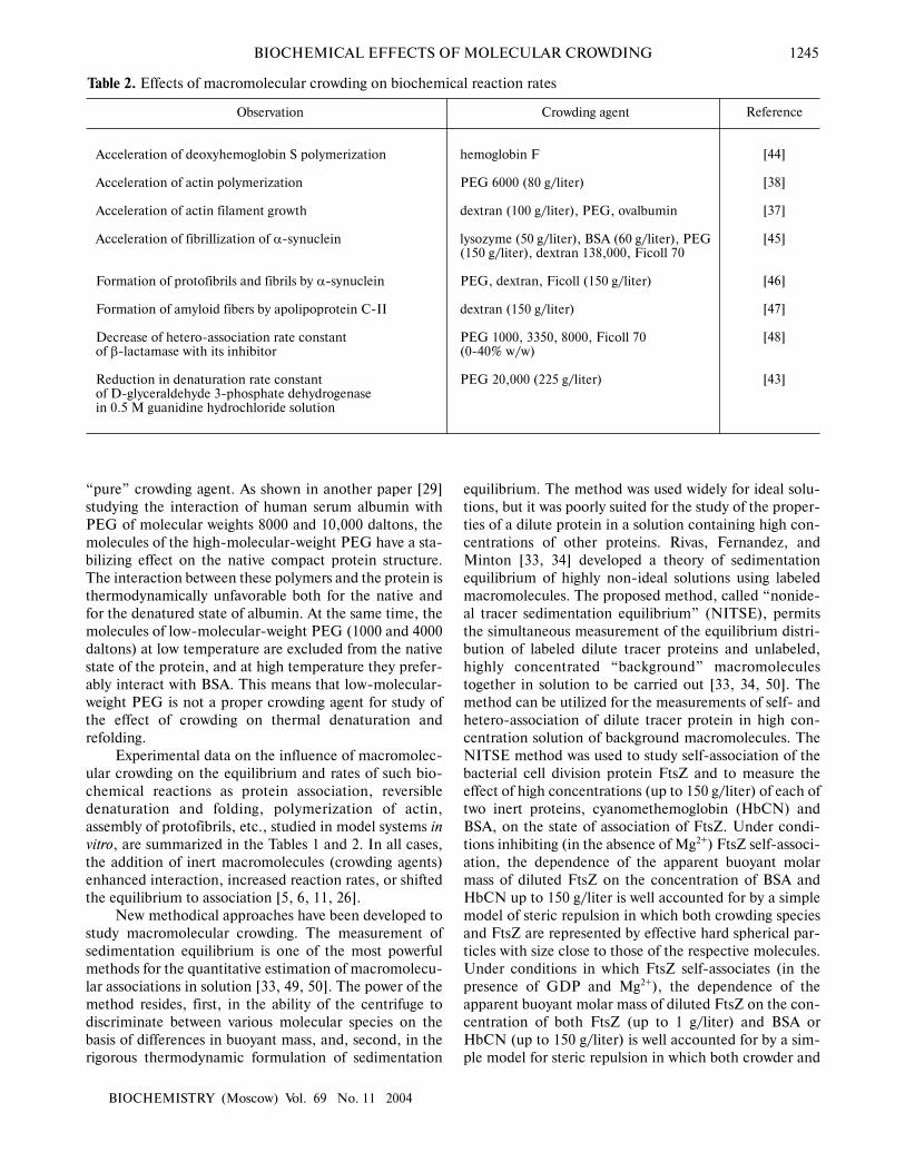
Table 1. Experimentally observed effects of macromolecular crowding on equilibrium of biochemical reactions
Stabilization and compactization of macromolecules
Association and self-assembly of macromolecules
BIOCHEMICAL EFFECTS OF MOLECULAR CROWDING 1245
BIOCHEMISTRY (Moscow) Vol. 69 No. 11 2004
“pure” crowding agent. As shown in another paper [29]studying the interaction of human serum albumin withPEG of molecular weights 8000 and 10,000 daltons, themolecules of the high-molecular-weight PEG have a sta-bilizing effect on the native compact protein structure.The interaction between these polymers and the protein isthermodynamically unfavorable both for the native andfor the denatured state of albumin. At the same time, themolecules of low-molecular-weight PEG (1000 and 4000daltons) at low temperature are excluded from the nativestate of the protein, and at high temperature they prefer-ably interact with BSA. This means that low-molecular-weight PEG is not a proper crowding agent for study ofthe effect of crowding on thermal denaturation andrefolding.
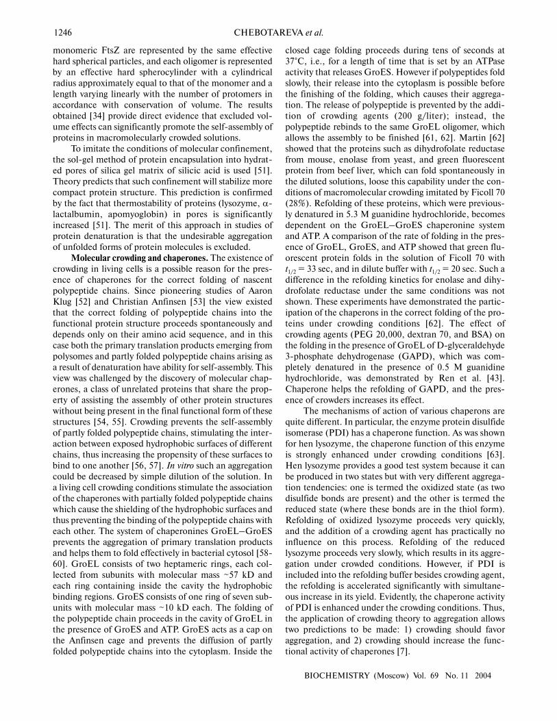
Experimental data on the influence of macromolec-ular crowding on the equilibrium and rates of such bio-chemical reactions as protein association, reversibledenaturation and folding, polymerization of actin,assembly of protofibrils, etc., studied in model systems invitro, are summarized in the Tables 1 and 2. In all cases,the addition of inert macromolecules (crowding agents)enhanced interaction, increased reaction rates, or shiftedthe equilibrium to association [5, 6, 11, 26].
New methodical approaches have been developed tostudy macromolecular crowding. The measurement ofsedimentation equilibrium is one of the most powerfulmethods for the quantitative estimation of macromolecu-lar associations in solution [33, 49, 50]. The power of themethod resides, first, in the ability of the centrifuge todiscriminate between various molecular species on thebasis of differences in buoyant mass, and, second, in therigorous thermodynamic formulation of sedimentation
equilibrium. The method was used widely for ideal solu-tions, but it was poorly suited for the study of the proper-ties of a dilute protein in a solution containing high con-centrations of other proteins. Rivas, Fernandez, andMinton [33, 34] developed a theory of sedimentationequilibrium of highly non-ideal solutions using labeledmacromolecules. The proposed method, called “nonide-al tracer sedimentation equilibrium” (NITSE), permitsthe simultaneous measurement of the equilibrium distri-bution of labeled dilute tracer proteins and unlabeled,highly concentrated “background” macromoleculestogether in solution to be carried out [33, 34, 50]. Themethod can be utilized for the measurements of self- andhetero-association of dilute tracer protein in high con-centration solution of background macromolecules. TheNITSE method was used to study self-association of thebacterial cell division protein FtsZ and to measure theeffect of high concentrations (up to 150 g/liter) of each oftwo inert proteins, cyanomethemoglobin (HbCN) andBSA, on the state of association of FtsZ. Under condi-tions inhibiting (in the absence of Mg2+) FtsZ self-associ-ation, the dependence of the apparent buoyant molarmass of diluted FtsZ on the concentration of BSA andHbCN up to 150 g/liter is well accounted for by a simplemodel of steric repulsion in which both crowding speciesand FtsZ are represented by effective hard spherical par-ticles with size close to those of the respective molecules.Under conditions in which FtsZ self-associates (in thepresence of GDP and Mg2+), the dependence of theapparent buoyant molar mass of diluted FtsZ on the con-centration of both FtsZ (up to 1 g/liter) and BSA orHbCN (up to 150 g/liter) is well accounted for by a sim-ple model for steric repulsion in which both crowder and
Observation
Acceleration of deoxyhemoglobin S polymerization
Acceleration of actin polymerization
Acceleration of actin filament growth
Acceleration of fibrillization of α-synuclein
Formation of protofibrils and fibrils by α-synuclein
Formation of amyloid fibers by apolipoprotein C-II
Decrease of hetero-association rate constantof β-lactamase with its inhibitor
Reduction in denaturation rate constantof D-glyceraldehyde 3-phosphate dehydrogenasein 0.5 M guanidine hydrochloride solution
Crowding agent
hemoglobin F
PEG 6000 (80 g/liter)
dextran (100 g/liter), PEG, ovalbumin
lysozyme (50 g/liter), BSA (60 g/liter), PEG(150 g/liter), dextran 138,000, Ficoll 70
PEG, dextran, Ficoll (150 g/liter)
dextran (150 g/liter)
PEG 1000, 3350, 8000, Ficoll 70(0-40% w/w)
PEG 20,000 (225 g/liter)
Reference
[44]
[38]
[37]
[45]
[46]
[47]
[48]
[43]
Table 2. Effects of macromolecular crowding on biochemical reaction rates
1246 CHEBOTAREVA et al.
BIOCHEMISTRY (Moscow) Vol. 69 No. 11 2004
monomeric FtsZ are represented by the same effectivehard spherical particles, and each oligomer is representedby an effective hard spherocylinder with a cylindricalradius approximately equal to that of the monomer and alength varying linearly with the number of protomers inaccordance with conservation of volume. The resultsobtained [34] provide direct evidence that excluded vol-ume effects can significantly promote the self-assembly ofproteins in macromolecularly crowded solutions.
To imitate the conditions of molecular confinement,the sol-gel method of protein encapsulation into hydrat-ed pores of silica gel matrix of silicic acid is used [51].Theory predicts that such confinement will stabilize morecompact protein structure. This prediction is confirmedby the fact that thermostability of proteins (lysozyme, α-lactalbumin, apomyoglobin) in pores is significantlyincreased [51]. The merit of this approach in studies ofprotein denaturation is that the undesirable aggregationof unfolded forms of protein molecules is excluded.
Molecular crowding and chaperones. The existence ofcrowding in living cells is a possible reason for the pres-ence of chaperones for the correct folding of nascentpolypeptide chains. Since pioneering studies of AaronKlug [52] and Christian Anfinsen [53] the view existedthat the correct folding of polypeptide chains into thefunctional protein structure proceeds spontaneously anddepends only on their amino acid sequence, and in thiscase both the primary translation products emerging frompolysomes and partly folded polypeptide chains arising asa result of denaturation have ability for self-assembly. Thisview was challenged by the discovery of molecular chap-erones, a class of unrelated proteins that share the prop-erty of assisting the assembly of other protein structureswithout being present in the final functional form of thesestructures [54, 55]. Crowding prevents the self-assemblyof partly folded polypeptide chains, stimulating the inter-action between exposed hydrophobic surfaces of differentchains, thus increasing the propensity of these surfaces tobind to one another [56, 57]. In vitro such an aggregationcould be decreased by simple dilution of the solution. Ina living cell crowding conditions stimulate the associationof the chaperones with partially folded polypeptide chainswhich cause the shielding of the hydrophobic surfaces andthus preventing the binding of the polypeptide chains witheach other. The system of chaperonines GroEL–GroESprevents the aggregation of primary translation productsand helps them to fold effectively in bacterial cytosol [58-60]. GroEL consists of two heptameric rings, each col-lected from subunits with molecular mass ~57 kD andeach ring containing inside the cavity the hydrophobicbinding regions. GroES consists of one ring of seven sub-units with molecular mass ~10 kD each. The folding ofthe polypeptide chain proceeds in the cavity of GroEL inthe presence of GroES and ATP. GroES acts as a cap onthe Anfinsen cage and prevents the diffusion of partlyfolded polypeptide chains into the cytoplasm. Inside the
closed cage folding proceeds during tens of seconds at37°C, i.e., for a length of time that is set by an ATPaseactivity that releases GroES. However if polypeptides foldslowly, their release into the cytoplasm is possible beforethe finishing of the folding, which causes their aggrega-tion. The release of polypeptide is prevented by the addi-tion of crowding agents (200 g/liter); instead, thepolypeptide rebinds to the same GroEL oligomer, whichallows the assembly to be finished [61, 62]. Martin [62]showed that the proteins such as dihydrofolate reductasefrom mouse, enolase from yeast, and green fluorescentprotein from beef liver, which can fold spontaneously inthe diluted solutions, loose this capability under the con-ditions of macromolecular crowding imitated by Ficoll 70(28%). Refolding of these proteins, which were previous-ly denatured in 5.3 M guanidine hydrochloride, becomesdependent on the GroEL–GroES chaperonine systemand ATP. A comparison of the rate of folding in the pres-ence of GroEL, GroES, and ATP showed that green flu-orescent protein folds in the solution of Ficoll 70 witht1/2 = 33 sec, and in dilute buffer with t1/2 = 20 sec. Such adifference in the refolding kinetics for enolase and dihy-drofolate reductase under the same conditions was notshown. These experiments have demonstrated the partic-ipation of the chaperons in the correct folding of the pro-teins under crowding conditions [62]. The effect ofcrowding agents (PEG 20,000, dextran 70, and BSA) onthe folding in the presence of GroEL of D-glyceraldehyde3-phosphate dehydrogenase (GAPD), which was com-pletely denatured in the presence of 0.5 M guanidinehydrochloride, was demonstrated by Ren et al. [43].Chaperone helps the refolding of GAPD, and the pres-ence of crowders increases its effect.
The mechanisms of action of various chaperons arequite different. In particular, the enzyme protein disulfideisomerase (PDI) has a chaperone function. As was shownfor hen lysozyme, the chaperone function of this enzymeis strongly enhanced under crowding conditions [63].Hen lysozyme provides a good test system because it canbe produced in two states but with very different aggrega-tion tendencies: one is termed the oxidized state (as twodisulfide bonds are present) and the other is termed thereduced state (where these bonds are in the thiol form).Refolding of oxidized lysozyme proceeds very quickly,and the addition of a crowding agent has practically noinfluence on this process. Refolding of the reducedlysozyme proceeds very slowly, which results in its aggre-gation under crowded conditions. However, if PDI isincluded into the refolding buffer besides crowding agent,the refolding is accelerated significantly with simultane-ous increase in its yield. Evidently, the chaperone activityof PDI is enhanced under the crowding conditions. Thus,the application of crowding theory to aggregation allowstwo predictions to be made: 1) crowding should favoraggregation, and 2) crowding should increase the func-tional activity of chaperones [7].
BIOCHEMICAL EFFECTS OF MOLECULAR CROWDING 1247
BIOCHEMISTRY (Moscow) Vol. 69 No. 11 2004
MOLECULAR CROWDINGINDUCED BY OSMOLYTES
In response to stress (osmotic, chemical, or ther-mal), many living organisms accumulate high concentra-tions of osmolytes. Osmolytes are small organic mole-cules, such as polyols, some amino acids, and methyl-amines. They protect proteins under stress conditions bystabilization of protein structure [64-68]. For instance,the tissues of sharks and rays contain urea in high con-centration. It is known that urea causes denaturation ofthe proteins, but this effect could be compensatedthrough supporting of the high concentration of thecounteracting osmolytes, especially trimethylamine N-oxide (TMAO), betaine, and sarcosine [64-68]. Amongorganisms concentrating proline are those that haveadapted to live in high salt environments, and they usethis osmolyte to offset the high external osmotic pressure[65]. Such polyols as sucrose probably have selective abil-ity to stabilize proteins at extremes of temperature [65,67].
Much experimental data has accumulated on theprotective action of osmolytes [67-76], but the molecularmechanisms of their action are not clear. The same exper-imental data can be explained very often using contradic-tory molecular mechanisms [19]. Let us discuss the effectof molecular crowding arising from the high concentra-tions of osmolytes on biochemical reactions (association,isomerization, protein folding and aggregation, interac-tion of macromolecules with ligands, etc.).
Molecular crowding could displace the equilibriumof reversibly associating protein towards the high-molec-ular-weight state, and the extent to which it influences theequilibrium state depends on the concentration of theinert cosolute [77-80]. For example, in the case of eno-lase, which reversibly associates as monomer−dimer,increase in the dimer fraction with increasing sucroseconcentration has been observed [78]. The fact that low-molecular-weight species can also participate in theeffects of excluded volume is demonstrated by a three-fold increase of dimerization constant for α-chy-motrypsin in the presence of 0.15 M sucrose [77]. Similareffects have been observed also for DNA–protein interac-tions [81, 82]. Carner and Rau [81] discovered a multipleincrease in the constant of binding of Gal repressor withDNA in the presence of sucrose, betaine, or triethyleneglycol. Poon and coworkers [82] showed an increase inthe apparent constant of binding of the regulatory proteinTyrR with DNA in the presence of sucrose and triethyl-ene glycol. In paper [83], the sedimentation equilibriummethod was used to quantify the action of high concen-trations of glucose, sucrose, and raffinose on thereversible dimerization of α-chymotrypsin. The dimer-ization constant (K2) was calculated from the sedimenta-tion equilibrium distributions of the enzyme in theabsence of osmolytes and the apparent dimerization con-
stant (K2,app) was calculated in the presence of osmolytes.To quantify the effect of osmolytes on the dimerizationreaction, the dependence (lnK2,app) was plotted againstthe molar sugar concentration (Cs), which appeared to belinear. To explain the effect of stabilization of the dimer ofα-chymotrypsin and to compare two models that inter-pret the effect of osmolytes either as a weak interaction (apreferential binding) or as a steric repulsion (effect ofexcluded volume), the authors used the magnitudeδlnK2,app/δCs (M–1). This experimentally evaluated valuewas compared with the value calculated from the models.It was shown that weak interactions suppose the δlnK2,app/δCs value to be less than that observed, whereas predic-tions based on the theory of excluded volume gives theδlnK2,app/δCs value exceeding this value. However, thetheory of excluded volume predicts rather well the lineardependence of lnK2,app on Cs and a change in the slope oflnK2,app on Cs with increase in the sugar molecule size. Toevaluate the expected effect of molecular crowding, thefollowing equations were used in [83]:
K2,app = K2(γ12/γ2) =
= K2 exp[(2U1s – U2s)Сs +…] (13)
or in logarithmic form:
lnK2,app = lnK2 + (2U1s – U2s)Сs +…, (14)
where the second virial coefficient (U1s for monomer andU2s for dimers) is expressed via excluded covolume (in theabsence of charge on sucrose). Stabilization parameterδlnK2,app/δCs is equivalent to the difference in 2U1s – U2s.
Under the assumption of spherical shape of α-chy-motrypsin monomer and saccharide, U1s can be calculat-ed as 4πN(r1 + rs)
3/3, where N is Avogadro’s number,enzyme monomer radius r1 is equal to 2.44 nm [77, 84],radii rs are equal to 0.25 nm for glucose, 0.34 nm forsucrose, and 0.43 nm for raffinose [83]. The value of U2s
can be calculated by approximating the protein dimerthrough the ellipsoid of rotation with the ratio of the axesequal to 2 [85, 86], and using the equation:
U2s = 4πNrs3/3 + 4πNab2/3 +
+ 2πNabrs[(1–ε2)1/2 + (sin–1ε)/ε] + (15)
+ 2πNars2[1 +{(1–ε2)/2ε}ln{(1+ε)/(1–ε)}],
where ε2 = 1 − (b/a)2, b = r1, a = 2r1.In comparing the experimental and calculated val-
ues, the authors of paper [83] concluded that molecularcrowding plays the major role in the enhancement of α-chymotrypsin dimerization. In article [79], the sedimen-tation velocity method was used to quantify the effect ofhigh concentrations of TMAO on the self-association of
1248 CHEBOTAREVA et al.
BIOCHEMISTRY (Moscow) Vol. 69 No. 11 2004
glycogen phosphorylase b (from rabbit skeletal muscle)induced by 1 mM AMP. Phosphorylase b associatesreversibly in a dimer–tetramer equilibrium, which can beconsidered as a reaction of the dimerization of dimers. Toquantify the effect of osmolyte on the reaction of dimer-ization, the dependence of the logarithm of the apparentassociation constant (lnK2,app) on the molar concentra-tion of the osmolyte (Cosm) was plotted, which appeared tobe linear. To evaluate the expected effect of molecularcrowding, relationships similar to those given above (14)were used in paper [79]:
ln[(K2,4)app /K2,4 ] = (2B2,osm – B4,osm)Сosm , (16)
where the second virial coefficient (B2,osm for dimer andB4,osm for tetramer) is expressed by the covolume exclud-ed. The values of B2,osm and B4,osm could be calculatedthrough approximation of the protein dimer and tetramerby the ellipsoid of rotation with an axial ratio of 1.9 andusing Eq. (15) [85, 86]. The comparison of the valueδln[(K2,4)app/K2,4]/δCosm (M–1) obtained experimentallywith the calculated value showed that the theory ofexcluded volume predicts a larger effect than that whichwas observed. The association constant K2,4 for dimer–tetramer equilibrium in the presence of 1 M TMAOincreases 2-fold. For explanation of the data, a reversiblestage of dimer isomerization, which is preceded by asso-ciation, was introduced. The small enhancement of theassociation degree of phosphorylase b in the presence ofTMAO as compared with the theoretical predictionsmeans that the effect of excluded volume on thedimer–tetramer equilibrium is compensated by the shiftof equilibrium in the isomerization reaction of the dimerT ↔ R towards the more compact, non-associating T-conformation [79].
The influence of TMAO on the self-association ofphosphorylase kinase from rabbit skeletal muscle hasbeen studied by sedimentation velocity and turbidimetry[80]. It was shown that TMAO (0.6-1.0 M) stimulates theassociation of phosphorylase kinase induced by Ca2+ andMg2+. In the presence of TMAO, besides associates con-sisting of relatively small number (n) of enzyme mole-cules (n = 2, 3, 4, …), large associates with sedimentationcoefficient s20,w = 189 and 385 S corresponding to 24- and70-mers of phosphorylase kinase were registered. The ini-tial rate of association increased nonlinearly with increas-ing osmolyte concentration (0.4-1.4 M).
A stabilizing effect of glycerol on the thermal inacti-vation and denaturation of creatine kinase was shown bydifferential scanning calorimetry [87]. Glycerol increasesthe temperature of denaturation of the enzyme (Tm). Astabilizing effect of 1 M glucose on the structure of acid-denatured ferricytochrome c was shown by Morar et al.[88]. The unfolded protein folded until the size and shapeof native cytochrome c was achieved in the presence of1 M glucose.
In review [19] the protective action of polyols [89] onthe isomerization equilibrium between folded (F) andunfolded (U) protein states is considered in detail.Protein stability is defined as the free energy change asso-ciated with the reaction F ↔ U: ∆G0 = –RTlnK; whereequilibrium constant K = CF/CU for dilute solution, CF
and CU are the concentrations of folded and unfoldedprotein state, respectively. In the presence of the highconcentrations of osmolytes the apparent isomerizationconstant was Kapp = CF/CU. The protective effect ofosmolyte is characterized as ∆∆G0/RT = ln(K/Kapp). Tocompare the effects of different osmolytes on the isomer-ization reaction, ln(Kapp/K) values were plotted againstmolar sugar concentration Cosm. Because all the plots arelinear, their slope [δ∆∆G0/δCosm] can be used as a meas-ure of protein stabilization. Stabilization increases withincreasing size of sugar during the transition from mono-to tetrasaccharide (glucose → trehalose → melesitose →stachiose) [19]. The models based on the concepts ofthermodynamic non-ideality (effects of excluded volumeand preferential hydration) and also the models consider-ing the preferential interaction of protein with osmolyteare reviewed in [19] for explanation of the effect ofosmolytes. The comparison of experimental and theoret-ical data showed that the δ∆∆G0/δCosm value calculatedon the basis of excluded volume theory exceeds the exper-imental value, and the other models give underestimatedvalue of δ∆∆G0/δCosm. Despite the fact that the degree ofthe displacement of equilibrium isomerization is notalways equivalent to the effect predicted on the basis ofthe excluded volume, the authors of the work [19] sup-posed that the effect of molecular crowding plays animportant role in protein stabilization.
In papers [90, 91], the effect of molecular crowdingon the kinetics of allosteric enzymes has been studied. Forreactions involving enzyme isomerization, be it preexist-ing [24] or substrate-induced [92], a crowded molecularenvironment should favor the smaller isomeric state.Thus, it was shown that the kinetics of pyruvate kinaseinhibition by phenylalanine is sigmoid, and after additionof 0.1 M proline, the sigmoid shape disappears [91]. Usingdifference sedimentation velocity analysis authors [91]showed that in the presence of allosteric inhibitor the sed-imentation coefficient of the enzyme decreases, and onthe addition of proline this difference disappears, which isindicative of the displacement of equilibrium between theenzyme isomers towards the more compact state.
Two kinds of models have been used in the literaturefor description of the interactions between osmolytes andproteins. One kind is based upon the effects of excludedvolume, and the other took into account the binding ofproteins with osmolytes. Weatherly and Pielak [93] study-ing the interactions of osmolytes with ferricytochrome cas a model protein concluded that the simple model can-not explain the interaction between osmolytes and pro-teins and interpreted the second virial coefficient as a
BIOCHEMICAL EFFECTS OF MOLECULAR CROWDING 1249
BIOCHEMISTRY (Moscow) Vol. 69 No. 11 2004
measure of interactions of the excluded volumes and pro-tein binding with osmolyte [93].
It should be noted that some researchers interpretthe influence of osmolytes on biochemical reactions interms of statistical mechanics using the theory of exclud-ed volume and calculating the second virial coefficients,which characterize the interactions of the molecules insolution [14-18, 82-84, 90, 91, 94-96]. Other researchersoperate with the terms of preferable hydration of the pro-tein and the free energy [66, 67, 69-71, 97-101]. Winzorand Wills analyzed both approaches and showed that theyare practically equivalent [14, 17].
The mechanism of the protective effect of osmolytescounteracting urea was studied with different model sys-tems. It was shown that the influence of “counteracting”osmolytes had a general character and was propagated onproteins, which were not exposed to urea in vivo [66, 68].Thus, Burg and Peters [68] showed that urea reduced thethermostability of ribonuclease A, whereas glycerophos-phocholine and TMAO compensated this effect. It wasdemonstrated [64, 102] that TMAO and β-alanine hadstabilizing effects on the structure of ribonuclease A,increasing its temperature of denaturation Tm. Dataobtained by NMR spectroscopy showed that the activityof ribonuclease A decreased in the presence of urea andwas completely restored on the addition of TMAO to urea(optimal ratio [urea]/[TMAO] is 1 : 1). Bolen and co-workers [101] showed that reduced carboxyamidatedribonuclease A (RCAM) exists in the unfolded state in thepresence of urea and refolds on the addition of stabilizingosmolytes (TMAO, sarcosine, sucrose, and proline).During refolding of the reduced carboxyamidated ribonu-clease T1 a more pronounced effect of osmolytes, favor-ing the protein refolding, was observed in the orderTMAO > sarcosine > sucrose > proline, where TMAO isthe more effective osmolyte and proline is the less effec-tive one [67]. The proteins are thermodynamically morestable in the presence of osmolyte than in water solution,because osmolytes increase Gibbs energy for the dena-tured state much stronger than for the native state [67, 70,71, 101, 103]. Bolen and Baskakov [67] named the non-favorable interactions between osmolyte and polypeptidebackbone as an “osmophobic effect”. Since the peptidebackbone is highly exposed to osmolyte in the denaturedstate, the osmophobic effect preferentially raises the freeenergy of the denatured state, shifting the equilibrium infavor of the native state.
Effects of hydration are very important also on pro-tein polymerization, when osmolytes are excluded fromthe nearest environment of the protein surface due toprotein–protein interactions and, in particular, canaccelerate the assembly of microtubules and increasetheir stability [104]. Yang et al. [105] showed that TMAOand glycerol accelerate the main steps in the amyloidalpathway (early nucleation, conformational changes, andalso the protofibril–fibril transformation).
It should be noted in conclusion that crowdingaffects all biochemical processes where changes inexcluded volume are observed. The collapse of newly syn-thesized polypeptide chains into compact functional pro-teins, protein unfolding, induced by chemical or thermalstress, the formation of oligomeric structures and mul-tienzyme complexes in metabolic pathways, the aggrega-tion of proteins into nonfunctional aggregates, such asbacterial inclusion bodies and plaques in human amyloiddiseases (Parkinson’s and Alzheimer’s diseases), arerelated to similar processes. To imitate the conditions invivo during the study of biochemical reactions in vitro,crowding agents should be used. Such experiments allowconclusions about the influence of crowding on biochem-ical processes occurring in the cell to be made.
This work was supported by the Russian Foundationfor Basic Research (grants 02-04-49099 and 02-04-48704), the Program for Fundamental Research“Molecular and Cell Biology” of the Russian Academy ofSciences, the Program for the Support of the LeadingScientific Schools Minpromnauka RF (grant813.2003.4), and by INTAS (grant 03-51-4813).
REFERENCES
1. Fulton, A. B. (1982) Cell, 30, 345-347.2. Zimmerman, S. B., and Trach, S. O. (1991) J. Mol. Biol.,
222, 599-620.3. Ellis, R. J., and Minton, A. P. (2003) Nature, 425, 27-28.4. Medalia, O., Weber, I., Frangakis, A. S., Nicastro, D.,
Gerisch, G., and Baumeister, W. (2002) Science, 298, 1209-1213.
5. Zimmerman, S. B., and Minton, A. P. (1993) Annu. Rev.Biophys. Biomol. Struct., 22, 27-65.
6. Minton, A. P. (2001) J. Biol. Chem., 276, 10577-10580.7. Ellis, R. J. (2001) Trends Biochem. Sci., 26, 597-604.8. Grunewald, K., Medalia, O., Gross, A., Steven, A., and
Baumeister, W. (2003) Biophys. Chem., 100, 577-591.9. Verkman, A. (2002) Trends Biochem. Sci., 27, 27-33.
10. Lyubarev, A. E., and Kurganov, B. I. (1996) in Organizationof Biochemical Systems: Structural and Regulatory Aspects(Kurganov, B. I., and Lyubarev, A. E., eds.) Nova Science,N. Y., pp. 1-81.
11. Hall, D., and Minton, A. P. (2003) Biochim. Biophys. Acta,1649, 127-139.
12. Minton, A. P. (1981) Biopolymers, 20, 2093-2120.13. Minton, A. P. (1983) Mol. Cell. Biochem., 55, 119-140.14. Wills, P. R., and Winzor, D. J. (1993) Biopolymers, 33,
1627-1629.15. Wills, P. R., Comper, W. D., and Winzor, D. J. (1993) Arch.
Biochem. Biophys., 300, 206-212.16. Winzor, C. L., Winzor, D. J., Paleg, L. G., Jones, G. P.,
and Naidu, B. P. (1992) Arch. Biochem. Biophys., 296, 102-107.
17. Winzor, D. J., and Wills, P. R. (1995) in Protein–SolventInteractions (Gregory, R. B., ed.) Marcel Dekker, N. Y., pp.483-520.
1250 CHEBOTAREVA et al.
BIOCHEMISTRY (Moscow) Vol. 69 No. 11 2004
18. Winzor, D. J., and Wills, P. R. (1995) Biophys. Chem., 57,103-110.
19. Davis-Searles, P. R., Saunders, A. J., Erie, D. A., Winzor,D. J., and Pielak, G. J. (2001) Annu. Rev. Biophys. Biomol.Struct., 30, 271-306.
20. Minton, A. P. (2001) Biophys. J., 80, 1641-1648.21. Laurent, T. C. (1963) Biochem. J., 89, 253-257.22. Ross, P. D., and Minton, A. P. (1977) J. Mol. Biol., 112,
437-452.23. Minton, A. P., and Wilf, J. (1981) Biochemistry, 20, 4821-
4826.24. Monod, J., Wyman, J., and Changeux, J. P. (1965) J. Mol.
Biol., 12, 88-118.25. Minton, A. P. (1990) Int. J. Biochem., 22, 1063-1067.26. Ralston, G. B. (1990) J. Chem. Educ., 67, 857-860.27. Hall, D., and Minton, A. P. (2004) Biophys. Chem., 107,
299-316.28. Farruggia, B., Nerli, B., and Pico, G. (2003) J. Chromatogr.
B. Analyt. Technol. Biomed. Life Sci., 798, 25-33.29. Farruggia, B., Garcia, G., D’Angelo, C., and Pico, G.
(1997) Int. J. Biol. Macromol., 20, 43-51.30. Tokuriki, N., Kinjo, M., Negi, S., Hoshino, M., Goto, Y.,
Urabe, I., and Yomo, T. (2004) Prot. Sci., 13, 125-133.31. Sasahara, K., McPhie, P., and Minton, A. P. (2003) J. Mol.
Biol., 326, 1227-1237.32. Minton, K. W., Karmin, P., Hahn, G. M., and Minton, A.
P. (1982) Proc. Natl. Acad. Sci. USA, 79, 7107-7111.33. Rivas, G., Fernandez, J. A., and Minton, A. P. (1999)
Biochemistry, 38, 9379-9388.34. Rivas, G., Fernandez, J. A., and Minton, A. P. (2001) Proc.
Natl. Acad. Sci. USA, 98, 3150-3155.35. Wilf, J., and Minton, A. P. (1981) Biochim. Biophys. Acta,
670, 316-322.36. Torbert, J. (1986) Biochemistry, 25, 5309-5314.37. Drenckhahn, D., and Pollard, T. D. (1986) J. Biol. Chem.,
261, 12754-12758.38. Tellam, R. L., Sculley, M. J., and Nichol, L. W. (1983)
Biochem. J., 213, 651-659.39. Bosma, H. J., Voordouw, G., de Kok, A., and Veeger, C.
(1980) FEBS Lett., 120, 179-182.40. Zimmerman, S. B., and Trach, S. O. (1988) Nucleic Acids
Res., 16, 6309-6326.41. Jarvis, T. C., Ring, D. M., Daube, S. S., and von Hippel, P.
H. (1990) J. Biol. Chem., 265, 15160-15167.42. Kurganov, B. I., Topchieva, I. N., Lisovskaya, N. P.,
Chebotareva, N. A., and Natarius, O. Ya. (1979)Biokhimiya, 44, 629-633.
43. Ren, G., Lin, Z., Tsou, C. L., and Wang, C. C. (2003) J.Prot. Chem., 22, 431-439.
44. Sunshine, H. R., Hofrichter, J., and Eaton, W. A. (1979) J.Mol. Biol., 133, 435-467.
45. Uversky, V., Cooper, M., Bower, K., Li, J., and Fink, A.(2001) FEBS Lett., 515, 99-103.
46. Shtilerman, M. D., Ding, T. T., and Lansbury, P. T., Jr.(2002) Biochemistry, 41, 3855-3860.
47. Hatters, D. M., Minton, A. P., and Howlett, G. J. (2002) J.Biol. Chem., 277, 7824-7830.
48. Kozer, N., and Schreiber, G. (2004) J. Mol. Biol., 336, 763-774.
49. Harding, S. E., Rowe, A. J., and Horton, J. C. (eds.) (1992)Analytical Ultracentrifugation in Biochemistry and PolymerScience, Royal Society of Chemistry, Cambridge.
50. Rivas, G., and Minton, A. P. (2003) Biochem. Soc. Trans.,31, 1015-1019.
51. Eggers, D. K., and Valentine, J. V. (2001) J. Mol. Biol., 314,911-922.
52. Caspar, D. L. D., and Klug, A. (1962) Cold Spring HarborSymp., 27, 1-24.
53. Anfinsen, C. B. (1973) Science, 181, 223-230.54. Ellis, R. J. (1987) Nature, 328, 378-379.55. Ellis, R. J., and Hemmingsen, S. M. (1989) Trends
Biochem. Sci., 14, 339-342.56. Hartl, F. U., and Hayer-Hartl, M. (2002) Science, 295,
1852-1858.57. Markossian, K. A., and Kurganov, B. I. (2004) Biochemistry
(Moscow), 69, 971-984.58. Agashe, V. R., and Hartle, F.-U. (2000) Semin. Cell Dev.
Biol., 11, 15-25.59. Sigler, P. B., Xu, Z., Rye, H. S., Burston, S. G., Fenton, W.
A., and Horwich, A. L. (1998) Annu. Rev. Biochem., 67,581-608.
60. Gottesman, M. E., and Hendrickson, W. A. (2000) Curr.Opin. Microbiol., 3, 197-202.
61. Ellis, R. J. (1997) Curr. Biol., 7, R531-R533.62. Martin, J. (2002) Biochemistry, 41, 5050-5055.63. Van den Berg, B., Wain R., Dobson, C. M., and Ellis, R. J.
(2000) EMBO J., 19, 3870-3875.64. Yancey, P. H., and Somero, G. N. (1979) Biochem. J., 183,
317-323.65. Yancey, P. H., Clark, M. E., Hand, S. C., Bowlus, R. D.,
and Somero, G. N. (1982) Science, 217, 1214-1222.66. Wang, A., and Bolen, D. W. (1997) Biochemistry, 36, 9101-
9108.67. Bolen, D. W., and Baskakov, I. V. (2001) J. Mol. Biol., 310,
955-963.68. Burg, M. B., and Peters, E. M. (1997) Am. J. Physiol., 273,
F1048-F1053.69. Baskakov, I. V., and Bolen, D. W. (1998) Biophys. J., 74,
2658-2665.70. Baskakov, I. V., and Bolen, D. W. (1998) J. Biol. Chem.,
273, 4831-4834.71. Liu, Y., and Bolen, D. W. (1995) Biochemistry, 34, 12884-
12891.72. Mashino, T., and Fridovich, I. (1987) Arch. Biochem.
Biophys., 258, 356-360.73. Meng, F.-G., Park, Y.-D., and Zhou, H.-M. (2001) Int. J.
Biochem. Cell Biol., 33, 701-709.74. Satoro, M. M., Liu, Y., Khan, S. M. A., Hou, L.-X., and
Bolen, D. W. (1992) Biochemistry, 31, 5278-5283.75. Yancey, P. H., and Somero, G. N. (1980) J. Exp. Zool., 212,
205-213.76. Zou, Q., Bennion, B. J., Daggett, V., and Murphy, K. P.
(2002) J. Am. Chem. Soc., 124, 1192-1202.77. Shearwin, K. E., and Winzor, D. J. (1988) Biophys. Chem.,
31, 287-294.78. Cann, J. R., Coombs, R. O., Howlett, G. R., Jacobsen, M.
P., and Winzor, D. J. (1994) Biochemistry, 33, 10185-10190.79. Chebotareva, N. A., Harding, S. E., and Winzor, D. J.
(2001) Eur. J. Biochem., 268, 506-513.80. Chebotareva, N. A., Andreeva, I. E., Makeeva, V. F.,
Kurganov, B. I., Livanova, N. B., and Harding, S. E. (2002)Progr. Colloid. Polym. Sci., 119, 70-76.
81. Carner, M. R., and Rau, D. C. (1995) EMBO J., 14, 1257-1263.
BIOCHEMICAL EFFECTS OF MOLECULAR CROWDING 1251
BIOCHEMISTRY (Moscow) Vol. 69 No. 11 2004
82. Poon, J., Bailey, M., Winzor, D. J., Davidson, B. E., andSawyer, W. H. (1997) Biophys. J., 73, 3257-3264.
83. Patel, C. N., Noble, S. M., Weatherly, G. T., Tripathy, A.,Winzor, D. J., and Pielak, G. J. (2002) Prot. Sci., 11, 997-1003.
84. Wills, P. R., Jacobsen, M. P., and Winzor, D. J. (1996)Biopolymers, 38, 119-130.
85. Ogston, A. G., and Winzor, D. J. (1975) J. Phys. Chem., 79,2496-2500.
86. Nichol, L. W., Jeffrey, P. D., and Winzor, D. J. (1976) J.Phys. Chem., 80, 648-649.
87. Meng, F.-G., Hong, Y.-K., He, H.-W., Lyubarev, A. E.,Kurganov, B. I., Yan, Y.-B., and Zhou, H.-M. (2004)Biophys. J., 87, 2247-2254.
88. Morar, A. S., Olteanu, A., Young, G. B., and Pielak, G. J.(2001) Prot. Sci., 10, 2195-2199.
89. Saunders, A. J., Davis-Searles, P. R., Allen, D. L., Pielak,G. J., and Erie, D. A. (2000) Biopolymers, 53, 293-307.
90. Lonhienne, T. G. A., and Winzor, D. J. (2001)Biochemistry, 40, 9618-9622.
91. Lonhienne, T. G. A., and Winzor, D. J. (2002)Biochemistry, 41, 6897-6901.
92. Koshland, D. E., Nemethy, G., and Filmer, D. (1966)Biochemistry, 5, 365-385.
93. Weatherly, G. T., and Pielak, G. J. (2001) Prot. Sci., 10,12-16.
94. Jacobsen, M. P., Wills, P. R., and Winzor, D. J. (1996)Biochemistry, 35, 13173-13179.
95. Hall, D. R., Jacobsen, M. P., and Winzor, D. J. (1995)Biophys. Chem., 57, 47-54.
96. Wills, P. R., Hall, D. R., and Winzor, D. J. (2000) Biophys.Chem., 84, 217-225.
97. Timasheff, S. N. (1992) Biochemistry, 31, 9857-9864.98. Timasheff, S. N. (1993) Annu. Rev. Biophys. Biochem.
Struct., 22, 67-97.99. Arakawa, T., Bhat, R., and Timasheff, S. N. (1985)
Biophys. J., 47, 411-414.100. Baskakov, I. V., and Bolen, D. W. (1998) Biophys. J., 74,
2666-2673.101. Qu, Y., Bolen, C. L., and Bolen, D. W. (1998) Proc. Natl.
Acad. Sci. USA, 95, 9268-9279.102. Palmer, H. R., Bedford, J. J., Leader, J. P., and Smith, R.
A. J. (2000) J. Biol. Chem., 36, 27708-27711.103. Lee, J. C., Gekko, K., and Timashev, S. N. (1979) Meth.
Enzymol., 61, 26-49.104. Sackett, D. L. (1997) Am. J. Physiol., 273, R669-R676.105. Yang, D.-S., Yip, C. M., Huang, T. H. J., and Chakrabatty,
A. (1999) J. Biol. Chem., 274, 32970-32974.
BIOCHEMISTRY (Moscow) Vol. 69 No. 11 2004
Fig. 1 (for Chebotareva et al.). Crowding conditions in the cytoplasm of an intact motile cell of Dictyostelium discoideum. The cytoplasmis filled with macromolecular components, creating the crowding conditions: actin filaments (reddish), ribosomes and other macromole-cular complexes (green), and membranes (blue) (reprinted with permission from article [4], 2002 AAAS).
Fig. 2 (for Chebotareva et al.). Scheme illustrated the concept of excluded volume. Large black spheres depict big ball-bearings, smallerbrown spheres depict grains of sand, and blue field indicates water. The red border demarcates a region of unit volume (reprinted with per-mission from article [11] 2003 Elsevier).
a b c