Embed Size (px)
Citation preview
BGU Handbook version 6.0 June 2011 1
BIOCHEMICAL GENETICS UNIT
METABOLIC ASSAYS
USERS HANDBOOK
Version 6.0
PLEASE DO NOT USE AFTER JUNE 2012
BGU Handbook version 6.0 June 2011
2
CONTENTS
CONTENTS 2
1 CONTACT DETAILS AND ENQUIRIES 3
2 GENERAL INFORMATION 4
3 SPECIMEN COLLECTION 5
4 ABBREVIATIONS 6
5 INDICATIONS FOR REQUESTING A METABOLIC SCREEN 7
6 FREQUENT ENQUIRIES 8
6.1 GLUTARIC ACIDURIA TYPE 1 8 6.2 HELLP 8 6.3 MEDIUM CHAIN ACYL-COA DEHYDROGENASE DEFICIENCY 8 6.4 SULPHITE OXIDASE/MOLYBDENUM COFACTOR DEFICIENCIES 8 6.5 VITAMIN B6 DISORDERS CAUSING NEONATAL SEIZURES 9 6.6 INVESTIGATION FOR SUSPECTED MITOCHONDRIAL DISORDERS 9 6.7 INVESTIGATION FOR A METABOLIC CAUSE OF RHABDOMYOLYSIS 9 6.8 WHEN TO MEASURE AMMONIA 10 6.9 WHEN TO MEASURE LACTATE 10 6.10 WHICH SAMPLE TYPE FOR THE INVESTIGATION OF PORPHYRIA? 10
7 IN-HOUSE METABOLIC INVESTIGATIONS 12
7.1 ACYLCARNITINES 12 7.2 AMINO ACIDS 12
7.2.a Plasma Amino Acids 12 7.2.b Urine Amino Acids 13 7.2.c CSF Amino Acids 14
7.3 BIOTINIDASE 15 7.4 CHITOTRIOSIDASE 15 7.5 CREATINE AND GUANIDINOACETATE 16 7.6 GALACTOSE-1-PHOSPHATE URIDYLTRANSFERASE 17 7.7 GLYCOSAMINOGLYCANS 17 7.8 HOMOCYSTEINE 18 7.9 ORGANIC ACIDS 19 7.10 OROTATE 20 7.11 VERY LONG CHAIN FATTY ACIDS, PRISTANATE & PHYTANATE 20 7.12 MONITORING TREATMENT OF PHENYLKETONURIA 21 7.13 SWEAT TESTS 22
8 REFERRED METABOLIC ASSAYS 23
8.1 3-HYDROXYBUTYRATE AND FREE FATTY ACIDS 23 8.2 7-DEHYDROCHOLESTEROL 23 8.3 BILE ACID METABOLITES 23 8.4 NEUROTRANSMITTERS 23 8.5 OLIGOSACCHARIDES AND SIALIC ACID 24 8.6 PORPHYRINS 24 8.7 PURINES AND PYRIMIDINES 25 8.8 THYMIDINE 25 8.9 TRANSFERRIN GLYCOFORMS 25 8.10 TRIMETHYLAMINE 26 8.11 WHITE CELL CYSTINE 26 8.12 WHITE CELL ENYZMES 26
9 QUALITY ASSURANCE SCHEMES 28
10 REFERENCES AND FURTHER INFORMATION 28
BGU Handbook version 6.0 June 2011
3
1 CONTACT DETAILS AND ENQUIRIES
Contact Details
Biochemical Genetics Unit Box 247
Addenbrooke’s Hospital Hills Road
Cambridge CB2 0QQ
Hospital Switchboard: 01223 245151
Clinical Enquiries
Dr Jacqui Calvin, Consultant Clinical Scientist Tel: 01223 257130 [email protected]
Ms Sarah Hogg, Principal Clinical Scientist Tel: 01223 596172 [email protected]
Laboratory Enquiries Tel: 01223 217160
Fax: 01223 216867
Mr Kieran McIntee, Senior Biomedical Scientist Tel: 01223 217160 [email protected]
Ms Vriti Hansraj, Senior Biomedical Scientist Tel: 01223 217160 [email protected]
BGU Handbook version 6.0 June 2011
4
2 GENERAL INFORMATION The Biochemical Genetics Unit (BGU), based at Addenbrooke’s Hospital in
Cambridge provides laboratory services for the investigation and monitoring of inborn errors of metabolism. The service includes newborn screening of
approximately 28,000 babies born in East Anglia per annum. They are screened for phenylketonuria (PKU), congenital hypothyroidism (CHT), cystic fibrosis (CF), sickle cell disease (SCD) and medium chain acyl-CoA dehydrogenase deficiency
(MCADD). The laboratory also provides core metabolic tests supporting the metabolic (adult and paediatric), genetic and neurology clinics both locally at
Addenbrooke’s and across the region. The laboratory has full CPA accreditation. The biochemical basis of inherited disorders includes many metabolic pathways and there is a vast array of specialist assays available throughout the UK. Please
contact one of the clinical scientists if you require information about assays or diseases not covered in this booklet.
Opening Times
The core opening hours of the BGU are 08:00 to 16:00 Monday to Friday. There is no formal out of hours service offered by this laboratory. If urgent assays / advice is required out of hours, please contact the on call biomedical scientist via
switchboard (01223 245151, bleep 156-0383) who will forward the request to the Duty Biochemist.
BGU Handbook version 6.0 June 2011
5
3 SPECIMEN COLLECTION
In patients with episodic illness it is necessary to collect specimens at a time when they are symptomatic, therefore it is important that the date and time of sampling are included on the sample and request form. Diagnoses may be
missed if the patient is well or if changes in treatment or diet are initiated prior to specimen collection. Drugs may interfere in the analytical processes or by in
vivo alteration of metabolic pathways; details of any drug treatments or special diets should be included on the request form. Exchange transfusions / blood transfusions may affect analytes measured in blood, especially enzymes in
erythrocytes.
Sample Identification & Policy on Unlabelled Specimens Each specimen must be clearly labelled with the patient’s demographic details. Unidentifiable samples are not suitable for analysis. All samples and request
forms must have 3 points of identification, and must include the NHS number. Sample Volumes
Ideal sample volumes are quoted for each analyte. However it may be possible to use a smaller volume, for urine samples the volume is dependent on the creatinine concentration. It may also be possible to analyse samples on dilution
where it is not possible to obtain another specimen. If in doubt please contact the laboratory. Request Forms A request form must accompany each sample (the exception to this rule is dried blood spot cards sent for routine newborn screening).
In addition to the demographic details, please provide: 1. Date and time of sample collection – particularly important where the
patient is undergoing active (and changing) treatment. 2. Clinical details – important for selecting the method of analysis (eg qualitative vs quantitative urine amino acids) and interpretation of the
results, particularly where there are mild or apparently non-specific abnormalities. Please include any suspected diagnosis so that the
presence or absence of pathognomic metabolites and any further tests required can be included in the report.
3. Details of any drug therapy.
Transport of Samples Urgent samples should be sent by courier but only after discussion with one of the clinical scientists. Non-urgent samples should be sent directly to the BGU by
1st class post or hospital transport. All samples should be packaged in accordance with UN3373 and Packaging Instruction 602.
Urgent Samples & Turnaround Times The average turnaround times are detailed with each test. If urgent results are
required, please contact one of the clinical scientists to discuss.
Result Reporting Reports are printed daily and dispatched by first class post.
BGU Handbook version 6.0 June 2011
6
4 ABBREVIATIONS
AA Amino acids AIP Acute Intermittent Porphyria BH4 Tetrahydrobiopterin
CDG Congenital Disorders of Glycosylation CF Cystic Fibrosis
FAOD Fatty Acid Oxidation Defect GAGS Glycosaminoglycans (mucopolysaccharides) GC-MS Gas Chromatography Mass Spectrometry
GE Glycine Encephalopathy (Non-Ketotic Hyperglycinaemia) HPLC High Performance Liquid Chromatography
LCHADD Long Chain Hydroxy Acyl CoA Dehydrogenase Deficiency LSD Lysosomal Storage Disorder MCADD Medium Chain Acyl CoA Dehydrogenase Deficiency
MMA Methylmalonic Aciduria MPS Mucopolysaccharides
MSUD Maple Syrup Urine Disease NKH Non Ketotic Hyperglycinaemia (Glycine encephalopathy) OA Organic Acids
PKU Phenylketonuria TLC Thin Layer Chromatography
VLCADD Very Long Chain Acyl CoA Dehydrogenase Deficiency
BGU Handbook version 6.0 June 2011
7
5 INDICATIONS FOR REQUESTING A METABOLIC SCREEN
It is difficult to provide an exhaustive list of the indications for screening a child (or adult) for an inborn error of metabolism. There are many diseases, most of which are very rare, associated with a variety of clinical signs and symptoms.
These may be vague and non specific, and may not always be present. Other considerations include family history (e.g. infant deaths), parental consanguinity,
previous unexplained episodes and regression. The presence or absence of other signs and symptoms and the age of the child should also always be taken into account. The presence of strange odours or coloured urine may provide
important clues to the presence of metabolic disease; however these characteristic odours are not always present.
First-line (core) metabolic tests should usually include: Urine organic acids
Blood spot acylcarnitines
Plasma amino acids
Urine amino acid screening has a fairly low yield but should be undertaken where a transport disorder (e.g. cystinuria, Hartnup disease) or renal tubular disorder is
suspected. However urine organic acids and amino acids are undertaken on all samples on which a ‘urine metabolic screen’ has been requested.
Urine Metabolic Screen
Initially urine samples will be screened for the following:
Glucose, ketones, pH - Multistix Amino Acids - desalted urine 2D TLC with ninhydrin detection
Organic Acids – GC-MS In addition urine glycosaminoglycans may be added depending on the clinical
details provided.
Indications: see above Sample Type: 10 mL Urine (plain) Average Turn Around Time: 7 days
Depending upon the results of the qualitative amino acid screen, samples may be referred for quantitative amino acid analysis.
External laboratory information: Freeze urine on receipt and dispatch frozen (1st class post or hospital transport)
BGU Handbook version 6.0 June 2011
8
6 FREQUENT ENQUIRIES 6.1 GLUTARIC ACIDURIA TYPE 1
Glutaric aciduria type 1 (GA-1) is due to deficiency of the enzyme glutaryl-CoA dehydrogenase, which leads to the build up of glutarate and 3-hydroxyglutarate (excreted in the urine). Clinical features include macrocephaly, subdural
haematoma, seizures and dystonia, often following an encephalopathic episode. It is an important diagnosis since GA-I can mimic non-accidental injury.
Tests to request: Urine organic acids (glutarate and 3-hydroxyglutarate) and blood spot acylcarnitines (glutarylcarnitine). It is stated in the literature that organic acids and acylcarnitines can be normal in GA-1.
If acylcarnitines and organic acids are normal but there is a strong clinical suspicion of this disorder, fibroblast glutaryl-CoA dehydrogenase
activity should be measured (skin biopsy), review by a paediatric neurologist is recommended. Please phone to discuss.
6.2 HELLP
Haemolysis, elevated liver enzymes and low platelets (HELLP) during the last
trimester of pregnancy is associated with long chain hydroxyacyl-CoA dehydrogenase deficiency (LCHADD) in the infant. The incidence of HELLP in
mothers whose babies have LCHADD is relatively high. Conversely, LCHADD is a rare cause of HELLP. Tests to request: Blood spot acylcarnitines.
6.3 MEDIUM CHAIN ACYL-CoA DEHYDROGENASE DEFICIENCY
MCADD is the most common of the fatty acid oxidation defects and affects fatty acids of carbon chain length C6 to C10. Clinical features include hypoketotic hypoglycaemia, however ketosis does not exclude a fatty acid oxidation defect.
Note: newborn screening started in East Anglia on 1st June 2008.
Tests to request: Blood spot acylcarnitine analysis (octanoylcarnitine) and urine
organic acids (dicarboxylic aciduria, hexanoylglycine, suberylglycine).
6.4 SULPHITE OXIDASE/MOLYBDENUM COFACTOR DEFICIENCIES
Sulphite oxidase deficiency is a cause of intractable seizures in infancy. Biochemical abnormalities include increased sulphite, thiosulphite, taurine and
sulphocysteine with a low plasma total homocysteine. Molybdenum cofactor deficiency is clinically indistinguishable from sulphite oxidase deficiency. In
addition to the biochemical abnormalities described above patients have raised urine xanthine and low uric acid in plasma and urine. Dip stick testing of urine sulphite is unreliable and is not recommended. Urine thiosulphate is also
unsatisfactory with non-specific increases in acutely ill infants.
Tests to request: Sulphocysteine is a stable metabolite and a useful marker of sulphite oxidase deficiency. This compound can be detected at the beginning of the quantitative amino acid trace. Unfortunately interfering substances in plasma
and urine can make interpretation difficult, however CSF often provides a cleaner sample.
BGU Handbook version 6.0 June 2011
9
6.5 VITAMIN B6 DISORDERS CAUSING NEONATAL SEIZURES
Pyridoxal-5-phosphate is an essential cofactor for various enzymes including those involved in serotonin and dopamine synthesis. Therefore pyridoxal phosphate deficiency is a cause of neonatal seizures. Two disorders have been
described; pyridoxal phosphate dependent epilepsy (due to pyridox(am)ine-5-phosphate oxidase deficiency) and pyridoxine-dependent epilepsy. The latter is
due to mutations in the antiquitin gene which result in the formation of an intermediate which binds to pyridoxal phosphate, rendering it inactive.
Tests to request: The laboratory investigation of these disorders is limited and a
clinical diagnosis is usually made based on response to therapeutic trials of pyridoxal phosphate or pyridoxine.
Pyridoxal phosphate dependent epilepsy: Pyridoxal phosphate is low in CSF, together with abnormal CSF neurotransmitter metabolites.
Pyridoxine-dependent epilepsy: Pipecolate and α-aminoadipate semialdehyde
(AASA) are increased, although AASA is not offered routinely within the UK. Pipecolate can be analysed in CSF (preferably) or plasma samples, but is not
specific for pyridoxine-responsive epilepsy.
Treatment with pyridoxal phosphate or pyridoxine may normalise the results.
6.6 INVESTIGATION FOR SUSPECTED MITOCHONDRIAL DISORDERS
Mitochondrial disorders are a clinically and genetically heterogeneous group of
disorders which can present in any system, at any age with any pattern of inheritance. Investigation of these disorders is complex; Dr Alasdair Parker
(Consultant Paediatric Neurologist) has developed investigation protocols for suspected mitochondrial disease. Included are a number of non-laboratory investigations (e.g. ophthalmology, audiology etc) as well as biochemistry,
histopathology and genetic investigations. Biochemical investigations are mainly used to provide further clues to a possible mitochondrial disorder (e.g evidence
of a tubulopathy) and to exclude other metabolic diseases which may mimic a mitochondrial disorder. The cardinal investigation for mitochondrial disease is measurement of plasma and CSF lactate, however it should be borne in mind
that even in proven mitochondrial disease plasma lactate can be normal, slightly raised or only intermittently raised. CSF lactate may be more useful, a normal
CSF lactate concentration in a fitting child is reassuring. Please phone one of the BGU clinical scientists or paediatric neurologists (01223 216662 / ext. 2662) to discuss.
6.7 INVESTIGATION FOR A METABOLIC CAUSE OF RHABDOMYOLYSIS
Once acquired causes of rhabdomyolysis have been excluded, metabolic causes should be considered. These include glycogen storage diseases and fatty acid
oxidation defects. In the first instance acylcarnitines (blood spot and plasma) and urine organic acids should be analysed. For further information please contact one of the BGU Clinical Scientists.
BGU Handbook version 6.0 June 2011
10
6.8 WHEN TO MEASURE AMMONIA
Ammonia is primarily produced from the catabolism of amino acids. It is neurotoxic and is detoxified by conversion to urea in the liver via the urea cycle. Hyperammonaemia can present as an acute overwhelming crisis in the neonatal
period or as a more insidious, episodic illness in children and adults.
Indications: any one of the following: − Neonate
unexplained neurological deterioration
− Infant unexplained illness, particularly if male,
history of sibling death or parental consanguinity − Infant or child
failure to thrive, feeding problems, vomiting, unexplained seizures
chronic neurological problems (including developmental delay or regression or ataxia)
− Child or adult unexplained episodic illness (lethargy, cyclical vomiting, ataxia,
seizures) particularly if precipitated by protein intake unexplained encephalopathy ‘encephalitis’, behavioural problems, psychosis
unexplained progressive quadriplegia and learning disability
6.9 WHEN TO MEASURE LACTATE
Indications: − Hypoglycaemia
− Hepatomegaly − Neurodegeneration − Encephalopathy − Muscle disease − Suspected mitochondrial disorder*
− Unexplained metabolic acidosis *See section 6.6 above 6.10 WHICH SAMPLE TYPE FOR THE INVESTIGATION OF PORPHYRIA?
If in doubt please phone one of the BGU clinical scientists to discuss prior to collecting samples.
Porphyria is caused by deficiency of one of the 8 enzymes in the haem biosynthetic pathway. It is traditionally grouped into acute and non-acute (cutaneous) porphyria.
The typical symptoms of the acute porphyrias include abdominal pain, vomiting
and tachycardia. The screening test for these disorders is urine porphobilinogen. A normal result excludes acute porphyria as the cause of current acute
symptoms, but does not exclude latent porphyria. Where non-acute porphyria is suspected in a patient with skin lesions, the first line test is red cell and plasma porphyrins.
BGU Handbook version 6.0 June 2011
11
Where there is a family history of porphyria DNA analysis may be the most
appropriate investigation, if the familial mutation is known.
If porphobilinogen is increased, further samples of urine, blood and/or faeces may be required. Where the first line test is normal, but there is a strong clinical
suspicion of porphyria please phone one of the Clinical Scientists to discuss possible further investigations. Note all samples for porphyrin investigations must be protected from light.
BGU Handbook version 6.0 June 2011
12
7 IN-HOUSE METABOLIC INVESTIGATIONS
7.1 ACYLCARNITINES
Analysis of the acylcarnitine profile is a powerful tool in the investigation of FAOD
(eg MCADD, LCHADD, VLCADD) and classical organic acidurias. β-oxidation of long chain fatty acids has an important role in energy production, a process that becomes critical during prolonged fasting. The clinical presentation of FAOD is
variable but they typically present in early childhood with hypoketotic hypoglycaemia.
NB: Ketonuria does not exclude a FAOD Diagnostic abnormalities may not be present in carnitine deficient individuals.
Indications: − Hypoglycaemia (usually
hypoketotic) − Cardiomyopathy − Hepatomegaly
− Hyperammonaemia
− Hypotonia − Muscle weakness − Rhabdomyolysis
Sample Type: Dried blood spot
Average Turn Around Time: 6 days Table 7.1: Blood Spot Acylcarnitine Reference Ranges
Acylcarnitine* (µµµµmol/L) Acylcarnitine* (µµµµmol/L)
Free 8 – 35 Hexanoyl < 0.2
Acetyl 5 – 27 Octanoyl < 0.2
Propionyl 0.13 – 4.00 Decanoyl < 0.2
Butyryl < 0.5 Myristoyl < 0.5
Isovaleryl < 0.3 Palmitoyl 0.6 – 4.5
* Please note: the above acylcarnitines are quantitated. Any other
acylcarnitines present in abnormal amounts will be reported qualitatively.
External laboratory information: Store at room temperature in glassine envelope. Send by 1st class post or hospital transport.
Method: Tandem Mass Spectrometry (underivatised)
7.2 AMINO ACIDS
7.2.a Plasma Amino Acids
Plasma amino acids may be abnormal in a variety of amino acid disorders, including urea cycle defects and some organic acidurias. Investigations should be carried out, as far as possible, on samples taken when the patient is
symptomatic. Dietary restrictions may cause characteristic patterns to disappear and result in false negative results. Plasma amino acids fluctuate
depending on the protein intake and whether the patient is in a fed or fasted state. Patients receiving an intravenous amino acid mixture may have an
BGU Handbook version 6.0 June 2011
13
abnormal amino acid pattern. Information on the type of diet and the timing of
the sample in relation to meals will aid interpretation. For the investigation of epileptic encephalopathy a paired plasma sample must
accompany any CSF. For information about sulphocysteine see 6.4 Sulphite oxidase deficiency / molybdenum cofactor deficiency.
Indications:
− Hyperammonaemia
− Lethargy progressing to coma, overwhelming illness in first few days of life − Unexplained seizures
− Episodic vomiting − Microcephaly − Epileptic encephalopathy
Sample Type: 0.5 mL Lithium heparin plasma
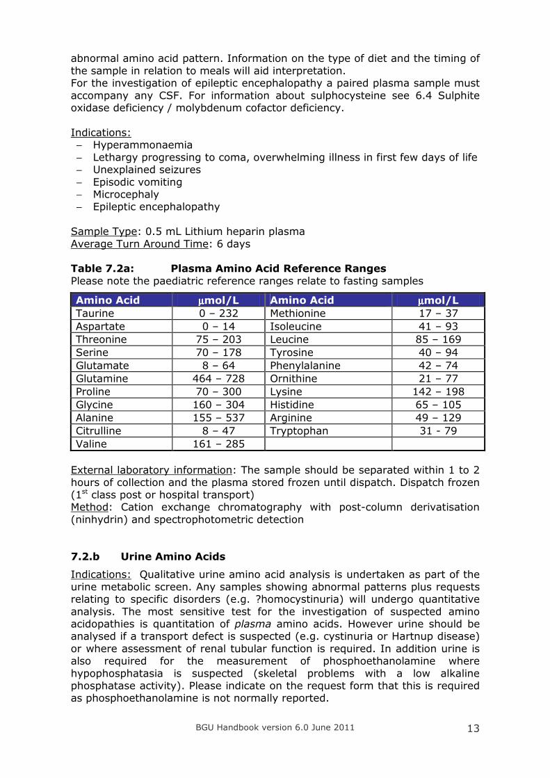
Average Turn Around Time: 6 days Table 7.2a: Plasma Amino Acid Reference Ranges
Please note the paediatric reference ranges relate to fasting samples
Amino Acid µµµµmol/L Amino Acid µµµµmol/L
Taurine 0 – 232 Methionine 17 – 37
Aspartate 0 – 14 Isoleucine 41 – 93
Threonine 75 – 203 Leucine 85 – 169
Serine 70 – 178 Tyrosine 40 – 94
Glutamate 8 – 64 Phenylalanine 42 – 74
Glutamine 464 – 728 Ornithine 21 – 77
Proline 70 – 300 Lysine 142 – 198
Glycine 160 – 304 Histidine 65 – 105
Alanine 155 – 537 Arginine 49 – 129
Citrulline 8 – 47 Tryptophan 31 - 79
Valine 161 – 285
External laboratory information: The sample should be separated within 1 to 2
hours of collection and the plasma stored frozen until dispatch. Dispatch frozen (1st class post or hospital transport) Method: Cation exchange chromatography with post-column derivatisation
(ninhydrin) and spectrophotometric detection
7.2.b Urine Amino Acids
Indications: Qualitative urine amino acid analysis is undertaken as part of the
urine metabolic screen. Any samples showing abnormal patterns plus requests relating to specific disorders (e.g. ?homocystinuria) will undergo quantitative
analysis. The most sensitive test for the investigation of suspected amino acidopathies is quantitation of plasma amino acids. However urine should be analysed if a transport defect is suspected (e.g. cystinuria or Hartnup disease)
or where assessment of renal tubular function is required. In addition urine is also required for the measurement of phosphoethanolamine where
hypophosphatasia is suspected (skeletal problems with a low alkaline phosphatase activity). Please indicate on the request form that this is required as phosphoethanolamine is not normally reported.
BGU Handbook version 6.0 June 2011
14
Monitoring Cystinuria
In patients with cystinuria only cystine, ornithine, arginine and lysine will be
reported. The cystine concentration will also be given in µmol/L, the solubility
of cystine in urine is approximately 1000 µmol/L. At concentrations above this, the patient is at high risk of stone formation.
Sample Type: 2 mL Urine (plain) Average Turn Around Times: Screening - 7 days
Quantitation – 13 days Table 7.2b: Paediatric Urine Amino Acid Reference Ranges
(µµµµmol/mmol creatinine)
0-7 days 8 days-1 month 1-4 months 4 months-2 years 2-4 years 4-6 years 6-10 years 10-18 years >18 years
Threonine 6-55 10-139 19-140 9-100 10-89 1-73 2-45 0-36 1-48
Serine 21-204 23-308 41-288 30-191 30-148 19-112 1-95 12-78 5-69
Glutamate 0-52 0-55 0-51 0-69 0-48 0-38 0-31 1-6 1-3
Glutamine 0-182 41-216 40-239 28-253 23-187 13-151 7-137 18-98 19-57
Proline 4-142 2-233 3-341 0-34 0-26 0-13 0-7 0-6 0-7
Glycine 0-1046 78-1259 105-796 40-616 76-516 24-397 20-201 18-252 12-199
Alanine 0-135 41-308 58-297 34-189 18-175 8-111 8-80 10-85 5-59
Valine 9-15 2-28 6-27 4-32 6-20 4-11 1-12 2-11 2-7
Cystine 0-65 2-52 7-54 0-36 5-25 4-22 3-17 5-16 1-19
Methionine 5-25 0-8 2-20 2-20 2-9 2-9 0-8 0-8 0-8
Isoleucine 3-9 1-18 0-26 0-10 4-14 2-12 0-7 0-7 1-5
Leucine 3-24 1-25 0-25 3-21 2-21 2-12 1-12 1-9 2-6
Tyrosine 0-29 9-55 13-75 10-72 0-71 8-51 6-42 6-37 5-27
Phenylalanine 0-34 3-34 3-40 5-37 0-41 2-25 1-20 1-17 2-11
Ornithine
Lysine 0-82 10-172 18-148 0-182 0-92 0-52 0-64 0-20 12-52
Arginine 2-8 0-20 0-19 0-19 1-13 1-7 0-9 0-8 1-7
Homocystine
0 - 30
0-1
External laboratory information: freeze urine on receipt and dispatch frozen
(1st class post or hospital transport) Methods:
Qualitative Analysis – two dimensional thin layer chromatography Quantitative Analysis – cation exchange chromatography with post-column derivatisation (ninhydrin) and spectrophotometric detection
7.2.c CSF Amino Acids
Indications: Intractable seizures. CSF amino acid analysis is required for the diagnosis of glycine encephalopathy (GE) (also known as non-ketotic
hyperglycinaemia or NKH) and 3-phosphoglycerate dehydrogenase deficiency. It may also be useful in the investigation of sulphite oxidase deficiency. A
paired plasma sample must always accompany the CSF. Sample Type: 0.5 mL CSF (plain, fluoride oxalate may also be used) with a
paired lithium heparin plasma sample (0.5 mL plasma). Note blood-stained CSF is not suitable for analysis.
Average Turn Around Time: 8 days CSF Amino Acid Reference Ranges
CSF serine 23 - 100 µmol/L
CSF glycine < 10 µmol/L CSF:plasma glycine ratio < 0.04
BGU Handbook version 6.0 June 2011
15
External laboratory information: freeze CSF and plasma on receipt and
dispatch frozen (1st class post or hospital transport) Method: Quantitative analysis – cation exchange chromatography with post-
column derivatisation (ninhydrin) and spectrophotometric detection
7.3 BIOTINIDASE
Biotin is a cofactor for multiple carboxylases and the recycling of biotin
requires the activity of the enzyme biotinidase. Typically biotinidase deficiency presents between 3-6 months of life with seizures. Treatment is with biotin
replacement, which should be initiated prior to the result being available. Biotinidase is a relatively unstable enzyme; low results should be checked on a fresh sample if clinically indicated.
Indications:
− Seizures − Ataxia − Hypotonia
− Alopecia − Skin rashes
Sample Type: 400 µL Lithium heparin plasma
Average Turn Around Time: 10 days
Table 7.3 Plasma Biotinidase Reference Ranges
Biotinidase Activity (nmol/mL/min)
Normal 4.4 – 12.0
Partial deficiency 0.7 – 2.1
Deficiency < 0.7
Obligate heterozygote 2.2 – 5.2
External laboratory information: Separate and freeze plasma as soon as possible. Dispatch frozen (1st class post or hospital transport) Method: Spectrophotometry
7.4 CHITOTRIOSIDASE
Gaucher disease is a lysosomal storage disorder resulting from an inherited
deficiency of the enzyme β-glucosidase. This deficiency results in impaired
breakdown of the lipid glucocerebrosidase and its subsequent accumulation in cells. Gaucher disease is characterised by markedly elevated chitotriosidase
activity; symptomatic Gaucher patients typically exhibit concentrations 100 times greater than the reference range. However, chitotriosidase may be mildly increased in a number of other lysosomal storage disorders and other
illnesses, such as sarcoidosis. Benign deficiency of chitotriosidase occurs in approximately 6% of Caucasians.
Indications: Diagnosis and monitoring of Gaucher disease
Sample Type: 100 µL Lithium heparin plasma or serum
Turn Around Time: 7 working days
Reference Range: 0 – 140 µmol/L/hour
BGU Handbook version 6.0 June 2011
16
External laboratory information: Store frozen, dispatch 1st class post or
hospital transport Method: Fluorimetric
7.5 CREATINE AND GUANIDINOACETATE
This relatively new group of disorders is characterised by cerebral creatine deficiency, the main symptoms of which are learning disability and speech
delay, and, in some patients intractable seizures. Of the three disorders described; arginine:glycine amidinotransferase (AGAT) deficiency and
guanidinoacetate methyltransferase (GAMT) deficiency show decreased creatine in the urine and plasma. Guanidinoacetate in urine and plasma is increased in GAMT deficiency and undetectable in AGAT deficiency. Defects in
the creatine transporter (an X-linked disorder) result in an increase in the urine creatine/creatinine ratio. Guanidinoacetate is stable in urine and plasma. Whilst
creatine is stable in plasma, it is unstable in urine and concentrations increase within 1-2 hours of collection, leading to potentially false positive results for the creatine transporter defect or spuriously normal results in AGAT and GAMT.
Indications: − Mental retardation
− Absent / delayed speech − Seizures − Movement disorder
Sample Type:
1 mL urine (plain) send to laboratory as soon as possible after collection (e.g. 1 to 2 hours)
100 µL Lithium heparin plasma (or serum)
Average Turn Around Time: 14 days
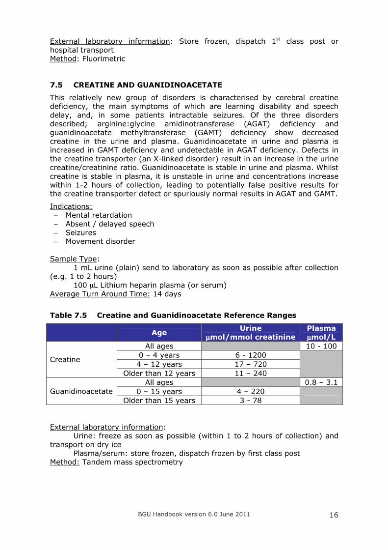
Table 7.5 Creatine and Guanidinoacetate Reference Ranges
Age
Urine
µµµµmol/mmol creatinine
Plasma
µµµµmol/L
All ages 10 - 100
0 – 4 years 6 - 1200
4 – 12 years 17 – 720 Creatine
Older than 12 years 11 – 240
All ages 0.8 – 3.1
0 – 15 years 4 – 220 Guanidinoacetate
Older than 15 years 3 - 78
External laboratory information: Urine: freeze as soon as possible (within 1 to 2 hours of collection) and
transport on dry ice Plasma/serum: store frozen, dispatch frozen by first class post Method: Tandem mass spectrometry
BGU Handbook version 6.0 June 2011
17
7.6 GALACTOSE-1-PHOSPHATE URIDYLTRANSFERASE
Classical galactosaemia is caused by a deficiency of the enzyme galactose-1-phosphate uridyltransferase. Galactose is produced in the small intestine from the breakdown of dietary lactose into galactose and glucose. The presence of
reducing substances in urine may be an important clue to diagnosis but equally can be misleading. False positives can occur in severe liver disease and false
negatives can occur if lactose is not present in the diet, and therefore blood spot galactose-1-phosphate uridyltransferase is the recommended screening test. Carriers of galactosaemia cannot be detected by this screening method.
Please note: results are invalid if the sample has been collected within 6 weeks of a blood transfusion. If galactosaemia is suspected in a child who has had a
blood transfusion please discuss alternative testing with one of the BGU clinical scientists.
Indications: − Hepatomegaly
− Prolonged jaundice with abnormal liver function tests
Sample Type: Dried blood spot Turn Around Time: 5 working days Reference Range: Qualitative result only
Normal results are reported as: ‘Galactose-1-phosphate uridyl transferase activity measured by the Beutler screening test appeared to be
within normal limits. Action taken on the strength of this result should recognise that it is a screening test.’ Deficient results are reported as: ‘There was no detectable galactose-1-
phosphate uridyl transferase activity when measured by the Beutler screening test. This enzyme is relatively unstable, particularly if the dried blood spot is
subjected to hot or humid conditions. The screening test also requires the presence and activity of endogenous blood glucose-6-phosphate dehydrogenase. Action taken on the strength of this result should recognise
that it is a screening test.’
Deficient results should be confirmed - the laboratory will contact you to arrange this.
External laboratory information: Store frozen, dispatch by first class post Method: Beutler screening test
7.7 GLYCOSAMINOGLYCANS
The mucopolysaccharidoses are a group of inherited disorders characterised by the accumulation of glycosaminoglycans in the lysosomes. Children may appear normal at birth but later develop progressive skeletal abnormalities,
coarse facies and hepatomegaly. Normal urine glycosaminoglycans consist mainly of chondroitin sulphate with traces of heparan and dermatan sulphates.
Mucopolysaccharidoses are characterised by abnormal patterns of glycosaminoglycans in urine. Initially, samples are screened for total glycosaminoglycan concentration. False positive results are common,
particularly in young infants. Positive results will be referred for typing by electrophoresis if clinical details suggestive of a mucopolysaccharidosis are
given. False negative quantitative results may also be encountered, therefore if
BGU Handbook version 6.0 June 2011
18
there is a strong clinical suspicion of a mucopolysaccharidosis, please
specifically request GAG typing. If urine glycosaminoglycan typing and white cell enzymes are normal and a storage disorder is still suspected clinically,
urinary oligosaccharide and sialic acid analysis should be considered (see section 8.7).
Indications:
− Hepatomegaly
− Skeletal deformities − Abnormal facies
− Behavioural problems
− Inguinal and umbilical hernias − Loss of developmental skills
Sample Type: 5 mL Urine (plain) Average Turn Around Time: GAG quantitation - 7 days
GAG typing – 21 days
Table 7.7: Urine GAG Reference Ranges
Age Glycosaminoglycans (mg/mmol creatinine)
1 week – 2 months < 70
2 months – 1 year < 39
1 –2 years < 29
2 –4 years < 26
4 –8 years < 24
8 - 12 years < 21
> 12 years < 13
All results within the reference range will be reported with the following comment: ‘This assay is a screening test only. If there is strong clinical
suspicion of a mucopolysaccharidosis, please contact Dr Jacqui Calvin or Sarah Hogg to discuss further investigations’. External laboratory information: Store frozen, dispatch frozen (1st class post or
hospital transport) Method: GAG quantitation: dimethylmethylene blue dye binding method with spectrophotometric detection
GAG typing: two dimensional electrophoresis
7.8 HOMOCYSTEINE
Measurement of total homocysteine is offered for the diagnosis and monitoring of inherited defects in homocystine metabolism, such as classical
homocystinuria and methionine synthase deficiency. Free homocystine is not recommended as it is only detectable in plasma when the binding capacity of
plasma proteins has been exceeded. The binding of homocystine to plasma protein, mainly albumin, seems to be saturable with a maximal capacity of
about 140 µmol/L total homocysteine. Likewise urine homocystine will only be
detectable when the binding capacity is exceeded.
Indications: − Marfanoid appearance
BGU Handbook version 6.0 June 2011
19
− Early onset vascular occlusive
disease
− Lens dislocation (usually downward)
− Early onset osteoporosis Sample Type: 0.5 mL Lithium heparin plasma, transport to laboratory urgently to allow separation of the plasma within one hour of venepuncture
Average Turn around time: 8 days Total homocysteine reference ranges: males: < 18 µmol/L females: < 16 µmol/L External laboratory information: separate plasma within one hour of collection
and store frozen. Dispatch frozen by 1st class post. Method: tandem mass spectrometry 7.9 ORGANIC ACIDS
Analysis of organic acids in urine can assist in the diagnosis of a number of
disorders including those of amino acid metabolism (e.g. MSUD, urea cycle defects). Orotate is quantitated using d2-orotate internal standard – see below for more information. Methylmalonate is quantitated using d3-methylmalonate
internal standard.
Indications: [Note: (+) indicates ‘occurring with other features’] − Recurrent episodic ketosis, acidosis, vomiting and dehydration − Reye-like syndrome
− Hypoglycaemia − Hyperammonaemia
− Seizures (+) − Seizures, ataxia, hypotonia − Macrocephaly, dystonia, seizures, neurodegeneration
− Cardiomyopathy − Unexplained lactic acidaemia
− Alopecia (+) − Failure to thrive (+) − Developmental Delay (+)
Table 7.9: Organic Acids Sometimes Requested Individually
Organic Acid Disease
N-Acetylaspartate Canavan Disease
Glutarate, 3-hydroxyglutarate Glutaric aciduria type 1
Homogentisate Alkaptonuria
4-Hydroxybutyrate 4-Hydroxybutyric aciduria
Orotate Urea cycle defects
Methylmalonate MMA
Mevalonate Mevalonic aciduria
Suberylglycine, hexanoylglycine MCADD
Succinylacetone Tyrosinaemia type I
BGU Handbook version 6.0 June 2011
20
Sample Type: Urine (plain) - volume is dependent on the urine creatinine
concentration (the more dilute the urine the larger the volume required). Usually 5 mL is sufficient for analysis.
Average Turn Around Time: 6 days Methylmalonate Reference range
0 - 1 yrs < 20 µmol/mmol creatinine
1 yr to adult < 10 µmol/mmol creatinine
Mild increases (up to 100 µmol/mmol creatinine) in methylmalonate are not uncommon, they are not believed to be significant unless the patient is a breast fed infant where there is maternal vitamin B12 deficiency. The advice is
usually to repeat in one month, although an earlier repeat is recommended if there is evidence of acidosis, lethargy, hypotonia or developmental delay.
External laboratory information: Freeze urine on receipt and dispatch frozen (1st class post or hospital transport)
Method: Solvent extraction followed by GC-MS of silylesters (qualitative)
7.10 OROTATE
Orotic acid is an intermediate in the synthesis of pyrimidine nucleotides. In most defects of the urea cycle carbamoyl phosphate accumulates. This feeds
into the pyrimidine biosynthetic pathway resulting in an excess of orotic acid. Mildly raised values have also been reported in mitochondrial disease. Note:
samples collected following an allopurinol load test will be referred to Guy’s Hospital for measurement of orotic acid and orotidine. (The Guy’s protocol for this test is available from one of the BGU Clinical Scientists).
Indications:
− Differential diagnosis of urea cycle defects − Disorders of pyrimidine metabolism
Sample Type: 5 mL Urine (plain) Average Turn Around Time: 6 days
Reference Range:
0 - 2 years: 0 – 6 µmol/mmol creatinine
Older than 2 years: 0 - 3 µmol/mmol creatinine External laboratory information: Store frozen, dispatch frozen (1st class post
or hospital transport) Method: Solvent extraction followed by GC-MS of silylesters
7.11 VERY LONG CHAIN FATTY ACIDS, PRISTANATE & PHYTANATE
Peroxisomes are responsible for β−oxidation of very long chain fatty acids (fatty acids with a carbon length more than 22), bile acid metabolism and plasmalogen synthesis. Peroxisomal disorders can be classified into 2
categories; defects in peroxisomal biogenesis disorders (eg Zellweger syndrome, infantile Refsum disease) and defects in specific peroxisomal
enzymes. Very long chain fatty acids are very sensitive for the diagnosis of X-
BGU Handbook version 6.0 June 2011
21
linked adrenoleukodystrophy in males. However approximately 15% of
symptomatic female carriers have normal very long chain fatty acids.
Phytanate and pristanate are assayed as part of the plasma very long chain
fatty acid profile. They are useful in the diagnosis of Refsum disease, α-methyl-acylCoA racemase deficiency and rhizomelic chondrodysplasia punctata
(depending on the age of the patient). Pristanate and phytanate may be normal in young infants with peroxisomal biogenesis defects as both
compounds are derived from exogenous, dietary sources. Indications: [Note: (+) indicates ‘occurring with other features’]
− Idiopathic adrenal insufficiency in males
− Neurological abnormalities − Leukodystrophy − Ataxia
− Seizures (+)
− Hypotonia − Ocular abnormalities
− Skeletal abnormalities − Dysmorphic features − Liver dysfunction (+)
− Hepatomegaly
Sample Type: 0.5 mL EDTA plasma, send to laboratory as soon as possible Average Turn Around Time: 10 days
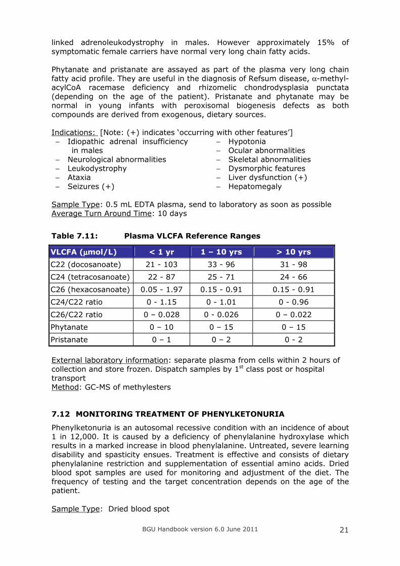
Table 7.11: Plasma VLCFA Reference Ranges
VLCFA (µµµµmol/L) < 1 yr 1 – 10 yrs > 10 yrs
C22 (docosanoate) 21 - 103 33 - 96 31 - 98
C24 (tetracosanoate) 22 - 87 25 - 71 24 - 66
C26 (hexacosanoate) 0.05 - 1.97 0.15 - 0.91 0.15 - 0.91
C24/C22 ratio 0 - 1.15 0 - 1.01 0 - 0.96
C26/C22 ratio 0 – 0.028 0 - 0.026 0 – 0.022
Phytanate 0 – 10 0 – 15 0 – 15
Pristanate 0 – 1 0 – 2 0 - 2
External laboratory information: separate plasma from cells within 2 hours of collection and store frozen. Dispatch samples by 1st class post or hospital
transport Method: GC-MS of methylesters
7.12 MONITORING TREATMENT OF PHENYLKETONURIA
Phenylketonuria is an autosomal recessive condition with an incidence of about 1 in 12,000. It is caused by a deficiency of phenylalanine hydroxylase which results in a marked increase in blood phenylalanine. Untreated, severe learning
disability and spasticity ensues. Treatment is effective and consists of dietary phenylalanine restriction and supplementation of essential amino acids. Dried
blood spot samples are used for monitoring and adjustment of the diet. The frequency of testing and the target concentration depends on the age of the patient. Sample Type: Dried blood spot
BGU Handbook version 6.0 June 2011 22
Turn Around Time: 2 working days
External laboratory information: send by 1st class post
Method: tandem mass spectrometry
7.13 SWEAT TESTS
The determination of sweat chloride concentration is useful in the diagnosis of cystic fibrosis. Sweat testing can be performed after 2 weeks of age on infants greater than 3 kg that are normally hydrated and without significant systemic
illness. If clinically important, sweat testing can be attempted after one week of age but will need repeating if insufficient sweat is collected. A repeat test is
recommended when the result is abnormal or borderline and the genotype is not confirmatory.
Indications: − Phenotype suggestive of CF (respiratory infection, exocrine pancreatic
insufficiency) − Positive newborn screening test
Sample Type: Sweat collected into a Wescor Macroduct tube. National
guidelines state not less than 1g/m2/min. A minimum sweat volume of 60 µL
(approximately 185 mm) is required to enable duplicate analysis. Turn Around Time: 1 working day
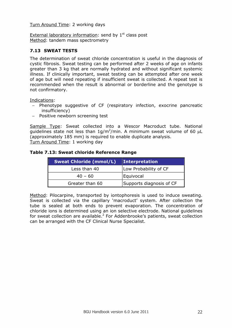
Table 7.13: Sweat chloride Reference Range
Sweat Chloride (mmol/L) Interpretation
Less than 40 Low Probability of CF
40 – 60 Equivocal
Greater than 60 Supports diagnosis of CF
Method: Pilocarpine, transported by iontophoresis is used to induce sweating. Sweat is collected via the capillary ‘macroduct’ system. After collection the
tube is sealed at both ends to prevent evaporation. The concentration of chloride ions is determined using an ion selective electrode. National guidelines for sweat collection are available.2 For Addenbrooke’s patients, sweat collection
can be arranged with the CF Clinical Nurse Specialist.
BGU Handbook version 6.0 June 2011 23
8 REFERRED METABOLIC ASSAYS
8.1 3-HYDROXYBUTYRATE AND FREE FATTY ACIDS
These intermediary metabolites may be useful in the investigation of unexplained hypoglycaemia. (Please request a simultaneous lab glucose analysis.)
Interpretation depends on the fed or fasted state of the patient. If hypoglycaemic, suppression of both 3OHB and FFA is consistent with
hyperinsulinism whereas in FAOD the ratio of FFA to 3OHB is typically greater than 3. However these two diagnoses may be more easily made by analysing
insulin at the time of hypoglycaemia and blood spot acylcarnitines, respectively.
Sample: 1 mL fluoride tube Transport: Store frozen, dispatch frozen 1st class post
Referral laboratory: Chemical Pathology, Sheffield Children’s Hospital
8.2 7-DEHYDROCHOLESTEROL
Smith-Lemli-Opitz syndrome (SLO) is an autosomal recessive disorder with multiple congenital malformations (microcephaly, 2,3 syndactyly, cleft palate,
congenital heart defects). There is a deficiency of sterol delta-7-reductase that causes an increase in the cholesterol precursor 7-dehydrocholesterol (7DHC). In SLO the total cholesterol is typically below 1.5 mmol/L (measured by GC-
MS), with an increase in the 7DHC:cholesterol ratio.
Sample Type: 0.5 mL lithium heparin or EDTA plasma, or serum Transport: 1st Class post
Referral laboratory: Chemical Pathology, Sheffield Children’s Hospital
8.3 BILE ACID METABOLITES
Inherited defects in bile acid biosynthesis cause cholestasis and malabsorption (due to bile acid deficiency), with progressive neurological dysfunction and
xanthomas (due to deposition of precursors). Bile acids are partly synthesised in the peroxisome. Analysis is also indicated in the workup of suspected peroxisomal biogenesis disorders where VLCFA are
abnormal.
Sample type: 10 mL plain urine and/or 2 mL lithium heparin plasma Sample storage: Store frozen prior to dispatch first class post Referral laboratory: Chemical Pathology, Sheffield Children’s Hospital
8.4 NEUROTRANSMITTERS
The following metabolites may be analysed in CSF, depending on the clinical details provided:
HVA, 5HIAA, 5-methyltetrahydrofolate (5MTHF), neopterin, dihydrobiopterin, tetrahydrobiopterin and pyridoxal phosphate.
BGU Handbook version 6.0 June 2011 24
Clinical indications include oculogyric crises, temperature instability, ptosis,
parkinsonian features and dystonia. This test is usually requested after the child has been reviewed by a paediatric neurologist.
Sample Type: CSF collected into a set of three 1 mL tubes containing special
preservatives and frozen on dry ice at the bedside. (Tubes and collection instructions are obtained from the Neurometabolic Unit, the National Hospital, Queen Square, London. Tel: 020 7837 3611 ext 3844).
Sample storage: Once collected CSF should be stored at –70oC and can be kept for more than a year
Transport: Courier - on dry ice Referral laboratory: Neurometabolic Unit, National Hospital, London
8.5 OLIGOSACCHARIDES AND SIALIC ACID
Oligosaccharides are low molecular weight carbohydrate polymers made up of
at least 3 monosaccharide subunits. The oligosaccharides in urine are derived from the incomplete breakdown of carbohydrate side chains of complex
glycoproteins. Abnormal oligosaccharides accumulate in a range of lysosomal storage diseases.
Unfortunately this screening test is insensitive with poor specificity. The
oligosaccharide excretion in glycoproteinoses may be variable and/or the abnormality subtle. Spurious results may be seen in patients infused with large
amounts of complex carbohydrates (eg dextran). Neonates and breast fed infants show patterns which would be considered abnormal in older children. For these reasons urine oligosaccharide chromatography is not offered
as a first line test by the BGU. If a lysosomal storage disorder is suspected clinically WCE analysis is recommended.
If the WCE analyses and urine GAGS are normal and a storage disease is still suspected urines will be referred to the Willink, Manchester for urine oligosaccharides and sialic acid, to investigate the possibility of sialidosis or
galactosialidosis.
8.6 PORPHYRINS
Urine, blood and faecal porphyrin analyses are available for the investigation of
the acute and non-acute porphyria; please see section 6.10 “Which sample for the investigation of porphyria?”.
Samples: Urine: 20 mL fresh, preferably early morning urine or crisis urine in plain white-topped universal Faeces: about 5-10 g (marble-sized) sample in faecal (blue) universal Blood: 5-10 mL whole blood EDTA Transport: First Class post Referral laboratory: Porphyria Service, University Hospital of Wales, Cardiff
BGU Handbook version 6.0 June 2011 25
8.7 PURINES AND PYRIMIDINES
Over 20 different disorders of purine and pyrimidine metabolism are known, of which several cause significant clinical disease. Three organ systems are
prominently affected: kidneys (renal stones), bone marrow (immunodeficiency
± megaloblastic anaemia) and brain (neurological problems, e.g. abnormal muscle tone, dystonia, autistic-like behavioural problems), some patients may
have more than one organ system affected. This test is often undertaken in children with unexplained neurological problems when first line investigations
have drawn a blank. Sample Type: 5 mL random urine, plain universal
Sample storage: Store frozen until sending to lab by 1st class post Referral laboratory: Purine Research Laboratory, St Thomas’ Hospital, London
8.8 THYMIDINE
Thymidine is increased in plasma and urine of patients with Myo-Neuro-Gastrointestinal-Encephalopathy (MNGIE) which is a mitochondrial disorder caused by mutations in thymidine phosphorylase. It usually presents in
adolescence with abdominal pain, bloating and malabsorption, with a neuropathy and encephalopathy.
Sample Type: 1mL random urine, plain universal & 0.5mL EDTA plasma/serum Sample storage: Store frozen until sending to lab by 1st class post Referral laboratory: Purine Research Laboratory, St Thomas’ Hospital, London
8.9 TRANSFERRIN GLYCOFORMS
The initial screening test for congenital defects of glycosylation (CDG) is
isoelectric focusing of transferrin glycoforms to detect abnormal patterns of glycosylation. Enzyme analysis is available for confirmatory testing of some
subtypes. The molecular basis of more than 20 defects have been identified however CDG Type Ia is the most common, accounting for approximately 80% of cases.
Please note this test is unreliable in the first 3 weeks of life (reflects maternal transferrin).
Indications: [Note: (+) indicates ‘occurring with other features’]
− Psychomotor retardation
− Seizures (+) − Strabismus
− Cerebellar hypoplasia − Dysmorphy (fat pads, inverted nipples) − Coagulopathy
− Protein-losing enteropathy (type 1b)
BGU Handbook version 6.0 June 2011 26
In view of the extremely broad clinical spectrum of CDG patients, it is
recommended that transferrin glycoform analysis is considered in any unexplained multisystem disorder.
Sample Type: 1 mL serum or lithium heparin plasma
Storage: Store frozen prior to dispatch by 1st Class Post Referral Laboratory: Neurometabolic Unit, National Hospital, London
8.10 TRIMETHYLAMINE
Patients with primary trimethylaminuria have defective trimethylamine N-oxide
synthetase activity. This deficiency does not produce disease but the strong, unpleasant fishy odour can lead to social ostracism and psychological disorders. Secondary trimethylaminuria also occurs and is caused by
enterobacterial overproduction of trimethylamine. A diet low in fish, liver and egg yolks usually improves the odour.
Sample Type: Preferred: 24 hour urine – collect into acid bottle (HCl). 10 mL
random urine, acidified with HCl to pH 1, also suitable. Storage: Store frozen prior to dispatch by 1st Class Post Referral Laboratory: Chemical Pathology, Sheffield Children’s Hospital
8.11 WHITE CELL CYSTINE
Cystinosis is a disorder of lysosomal membrane transport. The most severe infantile form of cystinosis starts with Fanconi syndrome at the age of 3-6 months. Untreated patients develop renal failure before the age of 10. Free
cystine accumulates within the lysosomes of most tissues at 10-1000 times normal.
Sample Type: 3 mL lithium heparin whole blood Storage: Store whole blood (do not separate) at room temperature prior to
dispatch by Special Delivery Referral Laboratory: WellChild Laboratory, Evelina Children’s Hospital
8.12 WHITE CELL ENYZMES
Lysosomal storage disorders result from a deficiency of a specific lysosomal enzyme, activator or transport protein, causing an accumulation of un-degraded substrates within the lysosomes. Characteristically patients develop
normally during the neonatal period but present with progressive deterioration in early childhood. Unlike ‘small molecule’ metabolic disorders, presentation
tends to be slow and more insidious. Symptoms include bone deformities and short stature, heart and respiratory difficulties, coarse facial features, an enlarged head, tongue, liver and spleen, and, in many patients, neurological
degeneration. There are no reliable screening tests for these disorders (apart from urine GAGS in suspected mucopolysaccharidoses) and white cell enzyme
analysis is often the first line test when there is a strong clinical suspicion of an LSD.
Indications for analysis include hepatosplenomegaly, developmental
regression, neurological deterioration, dysmorphic features, cherry red spot,
BGU Handbook version 6.0 June 2011 27
leukodystrophy and angiokeratoma. Please contact the laboratory to discuss
before taking samples.
Sample Type: 10 mL EDTA whole blood for the enzymes in the table below. For other enzymes please contact one of the BGU clinical scientists for sample
requirements. Sample storage: For Addenbrooke’s patients please send whole blood (at room temperature) to the BGU urgently. Samples must be received before 2pm to
enable same day despatch to Manchester. We are unable to accept samples on a Friday. Please phone in advance to enable the laboratory to arrange
transport. Referral laboratory: Willink Laboratory, Manchester Children’s Hospital
Table 8.11: The following white cell enzymes (by disease) are offered as a battery of tests by the Willink laboratory.
Enzyme Disease
Acid Esterase Wolman disease
α-Fucosidase Fucosidosis
α-Galactosidase Fabry disease
α-Mannosidase α-mannosidosis
α-N-acetylgalactosaminidase Schindler disease
Arylsulphatase A Metachromatic leukodystrophy
β-Galactosidase GM1 gangliosidosis
β-Glucosidase Gaucher disease
β−Glucuronidase Sly disease
β-Hexosaminidase Sandhoff Disease
β-Mannosidase β-Mannosidosis
Chitotriosidase Non-specific storage marker
Galactocerebrosidase Krabbe leukodystrophy
Glycoasparaginase Aspartylglucosaminuria
MUGS (methylumbelliferyl-N-
acetylglucosamine-6-sulphate substrate)
[for Hexosaminidase A]
Tay-Sachs
Sphingomyelinase Niemann-Pick A&B
Other enzymes are available on an individual basis and include:
− dried blood spot screens are available for α-galactosidase and α-
glucosidase (Pompe disease) − palmitoyl-protein thioesterase-1 and tripeptidyl peptidase 1 are enzymes
deficient in two of the neuronal ceroid lipofuscinoses (Batten disease), lithium heparin blood is required
− enzyme analysis for Niemann-Pick type C is not available; instead
cholesterol esterification studies and filipin staining are undertaken on fibroblasts
Please phone to discuss, prior to sending samples.
BGU Handbook version 6.0 June 2011 28
9 QUALITY ASSURANCE SCHEMES
The BGU participates in the following QA schemes: ERNDIM: Bloodspot acylcarnitine
Urine organic acids Quantitative organic acids (MMA)
Urine proficiency testing Plasma amino acids Special assays serum
Special assays urine Urine glycosaminoglycans (pilot scheme)
NEQAS: Newborn screening Urine orotic acid Quantitative phenylalanine
Sweat testing Metabolic cognitive scheme pilot
Willink: Urine glycosaminoglycans CDC: Newborn screening schemes WEQAS: Urine dipstick scheme
Urine porphyrin scheme
10 REFERENCES AND FURTHER INFORMATION
References
1. Vandemecum Metabolicum. Manual of Metabolic Paediatrics Zschocke & Hoffmann
2. National Guidelines for the Performance of the Sweat Test for the Investigation of Cystic Fibrosis, November 2003
Further Information National Metabolic Biochemistry Network
http://metbio.net/ British Inherited Metabolic Disease Group
http://www.bimdg.org.uk/
Online Mendelian Inheritance in Man http://www.ncbi.nlm.nih.gov/entrez/query.fcgi?db=OMIM
UK National Screening Committee http://www.nsc.nhs.uk/
UK Newborn Screening Programme Centre
http://www.newbornscreening-bloodspot.org.uk/ Sweat Testing Guidelines (available via The Association for Clinical
Biochemistry) http://acb.org.uk/
BGU Handbook version 6.0 June 2011 29
INDEX
3
3-Hydroxybutyrate ............................... 23
3-hydroxyglutarate ........................... 8, 19
4
4 Hydroxybutyric Aciduria...................... 19
5
5HIAA ................................................. 23
5-Methyltetrahydrofolate ....................... 23
7
7-Dehydrocholesterol ............................ 23
A
N-Acetylaspartate................................. 19
α-N-Acetylgalactosaminidase ................. 27
Acid Esterase ....................................... 27
Acylcarnitines....................................... 12
Alanine................................................ 13
Alkaptonuria ........................................ 19
Allopurinol load .................................... 20 α-Aminoadipate Semialdehyde .................9
Ammonia............................................. 10
Arginine .............................................. 13
Arylsulphatase A................................... 27
Aspartate ............................................ 13
Aspartylglucosaminuria ......................... 27
B
Batten disease ..................................... 27
Bile Acid Metabolites ............................. 23
Biotinidase........................................... 15
C
Canavan Disease .................................. 19
Chitotriosidase ..................................... 27
Cholesterol Esterification Studies............ 27
Citrulline ............................................. 13
Congenital Defects Of Glycosylation ........ 25
Creatine .............................................. 16
Creatine Transporter Defect ................... 15
CSF glycine.......................................... 14
CSF serine ........................................... 14
Cystine................................................ 26
Cystinosis ............................................ 26
Cystinuria ...................................7, 13, 14
D
Dihydrobiopterin................................... 23
F
Fabry disease .......................................27
Fatty Acid Oxidation Defects.................8, 9
Filipin Staining......................................27
Free Fatty Acids....................................23 α-Fucosidase........................................27
Fucosidosis ..........................................27
G
Galactocerebrosidase ............................27
Galactosaemia......................................17
Galactose-1-Phosphate Uridyl Transferase17
Galactosialodosis ..................................24 α-Galactosidase....................................27
β-Galactosidase ....................................27
Gaucher disease ............................. 15, 27 α-Glucosidase.......................................27
β-Glucosidase................................. 15, 27
β−Glucuronidase ...................................27
Glutamate............................................13
Glutamine............................................13
Glutarate .............................................19
Glutaric Aciduria Type 1..................... 8, 19
Glycine ............................................ 6, 13
Glycoasparaginase ................................27
Glycosaminoglycans ............... 7, 17, 18, 28
GM1 gangliosidosis ...............................27
Guanidinoacetate..................................16
H
Hartnup disease ...................................13
HELLP ...................................................8 β-Hexosaminidase.................................27
Histidine ..............................................13
Homocysteine .............................8, 18, 19
Homogentisate .....................................19
HVA ....................................................23
Hyperammonaemia.............. 10, 12, 13, 19
Hypophosphatasia.................................13
I
Infantile Refsum Disease .......................20
Isoleucine ............................................13
K
Krabbe leukodystrophy..........................27
L
Leucine................................................13
Lysine .................................................13
BGU Handbook version 6.0 June 2011 30
M
α-Mannosidase ..................................... 27
β-Mannosidase ..................................... 27
Medium Chain Acyl-CoA Dehydrogenase
Deficiency ..........................................8
Metabolic Screen ....................................7
Metachromatic leukodystrophy ............... 27
Methionine........................................... 13
Methylmalonate...............................19, 20
Mevalonic aciduria ................................ 19
Mitochondrial disorders ...........................9
MNGIE ................................................ 25
Molybdenum cofactor deficiency ...............8
Mucopolysaccharidoses ....................17, 26
MUGS ................................................. 27
MyoNeuroGastrointestinalEncephalopathy 25
N
Neopterin ............................................ 23
Neuronal Ceroid Lipofuscinoses .............. 27
Neurotransmitters ................................ 23
Niemann-Pick A&B ................................ 27
Niemann-Pick type C............................. 27
O
Oligosaccharides................................... 24
Organic Acids ...................... 7, 8, 9, 19, 28
Ornithine ............................................. 13
Orotate ............................................... 19
P
Palmitoyl-Protein Thioesterase-1 ............ 27
Peroxisomal Biogenesis Disorders ......20, 23
Phenylalanine....................................... 13
Phenylketonuria ............................... 6, 21
Phosphoethanolamine ........................... 13
Phytanate ............................................ 21
Pipecolate..............................................9
Pompe disease ..................................... 27
Porphobilinogen.................................... 10
Porphyria.............................. 6, 10, 11, 24
Pristanate............................................ 21
Proline ................................................ 13
Purines................................................ 25
Pyridoxal Phosphate.......................... 9, 23
Pyridoxal Phosphate Dependent Epilepsy ...9
Pyridoxine-Dependent Epilepsy ................9
Pyrimidines.......................................... 25
R
Refsum disease .................................... 21
Rhabdomyolysis .....................................9
Rhizomelic Chondrodysplasia Punctata .... 21
S
Sandhoff Disease..................................27
Schindler Disease .................................27
Serine .................................................13
Sialic Acid ...................................... 18, 24
Sialodosis ............................................24
Sly Disease ..........................................27
Smith-Lemli-Opitz syndrome..................23
Sphingomyelinase.................................27
Succinylacetone....................................19
Sulphite oxidase deficiency ................ 8, 13
Sulphocysteine ................................. 8, 13
Sweat chloride......................................22
T
Taurine................................................13
Tay-Sachs............................................27
Tetrahydrobiopterin ..............................23
Thiosulphite ...........................................8
Threonine ............................................13
Thymidine............................................25
Thymidine Phosphorylase.......................25
Transferrin Glycoforms ..........................25
Trimethylamine ....................................26
Tripeptidyl Peptidase 1 ..........................27
Tryptophan ..........................................13
Tyrosine ..............................................13
U
Urea Cycle Defects................................19
V
Valine..................................................13
Very Long Chain Fatty Acids............. 20, 21
Vitamin B6.............................................9
W
White Cell Enyzmes...............................26
Wolman disease ...................................27
X
X-Linked Adrenoleukodystrophy..............21
Z
Zellweger Syndrome .............................20