Embed Size (px)
Citation preview

Biochemical Properties of a Novel Cysteine Protease ofPlasmodium vivax, Vivapain-4Byoung-Kuk Na1,2., Young-An Bae1., Young-Gun Zo1, Youngchool Choe3, Seon-Hee Kim1, Prashant
V. Desai4, Mitchell A. Avery5, Charles S. Craik3, Tong-Soo Kim6, Philip J. Rosenthal7, Yoon Kong1*
1 Department of Molecular Parasitology and Center for Molecular Medicine, Samsung Biomedical Research Institute, Sungkyunkwan University School of Medicine,
Suwon, Korea, 2 Department of Parasitology and Institute of Health Sciences, Gyeongsang National University School of Medicine, Jinju, Korea, 3 Department of
Pharmaceutical Chemistry, University of California San Francisco, San Francisco, California, United States of America, 4 Department of Medicinal Chemistry, National Center
for Natural Products Research, University of Mississippi, University, Mississippi, United States of America, 5 Department of Chemistry and Biochemistry, University of
Mississippi, University, Mississippi, Unites States of America, 6 Department of Parasitology, Inha University College of Medicine, Incheon, Korea, 7 Department of Medicine,
University of California San Francisco, San Francisco, California, United States of America
Abstract
Background: Multiple cysteine proteases of malaria parasites are required for maintenance of parasite metabolichomeostasis and egress from the host erythrocyte. In Plasmodium falciparum these proteases appear to mediate theprocessing of hemoglobin and aspartic proteases (plasmepsins) in the acidic food vacuole and the hydrolysis of erythrocytestructural proteins at neutral pH. Two cysteine proteases, vivapain (VX)-2 and VX-3 have been characterized in P. vivax, butcomprehensive studies of P. vivax cysteine proteases remain elusive.
Findings: We characterized a novel cysteine protease of P. vivax, VX-4, of which orthologs appears to have evolveddifferentially in primate plasmodia with strong cladistic affinity toward those of rodent Plasmodium. Recombinant VX-4demonstrated dual substrate specificity depending on the surrounding micro-environmental pH. Its hydrolyzing activityagainst benzyloxycarbonyl-Leu-Arg-4-methyl-coumaryl-7-amide (Z-Leu-Arg-MCA) and Z-Phe-Arg-MCA was highest at acidicpH (5.5), whereas that against Z-Arg-Arg-MCA was maximal at neutral pH (6.5–7.5). VX-4 preferred positively charged aminoacids and Gln at the P1 position, with less strict specificity at P3 and P4. P2 preferences depended on pH (Leu at pH 5.5 andArg at pH 7.5). Three amino acids that delineate the S2 pocket were substituted in VX-4 compared to VX-2 and VX-3 (Ala90,Gly157 and Glu180). Replacement of Glu180 abolished activity against Z-Arg-Arg-MCA at neutral pH, indicating theimportance of this amino acid in the pH-dependent substrate preference. VX-4 was localized in the food vacuoles andcytoplasm of the erythrocytic stage of P. vivax. VX-4 showed maximal activity against actin at neutral pH, and that against P.vivax plasmepsin 4 and hemoglobin was detected at neutral/acidic and acidic pH, respectively.
Conclusion: VX-4 demonstrates pH-dependent substrate switching, which might offer an efficient mechanism for thespecific cleavage of different substrates in different intracellular environments. VX-4 might function as a hemoglobinase inthe acidic parasite food vacuole, a maturase of P. vivax plasmepsin 4 at neutral or acidic pH, and a cytoskeleton-degradingprotease in the neutral erythrocyte cytosol.
Citation: Na B-K, Bae Y-A, Zo Y-G, Choe Y, Kim S-H, et al. (2010) Biochemical Properties of a Novel Cysteine Protease of Plasmodium vivax, Vivapain-4. PLoS NeglTrop Dis 4(10): e849. doi:10.1371/journal.pntd.0000849
Editor: John Pius Dalton, McGill University, Canada
Received June 9, 2010; Accepted September 14, 2010; Published October 12, 2010
Copyright: � 2010 Na et al. This is an open-access article distributed under the terms of the Creative Commons Attribution License, which permits unrestricteduse, distribution, and reproduction in any medium, provided the original author and source are credited.
Funding: This work was financially supported by grants to Yoon Kong from Samsung Biomedical Research Institute (BA60023) and also by the Ministry of Healthand Social Welfare (A030075), Korean Government. Seon-Hee Kim is supported by the Brain Korea 21. National Institute of Health Grant, AI35707 supportedYoungchool Choe, Charles S Craik, and Philip J Rosenthal. Philip J Rosenthal is a Doris Duke Charitable Foundation Distinguished Clinical Scientist. The funders hadno role in study design, data collection and analysis, decision to publish, or preparation of the manuscript.
Competing Interests: The authors have declared that no competing interests exist.
* E-mail: [email protected]
. These authors contributed equally to this work.
Introduction
Plasmodium vivax, one of the most predominant human malaria
species worldwide, causes hundreds of millions of illnesses each
year, and can result in severe morbidity and mortality, especially
in children [1,2]. Emergence and spread of multidrug resistant
vivax malaria is an increasing problem, which is associated with
fatal disease [3–5].
Cysteine proteases of malaria parasites are intimately involved in
a variety of physiological processes essential for the parasite’s
survival. The potential roles of the cysteine proteases of P. falciparum
such as falcipain-2 (FP-2), FP-2B (synonym of FP-29) and FP-3 in
hemoglobin degradation in the acidic parasite food vacuole [6,7],
processing of food vacuole plasmepsins to active proteases [8], and
erythrocyte rupture via cleavage of cytoskeletal proteins followed by
merozoite release [9] have been well characterized. Genes encoding
three closely related FPs cluster on chromosome 11 within a 12-kb
stretch, called the cysteine protease island [10]. FP-2 and FP-2B
share similar primary structure and enzymatic properties [11].
Knockout of the FP-2 gene leads to a transient block in hemoglobin
www.plosntds.org 1 October 2010 | Volume 4 | Issue 10 | e849
hydrolysis, but parasites compensate for the loss through production
of FP-2B and/or FP-3, and they multiply at the same rate as wild
type parasites. In contrast, FP-3 appears to have an essential role, as
knockout of the FP-3 gene is lethal [12]. Another papain-like
cysteine protease FP-1, of which gene locates on chromosome 14,
appears to be active in early invasive merozoites and in oocyst
production in mosquitoes [13,14]. Plasmodium cysteine proteases
appear to display non-overlapping roles with differentiated bio-
chemical properties and expression patterns, as well as redundancy
and complementation [15].
Two cysteine proteases, vivapain (VX)-2 and VX-3, which are
encoded on chromosome 9, have been identified in P. vivax
[16,17]. VX-2 and VX-3 share a number of biochemical
properties with FP-2 and FP-3, including acidic pH optima,
requirement for reducing conditions for maximal enzyme activity,
and preference toward peptide substrates with positively charged
residues at the P1 position and Leu at P2 [17]. Structural modeling
of VX-2 and VX-3 has also revealed a topology similar to those of
FP-2 and FP-3; however, some substantial differences are detected
in the predicted sizes of the binding pockets and residues involved
in substrate binding [18]. A gene (XM_001612308) encoding a
protein with significant similarity to FP-1 has recently become
available in the nucleotide sequence of P. vivax (PVX_195290,
PVX_239290, and PVX_240290); its biochemical properties and
biological activity remain unclear.
Interests in specific inhibitors impeding plasmodial cysteine
proteases have focused on their chemotherapeutic applicability;
effective inhibitors impair normal parasite growth in vitro [19]. In
addition, rupture of the erythrocyte membrane by mature
parasites is inhibited by broad-spectrum inhibitors of serine and
cysteine proteases [20,21]. Identification and further character-
ization of P. vivax cysteine proteases will be helpful to investigate
their biological roles and to characterize targets for antimalarial
drugs. However, comprehensive studies of P. vivax cysteine
proteases have been hindered by an inability to culture P. vivax.
In the present study, we describe the biochemical properties of a
novel cysteine protease of P. vivax, designated vivapain-4 (VX-4),
which displays unusual pH-dependent substrate specificity.
Molecular modeling and subsequent mutation analysis demon-
strated that Glu180 is involved in the pH-dependent substrate
specificity of VX-4. The protease effectively hydrolyzed hemoglo-
bin at acidic pH, actin at neutral pH, and plasmepsin 4 at neutral
and acidic pHs, supporting its role in the maintenance of
metabolic homeostasis and architectural remodeling of the
parasite during growth and development.
Materials and Methods
Ethics StatementAll animals used in this study were housed in accordance with
guidelines from the Association for the Assessment and Accred-
itation of Laboratory Animal Care (AAALAC). All protocols were
approved by the Institutional Review Board and conducted in
the Laboratory Animal Research Center of Sungkyunkwan
University.
In silico Identification of Cysteine Protease GeneGenes putatively coding for cysteine proteases were identified
from primate and rodent Plasmodium sequences deposited in
PlasmoDB (http://plasmodb.org) and GenBank (http://www.
ncbi.nlm.nih.gov/) through BLAST searches. The amino acid
(AA) sequences of cysteine proteases of P. falciparum (FP-1, FP-2,
FP-2B, and FP-3), P. vivax (VX-2 and VX-3), P. yoelii (yoelipain
[YP]-1 and YP-2), and P. berghei (bergheipain [BP]-1 and BP-2)
were used in multiple queries, with a threshold at 0.001 (E-value
cut-off). After excluding redundancies, the AA sequences were
aligned with ClustalX and optimized with GeneDoc. The
alignment was used as an input in the construction of neighbor
joining and maximum likelihood trees using PHYLIP (ver. 3.6b)
and TREE_PUZZLE (ver. 5.2). The standard error in each of the
connecting nodes was estimated by bootstrapping of 1000
replicates. Two novel cysteine proteases isolated from P. vivax
were annotated as P. vivax cysteine protease 1 (VX-1; XP_
001615807) and 4 (VX-4; XP_001615272), according to their
clustering patterns in the trees.
Expression and Refolding of a Recombinant VX-4 (rVX-4)The open reading frame (ORF) of VX-4 was amplified with
forward (59- ATGGAATATCACATGGAGTACTCGAAC-39)
and reverse (59-CTAGTCAAGCAGGGGGACGTACGCCTC-
39) primers using Ex Taq DNA polymerase (Takara) and P. vivax
genomic DNA (100 ng) isolated from a Korean patient (a generous
gift from Dr. JS Yeom). The product was gel-purified, ligated into
the pCR2.1 vector (Invitrogen) and transformed into competent E.
coli Top10 cells (Invitrogen). The nucleotide sequence was
determined with an ABI PRISM 377 DNA sequencer (Applied
Biosystems). The DNA fragment harboring the mature region and
a portion of the prodomain from AA position 182 were ampli-
fied using 2 primers; 59-GAGCTCGAGATGCAGCAGAGGTA-
CCT-39 (contains a 59 Sac I site) and 59-CTGCAGCTAATC-
CACGAGCGCAACGA-39 (contains a 59 Pst I site). The PCR
product was ligated and transformed as described above, and
ligated into the pQE-30 expression vector (Qiagen). The plasmid
was transformed into competent E. coli M15 (pREP4) cells
(Qiagen), grown overnight in LB medium and induced with
1 mM isopropyl-1-thio-b-D-galactopyranoside for 3 h at 37uC.
The bacterial cells were suspended in lysis buffer and then
centrifuged. rVX-4 was purified from the supernatant by nickel-
nitrilotriacetic acid (Ni-NTA, Qiagen) chromatography, following
Author Summary
Plasmodium vivax affects hundreds of millions each yearand results in severe morbidity and mortality. Plasmodialcysteine proteases (CPs) play crucial roles during theprogression of malaria since inhibition of these moleculesimpairs parasite growth. These CPs might be targeted fornew antimalarial drugs. We characterized a novel P. vivaxCP, vivapain-4 (VX-4), which appeared to evolve differen-tially among primate Plasmodium species. VX-4 showedhighly unique substrate preference depending on sur-rounding micro-environmental pH. It effectively hydro-lyzed benzyloxycarbonyl-Leu-Arg-4-methyl-coumaryl-7-amide (Z-Leu-Arg-MCA) and Z-Phe-Arg-MCA at acidic pHand Z-Arg-Arg-MCA at neutral pH. Three amino acids(Ala90, Gly157 and Glu180) that delineate the S2 pocketwere found to be substituted in VX-4. Alteration of Glu180abolished hydrolytic activity against Z-Arg-Arg-MCA atneutral pH, indicating Glu180 is intimately involved in thepH-dependent substrate preference. VX-4 hydrolyzed actinat neutral pH and hemoglobin at acidic pH, andparticipated in plasmepsin 4 activation at neutral/acidicpH. VX-4 was localized in the food vacuoles and cytoplasmof the erythrocytic stage of P. vivax. The differentialsubstrate preferences depending on pH suggested ahighly efficient mechanism to enlarge biological implica-tions of VX-4, including hemoglobin degradation, matura-tion of plasmepsin, and remodeling of the parasitearchitecture during growth and development of P. vivax.
Plasmodium vivax Vivapain-4
www.plosntds.org 2 October 2010 | Volume 4 | Issue 10 | e849
the manufacturer’s instruction. Optimal refolding conditions for
rVX-4 were determined with 100 different buffer combinations in
a microplate format [22]. For large-scale refolding, purified rVX-4
(100 mg) was diluted 100-fold in optimized refolding buffer
(250 mM L-arginine, 1 mM ethylenediaminetetraacetic acid
[EDTA], 5 mM reduced glutathione [GSH], 1 mM oxidized
glutathione [GSSG], and 100 mM Tris-HCl, pH 8.0), and
incubated overnight at 4uC. To allow processing to the active
enzyme, the pH was adjusted to 5.5 in the presence of 10 mM
dithiothreitol (DTT), the sample was incubated at 37uC for 2 h,
and the pH was then readjusted to 6.5. The protein was
concentrated with a Centriprep concentrator (cut-off: 10 kDa,
Millipore).
N-terminal AA SequencingThe fully processed rVX-4 was separated by 12% SDS-PAGE.
The protein was transferred to a polyvinylidene difluoride (PVDF)
membrane (Millipore) and stained with Coomassie blue. The band
was excised and subjected to protein sequencing on an ABI model
477A protein sequencer and an ABI model 120A PTH analyzer
(Applied Biosystems) at the Korea Basic Science Institute
(Daejeon, Korea).
Specific AntibodiesSix-wk-old, specific pathogen free (SPF) BALB/c female mice
were subcutaneously immunized 3 times with the purified rVX-4
(30 mg per each mouse per each time) in Freund’s adjuvants
(Sigma-Aldrich) at 2-wk intervals. One week after the final
inoculation, 10 mg protein were injected via tail vein. One week
later, the blood was collected by heart puncture, after which the
antiserum was prepared. BALB/c mouse (6-wk-old) serum
obtained from SPF strain was used as a normal control.
Cysteine Protease Activity Assay and KineticsCysteine protease activity was ascertained by the hydrolysis of
benzyloxycarbonyl-L-leucyl-L-arginine 4-methyl-coumaryl-7-am-
ide (Z-LR-MCA) (Peptide International). Enzyme (30 ml; 200 nM)
was added to 100 mM sodium acetate (220 ml, pH 5.5) containing
5 mM Z-LR-MCA and 10 mM DTT. The release of fluorescence
was assessed at excitation and emission wavelengths of 355 nm
and 460 nm with a SpectraMAX Gemini fluorometer (Molecular
Devices). For activity gel electrophoresis, refolded rVX-4 was
mixed with SDS-PAGE sample buffer lacking 2-mercaptoethanol
and subjected to 12% SDS-PAGE co-polymerized with 0.1%
gelatin. The gel was washed with 2% Triton X-100 (30 min),
incubated overnight with 100 mM sodium acetate (pH 5.5)
containing 10 mM DTT at 37uC and stained with Coomassie
Blue. For kinetic analysis, the rVX-4 (25 nM) was incubated with
varying concentrations of peptide substrates at pH 5.5, 6.5 and 7.5
in appropriate buffers, each supplemented with 10 mM DTT. The
release of MCA was monitored over 10 min at room temperature
as described above. Activities were compared as fluorescence over
time. The kinetic constants Km and Vmax were determined using
PRISM (GraphPad Software). The optimal pH was assessed in
100 mM sodium acetate (pH 4.5–5.5), 100 mM sodium phos-
phate (pH 6.0–6.5) and 100 mM Tris-HCl (pH 7.0–8.5). The
enzyme (50 nM) was added to each buffer supplemented with
10 mM DTT and 5 mM Z-L-phenyl-L-arginine 4-methyl-cou-
maryl-7-amide (Z-FR-MCA), Z-leucyl-L-arginine-MCA (Z-LR-
MCA), or Z-L-arginyl-L-arginine 4-methyl-coumaryl-7-amide (Z-
RR-MCA). The appropriate buffers were separately employed as
controls at each pH. Enzyme activity was measured as described
above. The effects of reducing agents were examined under
various concentrations of GSH, and pH stability was examined at
pH 5.0 and 8.0 by incubating rVX-4 at 37uC in the appropriate
buffer. Active site titration was done using a specific inhibitor,
trans-epoxysuccinyl-L-leuciloamido-(4-guanidino) butane (E-64).
Positional Scanning of Tetrapeptide Substrate LibrariesTwo synthetic combinatorial libraries were used to determine
the substrate specificities of the S1–S4 subsite of rVX-4. To
determine P1 specificity, a P1 diverse library consisting of 20
sublibraries was employed. In each sublibrary, the P1 position
contained one native AA, and the P2, P3, and P4 positions were
randomized with equimolar mixtures of AAs for 6859 tetrapeptide
substrates sequenced per sublibrary (in each case, cysteine was
omitted and methionine was replaced by norleucine). A total of 20
aliquots (561029 M) of each sublibrary were dispensed into wells
of a 96-well microfluor-1 U-bottom plate (Dynex) at a final
concentration of 7.3 nM. To determine P2, P3, and P4 specificity,
a complete diverse library was used in which the P2, P3, or P4
position was spatially addressed with 20 AAs (norleucine was
substituted for cysteine) and the remaining 3 positions were
randomized. Aliquots (2.561028 M) from each sublibrary were
added to 60 wells of a 96-well microfluor-1 U-bottom plate. Each
well contained 8000 compounds (final concentration of 30 nM).
Hydrolytic reactions were initiated by the addition of rVX-4
(10 nM) and monitored fluorometrically as described above.
Assays were performed at 37uC in 100 mM sodium acetate
(pH 5.5), 100 mM sodium phosphate (pH 6.5), or 100 mM Tris-
HCl (pH 7.5), in each case with 100 mM NaCl, 10 mM DTT,
1 mM EDTA, 0.01% Brij-35 and 1% dimethylsulfoxide (DMSO).
Hydrolysis of Macromolecular SubstratesTo observe possible roles of VX-4 in the processing of
plasmepsin (PM), we cloned P. vivax plasmepsin (PvPM) 4
(XM_001616821) employing P. vivax genomic DNA obtained
from the Korean patient as previously described [23]. Recombi-
nant PvPM4 expressed in E. coli cells was purified by Ni-NTA
chromatography (Qiagen) and refolded as described above. rVX-4
(50 nM) was incubated with PvPM4 (20 mg) in 100 mM sodium
acetate (pH 5.0–5.5), 100 mM sodium phosphate (pH 6.0–6.5), or
100 mM Tris-HCl (pH 7.0–7.5) supplemented with 10 mM DTT
for 3 h. The experiments were also performed in the presence of
E-64 (1 mM) and/or pepstatin A (10 mM). Hemoglobinase activity
of rVX-4 (30 nM), as well as those of rVX-2 and rVX-3 expressed
as previously described [17], was assessed using human hemoglo-
bin (Sigma-Aldrich) in different pHs (5.0–7.5) in the presence of
1 mM GSH at 37uC. Erythrocyte ghosts purified from fresh
human blood by hypotonic lysis were incubated with rVX-4
(200 nM) at pH 7.0 or 7.5 at 37uC for 3 h, after which reaction
products were analyzed by SDS-PAGE. For immunoblotting, the
electrophoretically resolved proteins were transferred to PVDF
membranes (Millipore) followed by blocking with 0.05% Tween
20 in phosphate buffered saline (PBST) containing 2% bovine
serum albumin. The membrane was incubated with appropriate
antibodies including anti-human spectrin (Sigma-Aldrich, 1:500
dilutions), anti-human band 3 (Sigma-Aldrich, 1:3000 dilutions),
or anti-human actin (Sigma-Aldrich, 1:1000 dilutions). Blots were
subsequently incubated with horseradish peroxidase-conjugated
host specific antibodies. The immunoreactive bands were
visualized using 4-chloro-1-naphthol (4C1N; Sigma-Aldrich)
supplemented with 3% hydrogen peroxide.
Comparative Protein Structure ModelingComputational analyses were accomplished in a Silicon
Graphics Octane 2 workstation, equipped with two parallel
R12000 processors (SGI). Homology modeling was orchestrated
Plasmodium vivax Vivapain-4
www.plosntds.org 3 October 2010 | Volume 4 | Issue 10 | e849

within the SYBYL 6.9 COMPOSER module (Tripos Associates,
MO). Energy minimization and molecular dynamic studies were
performed with the DISCOVER module of InsightII 2000
(Accelrys). The geometrical and local environmental consistency
of the model was assessed within the PROSTAT and InsightII
2000 Profiles-3D modules, together with the SYBYL 6.9
Matchmaker module. Structural models of FP-2, FP-3, VX-2,
VX-3 and VX-4 mature domains were prepared on the basis of
their sequence homology with several cysteine proteases using an
analogous approach [18]. More than 35% sequence identity was
observed between the protein homologs and the target AA
sequence. The homologs used in this analysis included human
cathepsins K (1ATK), V (1FH0) and S (1MS6); cruzain (1AIM), a
cysteine protease from Ginger rhizome (1CQD) and actinidin
(1AEC). Terms in parentheses refer to the Protein DataBank
accession numbers for the corresponding crystal structures.
Mutation AnalysesSite-directed mutagenesis was performed using a QuickChange
II Site-Directed Mutagenesis Kit (Stratagene). A pair of comple-
mentary primers with 39 bases was designed and a mutation to
replace Ala90 to Ile (A90I), Gly154 to Ser (G154S) or Glu180 to
Ala (E180A) was placed in the middle of the primers. Parental
DNA inserted in pQE-30 was amplified using Pfu Ultra HF DNA
polymerase with these primers for 16 cycles in a DNA thermal
cycler (Perkin-Elmer). After digestion of the parental DNA with
Dpn I, the amplified DNA with nucleotide substitution was
incorporated and transformed into E. coli XL1-Blue (Stratagene).
The mutations were verified by DNA sequencing. Double and
triple point mutagenesis of A90I, G154S, and E180A were also
done as described above. Each mutant plasmid was transformed
into competent E. coli M15 (pREP4) cells (Qiagen). Each
recombinant protein was individually expressed, purified and
refolded as described above.
Immunocytochemical StainingThin blood smears (2 ml) were prepared from EDTA-containing
venipuncture blood immediately after sampling from patients
infected with P. vivax (gift from Dr. JS Yeom). A part of the slides
were stained with 3% Giemsa, rinsed and air dried. The unstained
thin films were treated with 3% H2O2 for 5 min and incubated
with 1% bovine serum albumin. The films were incubated with
mouse anti-rVX-4 antibody (1:500 dilutions in PBS). The
reactions were visualized with an avidin-biotin complex (DAKO)
and examined under a light microscope (Axiophot, Carl Zeiss).
Results and Discussion
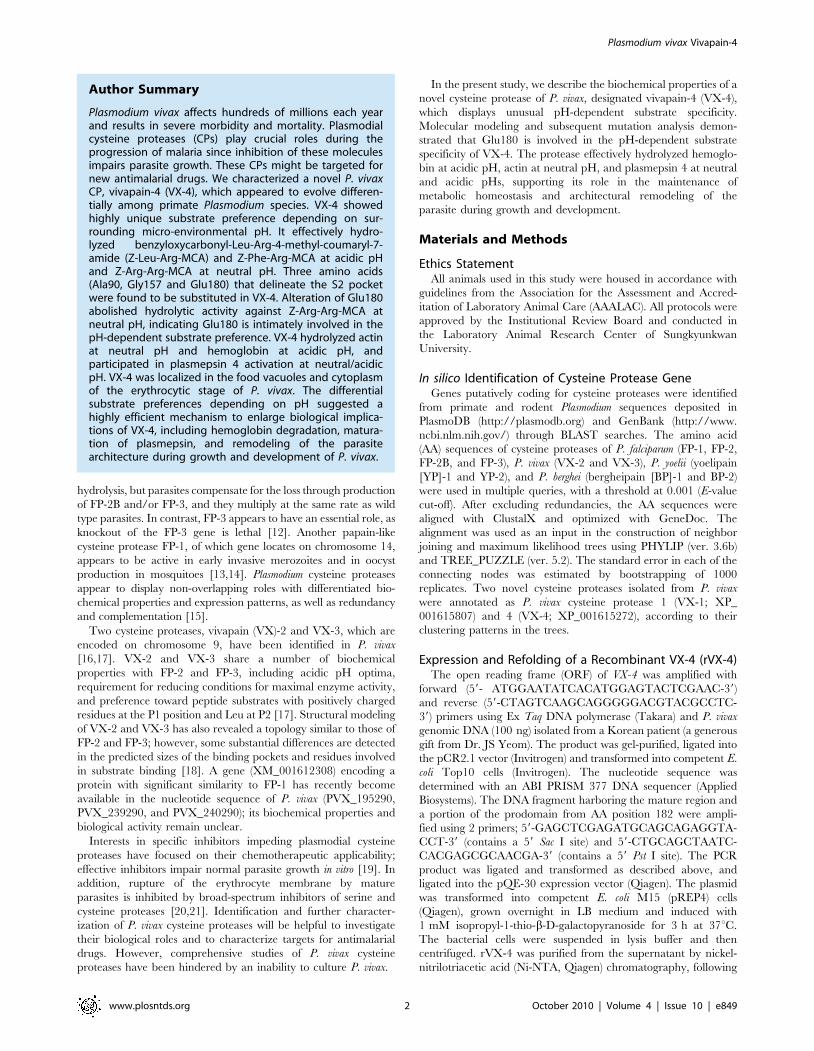
The P. vivax Genome Encodes Four Closely RelatedVivapains
By data-mining of the P. vivax genome (TIGR, Release 2.0), we
identified two genes putatively coding for novel cysteine proteases,
in addition to the previously identified genes encoding VX-2
(PlasmoDB code PVX_091415) and VX-3 (PVX_091410). We
designated these genes as VX-1 (PVX_195290) and VX-4
(PVX_091405). The other primate Plasmodium genomes examined,
such as P. falciparum, P. reichenowi and P. knowlesi, also harbored four
closely related cysteine protease genes. Conversely, avian and
rodent malaria parasites including P. gallinaceum, P. yoelii, and P.
berghei possessed only two paralogous genes (Figure 1). The
deduced AA sequence of VX-4 (TC5625, 484 AAs) revealed
considerable degrees of identity to that of VX-2 (TC5622, 59%)
and VX-3 (TC5618, 48%), while that of VX-1 (TC5613, 583 AAs)
was highly related to the FP-1-like proteases of P. falciparum, P.
knowlesi, P. ovale and P. fragile (37–77% identity). The greater length
of VX-1 might be attributable to an N-terminal extension [24].
Physiological implications and specific domain(s)/signature(s) of
VX-1 remain largely elusive. The primary structure of VX-4
tightly conserved the AA residues lining the catalytic site (Gln, Cys,
His, Asn and Trp) that are essential for the stabilization of a
thiolate-imidazolium ion pair and/or the transition state of the
catalytic site (AA positions highlighted in blue in Supplementary
Figure S1). The regulatory motifs of the plasmodial cysteine
proteases such as a bipartite trafficking domain, inhibitor domain
with ERFNIN signature and hemoglobin-binding FP2 arm were
also clearly identified in each of the corresponding regions
(Supplementary Figure S1) [25,26]. The eight Cys residues, which
are involved in the maintenance of structural geometry, were well
conserved in these proteins, whereas the last Cys was replaced by
Asn in VX-4 and KP-4 (arrowheads in Supplementary Figure S1).
Given the fact that a disulfide bridge between the seventh and
eighth Cys residues is intimately engaged in the stabilization of the
S2 and S19 sites of FP-2 [27], the more flexible binding pocket of
VX-4 might allow broader accessibility of proteolytic substrates. In
addition, several AA substitutions found in critical domains of VX-
4 suggest a distinctive physiological role for this protease
(Supplementary Figure S1). These collective data demonstrate
that VX-4 is a distinct cysteine protease that shares significant
identity with, but clearly differs from previously characterized P.
vivax cysteine proteases.
Plasmodium Cysteine Proteases Exhibit DifferentialEvolutionary Episodes Along with Their Donor Organisms
A neighbor-joining tree of VX-1 and VX-4 homologs, which
were retrieved from PlasmoDB and GenBank, was constructed
employing the AA sequences of mature domains (Figure 1). The
Plasmodium proteases were largely separated into two distinct
clusters consistent with their predicted biological roles: FP-1 clade,
of which members are implicated in host cell invasion [13] and
oocyst production [14], and FP-2 clade, the majority of which play
central roles in hemoglobin degradation [6,11,28]. An overall
topology similar to that of the neighbor-joining tree was observed
in a quartet maximum likelihood tree (TREE_PUZZLE program;
data not shown) and the major branching nodes were supported
by significant bootstrapping or quartet values. The falcipain
homolog genes appeared to have duplicated from a common
ancestor before diverging into each of the avian and mammalian
parasite lineages. The FP-1 family proteins seemed to have
diverged along with their specific donor organisms without any
provocative genetic event. Meanwhile, members of FP-2 clade
might have more complicated evolutionary pathways, including
either multiplication(s) in primate malaria or deletion(s) in rodent
malaria. The genes orthologous to VX-2 and VX-3 may have been
deleted in the rodent parasites, considering the polytomic
relationships among the P. vivax and P. knowlesi paralogs and the
tight clustering of VX-4/KP-4 with rodent malarial proteins. This
suggestion is further supported by the fact that P. falciparum and P.
reichenowi, which comprise a basal clade in mammalian Plasmodium
lineages [29,30], contain three paralogous genes. The three
paralogous genes occupying distinct but highly linked genomic
loci (cysteine protease island) may have undergone a kind of
convergent evolution events in these basal malaria genomes.
Adding to increased genic dosage, the degree of sequence
divergence was prominent among the primate FP-2 clade
members (0.81260.078), compared to related rodent proteins
(0.27160.034). The members of primate (0.26660.035) and
rodent (0.37760.056) FP-1 clade displayed values similar to that
of the rodent FP-2-like proteins (Supplementary Table S1).
Plasmodium vivax Vivapain-4
www.plosntds.org 4 October 2010 | Volume 4 | Issue 10 | e849
Alteration in gene copy number provides a simple way to change
expression levels or to enlarge protein pools with non-overlapping
functions. Biochemical studies have demonstrated that the primate
malaria proteins belonging to the FP-2 clade exhibit similar
enzymatic properties; however, those of P. vinckei (VP-2) and P.
berghei (BP-2) demonstrated quite dissimilar features, particularly in
terms of their substrate preference and inhibitor specificity [31,32].
Therefore, the large divergence among the primate FP-2 proteins
and tight clustering of VX-4 and KP-4 with rodent Plasmodium
proteins (bootstrapping value 76) further suggest biological roles of
VX-4 that are distinct from those previously described for VX-2
and VX-3 [16,17].
Recombinant VX-4 Shows Different SubstratePreferences Depending on pH
The full-length VX-4 gene amplified from a Korean P. vivax
patient’s blood displayed nucleotide sequence identical to that of
the reference Sal I strain (nucleotide sequence data is available in
the GenBank under the accession no. AY584068). A rVX-4
protein comprising a portion of the prodomain and entire mature
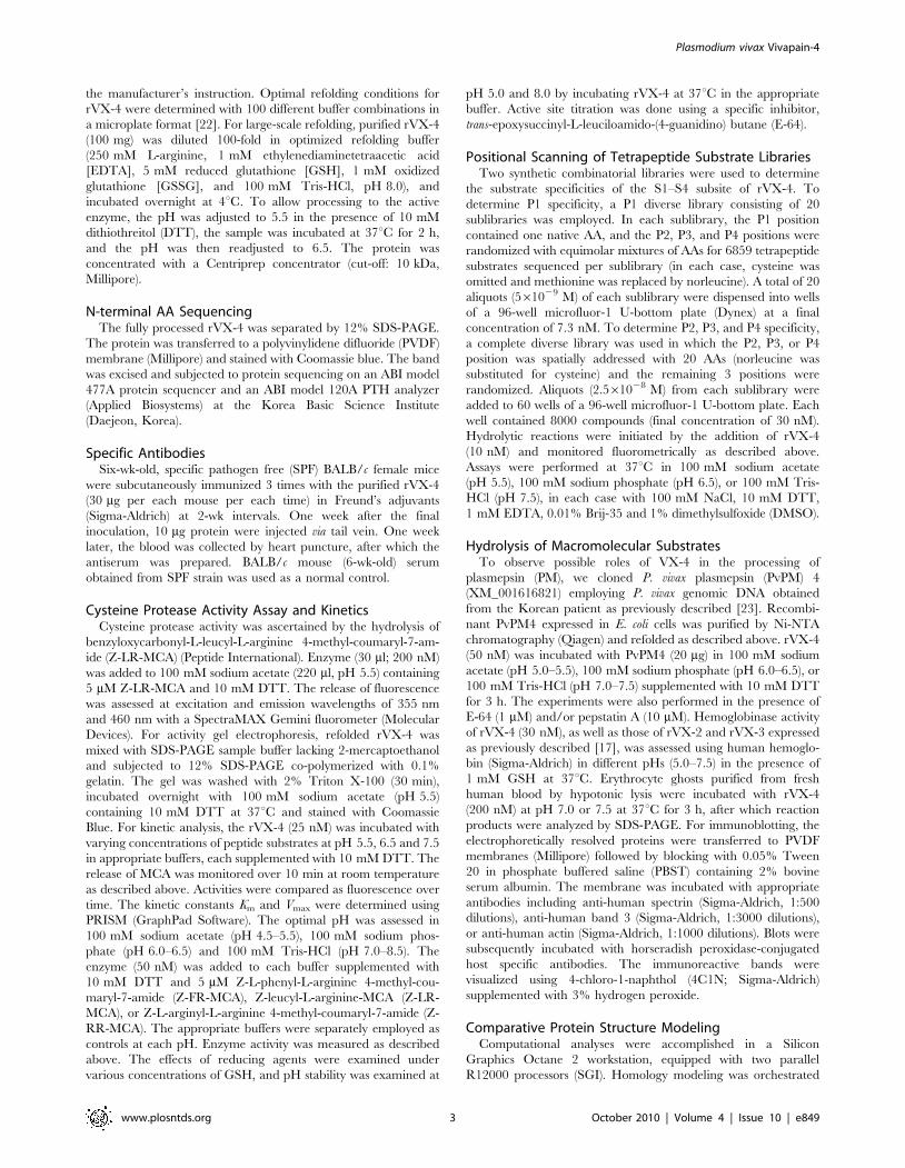
domain was expressed in E. coli (Figure 2A). Purified rVX-4 was
refolded followed by maturation under reducing and mild acidic
(pH 5.5) conditions. The fully processed 28 kDa protein (left
panel, Figure 2B) exhibited protease activity by gelatin-gel
electrophoresis (right panel, Figure 2B), which was completely
inhibited by the cysteine protease inhibitor E-64 (data not shown).
The N-terminal sequence of fully processed rVX-4 was NSPYV
(Supplementary Figure S1).
rVX-4 hydrolyzed synthetic dipeptidyl substrates with hydro-
phobic AA residues at their P2 site such as Z-LR-MCA and Z-FR-
MCA under acidic conditions. Activity was highest at pH 5.5. The
pH-optimum was substantially different with a substrate contain-
ing a basic AA at P2 (Z-RR-MCA) (Figure 2C) with maximal
activity at pH 6.5, and activity seen above pH 8. These results
suggest either that electrostatic conditions near the S2 site are
highly dependent on the surrounding pH or that the geometry of
the catalytic site can be changed in a pH-dependent manner.
rVX-4 was relatively stable after incubation at acidic and neutral
pH, while it was highly unstable under alkaline conditions (pH 8.0
and 8.5) against Z-LR-MCA (Figure 2D), which was similar to
results observed for FP-2 and FP-3 [28,32]. These data suggest
that decreased hydrolyzing activity of rVX-4 against Z-LR-MCA
and Z-FR-MCA at alkaline conditions might be due to an
irreversible change of the protein conformation. The requirement
for increased concentrations of E-64 to inhibit rVX-4 at higher pH
also supports altered structural geometry as the explanation for
altered substrate preference (Figure 2E).
Steady-state kinetic analyses confirmed varied substrate utiliza-
tion depending on pH (Table 1). rVX-4 showed a similar catalytic
efficiency against three peptide substrates at pH 5.5. However, at
pH 7.5 kcat/Km against Z-RR-MCA increased 2.6-fold whereas
that against Z-LR-MCA decreased 5.5-fold and Z-FR-MCA was
not hydrolyzed. The rVX-2 and rVX-3 exhibited much higher
kcat/Km values than that of rVX-4 toward Z-LR-MCA at the pH
conditions selected, although the optimal pH for rVX-2 was 6.5,
rather than 5.5. Interestingly, rVX-2 and rVX-3 could not
hydrolyze Z-FR-MCA or Z-RR-MCA. Phe has a large aromatic
R group, and it might not fit into the S2 pocket of rVX-2 and
rVX-3, which are stabilized by the disulfide bond between the
seventh and eighth Cys residues (Supplementary Figure S1).
Substrate preferences were further evaluated using a combina-
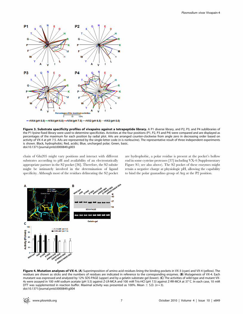
torial tetrapeptide library (Figure 3). AA utilization patterns of
vivapains at the P1, P3 and P4 positions were comparable to those
of FP-2 and FP-3 [15]. pH changes did not lead to significant
alterations in the substrate preference of VX-4 at these sites,
except that for tetrapeptides with Arg at P3; VX-4 was active at
pH 5.5, with substantial decreases as pH increased. The most
striking specificity was observed with substrates with different AAs
at P2. The vivapains preferred hydrophobic AAs such as Leu and
Val under acidic (VX-3 and VX-4, pH 5.5) and neutral (VX-2,
pH 6.5) conditions. VX-4 also exhibited significant activity
against P2 Met and His under acidic conditions. However,
relative activity against these hydrophobic AA residues was
significantly decreased, while that against Arg increased at pH 6.5
and 7.5, consistent with results obtained using synthetic peptide
substrates (Table 1).
Figure 1. Phylogenetic analysis of malaria cysteine proteases including VX-4. The phylogeny was based on the AA sequence alignment ofmature regions. Divergence rates were calculated with the Jones-Taylor-Thornton (JTT) substitution model, and the tree was constructed using theneighbor joining algorithm. The tree was rooted with GP-1 of Plasmodium gallinaceum, which was taken as an out-group. The number at each of thebranching nodes indicates the likelihood (percentage) of its appearance in the bootstrapping analysis with 1000 replicates. The enzymes from P. vivaxare in bold. The box indicates AAs found in the S2 pocket of primate plasmodial proteases, with position numbers based on mature VX-4. Red, blue,and black AAs are acidic, uncharged polar and hydrophobic, respectively. Note: The vivax protein with accession no. XP_001615272 was annotated asVX-2 during primary analysis of the whole genome sequence of the P. vivax Sal I strain. The name is changed to VX-4 according to our current result(AAT91956).doi:10.1371/journal.pntd.0000849.g001
Plasmodium vivax Vivapain-4
www.plosntds.org 5 October 2010 | Volume 4 | Issue 10 | e849
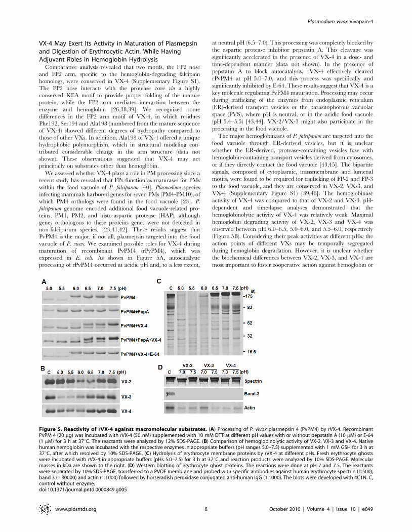
Glu180 Contributes to the pH-dependent SubstratePreferences of VX-4
Homology modeling of VX-4 demonstrated an overall topology
similar to those of FP-2, FP-3, VX-2 and VX-3 with the average
pairwise RMSD of 0.98 for the Ca atoms (data not shown).
However, a number of substitutions are recognized between VX-4
and the other VXs, including three prominent AA residues
delineating the S2 pocket (Ala90, Gly154 and Glu180; numbering
from the mature domain of VX-4) (Figure 4A; see also box in
Figure 1 and Supplementary Figure S1). The substrate preferences
of VX-4 were found to depend on AA residues occupying P2 site
and thus, the diagnostic AA substitution might be relevant to the
differential biochemistry of VX-4 compared to those of VX-2 and
VX-3. Seven mutant forms of VX-4, in which these three AA
residues were substituted by single, double, or triple site-directed
mutagenesis (A90I, G154S, E180A, A90I/G154S, A90I/E180A,
G154S/E180A, and A90I/G154S/E180A), were expressed in E.
coli, and their proteolytic activities were examined. All of the
refolded recombinant proteins showed hydrolytic activity against
gelatin (lower panel, Figure 4B). In assays against peptide
substrates, all of the mutants harboring A90I and G154S by
single or multiple substitutions maintained the pH-dependent
substrate specificity of wild-type VX-4. Conversely, those
containing E180A lost activity against Z-RR-MCA at pH 7.5
(Figure 4C). These results demonstrate that Glu180 plays a key
role in the pH-mediated switching of substrate specificity of VX-4.
The impact of a single AA substitution at a critical position has
been shown in a Leishmania major cathepsin B-like protease, in
which a Gly residue at the putative S2 pocket provided no
detectable proteolytic activity against Z-RR-AMC, while its
replacement with Glu restored activity [33]. A similar result was
also observed for papain, which exhibited a preference for Phe
over Arg at the P2 position, but exhibited cathepsin B-like
specificity when the S2 subsite was altered [34]. A cathepsin B-like
cysteine protease of Giardia lamblia that harbors a Glu residue at the
S2 pocket was active against both Z-FR-MCA and Z-LR-MCA
[35]. The crystallographic structure of cruzain, an essential
cysteine protease of Trypanosoma cruzi, demonstrated that the side
Figure 2. Biochemical properties of recombinant VX-4. (A) Expression and purification of rVX-4. Proteins were analyzed by 12% SDS-PAGE andstained with Coomassie blue. Lanes a, uninduced E. coli lysate; b, IPTG-induced E. coli lysate; c, Ni-NTA purified rVX-4. Mr, molecular masses in kDa.(B) Processing of the refolded rVX-4. The purified rVX-4 was refolded and activated, and aliquots collected every 30 min were analyzed by 12% SDS-PAGE with Coomassie staining (left). The proteolytic activity of fully processed rVX-4 was analyzed by a gelatin gel-zymogram (right). (C)Determination of pH optimum. The VX-4 enzyme activity was assayed in 100 mM sodium acetate (pH 4.5–5.5), sodium phosphate (pH 6.0–6.5) orTris/HCl (pH 7.0–8.5), each supplemented with 10 mM DTT. Activity was measured at 37uC each against Z-FR-MCA (&), Z-LR-MCA (#), and Z-RR-MCA(N). Maximal activity was presented as 100%. (D) Determination of enzyme stability. rVX-4 was incubated at different pHs in the respective buffers asin panel C. Residual activity was assayed with Z-LR-MCA in 100 mM sodium acetate (pH 5.5) supplemented with 1 mM DTT after indicatedincubations at pH 4.5 (N), 5.0 (#), 5.5 (m), 6.0 (n), 7.0 (q), 8.0 (%) and 8.5 (&). (E) Inhibition profile for E-64 was determined by incubating rVX-4(1 mM) with different concentrations of E-64 in 100 mM sodium acetate (pH 5.5; N), 100 mM sodium phosphate (pH 6.5; #) or 100 mM Tris-HCl(pH 7.5; &) at room temperature for 30 min. Residual activities (%) were determined using Z-LR-MCA as a substrate.doi:10.1371/journal.pntd.0000849.g002
Table 1. Comparison of substrate hydrolysis kinetics forvivapains.
kcat/Km (s21M21)
VX-2a VX-3a VX-4
pH 5.5 Z-FR-MCA NHb NH 1.556104
Z-LR-MCA 7.056105 8.626104 1.656104
Z-RR-MCA NH NH 1.346104
pH 6.5 Z-FR-MCA NH NH 5.526103
Z-LR-MCA 7.346105 6.366104 8.846103
Z-RR-MCA NH NH 3.496104
pH 7.5 Z-FR-MCA NH NH NH
Z-LR-MCA 4.156105 6.266103 3.056103
Z-RR-MCA NH NH 3.456104
Activity values for each enzyme represent mean from three independentexperiments.aValues for VX-2 and VX-3 were adopted from previous report (Na et al., 2004).bNH, no hydrolysis.doi:10.1371/journal.pntd.0000849.t001
Plasmodium vivax Vivapain-4
www.plosntds.org 6 October 2010 | Volume 4 | Issue 10 | e849
chain of Glu205 might vary positions and interact with different
substrates according to pH and availability of an electrostatically
appropriate partner in the S2 pocket [36]. Therefore, the S2 subsite
might be intimately involved in the determination of ligand
specificity. Although most of the residues delineating the S2 pocket
are hydrophobic, a polar residue is present at the pocket’s hollow
end in some cysteine proteases [37] including VX-4 (Supplementary
Figure S1; see also above). The S2 pocket of these enzymes might
retain a negative charge at physiologic pH, allowing the capability
to bind the polar guanadino group of Arg at the P2 position.
Figure 3. Substrate specificity profiles of vivapains against a tetrapeptide library. A P1 diverse library, and P2, P3, and P4 sublibraries ofthe P1-lysine fixed library were used to determine specificities. Activities at the four positions (P1, P2, P3 and P4) were compared and are displayed aspercentages of the maximum for each position by radial plot. AAs are arranged counter-clockwise from angle zero in decreasing order based onactivity of VX-4 at pH 7.5. AAs are represented by the single-letter code (n is norleucine). The representative result of three independent experimentsis shown. Black, hydrophobic; Red, acidic; Blue, uncharged polar; Green, basic.doi:10.1371/journal.pntd.0000849.g003
Figure 4. Mutation analyses of VX-4. (A) Superimposition of amino acid residues lining the binding pockets in VX-3 (cyan) and VX-4 (yellow). Theresidues are shown as sticks and the numbers of residues are indicated in reference to the corresponding enzymes. (B) Mutagenesis of VX-4. Eachmutatant was expressed and analyzed by 12% SDS-PAGE (upper) and by a gelatin substrate gel (lower). (C) The activities of wild-type and mutant VX-4s were assayed in 100 mM sodium acetate (pH 5.5) against Z-LR-MCA and 100 mM Tris-HCl (pH 7.5) against Z-RR-MCA at 37uC. In each case, 10 mMDTT was supplemented in reaction buffer. Maximal activity was presented as 100%. Mean 6 S.D. (n = 3).doi:10.1371/journal.pntd.0000849.g004
Plasmodium vivax Vivapain-4
www.plosntds.org 7 October 2010 | Volume 4 | Issue 10 | e849
VX-4 May Exert Its Activity in Maturation of Plasmepsinand Digestion of Erythrocytic Actin, While HavingAdjuvant Roles in Hemoglobin Hydrolysis
Comparative analysis revealed that two motifs, the FP2 nose
and FP2 arm, specific to the hemoglobin-degrading falcipain
homologs, were conserved in VX-4 (Supplementary Figure S1).
The FP2 nose interacts with the protease core via a highly
conserved KEA motif to provide proper folding of the mature
protein, while the FP2 arm mediates interaction between the
enzyme and hemoglobin [26,38,39]. We recognized some
differences in the FP2 arm motif of VX-4, in which residues
Phe192, Ser194 and Ala198 (numbered from the mature sequence
of VX-4) showed different degrees of hydropathy compared to
those of other VXs. In addition, Ala198 of VX-4 offered a unique
hydrophobic polymorphism, which in structural modeling con-
tributed considerable change in the arm structure (data not
shown). These observations suggested that VX-4 may act
principally on substrates other than hemoglobin.
We assessed whether VX-4 plays a role in PM processing since a
recent study has revealed that FPs function as maturases for PMs
within the food vacuole of P. falciparum [40]. Plasmodium species
infecting mammals harbored genes for seven PMs (PM4-PM10), of
which PM4 orthologs were found in the food vacuole [23]. P.
falciparum genome encoded additional food vacuole-related pro-
teins, PM1, PM2, and histo-aspartic protease (HAP), although
genes orthologous to these proteins genes were not detected in
non-falciparum species. [23,41,42]. These results suggest that
PvPM4 is the major, if not all, plasmepsin targeted into the food
vacuole of P. vivax. We examined possible roles for VX-4 during
maturation of recombinant PvPM4 (rPvPM4), which was
expressed in E. coli. As shown in Figure 5A, autocatalytic
processing of rPvPM4 occurred at acidic pH and, to a less extent,
at neutral pH (6.5–7.0). This processing was completely blocked by
the aspartic protease inhibitor pepstatin A. This cleavage was
significantly accelerated in the presence of VX-4 in a dose- and
time-dependent manner (data not shown). In the presence of
pepstatin A to block autocatalysis, rVX-4 effectively cleaved
rPvPM4 at pH 5.0–7.0, and this process was specifically and
significantly inhibited by E-64. These results suggest that VX-4 is a
key molecule regulating PvPM4 maturation. Processing may occur
during trafficking of the enzymes from endoplasmic reticulum
(ER)-derived transport vesicles or the parasitophorous vacuolar
space (PVS), where pH is neutral, or in the acidic food vacuole
(pH 5.4–5.5) [43,44]. VX-2/VX-3 might also participate in the
processing in the food vacuole.
The major hemoglobinases of P. falciparum are targeted into the
food vacuole through ER-derived vesicles, but it is unclear
whether the ER-derived, protease-containing vesicles fuse with
hemoglobin-containing transport vesicles derived from cytosomes,
or if they directly contact the food vacuole [43,45]. The bipartite
signals, composed of cytoplasmic, transmembrane and lumenal
motifs, were found to be required for trafficking of FP-2 and FP-3
to the food vacuole, and they are conserved in VX-2, VX-3, and
VX-4 (Supplementary Figure S1) [39,46]. The hemoglobinase
activity of VX-4 was compared to that of VX-2 and VX-3. pH-
dependent and time-lapse analyses demonstrated that the
hemoglobinolytic activity of VX-4 was relatively weak. Maximal
hemoglobin degrading activity of VX-2, VX-3 and VX-4 was
observed between pH 6.0–6.5, 5.0–6.0, and 5.5–6.0, respectively
(Figure 5B). Considering their peak activities at different pHs, the
action points of different VXs may be temporally segregated
during hemoglobin degradation. However, it is unclear whether
the biochemical differences between VX-2, VX-3, and VX-4 are
most important to foster cooperative action against hemoglobin or
Figure 5. Reactivity of rVX-4 against macromolecular substrates. (A) Processing of P. vivax plasmepsin 4 (PvPM4) by rVX-4. RecombinantPvPM 4 (20 mg) was incubated with rVX-4 (50 nM) supplemented with 10 mM DTT at different pH values with or without pepstatin A (10 mM) or E-64(1 mM) for 3 h at 37uC. The reactants were analyzed by 12% SDS-PAGE. (B) Comparison of hemoglobinolytic activity of VX-2, VX-3 and VX-4. Nativehuman hemoglobin was incubated with the respective enzymes in appropriate buffers (pH ranges 5.0–7.5) supplemented with 1 mM GSH for 3 h at37uC, after which resolved by 10% SDS-PAGE. (C) Hydrolysis of erythrocyte membrane proteins by rVX-4 at different pHs. Fresh erythrocyte ghostswere incubated with rVX-4 in appropriate buffers (pHs 5.0–7.5) for 3 h at 37uC and reaction products were analyzed by 10% SDS-PAGE. Molecularmasses in kDa are shown to the right. (D) Western blotting of erythrocyte ghost proteins. The reactions were done at pH 7 and 7.5. The reactantswere separated by 10% SDS-PAGE, transferred to a PVDF membrane and probed with specific antibodies against human erythrocyte spectrin (1:500),band 3 (1:30000) and actin (1:1000) followed by horseradish peroxidase conjugated anti-human IgG (1:1000). The blots were developed with 4C1N. C,control without enzyme.doi:10.1371/journal.pntd.0000849.g005
Plasmodium vivax Vivapain-4
www.plosntds.org 8 October 2010 | Volume 4 | Issue 10 | e849
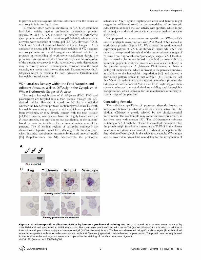
to provide activities against different substrates over the course of
erythrocytic infection by P. vivax.
To consider other potential substrates for VX-4, we examined
hydrolytic activity against erythrocyte cytoskeletal proteins
(Figures 5C and D). VX-4 cleaved the majority of erythrocytic
ghost proteins under acidic conditions (pH 5.0–6.0), whereas some
activities were negligible at neutral pH (6.5–7.5). However, VX-2,
VX-3, and VX-4 all degraded band-3 (anion exchanger 1, AE1)
and actin at neutral pH. The proteolytic activities of VX-4 against
erythrocyte actin and band-3 suggest an additional role for the
protease in remodeling of erythrocyte cytoskeleton during the
process of egress of merozoites from erythrocytes at the conclusion
of the parasite erythrocytic cycle. Alternatively, actin degradation
may be directly related to hemoglobin transport into the food
vacuole, as a recent study showed that actin filament turnover in P.
falciparum might be essential for both cytostome formation and
hemoglobin translocation [44].
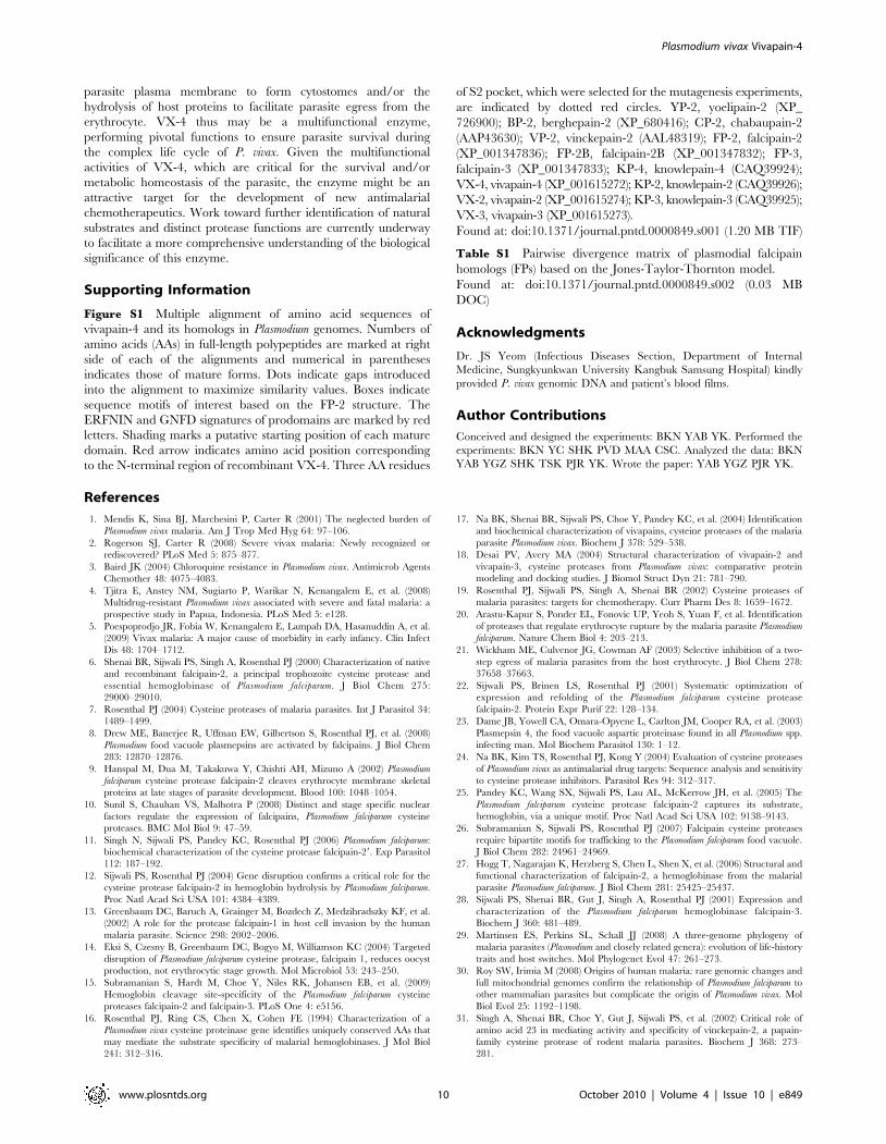
VX-4 Localizes Densely within the Food Vacuoles andAdjacent Areas, as Well as Diffusely in the Cytoplasm inWhole Erythrocytic Stages of P. vivax
The major hemoglobinases of P. falciparum (FP-2, FP-3 and
plasmepsins) are targeted into a food vacuole through the ER-
derived vesicles. However, it could not be clearly concluded
whether the ER-derived, protease-containing vesicles are fuse with
hemoglobin-containing transport vesicles, which were pinched off
from cytosomes, or they directly contact with the food vacuole
[43,45]. However, investigations have been highly limited with the
P. vivax proteins, not only due to low parasitemia in the patients’
blood, but also due to failure of experimental maintenance of the
parasite. The N-terminal regions of vavapains conserved the
characteristic bipartite signal for trafficking to the food vacuole,
which included cytoplasmic, transmembrane and lumenal motifs
[26] (Supplementary Fig. S1). Alternatively, the proteolytic
activities of VX-4 against erythrocytic actin and band-3 might
suggest its additional role(s) in the remodeling of erythrocytic
cytoskeleton, although the low activity with spectrin, which is one
of the major cytoskeletal proteins in erythrocyte, makes it unclear
(Figure 5D).
We prepared a mouse antiserum specific to rVX-4, which
showed negligible cross-reactions with rVX-2 and rVX-3 as well as
erythrocyte proteins (Figure 6A). We assessed the spatiotemporal
expression pattern of VX-4. As shown in Figure 6B, VX-4 was
shown to be expressed through all of the intraerythrocytic stages of
P. vivax, from ring to schizont/gametocyte stages. VX-4 localiza-
tion appeared to be largely limited to the food vacuoles with dark
homozoin pigment, while the protein was also labeled diffusely in
the parasite cytoplasm. P. falciparum FP-3 seemed to have a
biological implication(s), which is pivotal to the parasite’s survival,
in addition to the hemoglobin degradation [46] and showed a
distribution pattern similar to that of VX-4 [47]. Given the fact
that VX-4 has hydrolytic activity against cytoskeletal proteins, the
cytoplasmic distributions of VX-4 and FP-3 might suggest their
cytosolic roles such as cytoskeletal remodeling and hemoglobin
transportation, which is pivotal for the maintenance of intraeryth-
rocytic stage of the parasites.
Concluding RemarksThe substrate specificity of proteases depends largely on
interactions between a substrate and the enzyme active site. The
binding efficiency is greatly affected by the physicochemical
micromilieu. The reaction pH may confer substrate preference, as
has been seen with cruzain [36]. The pH-dependent substrate
switching of VX-4 might be relevant to its multiple biological roles;
the protein might function as a maturase of PvPM4 in the plasma
membrane or cytosomes at neutral pH, while it participates in the
degradation of hemoglobin in the acidic food vacuole. VX-4 might
also be involved in cytoskeletal remodeling for the invagination of
Figure 6. Spatiotemporal Localization of VX-4 by immunocytochemical staining. (A) rVX-2, rVX-3 and rVX-4 proteins were separated by12% SDS-PAGE and transferred to PVDF membrane. The membrane was incubated with anti-rVX-4 (1:1000 dilutions) for 4 h, with an additionalincubation with peroxidase-conjugated anti-mouse IgG (1:2000 dilutions) for 4 h. The blot was developed using 4C1N chromogen. (B) A thin bloodsmear from a patient with vivax malaria was stained with anti-rVX-4 conjugated with avidin-biotin complex system. The protein was densely labeledin the food vacuoles and adjacent areas, as compared to the staining of the dark hemozoin pigment.doi:10.1371/journal.pntd.0000849.g006
Plasmodium vivax Vivapain-4
www.plosntds.org 9 October 2010 | Volume 4 | Issue 10 | e849
parasite plasma membrane to form cytostomes and/or the
hydrolysis of host proteins to facilitate parasite egress from the
erythrocyte. VX-4 thus may be a multifunctional enzyme,
performing pivotal functions to ensure parasite survival during
the complex life cycle of P. vivax. Given the multifunctional
activities of VX-4, which are critical for the survival and/or
metabolic homeostasis of the parasite, the enzyme might be an
attractive target for the development of new antimalarial
chemotherapeutics. Work toward further identification of natural
substrates and distinct protease functions are currently underway
to facilitate a more comprehensive understanding of the biological
significance of this enzyme.
Supporting Information
Figure S1 Multiple alignment of amino acid sequences of
vivapain-4 and its homologs in Plasmodium genomes. Numbers of
amino acids (AAs) in full-length polypeptides are marked at right
side of each of the alignments and numerical in parentheses
indicates those of mature forms. Dots indicate gaps introduced
into the alignment to maximize similarity values. Boxes indicate
sequence motifs of interest based on the FP-2 structure. The
ERFNIN and GNFD signatures of prodomains are marked by red
letters. Shading marks a putative starting position of each mature
domain. Red arrow indicates amino acid position corresponding
to the N-terminal region of recombinant VX-4. Three AA residues
of S2 pocket, which were selected for the mutagenesis experiments,
are indicated by dotted red circles. YP-2, yoelipain-2 (XP_
726900); BP-2, berghepain-2 (XP_680416); CP-2, chabaupain-2
(AAP43630); VP-2, vinckepain-2 (AAL48319); FP-2, falcipain-2
(XP_001347836); FP-2B, falcipain-2B (XP_001347832); FP-3,
falcipain-3 (XP_001347833); KP-4, knowlepain-4 (CAQ39924);
VX-4, vivapain-4 (XP_001615272); KP-2, knowlepain-2 (CAQ39926);
VX-2, vivapain-2 (XP_001615274); KP-3, knowlepain-3 (CAQ39925);
VX-3, vivapain-3 (XP_001615273).
Found at: doi:10.1371/journal.pntd.0000849.s001 (1.20 MB TIF)
Table S1 Pairwise divergence matrix of plasmodial falcipain
homologs (FPs) based on the Jones-Taylor-Thornton model.
Found at: doi:10.1371/journal.pntd.0000849.s002 (0.03 MB
DOC)
Acknowledgments
Dr. JS Yeom (Infectious Diseases Section, Department of Internal
Medicine, Sungkyunkwan University Kangbuk Samsung Hospital) kindly
provided P. vivax genomic DNA and patient’s blood films.
Author Contributions
Conceived and designed the experiments: BKN YAB YK. Performed the
experiments: BKN YC SHK PVD MAA CSC. Analyzed the data: BKN
YAB YGZ SHK TSK PJR YK. Wrote the paper: YAB YGZ PJR YK.
References
1. Mendis K, Sina BJ, Marchesini P, Carter R (2001) The neglected burden of
Plasmodium vivax malaria. Am J Trop Med Hyg 64: 97–106.
2. Rogerson SJ, Carter R (2008) Severe vivax malaria: Newly recognized or
rediscovered? PLoS Med 5: 875–877.
3. Baird JK (2004) Chloroquine resistance in Plasmodium vivax. Antimicrob Agents
Chemother 48: 4075–4083.
4. Tjitra E, Anstey NM, Sugiarto P, Warikar N, Kenangalem E, et al. (2008)
Multidrug-resistant Plasmodium vivax associated with severe and fatal malaria: a
prospective study in Papua, Indonesia. PLoS Med 5: e128.
5. Poespoprodjo JR, Fobia W, Kenangalem E, Lampah DA, Hasanuddin A, et al.
(2009) Vivax malaria: A major cause of morbidity in early infancy. Clin Infect
Dis 48: 1704–1712.
6. Shenai BR, Sijwali PS, Singh A, Rosenthal PJ (2000) Characterization of native
and recombinant falcipain-2, a principal trophozoite cysteine protease and
essential hemoglobinase of Plasmodium falciparum. J Biol Chem 275:
29000–29010.
7. Rosenthal PJ (2004) Cysteine proteases of malaria parasites. Int J Parasitol 34:
1489–1499.
8. Drew ME, Banerjee R, Uffman EW, Gilbertson S, Rosenthal PJ, et al. (2008)
Plasmodium food vacuole plasmepsins are activated by falcipains. J Biol Chem
283: 12870–12876.
9. Hanspal M, Dua M, Takakuwa Y, Chishti AH, Mizuno A (2002) Plasmodium
falciparum cysteine protease falcipain-2 cleaves erythrocyte membrane skeletal
proteins at late stages of parasite development. Blood 100: 1048–1054.
10. Sunil S, Chauhan VS, Malhotra P (2008) Distinct and stage specific nuclear
factors regulate the expression of falcipains, Plasmodium falciparum cysteine
proteases. BMC Mol Biol 9: 47–59.
11. Singh N, Sijwali PS, Pandey KC, Rosenthal PJ (2006) Plasmodium falciparum:
biochemical characterization of the cysteine protease falcipain-29. Exp Parasitol
112: 187–192.
12. Sijwali PS, Rosenthal PJ (2004) Gene disruption confirms a critical role for the
cysteine protease falcipain-2 in hemoglobin hydrolysis by Plasmodium falciparum.
Proc Natl Acad Sci USA 101: 4384–4389.
13. Greenbaum DC, Baruch A, Grainger M, Bozdech Z, Medzihradszky KF, et al.
(2002) A role for the protease falcipain-1 in host cell invasion by the human
malaria parasite. Science 298: 2002–2006.
14. Eksi S, Czesny B, Greenbaum DC, Bogyo M, Williamson KC (2004) Targeted
disruption of Plasmodium falciparum cysteine protease, falcipain 1, reduces oocyst
production, not erythrocytic stage growth. Mol Microbiol 53: 243–250.
15. Subramanian S, Hardt M, Choe Y, Niles RK, Johansen EB, et al. (2009)
Hemoglobin cleavage site-specificity of the Plasmodium falciparum cysteine
proteases falcipain-2 and falcipain-3. PLoS One 4: e5156.
16. Rosenthal PJ, Ring CS, Chen X, Cohen FE (1994) Characterization of a
Plasmodium vivax cysteine proteinase gene identifies uniquely conserved AAs that
may mediate the substrate specificity of malarial hemoglobinases. J Mol Biol
241: 312–316.
17. Na BK, Shenai BR, Sijwali PS, Choe Y, Pandey KC, et al. (2004) Identification
and biochemical characterization of vivapains, cysteine proteases of the malaria
parasite Plasmodium vivax. Biochem J 378: 529–538.
18. Desai PV, Avery MA (2004) Structural characterization of vivapain-2 and
vivapain-3, cysteine proteases from Plasmodium vivax: comparative protein
modeling and docking studies. J Biomol Struct Dyn 21: 781–790.
19. Rosenthal PJ, Sijwali PS, Singh A, Shenai BR (2002) Cysteine proteases of
malaria parasites: targets for chemotherapy. Curr Pharm Des 8: 1659–1672.
20. Arastu-Kapur S, Ponder EL, Fonovic UP, Yeoh S, Yuan F, et al. Identification
of proteases that regulate erythrocyte rupture by the malaria parasite Plasmodium
falciparum. Nature Chem Biol 4: 203–213.
21. Wickham ME, Culvenor JG, Cowman AF (2003) Selective inhibition of a two-
step egress of malaria parasites from the host erythrocyte. J Biol Chem 278:
37658–37663.
22. Sijwali PS, Brinen LS, Rosenthal PJ (2001) Systematic optimization of
expression and refolding of the Plasmodium falciparum cysteine protease
falcipain-2. Protein Expr Purif 22: 128–134.
23. Dame JB, Yowell CA, Omara-Opyene L, Carlton JM, Cooper RA, et al. (2003)
Plasmepsin 4, the food vacuole aspartic proteinase found in all Plasmodium spp.
infecting man. Mol Biochem Parasitol 130: 1–12.
24. Na BK, Kim TS, Rosenthal PJ, Kong Y (2004) Evaluation of cysteine proteases
of Plasmodium vivax as antimalarial drug targets: Sequence analysis and sensitivity
to cysteine protease inhibitors. Parasitol Res 94: 312–317.
25. Pandey KC, Wang SX, Sijwali PS, Lau AL, McKerrow JH, et al. (2005) The
Plasmodium falciparum cysteine protease falcipain-2 captures its substrate,
hemoglobin, via a unique motif. Proc Natl Acad Sci USA 102: 9138–9143.
26. Subramanian S, Sijwali PS, Rosenthal PJ (2007) Falcipain cysteine proteases
require bipartite motifs for trafficking to the Plasmodium falciparum food vacuole.
J Biol Chem 282: 24961–24969.
27. Hogg T, Nagarajan K, Herzberg S, Chen L, Shen X, et al. (2006) Structural and
functional characterization of falcipain-2, a hemoglobinase from the malarial
parasite Plasmodium falciparum. J Biol Chem 281: 25425–25437.
28. Sijwali PS, Shenai BR, Gut J, Singh A, Rosenthal PJ (2001) Expression and
characterization of the Plasmodium falciparum hemoglobinase falcipain-3.
Biochem J 360: 481–489.
29. Martinsen ES, Perkins SL, Schall JJ (2008) A three-genome phylogeny of
malaria parasites (Plasmodium and closely related genera): evolution of life-history
traits and host switches. Mol Phylogenet Evol 47: 261–273.
30. Roy SW, Irimia M (2008) Origins of human malaria: rare genomic changes and
full mitochondrial genomes confirm the relationship of Plasmodium falciparum to
other mammalian parasites but complicate the origin of Plasmodium vivax. Mol
Biol Evol 25: 1192–1198.
31. Singh A, Shenai BR, Choe Y, Gut J, Sijwali PS, et al. (2002) Critical role of
amino acid 23 in mediating activity and specificity of vinckepain-2, a papain-
family cysteine protease of rodent malaria parasites. Biochem J 368: 273–
281.
Plasmodium vivax Vivapain-4
www.plosntds.org 10 October 2010 | Volume 4 | Issue 10 | e849
32. Chan C, Goh LL, Sim TS (2005) Differences in biochemical properties of the
Plasmodial falcipain-2 and berghepain-2 orthologues: implications for in vivoscreens of inhibitors. FEMS Microbiol Lett 249: 315–321.
33. Chan VJ, Selzer PM, McKerrow JH, Sakanari JA (1999) Expression and
alteration of the S2 subsite of the Leishmania major cathepsin B-like cysteineprotease. Biochem J 340: 113–117.
34. Khouri HE, Vernet T, Menard R, Parlati F, Laflamme P, et al. (1991)Engineering of papain: selective alteration of substrate specificity by site-directed
mutagenesis. Biochemistry 17: 8929–8936.
35. DuBois KN, Abodeely M, Sajid M, Engel JC, McKerrow JH (2006) Giardia
lamblia cysteine proteases. Parasitol Res 99: 313–316.
36. Gillmor SA, Craik CS, Fletterick BJ (1997) Structural determinants of specificityin the cysteine protease cruzain. Protein Sci 6: 1603–1611.
37. Sajid M, McKerrow JH (2002) Cysteine proteases of parasitic organisms. MolBiochem Parasitol 120: 1–21.
38. Sijwali PS, Shenai BR, Rosenthal PJ (2002) Folding of the Plasmodium falciparum
cysteine protease falcipain-2 is mediated by a chaperone-like peptide and not theprodomain. J Biol Chem 277: 14910–14915.
39. Wang SX, Pandey KC, Somoza JR, Sijwali PS, Kortemme T, et al. (2006)Structural basis for unique mechanisms of folding and hemoglobin binding by a
malarial protease. Proc Natl Acad Sci USA 103: 11503–11508.
40. Drew ME, Banerjee R, Uffman EW, Gilbertson S, Rosenthal PJ, et al. (2008)Plasmodium food vacuole plasmepsins are activated by falcipains. J Biol Chem
283: 12870–12876.
41. Francis SE, Gluzman IY, Oksman A, Knickerbocker A, Mueller R, et al. (1994)
Molecular characterization and inhibition of a Plasmodium falciparum aspartic
hemoglobinase. EMBO J 13: 306–317.
42. Banerjee R, Liu J, Beatty W, Pelosof L, Klemba M, et al. (2002) Four
plasmepsins are active in the Plasmodium falciparum food vacuole, including a
protease with an active-site histidine. Proc Natl Acad Sci USA 99: 990–995.
43. Klonis N, Tan O, Jackson K, Goldberg D, Klemba M, et al. (2007) Evaluation
of pH during cytostomal endocytosis and vacuolar catabolism of haemoglobin in
Plasmodium falciparum. Biochem J 407: 343–354.
44. Lazarus MD, Schneider TG, Taraschi TF (2008) A new model for hemoglobin
ingestion and transport by the human malaria parasite Plasmodium falciparum.
J Cell Sci 121: 1937–1949.
45. Dasaradhi PV, Korde R, Thompson J K, Tanwar C, Nag TC, et al. (2007) Food
vacuole targeting and trafficking of falcipain-2, an important cysteine protease of
human malaria parasite Plasmodium falciparum. Mol Biochem Parasitol 156:
12–23.
46. Sijwali PS, Koo J, Singh N, Rosenthal PJ (2006) Gene disruptions demonstrate
independent roles for the four falcipain cysteine proteases of Plasmodium
falciparum. Mol Biochem Parasitol 150: 96–106.
47. Dahl EL, Rosenthal PJ (2005) Biosynthesis, localization, and processing of
falcipain cysteine proteases of Plasmodium falciparum. Mol Biochem Parasitol 139:
201–212.
Plasmodium vivax Vivapain-4
www.plosntds.org 11 October 2010 | Volume 4 | Issue 10 | e849
![PARTIAL PURIFICATION OF CYSTEINE PROTEASE FROM · during seed germination to the initiation of cell death and senescence programs [7]. The study reveals that the protease inhibition](https://img.pdfslide.net/doc/110x75/5e856b9b52266469734617a4/partial-purification-of-cysteine-protease-during-seed-germination-to-the-initiation.jpg)


![Scholars Research Library...Several results on cloning and expression of protease gene in E. coli were reported such as intracellular protease from Pyrococcus furiosus [9], cysteine](https://img.pdfslide.net/doc/110x75/60a917e19c154a2ec963d882/scholars-research-library-several-results-on-cloning-and-expression-of-protease.jpg)