Embed Size (px)
Citation preview
VOL. 46, 1960 BIOCHEMISTRY: ZUCKERKANDL ET AL. 1349
8 Volkin, E., in Fourth International Congress of Biochemistry (New York: Pergamon Press,1959), vol. 7, p. 212.
1 Volkin, E., L. Astrachan, and J. L. Countryman, Virology, 6, 545 (1958).'0 Astrachan, L., and E. Volkin, Biochim. et Biophys. Acta, 29, 536 (1958).11 Countryman, J. L., and E. Volkin, J. Bacteriol., 78, 41 (1959).12 Volkin, E., and L. Astrachan, in The Chemical Basis of Heredity, eds. W. D. McElroy and B.
Glass (Baltimore: The Johns Hopkins Press, 1957) p. 686.13 Nomura, M., B. D. Hall, and S. Spiegelman, Fed. Proc., 19, Part 1, 315 (1960).14 Watanabe, I., Y. Kiho, and K. Miura, Nature, 181, 1127 (1958).15 Jeener, R., Biochim. et Biophys. Acta, 27, 665 (1958).16 Pardee, A. B., and L. S. Prestidge, Biochim. et Biophys. Acta, 37,544 (1960).17 Astrachan, L., and E. Volkin, Biochim. et Biophys. Acta, 32, 456 (1959).18 Volkin, E., unpublished observations.19 Gollub, E. G., and J. S. Gots, J. Bacteriol., 78,320 (1959).20 Herriott, R. M., and J. L. Barlow, J. Gen. Physiol., 36, 17 (1952).21 Adams, M. H., in Methods in Medical Research, ed. J. H. Comroe, Jr. (Chicago: Year Book
Publishers, 1950), vol.2, pp. 1-73.22 Hershey, A. D., J. Dixon, and M. Chase, J. Gen. Physiol., 36,777 (1953).23 Tyner, E. P., C. Heidelberger, and G. A. LePage, Cancer Research 13, 186 (1953).24 Cohn, W. E., J. Am. Chem. Soc., 72, 1471 (1950); also J. Cell. and Comp. Physiol., 38, Suppl.
1,21 (1951).25 Griswold, B. L., F. L. Humoller, and A. R. McIntyre, Anal. Chem., 23, 192 (1951)." Astrachan, L , unpublished observations.27 Ernster, L., R. Zetterstrom, and 0. Lindberg, Acta Chem. Scand., 4, 942 (1950)." Evans, E. A., Jr., Fed. Proc., 15,827 (1956).29 Colowick, S. P., and N. 0. Kaplan, eds., Methods in Enzymology (New York: Academic
Press, 1955), vol. 2, pp. 448, 456, and 468.30 Hershey, A. D., and N. E. Melechen, Virology, 3, 207 (1957).31 Tomizawa, J., and S. Sunakawa, J. Gen. Physiol., 39, 553 (1956).32 Burton, K., Biochem. J., 61, 473 (1955).33 Neidhardt, F. C., and F. Gros, Biochim. et Biophys. Acta, 25, 513 (1957).34 Kornberg, A., S. B. Zimmerman, S. R. Kornberg, and J. Josse, these PROCEEDINGS 45, 772
(1959).3 Flaks, J. G., J. Lichtenstein, S. S., Cohen, J. Biol. Chem., 234, 1507 (1959).36 Bessman, M. J., J. Biol. Chem., 234, 2735 (1959).37 Sommerville, R. and G. R. Greenberg, Fed. Proc., 18, 327 (1959).38 Keck, K., H. R. Mahler, and D. Fraser, Arch. Biochem. Biophys., 86, 85 (1960).
A COMPARISON OF ANIMAL HEMOGLOBINS BY TRYPTIC PEPTIDEPATTERN ANALYSIS*
BY EMILE ZUCKERKANDL,t RICHARD T. JONES, AND LINUS PAULING
DIVISION OF CHEMISTRY AND CHEMICAL ENGINEERING, t CALIFORNIA INSTITUTE OF TECHNOLOGY
Communicated August 22, 1960
The complete amino acid sequences (primary structure) of hemoglobins can,in principle, be determined by methods currently available. Although detailedstudies of the primary structure of human and horse hemoglobins are in progressin several laboratories,' the methods are so laborious that complete sequences havenot yet been established. Important questions in the realm of genetics and evolu-tion require the immediate examination of the structure, primary and other, of
1350 BIOCHEMISTRY: ZUCKERKANDL ET AL. PROC. N. A. S.
many different hemoglobins. The application of methods that are quicker, thoughless informative and reliable, than the techniques required for complete sequencedetermination is therefore in order as a provisional means of securing useful infor-mation. Such a method is the analysis of peptide patterns obtained by combinedpaper electrophoresis and chromatography of tryptic hydrolysates of denaturedhemoglobin.2 Of particular interest are comparisons between hemoglobin com-ponents present in (a) organisms of one animal species at a given time in develop-ment, (b) organisms of one species at different stages of development, and (c)organisms of different species. The present paper is concerned exclusively with thelast type of comparison. In order to scan the range of variation of hemoglobinstructure throughout evolution, hemoglobins from a number of animals both closelyand distantly related to man have been selected and compared as to tryptic peptidepatterns with human hemoglobin A. Whole hemoglobin preparations from adultanimals have been studied throughout. The problem of individual heterogeneitywill be treated elsewhere.
Materials and Methods.-Ape bloods3a were anticoagulated and transported inAlsever's solution. The apes studied included two lowlanci gorillas (Gorilla gorilla),two chimpanzees (Pan troglodytes), and three orangutans (Pongo pygmaeus).Erythrocytes from Rhesus monkeys (Macaca mulatta) were obtained from clottedblood.3b Heparinized porcine and bovine bloods were secured during bleedings atLos Angeles slaughter houses. Heparinized bloods were obtained from the marinelungfish, Pimelometopon pulcher (sheepshead), and from the cartilaginous fishCephaloscyllium uter (swell shark)." Blood was also obtained from a live specimenof Lepidosiren paradoxa (Dipneust, South American fresh water lungfish).3C dContamination of the latter blood by tissue fluid was unavoidable. Blood of thePacific hagfish, Polistotrema stouti, was also examined.4 Coelomic cells (hemoglobincells) were obtained from Urechis caupo (Echiurid marine "worm")5 collected atlow tide from mud flats near the Kerckhoff Marine Laboratory at Corona del Mar,California. 3e
In general, the hemoglobin preparations examined were from single individuals.However, in the case of Urechis single as well as pooled samples of coelomic cellswere used without apparent differences. Only a pooled sample of blood fromtwelve hagfish was examined. All of the animals studied were judged to be adultseither from their size or their known age. The youngest apes were one orangutan2 years old and one gorilla 2 years old. Peptide patterns of their hemoglobinswere indistinguishable from those of older individuals of the same species. TheLepidosiren, 14 inches long, was at a minimum 8 months of age, and might be con-sidered a subadult.The erythrocytes were washed four times with cold 0.9%70 NaCl with the ex-
ception of Pimelometopon and Cephaloscyllium cells, which were washed with 1.2%NaCl.6 The red cells from hagfish were washed with chilled 3% NaCl and thecoelomic cells of Urechis with 3.2%o NaCl. Washing with 0.9% NaCl led to con-siderable hemolysis in the case of the fresh water fish Dipneust.The washed cells were hemnolyzed in a standard fashion with distilled water and
toluene7 with the exception of Urechis, where ether was used instead of toluene.The washed red cells of hagfish were stored for one day at 4°C before lysing. Amajor portion of the hemoglobin obtained was insoluble possibly due to acidifica-
VOL. 46, 1960 BIOCHEMISTRY: ZUCKERKANDL ET AL. 1351
tion during storage. The peptide pattern shown below was obtained from theremaining soluble fraction. After lysis, each sample was centrifuged at high speedsin order to remove solid debris and the toluene phase. The resulting hemoglobinsolutions were saturated with carbon monoxide and dialyzed for a week againstdistilled water saturated with carbon monoxide (three changes of water with theratio of hemoglobin solution to water of the order of 1:100). This prolonged dialy-sis was employed to permit the flocculation of non-hemoglobin proteins.6 A crystalof thymol was added to one Urechis preparation in order to eliminate the possibilityof the formation of peptides by bacterial contamination. No differences werefound in the peptide patterns obtained with and without thymol.With species closely related to man the hemoglobin concentration in the final
preparation could be assayed approximately by the use of spectrophotometricconstants established for human hemoglobins. In other species the quantities ofmaterial to be used on peptide patterns were estimated roughly from the consump-tion of sodium hydroxide during tryptic digestion.
Hydrolysis with trypsin of heat-denatured hemoglobin preparations was per-formed at constant pH using a Radiometer automatic titrator as a pH-stat. Thehydrolyses were carried out at 40'C at pH 8.0 in the presence of Ca ion (0.01 M)with an enzyme to hemoglobin ratio of between 1 and 2% by weight (Worthingtontwice crystallized trypsin). Ninety minutes was allowed for the hydrolysis exceptin the case of the apes, where the digestion was stopped after sixty minutes.Those tryptic peptides which are soluble at pH 6.5 were analyzed by a combina-
tion of electrophoresis and chromatography on paper. These peptide patternswere obtained essentially as specified by Ingram2 except that the voltage rate was900-1,000 V for a duration of 31/2 hours. In general, a human control samplewas run as a pair mate with every animal hemoglobin in order to have a referencepattern for each unknown. Even with such parallel determinations, the spreadingoccurring during electrophoresis was not always identical. The polarity of theelectrophoretic field is marked on each pattern and the point of application of thesample is designated by a cross (+). The peptide spots resulting from tryptichydrolysis of human hemoglobin have been assigned arbitrary numbers by Ingram.2This convention has been employed in the present study. The neutral band regionis comprised of peptide spots 1 through 7. These peptides do not migrate to anyextent in the electric field, but do move some from the point of application due tofluid movement. Phenylalanine and lysine have been added as reference spots atleast on one pattern in the case of every species. Chromatography was ascendingin direction and the peptides were detected mainly by their reaction with ninhydrin.The Sakaguchi8 test for arginine and the cinnamaldehyde9 reaction for trypophanwere also employed. Patterns from each species were obtained at least in triplicate.
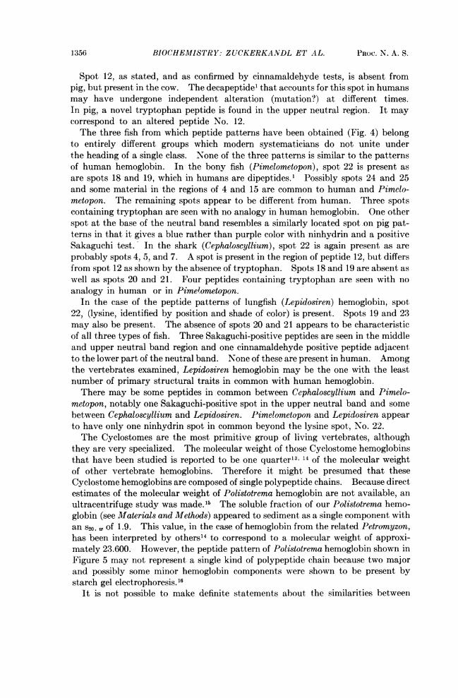
Results.-Figure 1 is a peptide pattern of human hemoglobin A as currentlyobtained in our laboratories. A diagram indicating the peptide spot numberingsystem is also included.
Figure 2 shows peptide patterns from three different apes (gorilla, chimpanzee,and orangutan) and one monkey (Rhesus). The gorilla, chimpanzee, and humanpatterns are almost identical in appearance. In the case of the gorilla peptidepatterns, spot numbers 12 and 24 both appear to be double. In humans the pep-tides that make up these two spots are from the beta chain. No difference was

Cathode AnodeFIG. 1.-A tryptic peptide pattern of adult human hemoglobin and its schematic representation.
The spots are numbered according to the convention introduced by Ingram.2 The site of spot 2(is shown on the diagram, although not seen on the pattern represented. The locations of fourhitherto undescribed spots usually found on patterns obtained in our laboratories are indicated.

VOL. 46, 1960 BIOCHEMISTRY: ZUCKERANDL, ETAL. 1353
PRIMATE HEMOGLOBINS
Human Chimpanzee
#..q,%.is||.....4~~~~~~~~~~~~~~~~~~~~~~~~~~~~~~~~~~~~~~~~~~~~~~~~~~~~~~~~~~~~~~~~~~~~~~~~~~~~~~~~~~Arca.."
OW".bs*|
Gorilla Orangutan Rhesus MonkeyFIG. 2.-Tryptic peptide patterns of primate hemoglobins. The circled spot on the Rhesus
monkey pattern represents phenylalanine added two and a half inches to the anodal side of thepoint of application of the peptide mixture.
observed between two gorillas, one male and one female. Spots 12 and 24 alsoseem to be double in the peptide patterns of chimpanzee. In addition, extramaterial may be present in the 15-16 region. In humans at least four majorpeptides are present in this region.' Two chimpanzees, both females, were foundto have identical patterns. Of course further differences between these two typesof apes and humans may be discovered upon the analysis of the individual pep-tides or the study of the protein residue which remains insoluble after hydrolysiswith trypsin.The difference from human patterns is somewhat greater for orangutan peptide
patterns than for the patterns of the two apes just mentioned. In two orangutans(one male and one female) spots 12 and 24 are double. The third orangutan(female), whose pattern is illustrated in Figure 2, was observed to have single 12and 24 spots. Further differences between the patterns of the individual orangu-tans are not apparent; however, each differs from human patterns by an apparentincrease in peptides in the 15-16 region. The orangutans also differ from humansand the other two apes studied by the appearance of two new spots, one anodal tospot 20 and the other cathodal and below spot 10. The latter does not appearto be the same as spot 9 of human. All of the spots containing arginine and trypto-phan are the same as in human.
Spot 12 is absent in the peptide patterns of Rhesus monkey hemoglobin. Anew spot, possibly representing a modification of the peptide of spot 12, is present
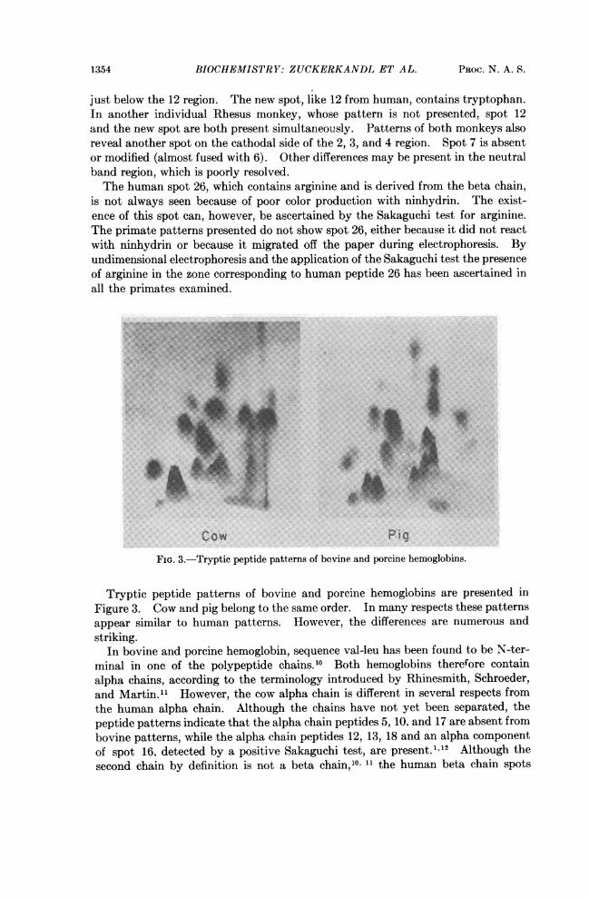
1354 BIOCHEMISTRY: ZUCKERKANDL ET AL. PROC. N. A. S.
just below the 12 region. The new spot, like 12 from human, contains tryptophan.In another individual Rhesus monkey, whose pattern is not presented, spot 12and the new spot are both present simultaneously. Patterns of both monkeys alsoreveal another spot on the cathodal side of the 2, 3, and 4 region. Spot 7 is absentor modified (almost fused with 6). Other differences may be present in the neutralband region, which is poorly resolved.The human spot 26, which contains arginine and is derived from the beta chain,
is not always seen because of poor color production with ninhydrin. The exist-ence of this spot can, however, be ascertained by the Sakaguchi test for arginine.The primate patterns presented do not show spot 26, either because it did not reactwith ninhydrin or because it migrated off the paper during electrophoresis. Byundimensional electrophoresis and the application of the Sakaguchi test the presenceof arginine in the zone corresponding to human peptide 26 has been ascertained inall the primates examined.*: ~~~~~~~~~~~~~~~~~~~~~~~.:;....
Cow PigFIG. 3.-Tryptic peptide patterns of bovine and porcine hemoglobins.
Tryptic peptide patterns of bovine and porcine hemoglobins are presented inFigure 3. Cow and pig belong to the same order. In many respects these patternsappear similar to human patterns. However, the differences are numerous andstriking.
In bovine and porcine hemoglobin, sequence val-leu has been found to be N-ter-minal in one of the polypeptide chains.'0 Both hemoglobins therefore containalpha chains, according to the terminology introduced by Rhinesmith, Schroeder,and Martin." However, the cow alpha chain is different in several respects fromthe human alpha chain. Although the chains have not yet been separated, thepeptide patterns indicate that the alpha chain peptides 5, 10, and 17 are absent frombovine patterns, while the alpha chain peptides 12, 13, 18 and an alpha componentof spot 16. detected by a positive Sakaguchi test, are present."'2 Although thesecond chain by definition is not a beta chain,0' 11 the human beta chain spots
VOL. 46, 1960 BIOCHEKISTRY: ZUCKERKANDL ET AL. 1355
12 and 19 are seen clearly. The human pure beta spots 2, 4, 14 (as ascertained by anegative reaction with cinnamaldehyde) and 25 are absent.The porcine alpha chain may be somewhat more similar to the human alpha
chain since the alpha spots 10, 13, and 18 are seen, and the presence of the alphaspots 11, 17, and 23 is suspected. The presence of an alpha-component in thecomposite spot 15 is likely because of a positive cinnamaldehyde test, and thepresence of an alpha-component in the composite spot 16 is indicated by a posi-tive Sakaguchi test. The alpha spot 5 is absent. Among the beta spots, 14 (cin-namaldehyde positive), 26 (Sakaguchi positive), 4, 19, 24, and possibly 25 arepresent; while 12 is absent. Like bovine hemoglobin, porcine hemoglobin doesnot contain beta chains; however, there are a number of tryptic peptides-similarto those from the human beta chain observed.The composite human spots 13-14, 15-16, 20 and 21 as well as the lysine spot 22
(arising from both a and f3 chains') are seen in pig as well as in cow. Thus the~~~~~~~~~~~~~~~~~~~~~~~~~~. S;...
*...zay88.,,s,.:..ss~~~~~~~~~~~~~~~~~.
Bony Fish Lungfish
.4.
SharkFIG. 4.-Tryptic peptide patterns of "fish" hemoglobins. The circled
spot on the shark pattern represents phenylalanine added two and a halfinches to the anodal side of the point of application of the peptidemixturee
apparent similarities between bovine and human patterns are in many ,respects thesame as those noted between porcine and human; however, the differences are notthe same. From three independent pattern studies of the same bovine sample itappears that many of the new spots are slightly on the anodal side of the neutralband region, whereas in porcine patterns most of the new spots are on the cathodalside. Arginine is present in at least two of the new porcine peptides, one neutraland one acidic.
1356 BIOCHEMISTRY: ZUCKERKANDL ET AL. PROC. N. A. S.
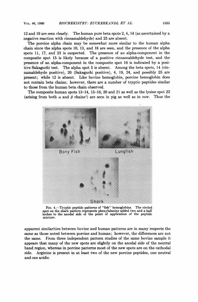
Spot 12, as stated, and as confirmed by cinnamaldehyde tests, is absent frompig, but present in the cow. The decapeptidel that accounts for this spot in humansmay have undergone independent alteration (mutation?) at different times.In pig, a novel tryptophan peptide is found in the upper neutral region. It maycorrespond to an altered peptide No. 12.The three fish from which peptide patterns have been obtained (Fig. 4) belong
to entirely different groups which modern systematicians do not unite underthe heading of a single class. None of the three patterns is similar to the patternsof human hemoglobin. In the bony fish (Pimelometopon), spot 22 is present asare spots 18 and 19, which in humans are dipeptides.1 Possibly spots 24 and 25and some material in the regions of 4 and 15 are common to human and Pimelo-metopon. The remaining spots appear to be different from human. Three spotscontaining tryptophan are seen with no analogy in human hemoglobin. One otherspot at the base of the neutral band resembles a similarly located spot on pig pat-terns in that it gives a blue rather than purple color with ninhydrin and a positiveSakaguchi test. In the shark (Cephaloscyllium), spot 22 is again present as areprobably spots 4, 5, and 7. A spot is present in the region of peptide 12, but differsfrom spot 12 as shown by the absence of tryptophan. Spots 18 and 19 are absent aswell as spots 20 and 21. Four peptides containing tryptophan are seen with noanalogy in human or in Pime'ometopon.
In the case of the peptide patterns of lungfish (Lepidosiren) hemoglobin, spot22, (lysine, identified by position and shade of color) is present. Spots 19 and 23may also be present. The absence of spots 20 and 21 appears to be characteristicof all three types of fish. Three Sakaguchi-positive peptides are seen in the middleand upper neutral band region and one cinnamaldehyde positive peptide adjacentto the lower part of the neutral band. None of these are present in human. Amongthe vertebrates examined, Lepidosiren hemoglobin may be the one with the leastnumber of primary structural traits in common with human hemoglobin.There may be some peptides in common between Cephaloscyllium and Pimelo-
metopon, notably one Sakaguchi-positive spot in the upper neutral band and somebetween Cephaloscyllium and Lepidosiren. Pimelometopon and Lepidosiren appearto have only one ninhydrin spot in common beyond the lysine spot, No. 22.The Cyclostomes are the most primitive group of living vertebrates, although
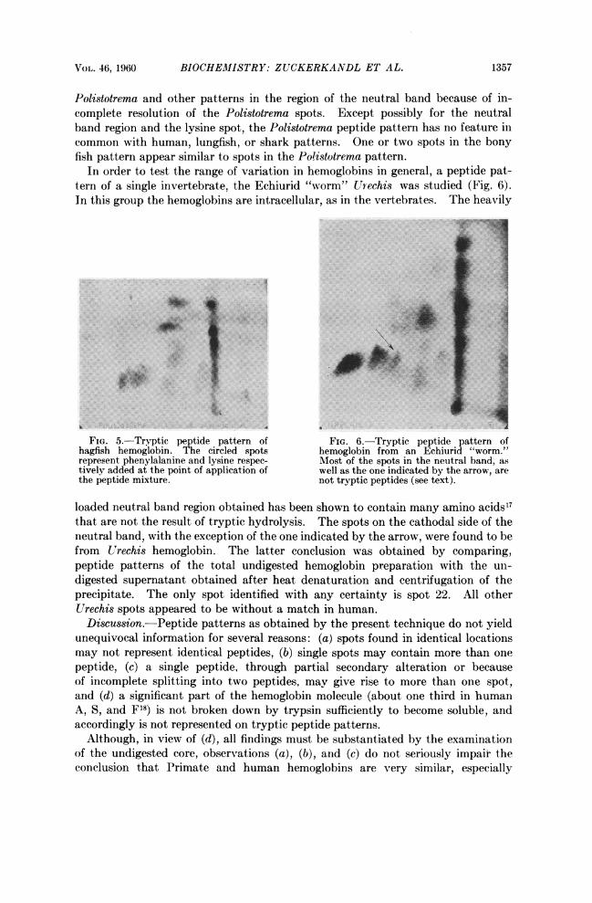
they are very specialized. The molecular weight of those Cyclostome hemoglobinsthat have been studied is reported to be one quarter'3 14 of the molecular weightof other vertebrate hemoglobins. Therefore it might be presumed that theseCyclostome hemoglobins are composed of single polypeptide chains. Because directestimates of the molecular weight of Polistotrema hemoglobin are not available, anultracentrifuge study was made.'5 The soluble fraction of our Polistotrema hemo-globin (see Materials and Methods) appeared to sediment as a single component withan S20, of 1.9. This value, in the case of hemoglobin from the related Petromyzon,has been interpreted by others'4 to correspond to a molecular weight of approxi-mately 23.600. However, the peptide pattern of Polistotrema hemoglobin shown inFigure 5 may not represent a single kind of polypeptide chain because two majorand possibly some minor hemoglobin components were shown to be present bystarch gel electrophoresis.'6
It is not possible to make definite statements about the similarities between
VOL. 46, 1960 BIOCHEM1ISTRY: ZUCKERKANDL ET AL. 1357
Polistotrema and other patterns in the region of the neutral band because of in-complete resolution of the Polistotrema spots. Except possibly for the neutralband region and the lysine spot, the Polistotrema peptide pattern has no feature incommon with human, lungfish, or shark patterns. One or two spots in the bonyfish pattern appear similar to spots in the Polistotrema pattern.
In order to test the range of variation in hemoglobins in general, a peptide pat-tern of a single invertebrate, the Echiurid "worm" Urechis was studied (Fig. 6).In this group the hemoglobins are intracellular, as in the vertebrates. The heavily
Y.i..f.,4-.i! ~.7 a
AIL.
FIG. 5.-Tryptic peptide pattern of FIG. 6.-Tryptic peptide pattern ofhagfish hemoglobin. The circled spots hemoglobin from an Echiurid "worm."~represent phenylalanine and lysine respec- Most of the spots in the neutral band, astively added at the point of application of well as the one indicated by the arrow, arethe peptide mixture. not tryptic peptides (see text).
loaded neutral band region obtained has been shown to contain many amino acids17that are not the result of tryptic hydrolysis. The spots on the cathodal side of theneutral band, with the exception of the one indicated by the arrow, were found to befrom Urechis hemoglobin. The latter conclusion was obtained by comparing,peptide patterns of the total undigested hemoglobin preparation with the un-digested supernatant obtained after heat denaturation and centrifugation of theprecipitate. The only spot identified with any certainty is spot 22. All otherUrechis spots appeared to be without a match in human.Discussion.-Peptide patterns as obtained by the present technique do not yield
unequivocal information for several reasons: (a) spots found in identical locationsmay not represent identical peptides, (b) single spots may contain more than onepeptide, (c) a single peptide, through partial secondary alteration or becauseof incomplete splitting into two peptides, may give rise to more than one spot,and (d) a significant part of the hemoglobin molecule (about one third in humanA, S, and F18) is not broken down by trypsin sufficiently to become soluble, andaccordingly is not represented on tryptic peptide patterns.
Although, in view of (d), all findings must be substantiated by the examinationof the undigested core, observations (a), (b), and (c) do not seriously impair theconclusion that Primate and human hemoglobins are very similar, especially
1358 BIOCHEMISTRY: ZUCKERKANDL ET AL. PROC. N. A. S.
gorilla, chimpanzee and human. There is no doubt that the identification of indi-vidual spots needs to be based on data more specific than position, shape, and(sometimes) color shade of the ninhydrin-developed spots, or even reaction withreagents specific for certain amino acid residues. However, when two complexpeptide patterns look similar as a whole the probability that most of the spotsactually represent identical or highly similar sequences becomes high.That chimpanzee should be extremely close to man while orangutan and Rhesus
monkey are somewhat more distant when examined by hemoglobin peptide pat-terns is in accord with serological data." On the other hand, while human hemo-globin strongly crossreacts with Rhesus monkey hemoglobin, it has been observednot to do so with porcine hemoglobin and only exceptionally crossreacts withcattle hemoglobin.20 The similarity between human and Primate hemoglobins sug-gests that these hemoglobins probably have not been modified extensively sincethe times of their common ancestor. It is possible that the genes for the a and ,3chains of a normal adult human hemoglobin are more stable than the mutatedgenes formed from them by simple mutations, and that the mutated genes oftenundergo back-mutation to the more stable normal genes. In addition to naturalselection, the thermodynamic stability of the genes themselves may be an impor-tant factor in determining the distribution of alleles in a population. Our resultsindicate that stable hemoglobin genes had been developed before the separationof humans and anthropoids from their common stock.The idea that the normal genes have greater thermodynamic stability than their
mutant alleles, and that in consequence the mutant alleles have a greater mutationrate than the normal genes, provides an explanation of the fact that some of theabnormal hemoglobins have their abnormalities at the same site.2' The allelescorresponding to one site may represent one mutant from the normal gene andother alleles formed by second mutations from this mutant, and involving itsunstable site.Human adult hemoglobin peptide patterns actually differ more from human
fetal patterns than from adult gorilla and chimpanzee patterns. In view of thetheory of recapitulation it would not appear paradoxical that an adult organismshould be in a sense a more distant "relative" of its own embryo than of otherclosely related adult organisms. Adaptative factors in response to the environmentmay play an equally important role in establishing the differences. In the onlyanimal examined (sheep) fetal hemoglobin differs from adult hemoglobin in molecu-lar shape.22As one gets further away from the group of Primates, the amount of primary
structure that is shared with human hemoglobin decreases. Different primarystructures may be compatible with rather constant tertiary structures.23 Thelimits to this statement remain to be investigated.The observation that pig and human on one hand and steer and human on the
other show to a large extent apparent identity of the same peptides but divergingdifferences for the others may again indicate the existence in the hemoglobinmolecule of zones more prone to mutation than others, or of zones where the oc-curring mutations are more liable to be preserved by natural selection. Part ofthe Primate pattern may have evolved relatively early in mammalian history, if notbefore, while another part may have varied more frequently throughout the groups.
VOL. 46, 1960 BIOCHEMISTRY: ZUCKERKANDL ET AL. 1359
Sections of both the alpha and beta chains, or of chains taking their place, appearto be involved. So far there is no evidence that one of the chains remains morestable through evolution than the other.Firm conclusions about the partial stability of chains are premature before the
sequence of each of the tryptic peptides that are common to different hemoglobinsis determined. On the basis of a chance distribution of amino acids in the hemo-globin molecule, it is evident that the smaller the peptide, the greater the chanceof finding it in different species. The presence of spot 22 seems to be the onlytrait shared by all hemoglobins. Because 22 is pure lysine, this only means that inall of the hemoglobins examined there is, in at least one place, a lysine next toanother lysine or an arginine. In human hemoglobin a lys-lys sequence has beenfound to occur in both the alpha and beta chains.' The frequently seen spots 18and 19 are dipeptides.1 Spots 20 and 21 are a tetrapeptide and a pentapeptide,respectively, which are closely related.' It is true that some of the spots that maybe common to pig, steer, and human (spots 10, 11, 24, and 25) contain much higherpeptides.However, the significance of the similarities observed between pig, steer, and
human hemoglobins is increased by the observation that many of these similaritiesare not shared by the three fish and none of them (except the lysine spot) by theCyclostome and the Urechis "worm." The three "fish" patterns differ amongthemselves considerably more than the mammalian patterns examined. Althoughshark and bony fish as well as shark and lungfish hemoglobins may share a smallnumber of tryptic peptides, bony fish patterns appear about as dissimilar fromlungfish as from human patterns. The findings are consistent with the view thatthese "fish" belong to widely divergent evolutionary lines.
Unless a greater constancy were found in the undigested core than in the restof the molecule, no large tryptic peptide would be constant throughout the verte-brate series. Thus, most of the hemoglobin molecule, at least in the portion solu-bilized by trypsin, has been subject to successful mutation at some time duringvertebrate evolution. Successful mutations in each tryptic peptide region haveprobably occurred more than once considering the differences between groups offish and between Cyclostomes and fish. This observation, to be sure, does notexclude the existence of preferential sites of (successful) mutation. But such sites,if they exist, must be distributed throughout the molecule and not be concentratedin any one part of it, in contradistinction to what has so far been observed withinsulin. 24
* This work was supported in part by Grant No. H3136 from the National Institutes of Health,U.S. Public Health Service, and was presented in part at the 138th meeting of the AmericanChemical Society.
t On leave from Centre National de la Recherche Scientifique, Paris.t Contribution No. 2618.1 Groups investigating the amino acid sequence of human hemoglobin include Dr. G.
Braunitzer and co-workers, Drs. R. J. Hill and W. Koingsberg, Drs. V. M. Ingram and J. A.Hunt, and Dr. W. A. Schroeder and co-workers. Information concerning the tryptic peptides isfrom unpublished sequence studies of W. A. Schroeder and co-workers.
2 Ingram, V. M., Biochim. Biophys. Acta, 28, 539-545 (1958).3Various hemoglobin specimens have been supplied for this research. The authors wish to
acknowledge the very valuable help in this respect of (a) Dr. W. P. Henschele, Head Veterinarian,San Diego Zoo; (b) Dr Renato Dulbecco, Division of Biology, California Institute of Tech-

1360 CHEMISTRY: ROSS AND STURTEVANT PROC. N. A. S.
nology; (c) Dr. K. S. Norris, Curator of Marineland of the Pacific, Palos Verdes, California;(d) Dr. W. H. Hildemann, Department of Zoology, University of California at Los Angeles, whodrew the lungfish blood; and (e) Dr. A. Tyler, Division of Biology, California Institute of Tech-nology.
4 Specimens of the Pacific hagfish were caught and bled by Dr. D)avid Jensen of the Oceanog-raphy Institute, La Jolla, California. The special methods employed were devised by him.
5 Redfield, A. C., and M. Florkin, Biol. Bull., 61, 185-210 (1931).6 Roche, J., Y. Derrien, and M. S. Chouaiech, Ann. Inst. Oceanograph., 20, 97-113 (1940).7 Drabkin, D. L., Arch Biochem., 21, 224 (1949).8Acher, R., and C. Crocker, Biochim. Biophys. Acta, 9, 704 (1952).9 Method of J. Harley-Mason, Biochem. J., 69, 60 P (1958), modified by Kenneth N. F. Shaw
(personal communication).10 Ozawa, H., and K. Satake, J. Biochem., 42, 641-647 (1955).11 Rhinesmith, Herbert S., W. A. Schroeder, and Nancy Martin, J. Abn. Chem. Soc., 80, 3358
(1958).12Hunt, J. A., Nature 183, 1373-1375 (1959).1'Svedberg, The, J. Biol. Chem., 103, 311-325 (1933).14 Lenhert, P. G., W. E. Love, and F. D. Carlson, Biol. Bull., 111, 293-294 (1956).16 Made under the direction of Mr. John E. Hearst in the laboratory of Dr. J. Vinograd.16Performed by Mr. Donald Sheffler in the laboratory of Dr. Ray Owen.17 The identification of these amino acids was made by Dr. Thomas L. Perry and I)r. Kenneth
Shaw.18 Barrett, H. W., and W. A. Schroeder, unpublished.19 Mollison, Th., quoted by P. Kramp in Primatologia I, ed. H. Hofer, A. Schultz, and D.
Starck (New York: S. Karker, Basel, 1956), pp. 1015-1034.20 Hektoen, L., and A. K. Boor, J. Infect. Dis., 49, 29-36 (1931).21 Ingram, V. M., Brit. Med. Bull., 15, 27-32 (1959), and unpublished work from this laboratory.22 Bragg, W. L., and M. F. Perutz, Acta Crzstall., 5, 323-328 (1952).23 Perutz, M. F., M. G. Rossmann, A. F. Cullis, H. Muirhead, G. Will, and A. C. T. North,
Nature, 185, 416-422 (1960).24 Harris, J. I., F. Sanger, and M. A. Naughton, Arch. Biochem. Biophys., 65, 427-438 (1956).
THE KINETICS OF DOUBLE HELIX FORMATION FROMPOLYRIBOADENYLIC ACID AND POLYRIBOURIDYLIC ACID*
BY PHILIP D. Rosst AND JULIAN MI. STURTEVANT
STERLING CHEMISTRY LABORATORY, YALE UNIVERSITY
Communicated by Raymond M. Fuoss, August 17, 1960
Synthetic polyadenylic acid (poly A) and polyuridylic acid (poly U), which arelargely in random coil conformation in solution at neutral pH, have been shownto react to form a double-stranded helical complex, poly (A + U), which has astructure similar to that of natusally occurring deoxyribonucleic acid (DNA).1-4The reaction is accompanied by a decrease in the optical density at 259 mrs. Undermost circumstances the reaction is too rapid for kinetic study by conventionalspectrophotometric means, but it can be readily followed by the stopped-flowtechnique.5 6 We report here the results of measurements made by this techniqueat various polymer concentrations, ionic strengths, and temperatures.The polymer samples employed were kindly supplied by Dr. A. Rich, who de-
termined the mean sedimentation coefficients to be 6.25 for the poly A and 4.52