Embed Size (px)
Citation preview
Biochimica et Biophysica Acta 1782 (2008) 664–670
Contents lists available at ScienceDirect
Biochimica et Biophysica Acta
j ourna l homepage: www.e lsev ie r.com/ locate /bbadis
3D mapping of glycogenosis-causing mutations in the large regulatory alpha subunitof phosphorylase kinase☆
Cathelène Carrière, Slavica Jonic, Jean-Paul Mornon, Isabelle Callebaut ⁎Université Pierre et Marie Curie-Paris 6, IMPMC-UMR7590, Paris, F-75005, FranceCNRS, Paris, F-75016, FranceUniversité Paris Diderot-Paris 7, Paris, F-75013, FranceInstitut de Physique Globe de Paris-IPGP, Paris, F-75005, France
Abbreviations: PhK, Phosphorylase Kinase; XLG, X-lCalcineurin B-Like; GH, Glycoside Hydrolase; HCA, Hydr☆ This article is dedicated to the memory of our colleagaway on January 4, 2008.⁎ Corresponding author. IMPMC-UMR7590, 140 rue
France. Tel./fax: +33 1 44 27 45 87.E-mail address: [email protected]
0925-4439/$ – see front matter © 2008 Elsevier B.V. Adoi:10.1016/j.bbadis.2008.09.011
a b s t r a c t
a r t i c l e i n f oArticle history:
Mutations in the liver isofo Received 13 August 2008Received in revised form 15 September 2008Accepted 19 September 2008Available online 7 October 2008Keywords:Phosphorylase kinasePhosphorylase kinase deficiencyGlycogen storage diseaseX-linked liver GlycogenosisMolecular modeling
rm of the Phosphorylase Kinase (PhK) α subunit (PHKA2 gene) cause X-linkedliver glycogenosis (XLG), the most frequent type of PhK deficiency (glycogen-storage disease type IX). XLGpatients can be divided in two subgroups, with similar clinical features but different activity of PhK(decreased in liver and blood cells for XLG-I and low in liver but normal or enhanced in blood cells for XLG-II). Here, we show that the PHKA2 missense mutations and small in-frame deletions/insertions areconcentrated into two domains of the protein, which were recently described. In the N-terminalglucoamylase domain, mutations (principally leading to XLG-II) are clustered within the predictedglycoside-binding site, suggesting that they may have a direct impact on a possible hydrolytic activity ofthe PhK α subunit, which remains to be demonstrated. In the C-terminal calcineurin B-like domain (domainD), mutations (principally leading to XLG-I) are clustered in a region predicted to interact with the regulatoryregion of the PhK catalytic subunit and in a region covering this interaction site. Altogether, these resultsshow that PHKA2 missense mutations or small in-frame deletions/insertions may have a direct impact on thePhK α functions and provide a framework for further experimental investigation.
© 2008 Elsevier B.V. All rights reserved.
1. Introduction
Phosphorylase Kinase (PhK; EC 2.7.11.19) is a large hexadecamericcomplex made of four different subunits (αβγδ)4. It plays a key role inthe regulation of glycogenolysis, by catalyzing the phosphorylationand activation of glycogen phosphorylase [1].
The catalytic activity of PhK is conferred by the γ subunit, and ismodified by the phosphorylation state of the large regulatory α and βsubunits, which together, account for approximately 4/5 of themass ofthe holoenzyme and probably arose from gene duplication [2]. ThePhK γ subunit binds an intrinsic molecule of calmodulin, whichconstitutes the δ subunit of the multimeric assembly. PhK has awidespread tissue distribution and the α, β and γ subunits haveseveral tissue-specific isoforms. Some isoforms are encoded bydistinct genes, whereas other results from differential splicing of thesame gene [3,4]. Two genes (PHKA1 and PHKA2) encode the PhK αsubunit, one gene (PHKB) the PhK β subunit, two genes (PHKG1 and
inked Liver Glycogenosis; CBL,ophobic Cluster Analysisue Nicolas Boisset, who passed
de Lourmel, Paris, F-75015,
(I. Callebaut).
ll rights reserved.
PHKG2) the PhK γ subunit, whereas three genes (CALM1, CALM2 andCALM3) encode for identical calmodulin proteins (δ subunit).
Deficiency of PhK results in glycogen-storage disease (GSD) typeIX. It is the most frequent inherited disorder of glycogen metabolism,accounting for approximately 25% of all cases of GSDs (approximately1/100,000 births) [4]. Mutations of the liver isoforms of PhK α or γsubunits (PHKA2 and PHKG2 genes) cause liver-specific glycogenoses[3,5,6]. X-linked liver glycogenosis (XLG) is the most frequent type ofPhK deficiency [7] and is caused by mutations in the PhK αL isoform(PHKA2 gene) [3,5]. Mutations in the PhK αM isoform (PHKA1 gene)cause X-linked muscle specific PhK deficiency [8–11], whereasmutations in the ubiquitously expressed PhK β subunit (PHKB gene)cause combined liver and muscle PhK deficiency [12,13].
XLG (caused by mutations in the PHKA2 gene) can be divided intwo subtypes, XLG-I and XLG-II, with patients having very similarclinical symptoms (hepatomegaly and growth retardation duringchildhood) but different in vitro enzyme deficiency. Patients withXLG-I have indeed a deficiency in PhK activity in peripheral blood cellsand liver [7], whereas patients with XLG-II have normal PhK activity inperipheral blood cells and variable activity in liver [14]. Interestingly, ithas been suggested that most of the XLG-I mutations, principallyinducing truncation or disruption of the protein, may lead to anabsence of the α subunit and/or to an unstable PhK complex. Incontrast, XLG-II mutations, which are missense mutations or small in
665C. Carrière et al. / Biochimica et Biophysica Acta 1782 (2008) 664–670
frame deletions and insertions, may directly affect the function of theα subunit and lead to in vivo deregulation of the holoenzyme [3,15](Table 1).
In the past, the sequences of the PhK α and β subunits have notprovided much insight into their possible functions. However, a fewyears ago, Pallen predicted that their N-terminal extremitiescorrespond to glucoamylase-like domains (family 15 of glycosidehydrolases [16]), suggesting a previously overlooked amylase activity[17], which however still remains to be experimentally demon-strated. More recently, we reported that the C-terminal domains(that we called domains C and D) of PhK α ad β can be related tocalcineurin B-like (CBL) proteins [18]. These proteins, which aremembers of the EF hand family, are involved in the regulation ofkinases of the CIPK/PKS family, and relieve autoinhibition of theirtarget kinases by binding to their regulatory region [19]. Theprediction that we made of CBL-like domains in the large regulatorysubunits of PhK provided new perspectives for understanding themechanism by which the “CBL-like” domain D of PhK α mightregulate the activity of the γ subunit, through a direct interactionwith its regulatory C-terminal region [20].
Interestingly, missense mutations (and small in-frame deletions/insertions) affecting the PHKA2 gene are concentrated in either theGH15-like domain and the CBL-like domain D of the protein (Table1). In this study, we considered 3D structure models of these twodomains of PhK α, in order to predict the potential impact that thesemutations may have on its structure and/or function(s). The 3Dmodel of the GH15-like domain of PhK α was constructed on thebasis of a refined alignment that we made here of the PhK αsequence with a bacterial glucoamylase template, whereas the 3Dmodel of the PhK α CBL-like domain D originated from our previousanalysis [18].
Table 1Known missense mutations or small deletions/insertions in PhK αL subunit (PHKA2gene), which lead to XLG-I and/or XLG-II
Mutation Type of XLG Reference
GH15-like domainC91Y XLG-I [29]Y116d XLG-I [30]Y116–T120dup XLG-II [23]H132Y XLG-II [25]H132P XLG-II [25]F141del XLG-I [31]R186C XLG-II [3,15]R186H XLG-II [25]K189E XLG-II [5]K189_T190del XLG-II [3]G193V XLG-II [32]T251del XLG-II [15]R295H XLG-I/XLG-II [3]R295C XLG-II [33]D299G XLG-II [25]P399S XLG-I [5]
CBL-like domain DN953/L954I XLG-I [5]R1070del XLG-I [23]R1111_E1112insTR XLG-II [15]M1113I XLG-I [23]T1114I XLG-II [15]E1125K XLG-I [3]P1205L XLG-I [25,30,31]G1207W XLG-I [5]G1210E XLG-I [29]
Other domainsP498L XLG-II [23]I566N XLG-I [34]Q818_Y825del8 XLG-I [5]P869R XLG-I [23]R916W XLG-I [23]
The small in-frame deletions (del), insertions (ins) and duplications (dup) are indicatedin italics.
2. Material and methods
Models of the three-dimensional structures of the PhK α and βGH15-like domains and CBL-like domains D were constructed asdescribed in Supplementary data 1. Three-dimensional structureswere visualized using Swiss-PdbViewer [21].
3. Results and discussion
3.1. The GH15-like domain of PhK α and β: modeling and analysis of theputative active sites
The GH15-like domains of the PhKα and β subunits were modeledon the basis of the alignment shown in Fig. 1, using the experimentalstructure of the glucoamylase from Clostridium thermosaccharolyticumas template (pdb 1LF9, see Supplementary data 1 for details of theprocedure). Glucoamylases adopt a characteristic (α/α)6 barrelstructure (also known as six-helical hairpin toroids) [22] (Fig. 2).Helical hairpins are arranged into a two-layered toroid (with helices ofthe inner and outer layers designated, in Figs. 1 and 2, “int” and “ext”,respectively). Long loops connect external (outer) with internal(inner) helices, forming at one end of the toroid a narrow pocket inwhich the substrate binds. In contrast, connecting loops on the otherend of the toroid (thus connecting internal with external helices) arerelatively short, and do not protrude from the toroid core. GH15-likedomains of the PhK α and β subunits share these features, with aparticular good sequence conservation of loops connecting externalwith internal helices, forming the walls of the active site (Fig. 1).Noteworthy is that the small β-strands or extended regions existing inthese loops are particularly well conserved. Only the loop linking helixext1 with helix int1 is smaller in the PhK α and β subunits than in the1LF9 template (Fig. 1). The loop linking helix int3 with helix ext4 isalso smaller (with the two helices being probably smaller), but thisregion, which is the most difficult to align with accuracy, is locatedopposite to the predicted active site (Fig. 1).
In glucoamylases, two acidic residues (E438 and E636 in 1LF9)directly participate in the hydrolysis of the glycosidic bond of the non-reducing end of polysaccharides through a general acid-basemechanism [16]. As shown in Fig. 2 and Table 2, the GH15-likedomain of the PhK α subunit conserves not only these two catalyticresidues, but also most of the residues contacting the substrate in theactive site (black circles up to the 1LF9 sequence on Fig. 1), amongwhich several aromatic amino acids can be found. This suggests, asalready hypothesized by Pallen [17], that this domain might have acatalytic activity, even though to our knowledge, this activity has notyet been experimentally demonstrated. It is interesting to note thatthe aromaticity of the substrate-binding pocket is likely increased inthe PHK α and β GH15-like domains, as three residues of the bacterialglucoamylase template (1LF9 G392, L602 and E605) are substituted byaromatic residues (H132, F331 and Y334 in PHK α; H169, F368 andY371 in PhK β; highlighted with the symbol “+” in Fig. 1 and coloredsalmon in Fig. 2). The glucoamylase (1LF9) L652, which directlycontacts the substrate (Fig. 2 and Table 2), is also substituted by anaromatic residue in PhK (H400 in PHK α and F441 in PhK β).
In contrast to PHK α, the GH15-like domain of the PhK β subunitlacks some of the active site residues, in particular a glutamic acid (E),whichwould assist thehydrolysis byactingas abase. This one is replacedin PhK β by a lysine (K408, Figs. 1 and 2, Table 2). These features suggestthat the PhK β subunit might be devoid of catalytic activity and thereby,might rather play a regulatory role relative to the PhK α subunit.
3.2. Mapping of disease-causing mutations (missense and smallin-frame deletions/insertions)
Most of the missense mutations (amino acid substitutions) andsmall deletions (del), duplications (dup) and insertions (ins) in the
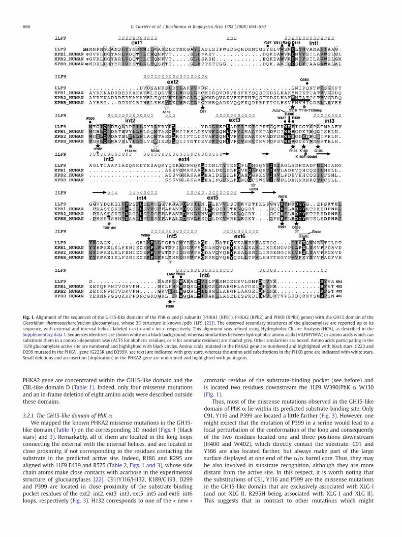
Fig. 1. Alignment of the sequences of the GH15-like domains of the PhK α and β subunits (PHKA1 (KPB1), PHKA2 (KPB2) and PHKB (KPBB) genes) with the GH15 domain of theClostridium thermosaccharolyticum glucoamylase, whose 3D structure is known (pdb 1LF9, [22]). The observed secondary structures of the glucoamylase are reported up to itssequence, with external and internal helices labeled « ext » and « int », respectively. This alignment was refined using Hydrophobic Cluster Analysis (HCA), as described in theSupplementary data 1. Sequences identities are shownwhite on a black background, whereas similarities between hydrophobic amino acids (VILFMYWW) or amino acids which cansubstitute them in a context-dependent way (ACTS for aliphatic residues, or H for aromatic residues) are shaded grey. Other similarities are boxed. Amino acids participating in the1LF9 glucoamylase active site are numbered and highlighted with black circles. Amino acids mutated in the PHKA2 gene are numbered and highlighted with black stars. G223 andD299 mutated in the PHKA1 gene (G223R and D299V, see text) are indicated with grey stars, whereas the amino acid substitutions in the PHKB gene are indicated with white stars.Small deletions and an insertion (duplication) in the PHKA2 gene are underlined and highlighted with pentagons.
666 C. Carrière et al. / Biochimica et Biophysica Acta 1782 (2008) 664–670
PHKA2 gene are concentrated within the GH15-like domain and theCBL-like domain D (Table 1). Indeed, only four missense mutationsand an in-frame deletion of eight amino acids were described outsidethese domains.
3.2.1. The GH15-like domain of PhK αWe mapped the known PHKA2 missense mutations in the GH15-
like domain (Table 1) on the corresponding 3D model (Figs. 1 (blackstars) and 3). Remarkably, all of them are located in the long loopsconnecting the external with the internal helices, and are located inclose proximity, if not corresponding to the residues contacting thesubstrate in the predicted active site. Indeed, R186 and R295 arealigned with 1LF9 E439 and R575 (Table 2, Figs. 1 and 3), whose sidechain atoms make close contacts with acarbose in the experimentalstructure of glucoamylases [22]. C91/Y116/H132, K189/G193, D299and P399 are located in close proximity of the substrate-bindingpocket residues of the ext2–int2, ext3–int3, ext5–int5 and ext6–int6loops, respectively (Fig. 3). H132 corresponds to one of the « new »
aromatic residue of the substrate-binding pocket (see before) andis located two residues downstream the 1LF9 W390/PhK α W130(Fig. 1).
Thus, most of the missense mutations observed in the GH15-likedomain of PhK α lie within its predicted substrate-binding site. OnlyC91, Y116 and P399 are located a little farther (Fig. 3). However, onemight expect that the mutation of P399 in a serine would lead to alocal perturbation of the conformation of the loop and consequentlyof the two residues located one and three positions downstream(H400 and W402), which directly contact the substrate. C91 andY166 are also located farther, but always make part of the largesurface displayed at one end of the α/α barrel core. Thus, they maybe also involved in substrate recognition, although they are moredistant from the active site. In this respect, it is worth noting thatthe substitutions of C91, Y116 and P399 are the missense mutationsin the GH15-like domain that are exclusively associated with XLG-I(and not XLG-II; R295H being associated with XLG-I and XLG-II).This suggests that in contrast to other mutations which might
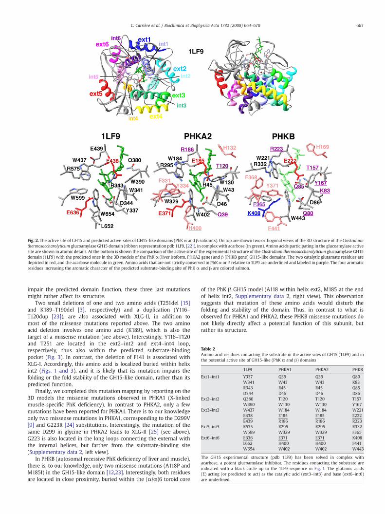
Fig. 2. The active site of GH15 and predicted active-sites of GH15-like domains (PhK α and β subunits). On top are shown two orthogonal views of the 3D structure of the Clostridiumthermosaccharolyticum glucoamylase GH15 domain (ribbon representation pdb 1LF9, [22]), in complexwith acarbose (in green). Amino acids participating in the glucoamylase activesite are shown in atomic details. At the bottom is shown the comparison of the active site of the experimental structure of the Clostridium thermosaccharolyticum glucoamylase GH15domain (1LF9) with the predicted ones in the 3D models of the PhK α (liver isoform, PHKA2 gene) and β (PHKB gene) GH15-like domains. The two catalytic glutamate residues aredepicted in red, and the acarbose molecule in green. Amino acids that are not strictly conserved in PhKα or β relative to 1LF9 are underlined and labeled in purple. The four aromaticresidues increasing the aromatic character of the predicted substrate-binding site of PhK α and β are colored salmon.
Table 2Amino acid residues contacting the substrate in the active sites of GH15 (1LF9) and inthe potential active site of GH15-like (PhK α and β) domains
1LF9 PHKA1 PHKA2 PHKB
Ext1–int1 Y337 Q39 Q39 Q80W341 W43 W43 K83R343 R45 R45 Q85D344 D46 D46 D86
Ext2–int2 Q380 T120 T120 T157W390 W130 W130 Y167
Ext3–int3 W437 W184 W184 W221E438 E185 E185 E222E439 R186 R186 R223
Ext5–int5 R575 R295 R295 R332W599 W329 W329 F365
Ext6–int6 E636 E371 E371 K408L652 H400 H400 F441W654 W402 W402 W443
The GH15 experimental structure (pdb 1LF9) has been solved in complex withacarbose, a potent glucoamylase inhibitor. The residues contacting the substrate areindicated with a black circle up to the 1LF9 sequence in Fig. 1. The glutamic acids(E) acting (or predicted to act) as the catalytic acid (ext3–int3) and base (ext6–int6)are underlined.
667C. Carrière et al. / Biochimica et Biophysica Acta 1782 (2008) 664–670
impair the predicted domain function, these three last mutationsmight rather affect its structure.
Two small deletions of one and two amino acids (T251del [15]and K189–T190del [3], respectively) and a duplication (Y116–T120dup [23]), are also associated with XLG-II, in addition tomost of the missense mutations reported above. The two aminoacid deletion involves one amino acid (K189), which is also thetarget of a missense mutation (see above). Interestingly, Y116–T120and T251 are located in the ext2–int2 and ext4–int4 loop,respectively, thus also within the predicted substrate-bindingpocket (Fig. 3). In contrast, the deletion of F141 is associated withXLG-I. Accordingly, this amino acid is localized buried within helixint2 (Figs. 1 and 3), and it is likely that its mutation impairs thefolding or the fold stability of the GH15-like domain, rather than itspredicted function.
Finally, we completed this mutation mapping by reporting on the3D models the missense mutations observed in PHKA1 (X-linkedmuscle-specific PhK deficiency). In contrast to PHKA2, only a fewmutations have been reported for PHKA1. There is to our knowledgeonly two missense mutations in PHKA1, corresponding to the D299V[9] and G223R [24] substitutions. Interestingly, the mutation of thesame D299 in glycine in PHKA2 leads to XLG-II [25] (see above).G223 is also located in the long loops connecting the external withthe internal helices, but farther from the substrate-binding site(Supplementary data 2, left view).
In PHKB (autosomal recessive PhK deficiency of liver and muscle),there is, to our knowledge, only two missense mutations (A118P andM185I) in the GH15-like domain [12,23]. Interestingly, both residuesare located in close proximity, buried within the (α/α)6 toroid core
of the PhK β GH15 model (A118 within helix ext2, M185 at the endof helix int2, Supplementary data 2, right view). This observationsuggests that mutation of these amino acids would disturb thefolding and stability of the domain. Thus, in contrast to what isobserved for PHKA1 and PHKA2, these PHKB missense mutations donot likely directly affect a potential function of this subunit, butrather its structure.
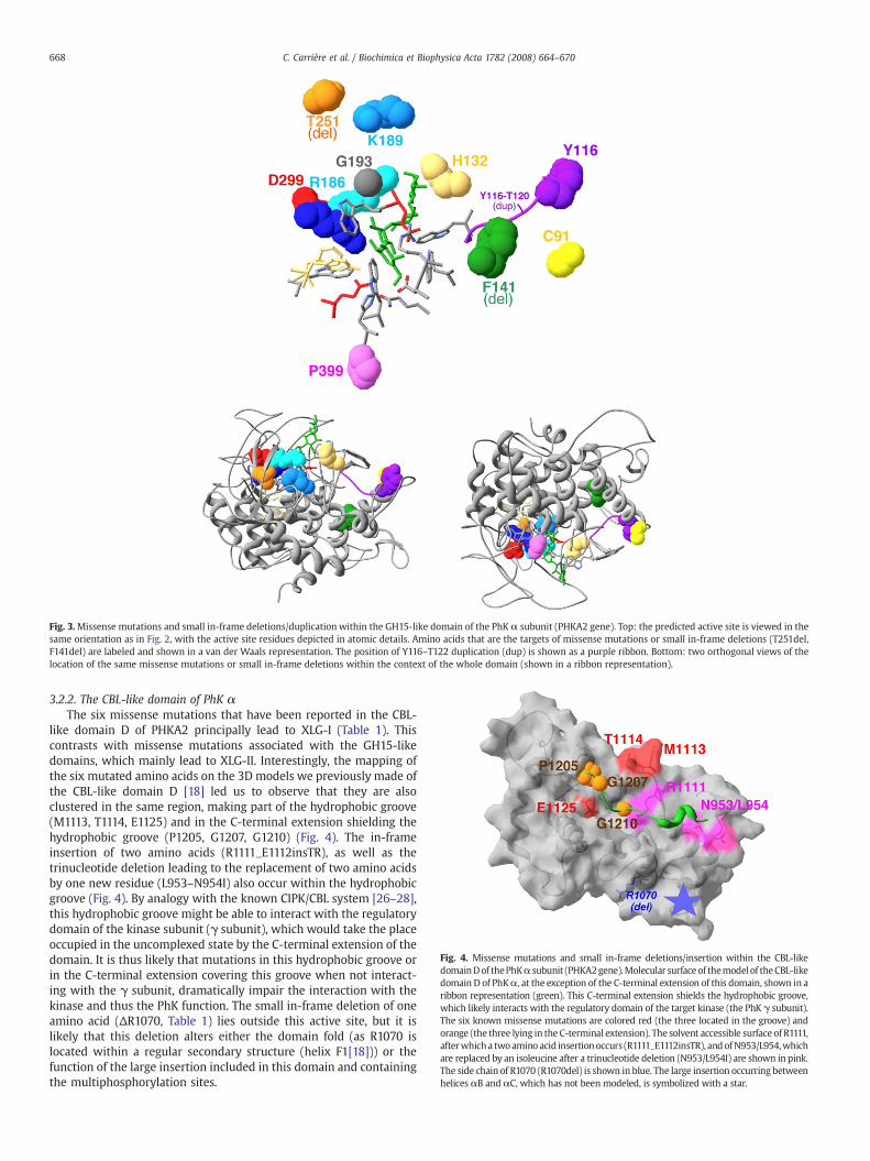
Fig. 3.Missense mutations and small in-frame deletions/duplication within the GH15-like domain of the PhK α subunit (PHKA2 gene). Top: the predicted active site is viewed in thesame orientation as in Fig. 2, with the active site residues depicted in atomic details. Amino acids that are the targets of missense mutations or small in-frame deletions (T251del,F141del) are labeled and shown in a van der Waals representation. The position of Y116–T122 duplication (dup) is shown as a purple ribbon. Bottom: two orthogonal views of thelocation of the same missense mutations or small in-frame deletions within the context of the whole domain (shown in a ribbon representation).
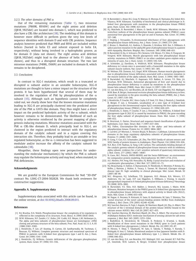
Fig. 4. Missense mutations and small in-frame deletions/insertion within the CBL-likedomainDof thePhKα subunit (PHKA2gene).Molecular surfaceof themodel of theCBL-likedomain D of PhKα, at the exception of the C-terminal extension of this domain, shown in aribbon representation (green). This C-terminal extension shields the hydrophobic groove,which likely interacts with the regulatory domain of the target kinase (the PhK γ subunit).The six known missense mutations are colored red (the three located in the groove) andorange (the three lying in the C-terminal extension). The solvent accessible surface of R1111,afterwhicha twoamino acid insertionoccurs (R1111_E1112insTR), andofN953/L954,whichare replaced by an isoleucine after a trinucleotide deletion (N953/L954I) are shown in pink.The side chain of R1070 (R1070del) is shown in blue. The large insertion occurring betweenhelices αB and αC, which has not been modeled, is symbolized with a star.
668 C. Carrière et al. / Biochimica et Biophysica Acta 1782 (2008) 664–670
3.2.2. The CBL-like domain of PhK αThe six missense mutations that have been reported in the CBL-
like domain D of PHKA2 principally lead to XLG-I (Table 1). Thiscontrasts with missense mutations associated with the GH15-likedomains, which mainly lead to XLG-II. Interestingly, the mapping ofthe six mutated amino acids on the 3D models we previously made ofthe CBL-like domain D [18] led us to observe that they are alsoclustered in the same region, making part of the hydrophobic groove(M1113, T1114, E1125) and in the C-terminal extension shielding thehydrophobic groove (P1205, G1207, G1210) (Fig. 4). The in-frameinsertion of two amino acids (R1111_E1112insTR), as well as thetrinucleotide deletion leading to the replacement of two amino acidsby one new residue (L953–N954I) also occur within the hydrophobicgroove (Fig. 4). By analogy with the known CIPK/CBL system [26–28],this hydrophobic groove might be able to interact with the regulatorydomain of the kinase subunit (γ subunit), which would take the placeoccupied in the uncomplexed state by the C-terminal extension of thedomain. It is thus likely that mutations in this hydrophobic groove orin the C-terminal extension covering this groove when not interact-ing with the γ subunit, dramatically impair the interaction with thekinase and thus the PhK function. The small in-frame deletion of oneamino acid (ΔR1070, Table 1) lies outside this active site, but it islikely that this deletion alters either the domain fold (as R1070 islocated within a regular secondary structure (helix F1[18])) or thefunction of the large insertion included in this domain and containingthe multiphosphorylation sites.
669C. Carrière et al. / Biochimica et Biophysica Acta 1782 (2008) 664–670
3.2.3. The other domains of PhK αOut of the remaining mutations (Table 1), two missense
mutations (P869R, R916W) and the eight amino acid deletion(Q818_Y825del) are located into domain C, which was predicted toalso have a CBL-like architecture [18]. The modeling of this domain ishowever more difficult to perform given the very low levels ofsequence identities with domain D and with CBL proteins. Sequenceanalysis however predicted that P869 and R916 are included withinhelices (buried in helix E3 and solvent exposed in helix E4,respectively), without being involved in a hydrophobic groove, asfor domain D (data not shown). The eight amino acid deletion(Q818_Y825del) might lead to the absence of helix F1 (data notshown), and thus to a disrupted domain structure. The two lastmissense mutations (P498L, I566N) are included in domain B, whichremains to be deciphered.
3.3. Conclusions
In contrast to XLG-I mutations, which result in a truncated ordisrupted α subunit and/or in an unstable holoenzyme, XLG-IImutations are thought to have a minor impact on the structure of theprotein. It has been hypothesized that several of them may beinvolved in the regulation of PhK by phosphorylation of the αsubunit [15]. Although such an hypothesis cannot be completelyruled out, we clearly show here that the known missense mutationsleading to XLG-II are principally clustered into the predicted activesite of the PhK α GH15-like domain. They thus might have a directimpact on the predicted hydrolytic activity of the α subunit, whichhowever remains to be demonstrated. The likelihood of such anactivity is otherwise reinforced by the present mapping of glyco-genosis-inducing mutations. Missense mutations associated with thePhK α CBL-like domain D, which principally lead to XLG-I, areclustered in the region predicted to interact with the regulatorydomain of the catalytic subunit and in a region covering thisinteraction site. Therefore, they probably lead to an unstable or lessregulated holoenzyme, as the α–γ interaction has been suggested tomodulate and/or increase the affinity of the catalytic subunit forcalmodulin [18].
Altogether, these findings open new perspectives for under-standing the molecular mechanism(s) by which the PhK α subunitmay regulate the holoenzyme activity andmay lead, whenmutated, toPhK deficiencies.
Acknowledgments
We are grateful to the European Commission for NoE “3D-EM”
contract No. LSHG-CT-2004-502828. We thank both reviewers forconstructive suggestions.
Appendix A. Supplementary data
Supplementary data associated with this article can be found, inthe online version, at doi:10.1016/j.bbadis.2008.09.011.
References
[1] R.J. Brushia, D.A. Walsh, Phosphorylase kinase: the complexity of its regulation isreflected in the complexity of its structure, Front. Biosci. 4 (1999) D618–D641.
[2] M.W. Kilimann, N.F. Zander, C.C. Kuhn, J.W. Crabb, H.E. Meyer, L.M. Heilmeyer Jr.,The alpha and beta subunits of phosphorylase kinase are homologous: cDNAcloning and primary structure of the beta subunit, Proc. Natl. Acad. Sci. U. S. A. 85(1988) 9381–9385.
[3] J. Hendrickx, P. Lee, J.P. Keating, D. Carton, I.B. Sardharwalla, M. Tuchman, C.Baussan, P.J. Willems, Complete genomic structure and mutational spectrum ofPHKA2 in patients with X-linked liver glycogenosis type I and II, Am. J. Hum.Genet. 64 (1999) 1541–1549.
[4] J. Hendrickx, P.J. Willems, Genetic deficiencies of the glycogen phosphorylasesystem, Hum. Genet. 97 (1996) 551–556.
[5] B. Burwinkel, L. Amat, R.G. Gray, N. Matsuo, K. Muroya, K. Narisawa, R.J. Sokol, M.A.Vilaseca, M.W. Kilimann, Variability of biochemical and clinical phenotype in X-linked liver glycogenosis with mutations in the phosphorylase kinase PHKA2gene, Hum. Genet. 102 (1998) 423–429.
[6] A.J. Maichele, B. Burwinkel, I. Maire, O. Søvik, M.W. Kilimann, Mutations in thetestis/liver isoform of the phosphorylase kinase gamma subunit (PHKG2) causeautosomal liver glycogenosis in the gsd rat and in humans, Nat. Genet. 14 (1996)337–340.
[7] F. Huijing, J. Fernandes, X-chromosomal inheritance of liver glycogenosis withphosphorylase kinase deficiency, Am. J. Hum. Genet. 21 (1969) 275–284.
[8] C. Bruno, G. Manfredi, A.L. Andreu, S. Shanske, S. Krishna, W.K. Ilse, S. DiMauro, Asplice junctionmutation in the alpha(M) gene of phosphorylase kinase in a patientwith myopathy, Biochem. Biophys. Res. Commun. 249 (1998) 648–651.
[9] B. Burwinkel, B. Hu, A. Schroers, P.R. Clemens, S.W.Moses, Y.S. Shin, D. Pongratz, M.Vorgerd, M.W. Kilimann, Muscle glycogenosis with low phosphorylase kinaseactivity: mutations in PHKA1, PHKG1 or six other candidate genes explain only aminority of cases, Eur. J. Hum. Genet. 11 (2003) 516–526.
[10] A. Schneider, J.J. Davidson, A. Wüllrich, M.W. Kilimann, Phosphorylase kinasedeficiency in I-strain mice is associated with a frameshift mutation in the alphasubunit muscle isoform, Nat. Genet. 5 (1993) 381–385.
[11] M. Wehner, P.R. Clemens, A.G. Engel, M.W. Kilimann, Human muscle glycogenosisdue to phosphorylase kinase deficiency associated with a nonsense mutation inthe muscle isoform of the alpha subunit, Hum. Mol. Genet. 3 (1994) 1983–1987.
[12] B. Burwinkel, A.J. Maichele, O. Aagenaes, H.D. Bakker, A. Lerner, Y.S. Shin, J.A.Strachan, M.W. Kilimann, Autosomal glycogenosis of liver and muscle due tophosphorylase kinase deficiency is caused by mutations in the phosphorylasekinase beta subunit (PHKB), Hum. Mol. Genet. 6 (1997) 1109–1115.
[13] I.E. van den Berg, E.A. van Beurden, J.B. de Klerk, O.P. van Diggelen, H.E. Malingré,M.M. Boer, R. Berger, Autosomal recessive phosphorylase kinase deficiency in liver,caused by mutations in the gene encoding the beta subunit (PHKB), Am. J. Hum.Genet. 61 (1997) 539–546.
[14] J. Hendrickx, P. Coucke, M.C. Hors-Cayla, G.P. Smit, Y.S. Shin, J. Deutsch, J. Smeitink,R. Berger, P. Lee, J. Fernandes, Localization of a new type of X-linked liverglycogenosis to the chromosomal region Xp22 containing the liver alpha-subunitof phosphorylase kinase (PHKA2), Genomics 21 (1994) 620–625.
[15] J. Hendrickx, E. Dams, P. Coucke, P. Lee, J. Fernandes, P.J. Willems, X-linked liverglycogenosis type II (XLG II) is caused by mutations in PHKA2, the gene encodingthe liver alpha subunit of phosphorylase kinase. Hum. Mol. Genet. 5 (1996)649–652.
[16] B. Henrissat, G. Davies, Structural and sequence-based classification of glycosidehydrolases, Curr. Opin. Struct. Biol. 7 (1997) 637–644.
[17] M.J. Pallen, Glucoamylase-like domains in the alpha- and beta-subunits ofphosphorylase kinase, Protein Sci. 12 (2003) 1804–1807.
[18] C. Carrière, J.P. Mornon, C. Venien-Bryan, N. Boisset, I. Callebaut, Calcineurin B-likedomains in the large regulatory alpha/beta subunits of phosphorylase kinase,Proteins 71 (2008) 1597–1606.
[19] D. Gong, Y. Guo, K.S. Schumaker, J.K. Zhu, The SOS3 family of calcium sensors andSOS2 family of protein kinases in Arabidopsis, Plant Physiol. 134 (2004) 919–926.
[20] N.A. Rice, O.W. Nadeau, Q. Yang, G.M. Carlson, The calmodulin-binding domain ofthe catalytic gamma subunit of phosphorylase kinase interacts with its inhibitoryalpha subunit: evidence for a Ca2+ sensitive network of quaternary interactions,J. Boil. Chem. 277 (2002) 14681–14687.
[21] N. Guex, M.C. Peitsch, SWISS-MODEL and the Swiss-PdbViewer: an environmentfor comparative protein modeling, Electrophoresis 18 (1997) 2714–2723.
[22] A.E. Aleshin, P.H. Feng, R.B. Honzatko, P.J. Reilly, Crystal structure and evolution ofa prokaryotic glucoamylase, J. Mol. Biol. 327 (2003) 61–73.
[23] N.J. Beauchamp, A. Dalton, U. Ramaswami, H. Niinikoski, K. Mention, P. Kenny, K.L.Kolho, J. Raiman, J. Walter, E. Treacy, S. Tanner, M. Sharrard, Glycogen storagedisease type IX: high variability in clinical phenotype. Mol. Genet. Metab. 92(2007) 88–99.
[24] M.C. Ørngreen, H.J. Schelhaas, T.D. Jeppesen, H.O. Akman, R.A. Wevers, S.T.Andersen, H.J. ter Laak, O.P. van Diggelen, S. DiMauro, J. Vissing, Is muscleglycogenolysis impaired inX-linked phosphorylase b kinase deficiency?Neurology70 (2008) 1876–1882.
[25] B. Burwinkel, Y.S. Shin, H.D. Bakker, J. Deutsch, M.J. Lozano, I. Maire, M.W.Kilimann, Mutation hotspots in the PHKA2 gene in X-linked liver glycogenosis dueto phosphorylase kinase deficiency with atypical activity in blood cells (XLG2),Hum. Mol. Genet. 5 (1996) 653–658.
[26] M. Nagae, A. Nozawa, N. Koizumi, H. Sano, H. Hashimoto, M. Sato, T. Shimizu, Thecrystal structure of the novel calcium-binding protein AtCBL2 from Arabidopsisthaliana, J. Biol. Chem. 278 (2003) 42240–42246.
[27] M.J. Sanchez-Barrena, H. Fujii, I. Angulo, M. Martinez-Ripoll, J.K. Zhu, A. Albert, Thestructure of the C-terminal domain of the protein kinase AtSOS2 bound to thecalcium sensor AtSOS3, Mol. Cell 26 (2007) 427–435.
[28] M.J. Sanchez-Barrena, M. Martinez-Ripoll, J.K. Zhu, A. Albert, The structure of theArabidopsis thaliana SOS3: molecular mechanism of sensing calcium for salt stressresponse, J. Mol. Biol. 345 (2005) 1253–1264.
[29] J. Rudolfová, R. Slovácková, M. Trbusek, K. Pesková, S. St'astná, L. Kozák,Identification of three novel mutations in the PHKA2 gene in Czech patientswith X-linked liver glycogenosis. J. Inherit. Metab. Dis. 24 (2001) 85–87.
[30] H. Hirono, Y. Shoji, T. Takahashi, W. Sato, E. Takeda, T. Nishijo, Y. Kuroda, T.Nishigaki, K. Inui, G. Takada, Mutational analyses in four Japanese families with X-linked liver phosphorylase kinase deficiency type 1. J. Inherit. Metab. Dis. 21(1998) 846–852.
[31] I.E. van den Berg, E.A. van Beurden, H.E. Malingré, H.K. van Amstel, B.T. Poll-The,J.A. Smeitink, W.H. Lamers, R. Berger, X-linked liver phosphorylase kinase

670 C. Carrière et al. / Biochimica et Biophysica Acta 1782 (2008) 664–670
deficiency is associated with mutations in the human liver phosphorylase kinasealpha subunit. Am. J. Hum. Genet. 56 (1995) 381–387.
[32] H. Hirono, K. Hayasaka, W. Sato, T. Takahashi, G. Takada, Isolation of cDNAencoding the human liver phosphorylase kinase alpha subunit (PHKA2) andidentification of a missense mutation of the PHKA2 gene in a family with liverphosphorylase kinase deficiency, Biochem. Mol. Biol. Int. 36 (1995) 505–511.
[33] K. Ban, K. Sugiyama, K. Goto, F. Mizutani, H. Togari, Detection of PHKA2 genemutation in four Japanese patients with hepatic phosphorylase kinase deficiency,Tohoku J. Exp. Med. 200 (2003) 47–53.
[34] F. Hidaka, H. Sawada, M. Matsuyama, H. Nunoi, A novel mutation of the PHKA2gene in a patient with X-linked liver glycogenosis type 1, Pediatr. Int. 47 (2005)687–690.


![Join domain dan sub domain [repaired]](https://img.pdfslide.net/doc/110x75/55ba68b5bb61eb41568b46a0/join-domain-dan-sub-domain-repaired.jpg)