Embed Size (px)
Citation preview
Biocompatibility of Materials
January 23, 2007Silicone Breast Implants
Estimated 1-2 Million women in the U.S. have had breast implants (1963-1998).90% Silicone Gel (about 10% are coated with polyurethane foam.)10% saline solution – 9% NaCl (still available without restriction).Reasons for implantation
Cosmetic enlargement or reshaping Reconstruction following mastectomy
Sheath can be made of poly dimethyl siloxane silicone (MW ~100,000). inside is low molecular weight silicone that provides the right feel (mw very low). Smooth surface and “hard” surfaceWater does not have proper feel (uses same envelope).
1951 Dr. PagmanPolyvinyl sponge (see figure)Fibroblasts grew into the sponge and became painful
1962 Dr. Cronin/GerowSilicone (see figure)
Jan 6, 1992Silicone was banned by FDA.2006 – FDA reinstated sale of silicone devices.
1976 Medical Device Safety ActFDA was given legal responsibility to regular surgical devices/implants
Grandfather clause enacted for then-current devices.1992 medical act was amended to add more power to the regulation.
USA did not require a law where a national implant database is maintained. 1992 this was changed for class 3 implants (most severe). Silicone implants are class3. class3 are devices that are actually penetrating the body.
1950s Japanese women injected low MW silicone gel injected into their breasts. Thousands of women developed necrotic breasts.Smooth surface implants allowed silicone fluid to diffuse out of the bag.
Freely injected fluidPossible carcinomasCancer Has been reported after silicone injectionDeaths have occurred – severe toxicity
Silicone Breast implant complaints:Reports of adverse effects (can show after a year or two)
Implant became hard and painfulo Fibrous encapsulation – eventually capsule contractions/hardens causing paino Capsular contracture – massaging was suggested to reorient the developing
fibroblastso Interference with mammogramso Silicone bleedo Capsule ruptureo Formation of calcium depositso Calcificationo Implant shifting positiono Polyurethane degradation (rough surface implants)
“All implants bleed Silicone gel through their outer envelope” FDA 1992
Countermeasures to prevent capsule formation Drains for hematoma and fluid accumulation Antibiotics Capsulotomy (exercise) Retromuscular positioning Steroids Polyurethane coating
Second lumen to prevent low MW silicone from passing throughUses fluorine – high electronegativity, large atomic radius => steric hindrance
Silicones:1940s F.S. Kipping, University College, England, WASPreparing small molecules for optical rotation studies, but was troubled by oils and “gunk” in his reaction flask (could polarize light)
1943 commercial production, commercial names silicone, silastic.
Of patients that require resurgery (independent experiments)Exp 1 57% fibrous encapsulation
Macrophages attack surface of material – o2 radicals, peroxides, etc. signaling cascade results in fibroblast action after inability to digest invader.
Exp 2 40% due to gel bleedingExp 3 15% calcification – polyurethane (rough surface) seemed to precipitate the formation of calcium phosphate.
Poly urethane problems (cleaves at ester group)Degradation – hydrolysis
By products eg 2,4 TDA, polymer fragmentsInflammatory reaction
24 toluene Diamine (TDA) was formed during enzymatic attack of polyurethane foam for first 4 days 9and goes away) –paper doneCounterpaper (1994) Luu and White
Found hydrolysis of polyester urethane foam in phosphate butter, ph 7.4Rate of TDA degradatin did not reach steady state equilibrium until after 120 days+
June 1988 breast implants designated as class 3 medical devicesApril 1991 fda requires safety and effectiveness dataJan 1993 voluntary moratorium on sale of silicone implants1996 sale prohibited (?)As a result – companies stopped providing materials, biomaterials field shrank, until law was passed to forbid filing of suits to material producing companies. Biomaterials Access Assurance Act of 1997
Studies have found that there is no correlation between silicone implants and rates of cancer. A number of surveys from reputable institutions have supported this
FDA draft guidance document (for testing)Chemistry (xlinking, heavy metals, saline filler, etc)Mechanical (fatigue, rupture, etc)
January 29, 2007Total Hip implant
Patients develop problems because they have been bedridden
Inveted by dr. John Charnley (british)
Teflon was recommendedPolytetrafluoroethylene = low coefficient of frictionFirst implants (hundreds) put in had to be taken off.PTFE creeps under load – deforms. Uneven shape increases wear, flaking off pieces which cause pain.
Material choice in US for last 35 years UHMWPE
Stainless steel femoral stem
InstallationHammer stem into bone – groves in the shaft lock into place on the bone, goes into marrowAdhesive – grouting material PMMA (Polymethylmethacrylaat) (plexiglass/Lucite)
Polymerization is exothermic reaction temp may rise to about 45-50cCartilage disintegrates at around 42cBone necroses at 50cStill being used today
Making the acetabular cupPowder – put into mold and compress with higher temp.Under an electron microscope – you see grain boundaries.
Temperature and pressure were not high enough to create large/single crystalsPowder – extruding machine. Extrude a tube, and then machine out the cup
Modularity – different sized heads and stems depending on nature of patient.
About one million steps per year for average person – one million articulations of the implant per year leading to abrasion.
Barium sulfate ring is placed in the acetabular cup – shows up on xrays and allows for examination of cup position
Securing the femoral stem (continued)More current procedure – deposit a microscopic porous coating on the surface of the shaft. Osteoblasts have a preference for pores of a diameter between 100-300 micronsb. Causes mechanical stabilization of the stem with natural bone.Ti 6Al 4V commonly used in orthopedic implantsPlasma spray procedure is used to apply coating to the stem.5 parameters can be used to control pore size.
Problems:Flakes of the PE cup can cause serious problems – can migrate into lymph node etc.Corrosion of the metal itself:
Stainless steel can corrodeTi 6al 4V is corrosion resistant. (see diagram) – W/exposure to air, TiO2 is formed – ceramic material. Used in shaftCorrosion w/SS Ni -> Ni++ oxidation. Releases atomic constituents that make up the product. The metal ions interact with surrounding tissue forming metallic-organic compounds.Nickel sensitivity: nickel is the most prevalent cause of sensitivity in patients. Many patients have pre-existing nickel sensitivity.
Fatigue fracture of the implant.Stress is placed on the stem w/each step.
Elastic Modulus of materialsBone: cortex 2-3 e6 psiStainless steel 25 e6Titanium alloys 12e6Bioglass 4585 5e6PE (high density) 85-160 e3
Mismatch of elastic moduliImplant is much stiffer tan the bone itself
When bone is overstressed and understressed, bone is resorbed (osteoclasts). However at proper stress levels bone reproduces.
Replacing polymer with ceramicAluminum oxide Al2O3Less deformation/flaking, but more brittle. No corrosion, less wearEuropean implants (head/cup, stem) tend to be ceramic either ZrO2 or Al2O3United states implants – 90% are metal stem, UHMWPE head. Ceramics are FDA approved.Concern about fracture and failure due to brittleness.
In USA (DR. Robert P Heaney)300,000 broken hips annually (acetabular cup and the femoral bone.)
80,000 are men1/3 die within one year. (w/o implant?)
Keeping motion of the acetabular cup to a minimumSquare shaped cupScrew threads
Hip Joint systems. Which system gives least wear1. M on M
a. Used to be popular, sometimes encounter galling – metals can flow and eventually lock together. Improved metallurgy may have overcome this problem
2. M on polymer3. C on polymer4. C on C5. C on M
Ceramics are good in compression.
Polymers1. PTFE2. UHMWPE MW=2-5 e6 daltons, n=100,000-200,0003. PMMA
Metal1. SS2. 90%Ti 6%Al 4%V – has been some concern about Vanadium in terms of its toxicity. No
confirmed studies about Ti6Al4V, but Nb has been substituted3. Cobalt-chromium-Mo chromium forms chromium oxide which provides corrosion
resistance
Ceramics1. Al2O32. ZrO2
.Find particles of UHMWPE, PMMA, Metals, bone at the implant site.
Wear: To impair, consume or diminish by use and friction. The potential for wear of medical devices exist when there is relative motion between two solids in contact under load.
Two types of wear:Adhesive wear: The mating surfaces of the two solids flow plastically under friction. Load forming junctions which break under tangential forces giving rise to wear debris.
Contact occurs only at a few asperities. DiagramAbrasive wear when a hard body slides over a soft surface (Co-Cr or SS (316L) over PE) A series of grooves or defects are ploughed in the softer material. This is two-body wear.Particles of wear debris or foreign particles which are trapped between the contacting, sliding surfaces and cause abrasive wear of the surfaces by ploughing action. Also called three body wear. What happens to the material that comes out of the grove?
Femoral surface also undergoes wear, even though it is much harder than the cup.
January 30, 2007Metal backing on the acetabular cup – prevents distortion and encourages ingrowth (plasma spray)
Fatigue wear: abrasion caused by cyclic loading and lost of material by spalling of surface layers. (spalling = peeling process)
Fretting wear: occurs as a result of frictional oscillatory movement of contacting solids
Abrasive index= Rx/RsRx = test specimenRs = standard material(Measure the number of cycles required to abrade .1 inch of material away.)
What kind of abrasion test should we use?1. pin-ondisc2. ring-ondisc3. journal-and-bush (rod in bushing)4. block on journal (block on rod)
measuring:1. weight loss2. volume loss3. delta thickness/length4. holographic surface measurements5. SEM6. light microscopy7. profilometer (can provide 3dview, valleys and peaks show wear)
TI 6Al 4V is not used in heads on femoral stems because of poor wear characteristics – see oxide needle model.
Particles that flake off are 50-200 um. Macrophages cannot ingest – form giant cells~10um, macrophages can engulf and digest.
1um = 1 micron = 10e-6 meters = 3.9e-5 inches
10g of tissue containing F75 alloy wear debris could contain 2.9e11 particles w/combined surface area of 1600cm^3.
Dr. Ian ClarkeClarke hypothesis:
1. regardless ofa. implant designb. material selectionc. fixation moderelative motion at articulating surfaces or micromotion at any interface may result in the release of microsized particles which provoke significant peri-implant bone loss
2. activated macrophages are primary agents in removing debris
where is wear taking place/-the bone/acrylic cement interface bond may be broken by micromotion leading to production of on a fibrous membrane
further micromotion will produce bone and acrylic debris resulting in macrophage activity, inflammation and bone resorption (leading to three body wear)
when PMMA bone junction is broken due to micromotion – can form fibrous layer and prevents mechanical bonding from taking place
90-95% hip implants in US use UHMWPE
Parameters to consider for UHMWPE Oxidation index – important in terms of wear and gamma radiation (used in sterilization).
Implant can crumble. Molecular weight affects TS, hardness, Elastic mod (2-6million typicall for these
products. Too high, gets brittle/stiffer) Molecular weight distribution – can purify by dissolving the polymer, heating to 135c.
run gel chromatography, or add alcohol and precipitate batches of material at different MW.
Microstructure – amorphous or crystalline? Desire crystalline. Amorphous glasslike does not have as desired properties
Is this sample branched or linear? Can affect packing desnsity. LDPE/HDPE Radical formation during storage
o Ethylene oxide sterilization – UHMWPE absorbed ethylene oxide and could diffuse out into the patient
o Irradiate in nitrogen atmosphere – inert and don’t generate free radicals.
Another case of wear debris: implant loosening Micromotion between femoral stem and the acrylic bone cement produces polymer and
metal particles Transport of these particles can result in accelerated wear of the UHMWPE acetabulum
Osteogenic regulatory molecules Bone morphogenetic proteins foster new bone regeneration (BMP-2) Sustained release carrier systems
o Biodegradable collagen scaffolds
rhBMP-2 induces Cell migration, proliferation Differentiation of mesenchymal cells New bone formation Increased vascularity
Delivery systems for bone morphogenetic proteins Absorbable collagen system (ACS) – BMP-2 Biodegradable polymer PLA-DX-PEG block copolymer containing BMP-2
Osteoconductive Provides a passive structure into which blood vessels may enter and new bone may form Graft osteoconduction: the facilitation of blood vessel incursion and new bone formation
into defined lattice structure
Osteoinductive Contains factors which induce the differentiation of mesenchymal cells into osteoblasts Graft osteoinduction: new bone produced the active recruitment of host mesenchymal
stem cells from the surrounding tissue, which differentiate into bone forming osteoblasts this process is facilitated by the presence of growth factors within the graft, principally BMPs
Recombinant human bone morphogenetic protein-2 RhBMP-2Can be produced in cell culture or body
Failure modes:Cyclic fatigueFractureSurgeon contribution to failure
Poor alignment, selection , etc
The most common cause of failure is aseptic loosening (loosening of stem itself)Characteristics
Fibrous membrane
Wear debris: PMMA, UHMWPE, metal; key factors in implant loosening Macrophage activity Inflammation Bone loss
February 5, 2007Dental implantsControversy with Calcium Hydroxyapatite (hydroxylapatite) HA
Testing:In-VitroIn-VivoClinical
Ti6Al4V 4 types of screws:Smooth surfaceRoughened surfacePlasma sprayed Ti6Al4V forms porous surfaceCaHA surface
Too much hydrogen in Ti tends to embrittle it.
Bone, Dentin, Enamel – all have about 36% Ca, ~17% P. Ca/P = ~1.6
CaHA C = 40%, P = 18.5% Ca/P (ratio) = 1.67Similar to natural material, hopefully does not elicit response from body.
Plasma spray parameters:Plasma currentGas flow rateGasesPowder flow rateGun distanceAngle of incidence.Control pore size and density
Want to allow osteoblasts to get into the material.
Coating thicknessPorous Ti6Al4V substrate 60-125 micronsHA 35-50Total 95-175 microns
What is the crystallinity of the HAUsed x-ray diffraction pattern to find the amount of crystallinity vs amorphous material.
Coating tensile strength was found and determined sufficient
In-vivo testing (dog)
Window cutout in the implant to allow for observation of cell growth
Osteoblasts are sensitive to the microstructure of implant – bare Titanium shows less bone growth (osseointegration) when compared to HA that is more crystalline in features. (65% crystallinity)
Clinical
Base/stem is installed into patient first, (3-months pre cap installation) to allow for the bone integration.
Cap the screw thread hole such that no tissue migrates into the hole, remove when abutment is installed.
Hydroxyapatite (HA) CoatingsWhat is the controversy?
Want to mechanically stabilize the device ASAP
Calcium PhospatesCeramics that includes a whole rage of amorphous anc rstalline materialsHA is in this group AKA bone mineralWhether HA is bioactive and reasons why some CaP are bioactive are not agreed upon. Do they cause more harm or good?
Question of solubilityHA is prone to disappear into solutionTriCalciumPhosphate is highly soluble. TCP Ca3(PO4)2Response to HA “too soluble” -> fluoroHA
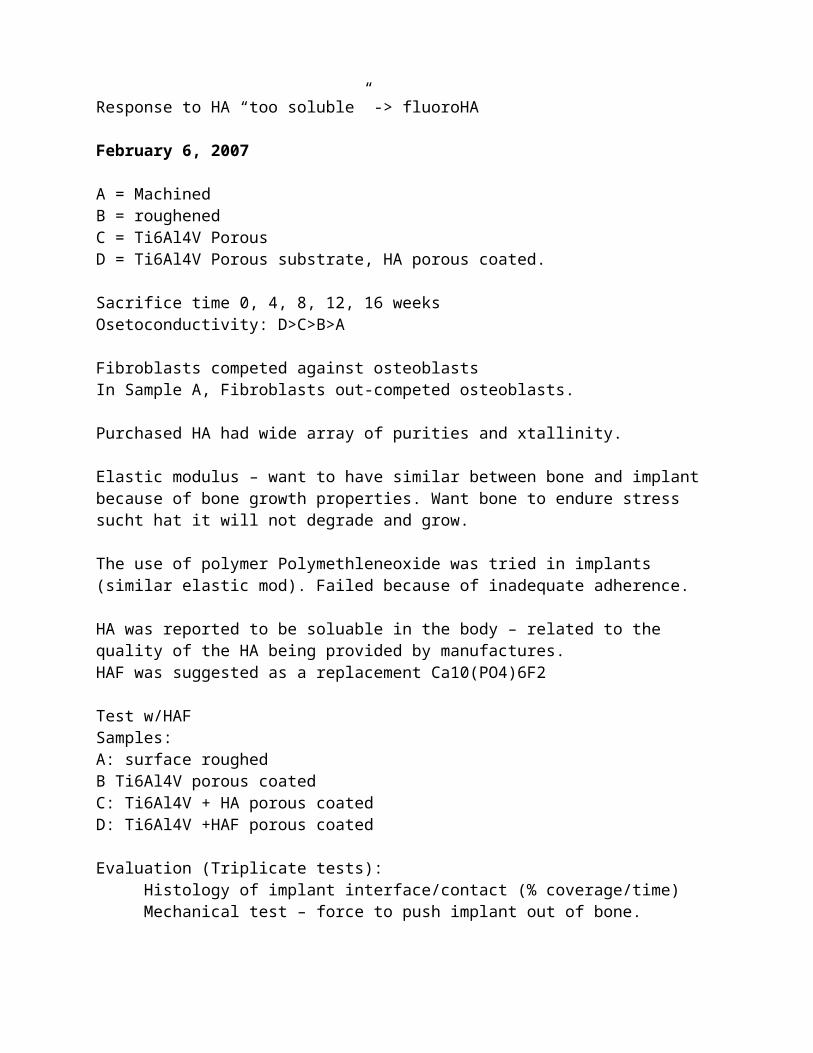
February 6, 2007
A = MachinedB = roughenedC = Ti6Al4V PorousD = Ti6Al4V Porous substrate, HA porous coated.
Sacrifice time 0, 4, 8, 12, 16 weeksOsetoconductivity: D>C>B>A
Fibroblasts competed against osteoblastsIn Sample A, Fibroblasts out-competed osteoblasts.
Purchased HA had wide array of purities and xtallinity.
Elastic modulus – want to have similar between bone and implant because of bone growth properties. Want bone to endure stress sucht hat it will not degrade and grow.
The use of polymer Polymethleneoxide was tried in implants (similar elastic mod). Failed because of inadequate adherence.
HA was reported to be soluable in the body – related to the quality of the HA being provided by manufactures.HAF was suggested as a replacement Ca10(PO4)6F2
Test w/HAFSamples:A: surface roughedB Ti6Al4V porous coatedC: Ti6Al4V + HA porous coatedD: Ti6Al4V +HAF porous coated
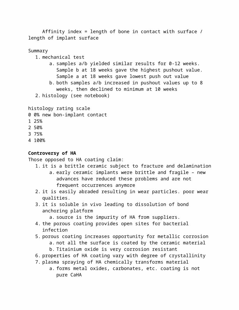
Evaluation (Triplicate tests):Histology of implant interface/contact (% coverage/time)Mechanical test – force to push implant out of bone. Affinity index = length of bone in contact with surface / length of implant surface
Summary1. mechanical test
a. samples a/b yielded similar results for 0-12 weeks. Sample b at 18 weeks gave the highest pushout value. Sample a at 18 weeks gave lowest push out value
b. both samples a/b increased in pushout values up to 8 weeks, then declined to minimum at 10 weeks
2. histology (see notebook)
histology rating scale0 0% new bon-implant contact1 25%2 50%3 75%4 100%
Controversy of HAThose opposed to HA coating claim:
1. it is a brittle ceramic subject to fracture and delaminationa. early ceramic implants were brittle and fragile – new advances have reduced these
problems and are not frequent occurrences anymore2. it is easily abraded resulting in wear particles. poor wear qualities.
3. it is soluble in vivo leading to dissolution of bond anchoring platforma. source is the impurity of HA from suppliers.
4. the porous coating provides open sites for bacterial infection5. porous coating increases opportunity for metallic corrosion
a. not all the surface is coated by the ceramic materialb. Titainium oxide is very corrosion resistant
6. properties of HA coating vary with degree of crystallinity7. plasma spraying of HA chemically transforms material
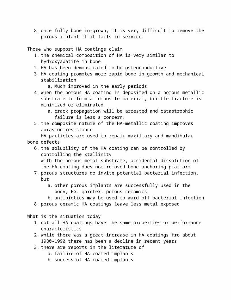
a. forms metal oxides, carbonates, etc. coating is not pure CaHA8. once fully bone in-grown, it is very difficult to remove the porous implant if it fails in
service
Those who support HA coatings claim1. the chemical composition of HA is very similar to hydroxyapatite in bone2. HA has been demonstrated to be osteoconductive3. HA coating promotes more rapid bone in-growth and mechanical stabilization
a. Much improved in the early periods4. when the porous HA coating is deposited on a porous metallic substrate to form a
composite material, brittle fracture is minimized or eliminateda. crack propagation will be arrested and catastrophic failure is less a concern.
5. the composite nature of the HA-metallic coating improves abrasion resistanceHA particles are used to repair maxillary and mandibular bone defects
6. the solubility of the HA coating can be controlled by controlling the xtallinitywith the porous metal substrate, accidental dissolution of the HA coating does not removed bone anchoring platform
7. porous structures do invite potential bacterial infection, buta. other porous implants are successfully used in the body, EG. goretex, porous
ceramicsb. antibiotics may be used to ward off bacterial infection
8. porous ceramic HA coatings leave less metal exposed
What is the situation today1. not all HA coatings have the same properties or performance characteristics2. while there was a great increase in HA coatings fro about 1980-1990 there has been a
decline in recent years3. there are reports in the literature of
a. failure of HA coated implantsb. success of HA coated implants
4. more long term, well designed, statistically controlled implant experiments are needed.
February 12, 2007Cardiovascular system
Ball and cage:1952: First heart valve was ball and cage. Dr. Hufnagel1960: Drs. Harken and Starr placed a heart valve into a patient.
Both had problems with thrombosis and blood coagulation
1969: Occluder disc valve – Bjork-Shiley1970s Bileaflet – introduced by St. Jude
glutaldehyde xlinks in the leaflets and makes it more durable
Mechanical route -> requires blood thinner for duration of implant.Chemicals:1) wafarin 2) coumadin
Porcine for older peopleMechanical for younger
Mitral -> LA to LVAortic -> LV to aortaTricuspid -> RA to RVPulmonary -> RV to pulmonary
Valve troubles are usually left side, mitral valve.
Valve replacements typicall last ~20 years now.
Valve functions:
3. to deliver the blood with minimal adverse effect
FailuresBall and cage: ring needs to be welded. Needs to be sewn in place.Ball swells – lipids are absorbed by the silicone materialUsed a metal instead. Stellite 21 Co-Cr-Mo (made noise)Textile was sewn around the ball to act as a cushion. Textile abrades away, causing clotting
Pyrolytic carbon was a good choice for material – reacted well in blood. Does not clot blood. Good against thrombosis
Hydrodynamics of blood flow is important. Don’t want turbulence or stagnation. Want uniform blood flow to minimize clotting risk.
Occluder valve -> minor orifice and major orifice. Blood flow is impeded by metal wires, also stagnation in the minor portion.Bileaflet – still major/minor, but no flow obstructions.
Problems resulting from adsorption on synthetic implants clotting mechanical impairment removal of components (important proteins, etc)
Blood clots occur on the sewing ring.Surgeon can affect performance with his sewing technique -> thread can collect protein.Fatigue fracture on the occluder strut. 40m cycles / yearFracture on the pyrolytic carbon disc
Porcine heart valve uses textile sewing ring
Large problem with porcine heart valve -> mineralization, calcificationSequence of events for intrinsic mineralization
1. leaflet tissue is infiltrated by host plasma proteins2. infiltration is a normal event and my provide internal lubricity3. in rare instances host proteins concentrate to the point at which fibrin forms and
mineralizes. Tissue surface is still intact4. Extension of mineralized areas disrupts leaflet
Mineralization control measures for synthetic filmes Avoid surface imperfections Avoid inclusions Avoid juvenile subjects – more calcification systems Design devices with maximum strain relief Use mineralization resistant materials Use anti-mineralization chemical treatments.
Crosslinking proteins in the porcine leaflet could possibly be related to calcification
Failure of cardiac valvesDesign, engineering: Wear, fracture, poppet escape, cuspal tear, calcification, hemolytic anemia, noiseUser: sutures, implantation
Amount of protein absorbedExtension of proteins on surface (height of polymer/protein above the surface)
Difference in surface energy is attracting other molecules
Imporatance of protein adsorption to synthetic implantsProtein depletion
Adsorption or adherence of physiological components to biomaterials surfaces is a key part of biocompatibility.
Protein adsorptionAlbumin -> no platelet adhesion
NonthrombogenicityFibrinogen, gamma globulin, prothrombin -> platelet adhesion, can lead to thrombosis
Artificial HeartsBarney Clark – first artificial heart. Dr. Robert Jarvik invented the heart.1982.
Biomaterials in the total artificial heart.Polyurethanes – blood chamber housing, flexing diaphragms, etcPoly carbonate: valve holders, air chamber basePVC – drive lines (lots of plasticizer, diffuses out)Polypropylene – suture materialWoven Dacron – outflow graftsDarcon Velcro – ventricular anchorDarcon velour – skin buttons,Silastic – skin buttonWoven silk – aspiration port sealing tie
Thrombus formation – patient surface geometryPatient thrombus potential, anticoagulation drugsSurface – surface quality, chemistryGeometry optimum flow conditions
TAH or VAD?
Christian Bernard 1964 first MD to do a human heart transplant
February 13, 2007Heart valvesSyntehtic materials
Polymers, metals/alloys Ceramics
Modified Natural materials (tissue valves) Bovine pericardium valve Porcine valve
Metals:Stellite 21Co-Cr-MoTi6Al4VStainless steel 316L
Polymers: (see figures)PTFRDarconDelrin
Ceramics:
Al2O3ZrO2Pyrolytic C
Low temperature isotropic ~1000c LTIUltra low temperature isotropic ~25c ULTI
General typesCaged-ballCaged Disk (similar to ball)Tilting-disk – cage is made of stelliteTissueBi-leaflet tilting-disk
First models: Medtronic (caged), St. Jude (bileaflet), Bjork-Shiley (mitral)
Low profile vs high profileHow far does it reach down into the heart chamber. Want low profile. Depends on shape of material, etcHigh profile -> silastic poppett
ProblemsBlood clotting
Occluder opening influences hydrodynamics Low or stagnant blood flow leads to thrombosis Bjork-shiley 60degree C/C heart valve improved hydrodynamics
Kay-Shhiley heart valveCaged disk.Problems: disk gets dislodged, large amount of turbulence, wearEntire occluder can wear on edges, and lock the valve in one position (partially open, etc)Delrin was not a satisfactory choice for the occluder
Tissue heart valvesPorcineBovine pericardial
Advantages Freedom from anticoagulant drug (warfarin/coumadin (rat poison)) Central orfice flow (hemodynamics) Cross-linking stabilization
Disadvantages steady degradation with time calcification (calcific degeneration) inflammatory cell infiltration fragmentation of collagen fibers leaflet perforation
Types of calcificationExtrinsic
thrombosis related calcification on surface of leaflets of valve hydroxyapatite deposit
intrinsic in interior of leaflet along collagen fibrils
See reaction (formation of chelated compound)
Effect on patient w/cardiovascular implant:1. protein deposits (within seconds)2. platelet activation (fibrinogen)3. blood clots4. embolism5. hemolysis (lysis of blood cells)
Affect on implantWearFatigue/fractureBlood clottingValve malfunctionHindered hydrodynamics
Dr. Fred Schoen studyIn 1980s:5-year survival 70-80%10 yr 55-70%Improved in the 1990s
Jarvik heart patients1. barney clark – 1982 oversized heart removed, lived 112 days2. William Schroeder lived 620 days, strokes, kidney failure, troubled history
Strokes, convulsions, fever (infection)
Biomed, Inc. Danvers, MA 20 years of research -> Abiocor heartAbiocor Artifical heart (2001)
Totally implantable Permanent Rechargeable internal battery Powered transcutaneously by external power pack Weight: 2pounds Size: small grapefruit Materials: Titanium pump housing and propeller Polymer ventricular chamber Constant flowrate
Picking materials
Non-thrombogenic – negative charge on endothelial lining. Repels platelets and blood cellsPTFE has negative chargePyrolytic carbon
Blood clotting: Albumin likes hydrophilic surfaces (wetting angle ->small as water spreads across surface)
Want hydrophilic –OH groups, something like PVA
February 19, 2007ThrombogenicityThrombus: blood clots, network of fibrin, platelets, erythrocytes, leucocytesThrombosis: formation of a blood clot
Thrombus formation on biomaterials1. Adsorption of plasma proteins
a. Fibrinnogen and thrombin2. adhesion of platelets3. aggregation of platelets4. fibrin formation5. mural thrombosis
endothelial cells typically have negative charge and repel blood cells. When injured, they may have a positive charge and attract platelets, etc to themselves.
Platelets are normally disc shaped. Under activation, they transform to pseudopods (long tubes..?)
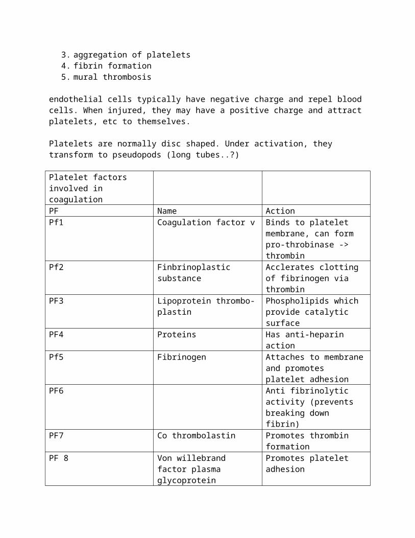
Platelet factors involved in coagulationPF Name ActionPf1 Coagulation factor v Binds to platelet membrane,
can form pro-throbinase -> thrombin
Pf2 Finbrinoplastic substance Acclerates clotting of fibrinogen via thrombin
PF3 Lipoprotein thrombo-plastin Phospholipids which provide catalytic surface
PF4 Proteins Has anti-heparin actionPf5 Fibrinogen Attaches to membrane and
promotes platelet adhesionPF6 Anti fibrinolytic activity
(prevents breaking down fibrin)
PF7 Co thrombolastin Promotes thrombin formationPF 8 Von willebrand factor plasma
glycoproteinPromotes platelet adhesion
Platelets contain glycoproteins, GPIIb/IIIa on cell membrane platelet receptors platelets must be activated for GPIIb/IIIa to bind to fibrinogen cross-linking of GPIIb/IIIa leads to platelet activation platelets do not possess a unique adhesive receptor (ie RGD) for albumin Platelets generate a chemical factor leading to formation of thrombin, then fibrin Activated platelets produce alpha-granule release (pro-coagulant.)
Preparing blood biocompatible surface Make it negatively charge Coat with albumin
Proteins compete for the surface of an implantWhich protein gets to the surface first makes a difference to the bio-compatibility outcomeAlbumin vs fibrogen albumin adsorption decreases platelet adsorption and decreases thrombogenicity(no receptor on platelet for albumin adhesion)
Albumin does not contain the RGP peptide sequence
AlbuminAdsoption decreases platelet adhesionDecrease thrombogencity blocks ibrinogen adsoption which attracts plateletsAbumin does not contain RGD (arginin-glycine-aspartic acid)sequence, an amono acid sequence common to adhesive proteinsAlbumin must occuy most of the surface (98%) to be anti thrombogenic
FibrinogenAdsorption (deposition)
Undergoes conformational changeA condition leading to platelet adsorptionFibrinogen adsorption increases thrombogenicity by generating fibrin
FibrinogenProvides signal to promote platelet adhesion
Thrombin links individual (3) chanins in a bibrinogen single molecule
sizeAlbumin 69000 MWHemoglobin 64450 MWGlobulin 165,000MWFibrinogen 400000 MW
Blood clotting factorsI. Fibrinogen
Provides signal to platelets for activation
Reacts with Prothrombin II -> fibrinII. Prothrombin
Reacts with proaccelerin (V + Ca2+) -> Thrombin IIaIII. Tissue factor
Key to extrinsic pathwayReactins with proconvertin (VII + Ca2+) to form complex
IV. Ca2+Acts as catalyst (400x quicker w/Ca2+)Only non-protein clotting factor Activates Stuart factor (X-Xa)
V. ProcaccelerinReacts with prothrombin II -> thrombin IIa
VI. Activated VReacts with prothrombin II -> Thrombin IIa
VII. ProconvertinReacts with Tissue Factor III + Ca+2 to form complex
VIII. Antihemophilic factorActivates stuart factor X Ca++ -> Xa
XI. Christmas Factoractivated IXa involved in activation of stuart factor X -> Xa
X. Stuart factorInvolved in conversion of prothrombin II -> Thrombrin IIa
XI. Plasma Prothrombo – plastin antecedentw/Ca++ activates Christmas factor IX -> IXa
XII. Hageman factorActivated XIIa is the key to the intrinsic pathway
XIII Fibrin stabilizing facorReacts with thrombin IIa for XIIIa Plays a role in Fibrin formation
Factors leading to the intrinsic pathway1. contact between blood elements and a surface2. damage to the wall of a blood vessel (endothelium)
a. activation of Hageman factor (XII) byi. exposure of a nonendothelial surface such as collagen (electro0negative
and thromboticii. platelet membrane-electronegative
iii. contact with a foreign substance (implant)
Will a Cl- ion get to the surface first?
Factors leading to the extrinsic pathway1. release of tissue thromboplastin (tissue factor III) from cells external to the vascular
processes. It is released when the tissue is damaged
a. in conjuction with proconvertin (factor VII), and Ca2+ it activates the Stuart factor (X).
February 20, 2007
Blood Flow rateLow Shear thrombosis (venous wounds)
Red thrombosis (erythrocytes + fibrin)High shear thrombosis (arterial wounds)
Shear stress > 3000 Dynes/cm^2White thrombosis (Platelets + Fibrin)
Vascular surface lined with endothelial cells subendothelium layer containing elastin (cross-linked polypeptides)
(factor III usually is related to events outside the vascular system)
Blood Compatiblity: Clinical manifestations Small diameter vascular grafts fail early due to thrombotic occlusion Synthetic venous prostheses do not exist Embolic complications are noted with artificial hearts Embolic problems are frequently observed with catheters Non-tissue heart valves require lifelong anticoagulation Sensors “foul” due to thrombus formation Long term implants are seen to be continuously platelet consumptive Significant blood damage is observed during hemodialysis and extracorporeal
oxygenation (also in heart valves)
Blood coagulation and electrostatic repulsion RBCs and platelets have a negative charge
Heparin sulfated molecule discovered at Hopkins early 20th century (Dr. Vincent Gott 1963)Sulfated muco poly saccharideContains –SO3(-1) groups -> key to the anti-thrombogenicityFound to interfere with factor XII
Factors for biocompatibility1. mechanical
a. tensile strengthb. elongationc. elastic modulusd. compressive strengthe. fatigue cyclingf. fracture toughness
2. Physical factorsa. Size
b. Shapec. Sharpness of corners (want round)
3. Electrical propertiesa. Surface chargeb. Dipole moment
4. chemical factorsa. compositionb. surface – hydrophilic/phobic, smooth/porousc. absorption of H2O, lipids. ElasticM goes downd. leaching -> rigid, stiffer, EM goes upe. oxidationf. xlink
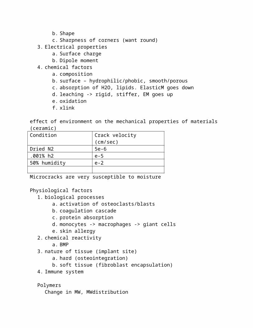
effect of environment on the mechanical properties of materials (ceramic)Condition Crack velocity (cm/sec)Dried N2 5e-6.001% h2 e-550% humidity e-2
Microcracks are very susceptible to moisture
Physiological factors1. biological processes
a. activation of osteoclasts/blastsb. coagulation cascadec. protein absorptiond. monocytes -> macrophages -> giant cellse. skin allergy
2. chemical reactivitya. BMP
3. nature of tissue (implant site)a. hard (osteointegration)b. soft tissue (fibroblast encapsulation)
4. Immune system
PolymersChange in MW, MWdistribution
Degradation products of materialsEither mechanical or chemically
Calcification
Physical1. change in size2. shape
3. surface topology4. optical properties (contact lens – protein adsorption)5. change in hydrophilicy/phobicity
Implant lifespan depends on:1. the surgeon2. patient3. implant site4. implant design5. implant material6. fabrication and processing conditions7. conditions of use
Sulzer swiss medical device company. Ceramic hip implants2006 experienced many failures due to improper cleaning/fabrication
February 26, 2007Effect of the implant on the body itself
Wound healingFibroblastCollagenMacrophage
Potential for infection
Biological1. bacterial infection2. macrophage action3. tissue ingrowth
a. mechanical stabilization4. tissue-implant bond
Bioglass fortified 45S5 (SiO2, p2O5 63%, CaO 34.5 %, Na2O)Within phase diagram: Good biocompatibility, good cell growth.
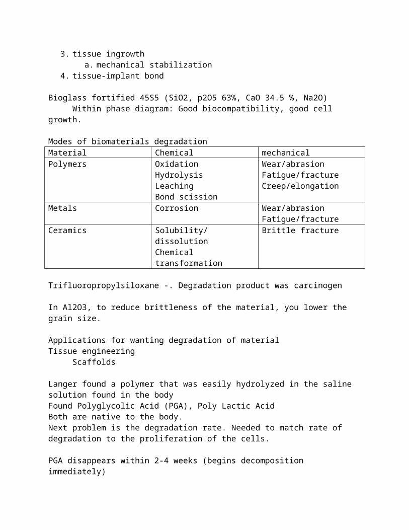
Modes of biomaterials degradationMaterial Chemical mechanicalPolymers Oxidation
HydrolysisLeachingBond scission
Wear/abrasionFatigue/fractureCreep/elongation
Metals Corrosion Wear/abrasionFatigue/fracture
Ceramics Solubility/dissolutionChemical transformation
Brittle fracture
Trifluoropropylsiloxane -. Degradation product was carcinogen
In Al2O3, to reduce brittleness of the material, you lower the grain size.
Applications for wanting degradation of materialTissue engineering
Scaffolds
Langer found a polymer that was easily hydrolyzed in the saline solution found in the bodyFound Polyglycolic Acid (PGA), Poly Lactic AcidBoth are native to the body.Next problem is the degradation rate. Needed to match rate of degradation to the proliferation of the cells.
PGA disappears within 2-4 weeks (begins decomposition immediately)PLA takes longer to degrade. “Well beyond 4 weeks”Study the kinetics of cell proliferation and degradation of the scaffold.Methyl group slows down the decomposition rate – not soluble in water.
ThrombogenicityCaused failure of jarvik, abiocor, heart valves.
Affect on implantWearFatigue/fractureBlood clotting
Valve malfunctionHindered hydrodynamics
Chemical1. oxidation2. hydrolysis3. adsorption (proteins) – change surface properties4. adsorption – swelling – plasticization (polymers)5. leaching – diffusion6. resorption – biodegrdation7. bond scission8. cross-linking9. change in molecular weight10. change in molecular weight distribution (caused by xlinking/scission)11. degradation products12. biodegradation – enzymatic13. calcification14. change in surface properties15. change in crystallinity
16. corrosion17. neutralization of surface charge.
Mechanical(mechanical degradation)
1. fatigue2. crack initiation and propagation3. fracture4. wear5. compliance
(electrica)1. anodic-cathodic reactions2. electrical stimuli (voltage can stimulate bone growth)
1. Creep (PTFE) UHMWPE2. Decrease in tensile strength breakage of bonds)
Physical1. change in size2. change in shape3. change in surface topology4. change in optical properties (contact lens- protein adsorption)5. change in hydrophilicity/phobicity
Polymer:Hydrophilic – changes to the external surfaceHydrophobic – changes to internal area
Promonocyte -> monocyte -> macrophage -> multinuclear giant cell.
The effect of the implant on the body:(local effects) Wear -> imflammation, macrophages, osteoclast activity (bone necrosis, infection)(systemic effects) Dr. Patrick Laing first raised the question about the degradation products of co-cr
Transport through the lymphatic system
February 27, 2007Degradation of products. (Chp16)
Polymers PMMA – monomer
o Inflammationo Cardiac arresto Hypertension
Ionic polymerization process – try to get polymerization as complete as possible.
Remove monomers(!) PVC
o Butyltin - Additive causes acid phospatase to collecto Plasticizers cause tubing to become softer, allowing for chains to slide over each
other Somewhat volatile and can come out of the tubing to contaminated
systems Silicon gel
o Lots of physiological interactions with injection
Metals Corrosion
o Cr+n, Co+2, Fe+2,+3, Ti+2o Too much breakup can cause tissue poisoning and necrosis
Effects of the implants on the body (no material is inert)Every implant elicits some physiological response (eg trauma, wound healing) Inflammation Systemic response Protein interaction Soft tissue encapsulation Hard tissue ingrowth, remodeling Change in pH Change in electrolyte concentration Change in pO2 Bone resorption Sensitivity Blood clotting
One form of host response: inflammation1. vasodilation2. increased vascular permeability3. Edema (fluid accumulation4. activation of cellular activity
1. initiation of inflammatory responsea. capillary dilationb. platelet activityc. coagulation factors
2. cellular activitya. neutrophils/leucocytesb. macrophages
3. remodelinga. tissue surrounding implant becomes granularb. collagen activity (often involved in xlinking process)c. fibroblasts (in soft tissue)
d. osteoblasts (hard tissue)4. capsular formation
a. fibroblastsb. osteoblasts
Signs for imflammation:Heat, redness, swilling, pain, loss of function
The role of the inflammatory response: isolate/encapsulate attack and destroy
any foreign object or device
Cells related to inflammation macrophages
o mononuclear monocytes transform to macrophages (large phagocytes – giant cells)
o attack and ingrest cellular debris and bacteria“we consider the macrophage to be the pivotal cell in determining the biocompatibility of implanted materials” – professor james m. Anderson – CWRU
foreign body giant cellso coalesced macrophages
Macrophages – cell receptorsCytokines: protein-> cellsSecrete chemical products: free radicals, peroxides,
O2., H2O2 – superoxide, OCl-Secret TGF-B – in the ECMActivate IL-12T-cell -> antigen
Role of metal ions in wound healingZinc – increases healing rate (positively affects tensile strength of tissue)
Why? May increase collagen cross-linking reactionMay be involved in enzyme reactions
Copper – enhances xlinking of collagen
Partial pressure of O2Increases with timeAt time of wound – 3 torrMacrophages appear – 10 torrFibroblast activity – 20-30 torrNormal pO2 – 45 torrpO2 related to collagen synthesis
1 ATM = 760 mm/Hg
Torr = international unit = 1/760 of a standard ATM, dynes/cm^2
pH ranges in the bodyblood 7.1-7.4urine 4.5-6.0gastric fluids 1.0intracellular 6.8interstitial 7.0
pH will:influence material/host interactionsaffect chemical reactionsinfluence chemical bonds and Van Der Waal forces
Bone response to implants metal implants
o bone plates – block periosteal arterioleso intramedullary nails – interfere with medullary arterial circulation
disrupt blood supply – damage blood vesselsrevascularization is necessary to maintain bone viability and healing
Bioelectric effectWolf’s law
Stress generated potential (SGP)Bone:
Tensile side is +Compressive side is –Piezoelectric
PTFE is also piezoelectric
Piezoelectric theoryFukuda/yasuda
Streaming potentialStreaming of ions (chlorides, etc) has affect one bone remodeling systems
Electrical stimulation (bone)- charge stimulates remodeling+ charge stimulates resorption
Electrical potential drops drastically at site of bone fracture
Polymer implant reactions:Minimal response:
Silicone rubber, PE, PP, PTFE, PMMA (questionable)Necrosis:
Some in stitu polymerizing materials (aka PMMA)
Shape and size of implant has effect on body.Acid phosphatase layer (enzymatic response) is less in a round shape than a rounded
triangular shape
Conclusions:Hard segments are most reactive (-CNO) ThrombogenicPEO rich surface (soft segment) have low platelet retentionThrombin adsorption is minimal on PEO segment.
Effects of biomaterials on the body: PMMA monomerCanine:Results in rapid dispersion in the body
- leaching of monomer from pmma- dose 2g/kg body weight- freshly mixed bone cement- femoral transcortical plug- detected monomer level 1 mg/100mg in vena cava in 2 minutes- peak concentration 3-4 mins, followed by decline
Human:- peak monomer concentration in 2 mins after implantation- similar results to canine exp
Infections often associated with prosthetic devicescirculartory shunt – meningitis type infectionsocular prosthesis – conjunctivitisdental implants - gingivitiscardiac pacemakers – pocket infections, bacteremia, endocarditisbreast implants – soft tissue infectionsjoint prostehesis – septic infection
March 5, 2007Assessment of biocompatibilitlyThe testing of biomaterials to determine their safety.
Testing:1. safety2. efficacy3. compliance
Modes of testing:1. in vitro
a. cell culture (2d environment)b. blood contact testsc. chemical, mechanical, physical
2. in-vivo (animalsa. host – what is the effect of the implant on the bodyb. material – what is the effect of the body on the material.
3. clinical tests.a. Functionality – material may be biocompatible but may not be carrying out its
intended function.b. Is the patient reporting pain, illness, blood clotting, etc. does patient actually die?
(artificial heart)4. Implant site
a. Subcutaneousb. Intramuscularc. Interperitoneal – inside the body cavityd. Transcortical (through the first layer of bone)e. Intramedullary
Principles1. determine property of the material
a. material characterization2. biocompatibility tests
a. every time the supplier/procedure is changed, new biocompatibility tests must be done. Must be tied into the quality control system.
3. manufacturer – needs an effective quality control system.
Organizations for testing:1. ASTM international
a. ASTM F 04 Materials and medical device committee. (Surgical comm.).i. Produces test methods
2. American association for medical instrumentation (AAMI)3. ISO TC-194 Biocompatiblity committee4. ISO TC-150 Surgical implants.
Maximum implantable dose (MID): maximum amount of implant material (does) that a test animal can tolerate without adverse physical or mechanical effects.
Carcinogenicity test: test to determine the tumorigenic potential of devices, materials, and/or extracts to either a single or multiple exposures over a period of the total life-span of the test animal.
Genotoxicity test: test that applies mammalian or non-mammalian cells, bateria, yeasts, or fungo to determine whether gene mutations, changes in chromosome structure, or other da or gene changes are caused by the test materials, device, etc.
Reproductive and developmental toxicity tests: tests to evaluate the potential effects of devices, materials, and or extracts on reproductive function, embryonic development (teratogenicity) and prenatal and early postnatal development.
Toxic agent: demonstrate an adverse effect on the animal – usually leading to cytotoxicity and cell necrosis.
Cytotoxicity:1. inflammation
a. rednessb. swellingc. edemad. paine. non-functionality
2. invasion of cellsa. leucocytesb. macrophagesc. lymphocytes
Rating scale:0 = No visible response1 = little2 = some3 = moderate4 = 5 = a lot
Classifying toxicity1. acute toxicity2. sub acute3. chronic
Sterilizing with Ethylene oxide (ETO)- polymers adsorb, upon implantation, the ETO diffuses out and causes
inflammation/necrosisw/ gamma exposure
- changes properties
Processing part (including sterilization) can affect the implant drastically.
F748 – a matrix that tells which road to take in terms of biocompatibility testing.
Testing for:1. skin irritation (typically using a rabbit) (F719)
Intact skin, abraded (24ours): patch testing.2. allergic (guinea picg) F7203. F756 Heymolysis
1. scope –2. References: astm standards, pharmacopia, fda protocols
3. protocol
Plasma hemoglobinKnowing the amount of material, can find the hemoglobin index.
Food and Drug AdministrationAfter 1976, medical devices came under regulation from FDA.510k = provide in vitro/vivo properties of devicePMA = Premarket approval (III) – requires clinical tests of humansGLP = Good laboratory practice. Documents tell you how to conduct tests, statistical analysis, etcGMP = Good manufacturing practiceBjork-Shiley heart valve. Problem was the with the welding of the struts to the ring, they polished over the bad welding.
FDA device classesClass1: General controlsNot for supporting or sustaining lifeNot for preventing impairment to healthNo unreasonable risk of iness or injuryEX: bandage
Class2: Performance standardsGeneral controls are insufficient to assure safety and effectivenessRequired to meet applicable standard (section 514)
Class3: premarket approvalClass 1 and 2 controls are insufficient to assure safety and effectivenessAre life-sustaining or life-supportingAre implanted in the bodyPresent unreasonable riskAll class 3 devices are subject to premarket approval (scientific review) requirements
Premarket approval (class 3) (most regulated devices)Must meet safety and effectiveness requirementsLaboratory studies
- invivo testing for toxicity and biocompatibility in tissue culture- animal studies: types, numbers, etc- clinical studies: compliance with IDE (investigational device exemption)
Finally, conduct clinical studies on humansEvaluate:
- safety and effectiveness- adverse reactions- complications- patient discontinuation
- device failure- replacements- analysis of results- contraindiciations- precautions
Can standards mitigate or eliminate medical device implant problems?Silicone gel-filled breast implants
Cyclic fatigue is now being testedBjork-shiley heart valves
Manufacture concealed the fact that there were defects in the structureTotal heart replacement devices
Blood clotting is the chief mode of failureBlood compatibility/clotting tests – would have learned that materials were
thrombogenicTemporomandibular joint implants
Used PTFE, which is bad in compression (creep).
Safe medical devices amendment of 1990Provides mandatory requirements for medical device tracking
FDA tracking system (proposed?)Device: Floow device from manufacturer to userPatient: Lifetime monitoring of device userHow can this be done realistically? What about multidevice patients?
By law, reports must be made if medical devices that contributed to:death (FDA & manufacturer0serious injury (manufacturer or FDA if manufacturer is unknown.Ser
Medical devices subject to trackingCertain vascular devices (grafts, VAD, pacemakers, heart valves)Silicone breast implants Live sustaining devices (ventilators, etc)
Tripartite Agreement (US, Canada, UK) used to regulate devices. Was eventually taken off the market.
Animal tests play a vital role in biocompatibility picture.
Animals in medical research16-25m animals sacrificed in animals shelters each year in USMedical research approximate 2% of these numbers3rd largest number of letters received by congressmen and senators are regarding animal testing. (behind national debt and health care)
Performance test methods (performance standards)1. duplicate body conditions2. realistic test methods
March 6, 2007
International standards for medical devicesISO TC-194 (technical committee 194)ISO10993 1-18 evaluation and testing, protocols, etc. 10993-7 “ETO sterilization and residuals”Identification and quantification of degradation products from polymers/ceramics/metals and alloys
FDA 510k documentPublished flowchart that guides certification processNeed to justify that the device is biocompatible
TestingStandard materialsCertified reference materialsReference material – not a national standard, “internal reference material”-Does your material give the same result as the standard reference material
Standard test methodsScientifically soundRepeatable/reproducible
- reproducible results through multiple labsHigh precision and accuracy (precision vs. accuracy)Passes interlaboratory testing criteria
Polymer processingMW -light scattering
-UltracentrifugationMv Viscosity, formula to determine MW
Biocompatibility testing: polymers, ceramics, metals/alloys, composite
Polymers: macromolecule built of many monomer unitsConfiguration vs conformationThe configuration of the chain refers to the arrangement of the subunits along the backbone of the polymer. Configuration is related to the internal structure of the chain while conformation is used to denote the physical outline or shape of the macromolecule.Chain folding
Isotactic: all methyl groups (R) are on the same side of the polymer chains
Syndiotactic: methyl groups are on alternate sides of the polymer chainAtactic: a random distribution of methyl group along the main chain
Amorphous polymersDescription: a mixture of long polymer chains with no particular orderTypical properties: often transparent, poor chemical resistance, softens with temperature, have a glass transition temperature, sensitive to creepEX: poly styrene, poly nitrile
Crystalline polymersDescription: a mixture of molecules which have ordered or aligned segments along with amorphous segments. Ordered areas are tightly packedProperties: typically opaque (dense packing), excellent chemical resistance, low friction, amorphous regions soften with temperature, a distinct melt temperature, creep due to amorphous regionsEX PE, PP, fluorocarbons (PTFE), nylon, acetal
Thermoplastic: amorphous and crystallineThermoset: cross-linked
Cross linked polymers:DESC: a single giant molecule interlinked by strong inter connecting bondsPROPs: typically transparent, excellent chemical resistance, does not soften with temperature., creep does not…
Nondegradable synthetics:Polyamides, polyesters, polyvinyl chloride, silicones, fluorocarbons, UHMWPEBiodegradables:PGA, PLA, etc
Environment changes the surface groups – ie OH or -CH2 groups on the surface
PTFE: fibersmicroporous fabric (goretex)sewing rings for valve struts
DacronSewing ring materialArtificial blood vessels
DelrinHemocampatible (76% of implants free of thrombosis)Poppet wear reportedAdsorption of water
Silastic (silicone)Early poppets: lipid absorption in starr0edwards valve
PolyurethaneCirculatory assist devices (LvAD)
PDMS coated polymer
Molecular weight are fundamental properties of a polymer sampleMw = Mn = weight of molecules / number of moleculesMWD – molecular weight distribuation
Determining molecular weight in a labMn = osmotic measurement. Measure the increase of osmotic pressure across a membraneMw =
Properties needed for engineering design of polymers:Tensile
Strength (ultimate, yield)ModulusCreep
Elongcation (ultimate, yield)Shear strengthCompressive (strength, modulus)Critical stress intensity factorCoefficient of frictionWear characteristicsGlass transitionFatigue lifeFracture toughness
March 19, 2007Metals and alloys for surgical implant applications
Properties -> performanceChemical compositionMechanical propertiesPhysical properties
Metal processes – to change the microstructure of the metal.Casting
A metal or alloy is cast (poured) into a mold. Allowed to coolWrought
Plastically deformed metal, shaped by hammering, beating, or pressingCold worked at room tempHot worked
ForgingA hot working operation to process metals and alloys
Heating and hammering a metal to shape. After foring operations the metal or alloy undergoes an annealing treatment consisting of heating to an optimum temperature and rapidly cooling to meet metallurgical requirements
Recorded history of metals for biomaterialsBC – egyptioans used gold for plates1829 – Levert, first recorded tolerance study1930 – Venable discovered Vitallium: Co-Cr-Mo for orthopedic devices1951 – Leventhal used Titaium for plates and in mesh1980-1990 Ti alloys with niobium, tungsten
Biocompatibility of Metallic implants is governed by:Mechanical factors:
FatigueFractureElastic modulusWear
Chemical factorsCorrosion
Physical factorsSurface characteristics
Physiological factors w/metalsHost responseChelation – metals become ionic when they corrode, look for free electrons in proteins, enzymesSensitivity – Ni+2Macrophage activationVital organsLocal Tissue responseEncapsulation – protein signals fibroblast cell which then deposits membrane around the insultGranulation – leads to scar tissueMacrophages – lead to giant cells to deal with objects that are too large for a single macrophageNecrosis (lysis)
Metals used in implants:Stainless steels – 316L, 302, 304Cobalt chrom molybdenum alloysTitanium and titanium alloys (corrosion resistant, TiO2)MP35N alloys Co-Cr-Ni-Mo add nickel to allow for easier machiningNitinol memory metal nickel titanium. Temperature dependent shape properties, stentsTantalum – used a sa 3d porous structure (scaffold)
Biocompatibility requirements for an implant made of an alloy1. should be corrosion resistant
2. implications for biocompatibilityo Local – necrosis, encapsulationo Systemic – heavy metals in system
3. should be non-toxic4. suitable mechanical properties
o wearo fatigueo fracture
5. microstructure – affects mechanical properties
Casting a femoral stem – use lost wax process
Fracture attributable to manufacturing defects:1. inclusions: reduced fatigue strength2. low Mo content (<2%): higher pitting rate3. poor design: sharp corners, holes too close4. excessive porosity: cast 316L SS (need Vacuum casting)5. severe cold working: produced surface cracks6. large grain size: lowered strength7. poor finishing: surface cracks8. improper electropolishing: surface porosity9. improper heat treatment:: lowered strength10. mixed metals (nail/screw: bone plate)11. out of round – femoral heads12. microstructural defects (large grain size)
Cahoon and Paxton, 1968 – 1970 studied hospital purchases, 50% had metallurgical defects
Large grain size becomes “landing strip” for various metals<.03% C, corrosion resistance of S.S. goes up – develops good Cr2O3 protection/passive layer>.03% C, form Cr23C6 in the grain boundaries. Depletes amount of Cr to form the passive layer
Characteristics of metals and alloys Good electrical and thermal conductorsMobile electronsTendency to corrode, oxidize, etcComparable tensile and compression strength (within the individual material)Crystallize readilyMore ductile than ceramicsContain “heavy” metal elementsExamples: S.S. chrome-cobalt alloys, titanium, etc
Energy levels:Primary Ionic bond 100kcal/moleSecondary bond 10kcal /mol
Metallic trace elements are essential to life
Vanadium, chromium, manganese, iron, cobalt, nickel, copper, zinc (Atomic numbers 23-30)When the concentration of the metal ion exceeds the tolerable maximum limit, is it potentially harmful?
Metals and alloys used in surgical implants:1. stainless steel
316LIron based alloys with minimum of 12% Cr to improve corrosion resistanceModulus 28 e6 psi (bone 2-5)TS 145000 psiElongation 10%
There is evidence that S.S. corrodes and metal is released/chelated into surrounding tissue.
Cobalt makes up most of a Co-Cr-Mo alloy ~60%
Haynes-Stellite 21 ASTM F-75Cast cobalt/S.S. has large grains – weakens materials, but is cheaperThermomechanically processed Co-Cr-Mo alloy, far different microstructure
Haynes-Stellite 25 ASTM F-90Co-Cr-W-Ni alloy
Is Co-Cr alloy a potential carcinogen?“there is a real problem associated with Co-Cr based alloys.”“estimates patient with Co-cr implant has 10x the normal chance of developing bone cancer.”“in animal studies Co-Ccr is associated with the highest cancer rates in an imals.”“Co-Cr implants have been put in for 50 years. Problems would have surface long before this.”
March 20, 2007
Metal alloys may be carcinogenic in human subjects, but is not totally substantiated.
Titanium and Titanium alloys1940s introduction as surgical implant1965 orthopedic implant
Characteristics; forms oxide layer corrosion resistance undergoes wear
Ti alloy is less dense than S.S.Strength is reasonable.
Types of Ti
Unalloyed titaniumAlloyed ti6al4v
Wrought, annealedCastForging
Ti6al4w(?) Wrought
4 grades of Ti, each with different amounts of O2. -> changes tensile strength of the alloyHigher O2 percentage leads to higher tensile strength and lower elongation
Ti 6Al 4V propertiesCorrosion resistanceBiocompatibilityDuctilityFabricablityHigh tensile/fatigue strengthLow densityBetter modulus match120,000-150,000 PSI, 8-10% elongation
Ti 6Al 4VTi 90%Al 6%V 4%
Titanium wearGenerally
titanium wears more than other metals and causes more wear of UHMWPE Nitriding and ion-implantation markedly reduce initial wear but long term wear rates may
be much less affected.Innmunogenicity associated with wear
Activated t-lyphocytes Associated macrophages Release of prostaglandin E2 and IL-1 (signs of activated immune system)
Conditions leading to the creation of an electrochemical cell on metallic implants1. two different metals in contact2. variations in O2 concentration3. variations in metal homogenicity4. anodic and cathodic reactions are going on simultaneously
Chemistry of Corrosion
Types of corrosion1. crevice corrosion2. fretting (disruption of any oxide layer that has formed)
3. pitting (low O2 concentration), in presence of Cl-4. intergranular5. stress cracking6. galvanic7. cyclic fatigue (goes back to stress cracking)8. generalized corrosion (everything except listed previously)9. erosion – can be chemical or mechanical
pourbaix diagrams – an equilibrium diagram which shows how metals react under conditions of potential and pH.The nernst equation is used to construct the pourbaix diagram.Useful for predicting
direction of reactions types of corrosion products effect of environment (ph, potential) on surface characteristics influence of environmental conditions
metallic corrosion leads to:1. local tenderness2. acute pain3. reddening4. swelling5. chronic inflammation6. changes in cellular metabolism, bone microstructure7. elemental sensitivity8. transport of metal ions9. cell necrosis
Hybrid metals1. drug eluting stents – metals/polymer2. infuse spinal cage
a. cage is Ti cpb. rhBMP-2 collagen sponge
galling – welds created on asperities due to heat from frictionmetal on metal implants, 1980 Sulzer
equal channel angular extrusion (ECAE)
now can take TiCP and match tensile strength of alloy. Can avoid Al, V
Ta has been vapor deposited onto polymers,Use in acteabular cup can stimulate ingrowth of bone
HippingP = 100MPa
T = 1000-1100 C.Alters microstructure of metal, giving finer grain size, distribution of particles, etc
March 26, 2007CeramicsWhat are ceramics?Any class of inorganic, nonmetallic products which are subjected to a temperature of 540C and above during the manufacture or use
Includes: metallic oxides, borides, carbides, nitrides, and mixtures of various compounds
Materials which are usually composed of compounds of metallic and non-metallic elementsAl2O3, ZrO2, Carbon (ie, pyrolytic), Glass (ie bioglass), calcium phosphates
Ceramics: Advantages largely biocompatible low coefficient of friction range of reactivity dense or porous forms (relating to brittleness, pores serve as crack arrestors) coatings over metal strong in compression
Disadvantages: Brittleness Low impact resistance High elastic modulus (wolf’s law) Weak in tension Surface defects (difficult to process and machine without implementing defects Low flexural properties. Difficult to fabricate
Metals ceramics Good electrical and thermal conductors Easily lose electrons Tendency to oxidize, corrode, etc Comparable tensile and compression
strength Crystallize readily More ductile Contain “heavy” metals
Good dielectrics Accept and share electrons Stable in chmical and thermal
environments Stronger in compression than tension Crystallize less readily More brittle Contain ions common in physiological
environments (eg Na+, K+, Ca++, Mg++, etc
Breadth of Ceramics FieldCeramics can come in two major types
Amorphous – glassesCrystalline – single and poly crystalline materials

Both types can be composites of sortsMultiphase glassesPolycrystalline ceramics, ie pure polycrystalline alumina, refractories
Crystal vs amorphous propertiesProperty Crystalline SiO2 Amorphous SiO2Tm/Tg Higher Tm Lower TgStrength Higher LowerSolubility Lower HigherThermal conductivity Higher LowerHardness Higher lower
Properties of ceramicsStrong inocovalent bonds tend to make ceramics
Strong, hard, brittle, electronically semi conducting through insulationThe ionocovalent bonds can exhibit a wide range of chemical solubilities.
Property developmentProperties are developed in different ways
Physical properties are developed by atomic bonding considerationsMicrostructural properties are developed by the processing history through ffiringPart properties are further developed through processing after figing
This is the history that gives us the property seen in an individual device
ProcessingCeramics are processed a number of ways to form the appropriate microstructures and parts - including
Beneficiating of raw materialsgreen formingSinteringFinishing
BeneficiationBeneficiating means taking the basic ores or chemicals and making powders or gels that can be used in a ceramic. These methods can include
Fusing of materials (ZrO2)Chemical precipitationSol gel precipitationVapor deposition methodsSolution growth
Green FormingIn green forming we take the ceramic stuff and form into the shape we want, processes include:
Cold isostatic pressing (CIP)Extrusion
Casting (gel, slip)
Sintering, also called “firing” of ceramic wareFiring is the method by which we make a hard productThis is where the ceramic green powders are coalesced into a single hard piece
FinishingFinishing is all the post firing stuff needed, in biomedical materials, including
GrindingPolishingJoiningSterilizationPackaging
Microstructural propertiesDeveloped through the processing history of a materialDependent upon
Grain size, orientation, boundariesNumber of phases
Types of crystalsGrain boundary/amorphous compositionPorosity is a phase
DopantsImpurityAlloying agent (metal gets mixed into material, ie ceramic)
Brittleness traceable to the microstructure and bonding:Ionic bonds with high degree of localization of electrical charges within the latticeElectrons not movileSmaller mobility of lattice defects/dislocations (low mobility tolerance)
How to deal with brittlenesscontrol grain size (smaller)deposit ceramic coating on metal substratecomposite material – add glass/metal fibers to the ceramic matrix. Increase tensile strength and serve as crack arrestors
Al2O3 coatingon 316L SS in water – change in morphology from round to fibrous natures
Wear rates are much lower in ceramic on ceramic articulating surfaces (100x lower on alumina/alumina vs Co-Cr-Mo alloy/UHMWPE)
Zirconias are also popular implant materialsZirconium Oxide ZrO2Calcium Zirconate CaO.ZrO2
European concern from alpha and gamma radiation was overcome by testing in US labs
Zirconium Oxide limitationsWear resistance – poor in comparison to Al2O3Decrease in strength of material while in a physiological environment, caused by phase transition in the crystal state. tetragonal -. Monoclinic, results in loss of tensile strengthSome of these limitations can be dealt with by stabilizing ZrO2 with Y2O3.
March 27, 2007Continuing on ceramics
Different types of Al2O3Density (g/cm^3) 3.99 3.96 3.87Grain size (um) 15-45 1-6 5-50Tensile (MPa) 206 310 262Youngs (GPa) 393 366
Alumina loses much (50+%) of its adherence strength to 316L SS almost immediately upon immersion in solution.Specifications of Al2O3
Corrosion resistance (ringers) 0.1 mg/m^2/day
Bone and Al2O3 interface: no fibrous interlayer can be seen at the interface. Good adherence.
(with stabilization of Y2O3) Al2O3 Zr2OE Mod (GPa 380 190Hardness (Mho) 9 6.5Wear ++ +
Reasons to use Zr2O (used in Europe): wear is not that significant
Hydroxylapatite: Percentage of HA in hard tissueBone 60-70%Cementum 70%Dentin 77%Enamel 98%
Wide variation in the mech. Properties of HA (different manufactures, grades, etc)Jarcho Cato
Compressive strength (mpa) 196 294
Ti6Al4V – immediately after placement, Titania(s) is formedTitanium oxide TiO2, TiO, Ti2O3Calcium Titanate 3CaO.2TiO2
Isotropic carbons used in clinical devices
Pryolytic carbon (low temperature isotropic, LTI) aka Pyrolite carbonGlassy carbon (vitreous carbon, polymeric carbon)Vapor-deposited carbon (ULTI carbon) aka Biolite carbon
Vapor deposition – typically deposit the carbon onto polymer/ceramic substrate
Carbon fibers and compositesIssues: characterization, strength, fracture toughness
ULTI carbon is strongest in tension among glassy, LTI, LTI w/Si carbons
Bioglass~45 % SiO2, 6% P2O5, 24.5% CaO, 24.5% Na2OWhy P2O5? -> calcium hydroxylapatite contains P.
By reducing CaO and adding CaF2 (12.25%), solubility was drastically changedA triangular phase diagram (SiO2, Na2O, CaO) was created to determine bone bonding results. Good results were found at somewhat equivalent amounts of the three materials (see composition above)
Mechanism of bonding between bone and glass ceramicsThre is a gap between bone tissue and the glass-ceramics immediately after operation. First the surface of the glass-ceramics becomes irregular because of dissolution between 5 and 10 days after implantation. Second a Ca-P layer is formed on the irregular surface of the glass-ceramics. The surface of the Ca-P layer at near the bone tissue is smooth. Bone tissue grows towards the Ca-P layer between 10 and 30 days after implantation.
Strong bonding adherence of collagen to bioglass surface.
CollagenCollagen represents about 25% human tissue15A wide, 400A periodicity of banded patterns.
Importance of TiO on top of Ti alloyFormation of Apatite on Ti and TiO2The oxide layer formed on Ti implants in the body increases in thickness and attracts minerals from the surrounding physiological solution.
Proteins are first adsorbed on TiO2, then mineral ions diffuse through the adsorbed protein layer.
Plasma Spraying – most of the coatings that are made from ceramics are made by some high energy phenomenon.
April 2, 2007Composites: materials composed of two or more different constituents, each of which contributes specific property characteristics that enhance the performance of the product.
Composites enable the design of surgical implants with mechanical and physical properties that are not attainable using single materials alone.
Other advantages of composites: Mechanical properties may be varied
Lower modulus of elasticity than metals High tensile, flexural, and fatigue strength
Improved biocompatibility Reduced corrosion
Types:Polymer – fiber, fillerBioglass – stainless steelAl2O3 and TiO2 – metal (plasma sprayed)Hydroxyapatite/fuorapatieBisphenol-a/glycidyl methacrylate (BIS-GMA)SiC/C - carbideCarbon Fiber/reinforced C
Matrix:Polymers
Thermoset – epoxy, phenolicThermoplastic – PMMA, PE, PP, Polysulphone, Polyester, Kevlar
MetalsAl, Ti, Ni
InorganicsCarbon, Al2O3
CombinationsPolymers/metals/inorganicsMetals/organics (coatings)M/M
Matrix FactorsModulusTransfers load to fibersSupports Fibers, maintains their position
Al2O3, ZrO2, and TiO2 coatings on metal implantsReduces corrosion and M releaseReduces M sensitivity and wear
ApplicationsSkeletal systems
Fixation devicesTotal hip endoprotheses
Bone cementTotal kneeEtc
Dental ImplantsResorbable Sutures – PLA/PGAArtificial skin – mixture of nylon and silicone
Characteristics:Less rigid than metalsHigh fatigue strengthPromotes bone healing/remodelingPain reduction (lower modulus)Good-excellent biocompatibility potential (aramid fiber/pmma)Wide latitude in design (shape, varying stiffness, fiber volume, etc)Chemically resistant (ie C-fiber/polysulfone)Eliminates metallic corrosion
Fiber-reinforced composite characteristicsImproved strengthFatigue resistanceReduced crack propagationAdjustable elastic modulusHigh strength to weight ratioStress transfer from matrix to fibers
Composite typesParticulates – isotropicFibers – isotropic or anisotropicLaminates – anisotropic
Fiber factorsLengthDiameterOrientationQuantity (%fiber content)Fiber properties – surface propertiesAspect ratio l/d - 3:1 ratio is a carcinogen (asbestos). Don’t want these fibers to be released
Mineral fibersNatural
Zeolites - mineralAsbestos
SyntheticBioglassCeramicCarbon
OrganicNatural
SilkCotton
Synthetic polymerMetallic fibers
SSTi 6/4Co-Cr-Mo
Carbon fiber reinforced bone cementAdvantages
Improved strength and fatigue lifeImproved modulus of elasticityHigh flexural strengthReduced exothermReduced thermal coefficient of expansion
DisadvantagesLoose fibersBonding difficulty
PMMAExothermal reaction – add a fillerImprove catalyst (benzoyl peroxide)Improve mech properties by selecting fibers/fillers
Kevlar, 5x TS of SS by weightIn contact with tissue, Kevlar forms a thin fibrous layer between polymer and tissue
Coated metal compositesBioglass +316L SS
Metal – bioglass interface is critical to performance of implantPretrement of metal surface assues proper bond to bioglass (cleaning, deoiling) 3% HF
Al2O3 and TiO2 coatings on metal implants (plasma spray technique)Reduces metallic corrosion and release of metal ionsMinimizes imflammatory responseMetal ion toxicity can be reduced (Ni, Cr)
Carbon fiber randomly reinforced UHMWPEChapped carbon fibers, wt%: 2-40UHMWPE: balanceCF/90% PE are in clinical use.
April 3, 2007More composites
Fiber directions
Composite factorsFibers must be firmly bonded to matrix
Glass fibers – silane coupling agentCarbon fibers – organic coatings
Coefficients of thermal expansion must be similar
Interface factorsNature of matrix/fiber bondIntegrity of bond
Usually the medical device composites consist of high strength fibers (polymers, metals or ceramics) in a ductile matrix (ie polymers) some fibers may also be incorporated in ceramics
Composite femoral stem – SS fibers with a polysulfone matrixFibers of the matrix separated under stress and was never successful (did not match elastic mod of bone)
Limitations hindering further development of composites as surgical implantsProblems dealing with adhesion of fibers/matrixPolymers – hydrophilic will swell (leading back to adhesion problems)Complex manufacturing proceduresNot widely accepted for use in implants.Scarcity of standards for testing – inspecting and testing adhesive bonds of surfaces.
Polypropylene matrix with untreated glass fibers:Fibers pull cleanly out of resin
With silane coating – failure is solvedSilane has =CH2 coupling site to polymer matrix
Other factors leading to good bonding between polymer and filamentLow contact angle between polymer and fiber
Hydrophilic surface has lower contact angle than hydrophobic (spreads out more)Want low viscosity resinNeeds to be clean and free of dust.Avoid surfaces with (micro)cracks and imperfectionsWant moderate roughness to the surface/fiber interface -> greater surface areaCoefficient of friction between filler and matrix should be the same
Tests on Molded compositesUniform dispersion of randomly oriented carbon fibersFiber size to be determined by composite manufacturerMechanical properties
UTSUYS
Ultimate elongationIZOD impact strength (ramrod attached to set of rates, collides into plastic material)
Biocompatibility in accordance with ASTM F-748Cell cultureIntramuscular implantationLong term implant(no site specific testing)
Biocompatibility of different classes of compositesClass I – minimal biological response
CF/reinforced CFCF/Polysulfone (biologically minimal response, but not mechanically)CF/PMMA
Class II – Active tissue responseBioglass SS fibersCA Phosphate/UHMWPE
Class III – Bioresorbable responsePLA/PGACaPhos/PLA
AcrlyNitrile – thermal decomposition for making graphitized structure (elemental carbon)Pyrolytic carbon formation
Dr. David HungerfordDr. FromdozaInterested in repairing femur with metal rod into intermedullary canal of femurWanted composite material to match elastic mod of bone, and be biocompatible
90% PEEK polymer10% glass fibersTook MG63 osteoblasts to evaluate biocompatibility.Osteocalcin is a marker of osteoblast activity
April 9, 2007Chimeric NeomorphogenesisThe process of engineering a tissue or organ in situ by placing dissociated cells onto synthetic biodegradable scaffolds and placement in a host to permit growth, function, and vascularization.
Tissue engineering: an inter disciplinary field that applies principles of engineering and the life sciences to the development of biological substitutes that restore, maintain and improve the function of damaged tissues and organs.
Goal of tissue engineering:Treat disease or malfunctioning body parts by transplanting specific cells and tissues that have been engineered in the laboratory.
Tissue engineering
-design-specification-fabrication
8-10 million transplants performed40-90 million hospital days
Liver:30,000 patients/year need liver attention3000 liver donations/year.
750k new diabetic patients are identified each year.150k die from diabetes.
1m die from cardiovascular disease/year
Burn patientsCurrent treatment
- autograft- allograft (cadaver skin)
– antigenic– potential disease carrier– limited supply
Tissue engineering:- derma graft
– fibroblast cells (secrete proteins and growth factors)– nylon mesh– silicone membrane
Tissue engineered implant:A biologic-biomaterial system designed to restore, modify or augment tissue or organ functions
Standards development:ASTM F04 – medical and surgical materials and devices Division IV: TEMPsFocus:
Biological components (cell, tissue, cellular, product and/or biomoleculeBiomaterials components (natural/synthetic)Preclinical/clinical assessmentsNomenclature
Limitations of current state of the art valves:Mechanical valves
Foreign body responseLack of growthMechanical failureNeed for lifelong anticoagulation
ThrombosisTissue valve (xenograft)
Foreign body responseLack of growthShort durabilityCalcification
Allogenic valve (homograft)Foreign body responseLack of growthDonor organ scarcityRejection
Porcine heart valve (aortic position)Cryolife, Inc. Atlanta GA: syner graft procine heart valve
Dr. Mark O’Brien performed first implants of tissue-engineered heart valvesTwo women patients in Brisbane, Australia
TechnologyPorcine heart valve depopulated of cellsOn implantation, will repopulate with patients heart cellsAllows trans-species transplant without use of immunosuppression
Tissue engineering system:Cells (millionsScaffoldsBioreactorsCulture vessels
Cell and tissue sourcing (problems)Autologous – aseptic harvesting, rigorous record keeping (time limits!)Allogeneic – aseptic harvesting, rigorous record keeping, adventitious agentsXenogeneic – aseptic harvesting, rigorous record keeping, adventitious agents, retroviruses.
Functional evaluation of tissue engineered cells/tissuesCell metabolism – glucose consumption, lactose productionCell damage – concentration of lactate dehydrogenaseExtracellular matrix – synthesis rates 9radio labeled)Measure mechanical properties (eg cartilage)Electrophysiological properties (eg cardiac tissue)
Scaffold requirements:Provide structural framework for selected cellsBiocompatibleResorbablePromote cell differentiation, function, and growthHigh porosityAllow exchange of nutrients and waste products
Prevent entry of immunoglobulins and lymphocytes of immune system
Scaffold design- surface chemistry- surface charge- surface morphology- resorbability- cytotoxicity
examples of resorbable polymer scaffoldspolyesters
- PGA, PLA- PGA/PLA co-polymers- Poly hydroxyl butryl acid (PHB)- Poly hydroxyl valeric acid (PHV)- PHB/PHV copolymers- Poly (e-caprolactone)
April 10, 2007Molecular weight Mv = molecular weight determined by viscosityMn = number average molecular weight
Tensile strength decreases with decreasing MWMW decreases with time in vivo/vitro.
Importance of resorbability- eliminates chronic foreign body reaction- reduces chance of infection- leads to restoration of normal tissue
Crystallinity and thermal properties tof PGA PLA and copolymers% xtalinity Tm Tg
PGA 46-52 225 3690:10 PGLA 40 210 3750:50 PGLA 0 None 55PLA 37 185 57dl-PLA 0 None N/A
Further addition of LA leads to totally amorphous material. Much more soluble than crystalline material. Gives a time window to match up cell proliferation rates with degradation of matrix.
Scaffold surface (pore size) important features- % porosity (usually between 70-80%)- Pore shape (round, square, etc)- Surface area/properties
Pore size5um – neovascularization , capillaries.5-15 um – fibroblast growth20-125um – epithelial cell growth (tissue engineered skin)100-300 um – osteoblast growth
Cell Adhesion – a key issue in tissue engineering- Polymer surfaces (scaffolds) generally do not have the ability to interact with cell-
surface recptors to affect cell adhesion- When implanted polymers adsorb adhesion proteins they support cell adhesions- Cells bind (adhesion) to adsorbed proteins rather than the polymer
Intrigens – surface receptors, peptide subunits, fibronectin.
Carbohydrates and cell adhesion- mono saccharides are incorporated into polymers to promote cell adhesion
– lactose bond to polystyrene binds mammalian hepatocytes- carbohydrates bind to lectins (cell adhesion proteins)
– lectin UEA-1 has been bound to polyethylene terephthalate increased endothelial cell attachment 100 fold fibroblasts, monocyte, smooth muscle adhesion was reduced
Oligopeptides – do not denature when they get to the surfaceCovalently bonded to scaffoldCell surface receptors
Oligopeptide sequences used for cell adhesionRGD – fibronectin, collagen, laminin
Integrins:Composed of alpha and beta transmembrane subunitsSelected from 16 alpha and 8 beta subunitsProduce more than 20 different receptors
Integrinn mediated adhesive reactions are involved in many cellular functionsLeukocyte homing and activationHemo statis (blood clotting)Bone resorptionResponse of cells to mechanical stressTumor growth and metastasisProgrammed cell death
Biological signalsCell signaling and cellular responsesSignal transmission by surfaces, bioactive agents, ligandsMechanisms
Biomaterial encapsulationPhagocyte responseBone depositionFailure of tissue engineered implants, eg immune response
What is a growth factor?A powerful regulator of biological functionA biologically active protein that promotes cell growth…
Polypeptide growth factorsIncreases cell proliferation, migration, aggregation, and differentiationBind to cell-surface receptors to stimulate cellular activity
FGF fibroblast growth factors, EGF epidermal growth factors, PDGF platelet derived growth factor
Tissue engineering bioreactorsIn vitro cell culture systems
Promote cell growth on 3d scaffoldsProvided needed nutrients and gasesEfficient mass transfer to cells/tisssues (flow and mixing)Subject developing cultures needed to physically stimulateStatic and rotating bioreactors
Bioreactors: Static/rotatingCartilage and cardiac tissues functionally superior in rotating bioreactors vs static
reactorsHydrodynamic forces affect cells via
Stress fields, stretching, pressure fluctuations.
Smooth muscleSeeded tubular silicone scaffoldsBuilt with Pulsatile radial stressProduced arteries (<6mm diam)High rupture strengthHigh suture retention
Bioreactors with mechanical stress componentsCartilageDynamic compression at physiological frequencies- promotes extraceullular matrix synthesis- increases glycoaminoglycan synthesis (GAG)- repaired bone defects in rabbitsCardiac tissue- increased size and function (contraction)- produced growth hormone
Tissue engineering of the liver – hepatocytesSeeded on polymer scaffold (5e7 cells/ml)Polymer scaffold: PGA PLA PGLA PVAScaffold implanted in hostHepatocyte growth factor (HGF)
- increased cels up to 30x in 4 weeks (rats)- sustained release of HGF
Islet cells for diabetic patientsDesmos, Inc. San Diego
Produces pancreatic isletsUses ECM protein laminin-5 as growth substrate
Microislet, La Jolla- Provides encapsulation technology- Alginate scaffold
Osteotech-provided HA and Ti plasma spray coatings for orthopedic and dental implants
Stryker biotech- markets a human recombinant osteogenic protein for nonunion fractures of long bone
Tissue regeneration- isolates mesenchymal cells of bone marrow, grows them on a scaffold in a bioreactor
obtaining a human ligament product
Bovine – derived cultured skinOrtec InternationalPorous matrix of bovine collagenSeeded with two layers of human dermal and epiththelial cells
Strategies:- replace only those specific cells needed for function- provide tissue inducing substances eg growth factors, signal molecules, etc- ccells incorporated in a matrix; synthetic or modified natural materials.
– Open cell matrix: implanted in body, become integrated with host tissue- Closed matrix: cells are isolated from the body by permeable membrane
– Permits nutrients, gas exchange to enter– Prevents entry of antibodies, immune cells
April 16, 2007Skin - organogenesis inc ApligrafCartilage carticel genzymeEncapsulated islet cells, hepatocytes, urothelial cells
Tissue engineering of bone – why is it important?- millions of surgical procedures, bone defects, non union- autogenous bone grafting- allogenic bone grafting
Scaffold: designed to be osteoconductive – addition of growth factors, etcSupports angiogeneisis
- promotes cell proliferation, differentiation
Distraction osetogenesis – application of dynamic force to cell colony- Pulsaltine- Cyclic mechanical testing- longitudinal force
Mesenchymal stem cells (MSC) (pluripotent)Osetoprogenitor cells- more advanced and can only produce bone- low population in bone marrow
Growth factors are specific proteins secreted by cells and are signaling molecules that interact with target cell receptors.
Bone Morphogenic factor- rhBMF 1-15 have been identified- BMF-2,3,7 involved in bone repair and stimulating MSC differtiation into bone- BMP-3,4,7,8 active during day 14-21
– Ostoblastic recruitment– Resorption calcified cartilage BMP -5,6-active during day 3-21
Vascular endothelial growth factor (VEGF)- stimulates vascularization- promotes new bone formation
transforming growth factor Beta (TGF-B)- stimulates MSC proliferation- stimulates bone regeneration- increased mechanical properties of new bone
Roles of growth factors (proteins)- cell adhesion- cell proliferation- cell migration- cell differentiation
Tissue engineering factors:- cells- scaffolds- bioreactor- upscaling (from “test tube” to large volume)- preservation
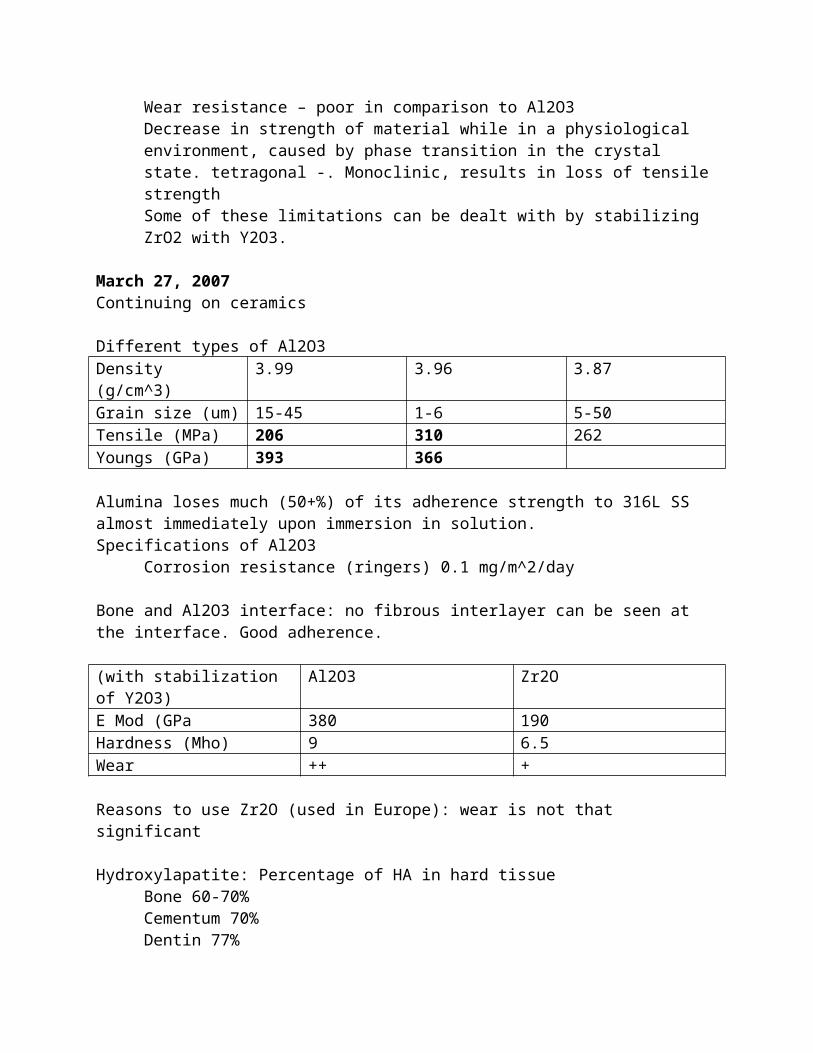
- imuno-compatibility
Osteogenic – bone syn cellsOsteoconducdtive
Stem Cells:1998
- James Thompson, University of Wisconsin established the first human embryonic stem cell line.
- US patent issued
Legal issues- Wisconsin alumni research foundation (WARF) licensed Geron Inc to commercialize
stem cell by-products: liver, muscle, nerve, pancreas, blood and bone- WARF is suing geron, Inc. contesting legality
Ethical issues- is it morally ethical to use human embryonic stem cells for research and applications- Dr. Roger Pedersen UCSF is moving his laboratory to Cambridge university, enging
in a more “receptive environment”- Patient’s right to know (patient autonomy)- Manufacturer’s obligation to patient- FDA’s oblicgation to protect and inform patients- Engineer’s obligation to
– Company– Patient– Engineers code of ethics
Other issues- the provider’s demand for any new inventions- etc
What is needed?Standards: reference biomaterials and test methods to improve precision and accuracy of laboratory test results
What is a reference material?A well characterized material that by a standard test has been determined to elicit a reproducible, quantifiable host or material response.
What is a standard test?A test based on established scientific and engineering princibples that has been demonstrated to yield accurate and reproducible results
Challenges for tissue engineering
RegulatoryAre products safe and effective?Mechanism for evaluationg “combination” products
Health insurance coverageAre these products significantly better than current treatments?Are they affordable?
Manufacturing and quality controlSource of materials (animals, variability of natural products, recombinant products).Complexity of material preparation. Scale up stem cell culture.Short shelf life
Fundamental understandingCell response to environmental stimuli: imperfectly understood.
Cell types:- fibroblasts- myoblasts- endothelial cells- valvular interstitial cells
Scaffolds:PGA (resorption time 2-4 weeks)PLA (18-36 months)PLGA (8-12 weeks)PHA PolyHydroxyAlkanoates – ex Poly-4-HydroxyButyratePGA-PHHB
NaturalCollagenAlginateHA
BioReactors:Want dynamic feature –
Some key points to improve performance and minimize failures:- rigorous design criteria and testing- meaningful materials specification requirements- standard test methods- quality control system
we need statistical data:- how many implants of a particular type are in patients?- How many implants are failing?- What is the mean time to failure- What is the principle mode of failure?
Need to develop a national and internation network of cooperating surgical implant retrieval and analysis groups to provide validated data on device failures and cause of failures
The future:Biomaterials and biotechnology espanding markets due to:
- graying population- monderization of developing nations health care systems- new tissue engineered product lines- increased use of drug delivery systems- research and development will lead to new innovations
Bioaterials and biotechnology- greater involvement of molecular biology, biochemistry, biomimetics,
nanobiomaterials and drug delivery systems will lead to remarkable advances- growth factors, cell signaling, stem cells, biocompatibility, improved synthetic
materials, bioinformatics
April 23, 2007Biocompatibility
Material failures- Surgeon- Patient- Processing
Breast implants – reports of adverse effects- fibrous encapsulation- capsular contracture- interference with mammograms- silicone bleed- capsule rupture- formation of calcium deposits- calcification- implant shifting position- silica filler- arthritis-like symptoms- corrective surgery- polyurethane degradation
Countermeasures to prevent capsule formation- drains for hematoma and fluid accumulation- antibiotics- capsulotomy (exercise)- retromuscular positioning- steroids- polyurethane coating
Silicone breast implants? Do they cause connective disease? Immune related disorders? Cancer?
“There is currently no evidence regarding silicone toxicity”
PTFE, UHMWPEPMMA bone cement (still in use)Porous surfaces – bypass PMMA depends on bone integration for mechanical stabilization
Metals 316L SS, Co-Cr-Mo, Ti6Al4VCeramics Al2O3 ZrO2
How can we overcome some of the wear problems with UHMWPE?- eliminate loosening of femoral stem- modify surface of acetabular cup- eliminate UHMWPE degradation via gamm sterilization- control Mwd of UHMWPE- strict quality control of processing conditions for production of UHMWPE- modify design of acetabular devices- improve surgical implantation technique- use another material
affinity index – length of bone in contact with surface/length of implant surface
Delrin
Affect on implantWear, fatigue/fracture, blood clotting, changing hydrodynamic flow of blood
Thrombus formationAdsorption of plasma proteins -> adhesion of platelets -> aggregation of platelets -> fibrin formation -> thrombus
AlbuminAdsorption decreases platelet adhesionDecrease thrombogenicity, blocks fibrinogen adsorption which attracts plateletsAlbumin does not contain RGD sequence, and amino acid sequence common to “adhesive proteins”Albumin must occupy most of the surface (>98%) to be anti-thrombogenic
Intrinsic pathway – XII Hageman factorExtrinsic – tissue factor, VII
Ti6Al4VCorrosion resistance, biocompatibility, ductility, fabricability, high tensile, fatigue strength, better modulus match to bone
Polymer additives – plasticizers, antioxidants, fillers, cross-linking agents, lubricants, mold release agents, etc