Embed Size (px)
Citation preview
BIOCONTROL OF SCLEROTINIA STEM ROT OF CANOLA BY BACTERIAL ANTAGONISTS AND STUDY OF BIOCONTROL
MECHANISMS INVOLVED
by Yilan Zhang
A Thesis
Submitted to the Faculty of Graduate Studies
In Partial Fulfillment of the Requirements for the Degree of
MASTER OF SCIENCE
Department of Plant Science
University of Manitoba
Winnipeg, Manitoba, Canada
© Yilan Zhang 2004
THE UNIVERSITY OF MANITOBA
FACULTY OF GRADUATE STUDIES *****
COPYRIGHT PERMISSION PAGE
Biocontrol of Sclerotinia Stem Rot of Canola
by Bacterial Antagonists and Study of
Biocontrol Mechanisms Involved
By
Yilan Zhang
A Thesis/Practicum submitted to the Faculty of Graduate Studies of the University of Manitoba in
partial fulfillment of the requirements of the degree of
Master of Science
Yilan Zhang © 2004
Permission has been granted to the Library of the University of Manitoba to lend or sell copies of the this thesis/practicum, to the National Library of Canada to
microfilm this thesis and to lend or sell copies of the film, and to the Univesity Microfilm Inc. to publish an abstract of this thesis/practicum.
The author reserves other publication rights, and neither this thesis/practicum nor
extensive extracts from it may be printed or otherwise reproduced without the author�s written permission.

II
ACKNOWLEGEMENTS
Fist of all, I would like to express my appreciation to my supervisor Dr. Dilantha Fernando for his guidance in my experiments, and support given at conference presentations and applying for several travel awards. Only with your patience, kindness and encouragement I could grow as a student and plant pathologist. I also appreciate the help of my co-supervisor Dr. Fouad Daayf, for the advice and support for my project during my Masters program. I would also like to thank both of you for your great input on my thesis writing. To two of my other committee members, Dr. Annemieke Farenhorst and Dr. Georg Hausner, thank you very much for your great effort on advising me in my project and thesis. Special thanks to Paula Parks, Alvin Iverson and Lorne Adam for your technical support. Also a big thank you to my lab mates for your continuous support and help. Special thanks to Clinton Jurke for providing breeder tents and some of the pictures. A very special thank you to Dr. Teresa de Kievit and Chrystal Berry for the work on southern blots. I thank Dr. Tim Paulitz for giving me the idea to investigate bacterial antibiotic-related genes using PCR. I also appreciate the staff and fellow graduate students in Plant Science who helped me on my project and shared the fun at BBQ�s and parties� I would like to thank my parents for their never changed love and support. The mountains and oceans would never separate our hearts being close to each other. Only when I live far away from you, I realize how much you sacrificed in making me an independent person. I also would like to thank the life itself. I had fully experienced the interesting, exciting and frustrations in life in Canada in the past two years. I survived and I did a good job! Nothing to regret!
III
TABLE OF CONTENTS
ACKNOWLEDGEMENTS ���������������������� II TABLE OF CONTENTS ...����������������...����� III LIST OF TABLES .������..�������������.�����.VI LIST OF FIGURES ����������������������.��..VII ABSTRACT ������������������������.��..�IX FORWARD ������������������������.���...XI 1.0 INTRODUCTION ������������������.����...1 2.0 LITERATURE REVIEW ������������������.��.4
2.1 Canola���������������������������4 2.1.1 Crop History��������������������...4 2.1.2 Crop usage��.������...�����������.�.6 2.1.3 Transgenic canola������������������...7 2.1.4 Production and economic importance��..��������..8 2.1.5 Growth stages and condition��������������..9 2.1.6 Diseases on canola������������������.10
2.2 Sclerotinia sclerotiorum ������������������.�.11 2.2.1 Introduction���������������������11 2.2.2 Taxonomy���������������������..12 2.2.3 Host range..���������������������12 2.2.4 Economic importance�����������������13 2.2.5 Disease cycle, infection and symptomology��������.13 2.2.6 Epidemiology������������������.��17
2.3 Management of S. sclerotiorum on canola��������.����..18 2.3.1 Host resistance�������������������...18 2.3.2 Cultural management�����������������.18 2.3.3 Disease forecasting�..���������������..�19 2.3.4 Chemical control�������������������20 2.3.5 Biological control������������������.. 20
2.4 Biological control���������������������.�.20 2.4.1 Introduction�����������������.����20 2.4.2 Biocontrol mechanisms����������������..21
2.4.2.1 Antibiosis������������������.21 2.4.2.1.1 Introduction���.�.����������21 2.4.2.1.2 Major bacterial antibiotics in biological control..22 2.4.2.1.3 Identification and characterization of antibiotic-
related genes / gene clusters��������.25 2.4.2.2 Plant induced resistance����������.�.�26

IV
2.4.2.2.1 Introduction��������������.26 2.4.2.2.2 Induced systemic resistance�����.�.�..27 2.4.2.2.3 Systemic acquired resistance�������...28 2.4.2.2.4 Plant defence-related secondary metabolites�...28 2.4.2.2.5 Plant phenolics�������������.30
2.4.2.3 Plant growth promoting rhizobacteria �������31 2.4.2.4 Competition�����������������.31 2.4.2.5 Parasitism and predation�����������..�32 2.4.2.6 Other mechanisms��������������...33
2.4.3 Biocontrol of S. sclerotiorum��������������.34
3.0 EVALUATION OF BIOLOGICAL CONTROL OF SCLEROTINIA SCLEROTIORUM ON CANOLA (BRASSICA NAPUS) BY BACTERIA THROUGH IN VITRO SCREENINGS, GREENHOUSE EXPERIMENTS, AND A FIELD STUDY���.������������������.38
3.1 Abstract��������������������������..38 3.2 Introduction�������������������������39 3.3 Materials and methods��������������������...41
3.3.1 Bacillus spp. isolation and storage�����������...41 3.3.2 Evaluation of bacterial antagonism through general plate
inhibition assays�������������������42 3.3.3 Antagonism through production of volatile compounds���..43 3.3.4 Testing for the presence of the oxalate oxidase enzyme���..44 3.3.5 Bacterial indentification���������������...44 3.3.6 Production of rifampicin-resistant strains and establishing of growth curves�������������������..45
3.3.7 Evaluation of biocontrol agents against sclerotinia on canola in the greenhouse������������������..46
3.3.8 Evaluation of the effect of time of application of Bacillus spp. BS6 against sclerotinia on canola and bacterial survival in the greenhouse���������������������47
3.3.9 Evaluation of biocontrol agents under field conditions����48
3.4 Results���������������������������51
3.4.1 Bacterial isolation and identification����������..51 3.4.2 Evaluation of bacterial antagonism through general plate
inhibition assays, inhibitory volatile production and oxidase enzyme presence������������������.51
3.4.3 Production of rifampicin-resistant strains and establishing of growth curves�������������������.55
V
3.4.4 Evaluation of biocontrol agents against sclerotinia on canola in the greenhouse�����������������..�.55
3.4.5 Effect of the time of application of strain BS6 against sclerotinia on canola in the greenhouse���������.56 3.4.6 Evaluation of biocontrol agents under field conditions��.�63
3.5 Discussion���������������������...���...64
4.0 DETECTION OF ANTIBIOTIC-RELATED GENES FROM BACTERIAL BIOCONTROL AGENTS USING POLYMERASE CHAIN REACTION ������������������������������.�.74
4.1 Abstract��������������������������...74 4.2 Introduction�������������������������.75 4.3 Materials and methods���������������������77
4.3.1 Genomic DNA extraction�������������..��79 4.3.2 DNA quantification���������������.��..80 4.3.3 PCR reactions��������������������80 4.3.4 Sequencing and BLAST search��������.�����82
4.4 Results���������������������������.82 4.4.1 Genomic DNA�����������������.��..82 4.4.2 PCR reactions������������������...�.83 4.4.3 Sequencing and BLAST search�������������.84
4.5 Discussion��������������������������88
5.0 PLANT INDUCED RESISTANCE MEDIATED BY SCLEROTINIA SCLEROTIORUM AND BACTERIAL BIOCONTROL AGENT BACILLUS AMYLOLIQUEFACIENS BS6 ������������������....91
5.1 Abstract����������������������.���..�.91 5.2 Introduction������������������������..�92 5.3 Materials and methods���������������������.93
5.3.1 Greenhouse experiment����������������.93 5.3.2 Phenolics extraction and fractionation����������...96 5.3.3 HPLC analysis�������������������...97
5.4 Results�������������������������..��98 5.4.1 Greenhouse experiment����..�������..���.�98 5.4.2 HPLC analysis���������������..���.�99
5.5 Discussion������������������������.....106 6.0 GENERAL DISCUSSION AND CONCLUSIONS..�.���������110 7.0 REFERENCE LIST������..�������������.��.�.115
VI
LIST OF TABLES
Tables Pages
2.1 Fungal diseases and pathogens on canola (Martens et al. 1994)��.���..�.11
3.1 Nineteen bacterial strains isolated from canola leaves����������...52
3.2 Bacterial sources and identifications�����������������..53
4.1 Bacterial antibiotic-gene-specific primers used in this study��������.78
.

VII
LIST OF FIGURES
Figures Pages 2.1 Life cycle of Sclerotinia sclerotiorum (Lib.) de Bary on canola�����...�14
3.1 S. sclerotiorum growth inhibition by strain BS6 on PDA and LBA�����..54 3.2 S. sclerotiorum growth inhibition by inhibitory volatiles produced by strains H and E12����.�����������������.������.54 3.3 Bacterial growth curves for rifampicin-resistant strains PA-23, #41, BS8, BS6, H, and E16�.�����..��������������������...57 3.4 Sclerotinia disease incidence (DI) in the greenhouse test using five bacterial antagonists�.��������������������������..58 3.5 Inhibition of sclerotinia disease progression in the greenhouse by using five
bacterial antagonists.�����.������������������..59
3.6 Canola yield harvested from sclerotinia diseased plants co-inoculated with bacterial antagonists in the greenhouse��.������.��������60
3.7 Canola seeds harvested from sclerotinia diseased plants inoculated with /without
B. amyloliquefaciens BS6����.�������������...���61 3.8 Sclerotinia disease incidence (DI) one week after ascospore inoculation��������������...�����������..�.62 3.9 Sclerotinia disease severity (DS) two weeks after ascospore inoculation�������������������..�����..�.�.62 3.10 Petal infestation level in Trial One 4 days after S. sclerotiorum ascospore inoculation (on July 12)���.�.�����������������...65 3.11 Sclerotinia disease incidence (DI) in Trial One observed right after canola
flowering stage (August 2)���������������������.65 3.12 Sclerotinia disease incidence (DI) in Trial One observed when plants reached full maturity���.�����������������������..66
3.13 Sclerotinia disease incidence (DI) in Trial Two observed when plants just reached maturity����������������...��������..66
VIII
3.14 Sclerotinia disease severity (DS) in Trial Two observed when plants reached maturity����������������������������.67 4.1 PCR products amplified with primers phz1/phz2 separated by agarose gel to
identify phenazine biosynthetic gene.��������������..��.85 4.2 PCR products amplified from strain P. chlororaphis PA-23 and P. fluorescens 2-79 using primers phz1/phz2 separated by agarose gel to identify and confirm the
presence of phenazine biosynthetic gene..���������������85
4.3 PCR products amplified from P. chlororaphis PA-23 and P. fluorescens Pf-5
with primers PrnAF/RF, PRND1/PRND2 and PrnCf/PrnCr separated by agarose gel to identify pyrrolnitrin biosynthetic gene�������.�����.�..86
4.4 PCR products amplified from strain P. chlororaphis PA-23 and P. fluorescens Q2-97 using primers BPF3/BPR2 separated by agarose gel to identify 2,4-DAPG biosynthetic gene.��������������..������.���..86 4.5 PCR products amplified from strain P. chlororaphis PA-23 and P. fluorescens Pf-5 using primers PLT BF/PLT BR separated by agarose gel to identify pyoluterorin biosynthetic gene�������������������..87
4.6 PCR products amplified with primers 678/677 separated by agarose gel to identify zwittermicin A self-resistant gene������������������.87 5.1 Diagram depicting inoculations on canola plants and leaves��������95 5.2 A-E: Typical disease symptoms on canola of Treatment A, B, C, D, and E,
respectively, observed 6 Days after disease inoculation (DAI); F: Disease symptoms of treatment A-E observed 25 DAI������������...100
5.3 Disease incidence (DI) observed on canola leaves 4 days after pathogen
inoculation (4DAI)�����������������������..101 5.4 Disease incidence (DI) observed on canola stems 7 days after pathogen
inoculation (DAI)������������������������101 5.5 Disease progress over time on canola plants�����..�������.....102 5.6 HPLC chromatograms from leaf samples 5 days after pathogen inoculation
(DAI)��������������������������..��.103 5.7 Spectrum index plot of 3 compounds from canola leaf sampled 5 days after
pathogen inoculation (DAI)��������������������105
IX
ABSTRACT
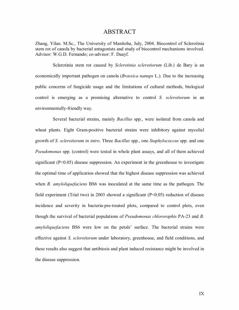
Zhang, Yilan. M.Sc., The University of Manitoba, July, 2004. Biocontrol of Sclerotinia stem rot of canola by bacterial antagonists and study of biocontrol mechanisms involved. Advisor: W.G.D. Fernando; co-advisor: F. Daayf.
Sclerotinia stem rot caused by Sclerotinia sclerotiorum (Lib.) de Bary is an
economically important pathogen on canola (Brassica nanups L.). Due to the increasing
public concerns of fungicide usage and the limitations of cultural methods, biological
control is emerging as a promising alternative to control S. sclerotiorum in an
environmentally-friendly way.
Several bacterial strains, mainly Bacillus spp., were isolated from canola and
wheat plants. Eight Gram-positive bacterial strains were inhibitory against mycelial
growth of S. sclerotiorum in intro. Three Bacillus spp., one Staphylococcus spp. and one
Pseudomonas spp. (control) were tested in whole plant assays, and all of them achieved
significant (P<0.05) disease suppression. An experiment in the greenhouse to investigate
the optimal time of application showed that the highest disease suppression was achieved
when B. amyloliquefaciens BS6 was inoculated at the same time as the pathogen. The
field experiment (Trial two) in 2003 showed a significant (P<0.05) reduction of disease
incidence and severity in bacteria-pre-treated plots, compared to control plots, even
though the survival of bacterial populations of Pseudomonas chlororaphis PA-23 and B.
amyloliquefaciens BS6 were low on the petals� surface. The bacterial strains were
effective against S. sclerotiorum under laboratory, greenhouse, and field conditions, and
these results also suggest that antibiosis and plant induced resistance might be involved in
the disease suppression.
X
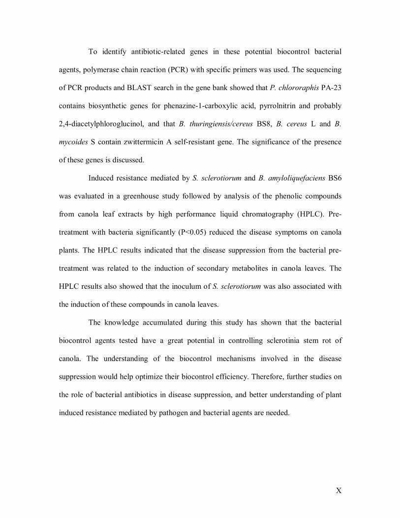
To identify antibiotic-related genes in these potential biocontrol bacterial
agents, polymerase chain reaction (PCR) with specific primers was used. The sequencing
of PCR products and BLAST search in the gene bank showed that P. chlororaphis PA-23
contains biosynthetic genes for phenazine-1-carboxylic acid, pyrrolnitrin and probably
2,4-diacetylphloroglucinol, and that B. thuringiensis/cereus BS8, B. cereus L and B.
mycoides S contain zwittermicin A self-resistant gene. The significance of the presence
of these genes is discussed.
Induced resistance mediated by S. sclerotiorum and B. amyloliquefaciens BS6
was evaluated in a greenhouse study followed by analysis of the phenolic compounds
from canola leaf extracts by high performance liquid chromatography (HPLC). Pre-
treatment with bacteria significantly (P<0.05) reduced the disease symptoms on canola
plants. The HPLC results indicated that the disease suppression from the bacterial pre-
treatment was related to the induction of secondary metabolites in canola leaves. The
HPLC results also showed that the inoculum of S. sclerotiorum was also associated with
the induction of these compounds in canola leaves.
The knowledge accumulated during this study has shown that the bacterial
biocontrol agents tested have a great potential in controlling sclerotinia stem rot of
canola. The understanding of the biocontrol mechanisms involved in the disease
suppression would help optimize their biocontrol efficiency. Therefore, further studies on
the role of bacterial antibiotics in disease suppression, and better understanding of plant
induced resistance mediated by pathogen and bacterial agents are needed.
XI
FOREWARD
This thesis is written in a manuscript style, with each manuscript having its own
abstract, introduction, materials and methods, results and discussion. There is a general
introduction and review of the literature prior to the manuscripts, followed by a general
discussion and conclusions and the references cited.

1
1.0 INTRODUCTION
Canola is a crop derived from rapeseed, which was cultivated as early as 5000
B.C. in China (Yan 1990). To be called �canola�, the oil component must contain less
than 2% erucic acid, and the solid component of the seed less than 30 µmol/g total
glucosinolates (Canola Council of Canada 2001e). Canola is now widely used as edible
oil in human consumption and an important source of proteins for animal feed. It is now
the most important oilseed crop in Canada, and is second only to soybean as the most
important source of vegetable oil in world production (Raymer 2002). The harvested area
of canola in 2003 was 11,587,200 acres in Canada, and the cash receipts totalled $483.6
million in 2002 (Manitoba Agriculture 2003; Canola Council of Canada 2004).
Sclerotinia stem rot caused by Sclerotinia sclerotiorum (Lib.) de Bary is one of
two most economically important diseases on canola in Canada, and losses ranging from
5-100% were reported in Western Canada (Manitoba Agriculture 2002). Sclerotinia
sclerotiorum is an ubiquitous pathogenic fungus that is known to infect over 400 plant
species (Boland and Hall 1994). There are more than 100 species of plants reported to be
the hosts for S. sclerotiorum in Canada (Boland and Hall 1994). The pathogen infects
canola mainly through its petals, which are infected by Sclerotinia ascospores
(Turkington and Morrall 1993). Canola cultivars currently registered in Canada have little
or no resistance to S. sclerotiorum (Kharbanda and Tewari 1996), and only limited
partially resistant cultivars are available in the world (Fuller et al. 1984; Wang et al.
2003; Kharbanda and Tewari 1996). The control of sclerotinia stem rot of canola is
mainly achieved by using fungicides, due to the difficulty of using cultural methods, such
2
as rotation and tillage to reduce the inoculum of the pathogen. However, public concern
of environmental pollution and pathogen resistance has facilitated research on alternative
ways such as biological control to control this disease (Bardin and Huang 2001).
Biological control is a method that is economical and most important,
sustainable, to achieve the goal of increasing crop yield (Cook and Baker 1983d). The
biocontrol agents used to control S. sclerotiorum are mainly parasitic fungi used in soil
applications, such as Coniothyrium minitans Campbell (Huang 1980). However, many
field studies failed to consistently control this pathogen (Hedke et al. 1999; McQuilken et
al. 1995), due to the fact that Sclerotinia ascospores can disperse long distance (Venette
1998) and that even a reduced number of sclerotia in a field can cause significant
infection and yield loss (Davies 1986). Therefore studies on foliar applied biocontrol
agents are worthwhile (Boyetchko 1999).
Bacterial biocontrol agents against S. sclerotiorum are rarely studied
(Boyetchko 1999). Some bacterial strains have shown antifungal activity to S.
sclerotiorum, such as Erwinia herbicola (Godoy et al. 1990c) and Bacillus spp. (Tu 1997;
Godoy et al. 1990c; Huang et al. 1993); however, most of the studies did not consistently
achieve disease suppression under field conditions (Yuen et al. 1992; Boland 1997).
Savchuck (2002) screened several bacterial antagonists against S. sclerotiorum in the lab
and greenhouse, and Sclerotinia control by Pseudomonas chlororaphis strain PA-23 and
Pseudomonas spp. strain #41 was evaluated under field conditions in 2001. A double
application of P. chlororaphis PA-23 showed significantly (P<0.05) lower disease
incidence of Sclerotinia on canola stem under field conditions in 2001, but biocontrol
3
mechanisms involved in the disease suppression were unclear. In order to continue the
field study of Pseudomonas chlororaphis PA-23 and Pseudomonas spp. #41, a similar
field study was carried out in 2002, but the experiment failed simply because of lack of
pathogen inoculum (ie. ascospores) at flowering.
An understanding of biocontrol mechanisms would benefit the utilization of
biocontrol agents. Several biological mechanisms might be involved in disease
suppression, which include parasitism, competition, antibiosis and plant induced
resistance, etc. (Handelsman and Stabb 1996).
The first objective of this study was to screen bacterial biocontol agents, mainly
Bacillus spp. in the lab and greenhouse, and evaluate their biocontrol abilities along with
Pseudomonas chlororaphis PA-23 and Pseudomonas spp. #41 under field conditions.
Methods of Sclerotinia inoculation and disease evaluation in the greenhouse and the field
will be developed, and bacterial population dynamics will be investigated.
The second objective was to study the biocontrol mechanisms involved in
disease control. Due to preliminary observations made during this study, antibiosis and
plant induced resistance in biocontrol will be investigated. The presence of antibiotic
biosynthetic genes or self-resistant genes will be studied by polymerase chain reaction
(PCR) and subsequent sequencing of the PCR-products. The plant induced resistance will
be studied in a greenhouse experiment by comparing the phenotypic changes among
different treatments, and the accumulation of phenolic compounds from leaf extracts will
be investigated over time.

4
2.0 LITERATURE REVIEW
2.1 Canola
2.1.1 Crop History
Canada plays a lead role in large-scale production of rapeseed-canola; however,
she is relatively new to the rapeseed cultivation. The earliest proof of rapeseed cultivation
has been found back to 5000 B.C. in China based on archaeological discoveries (Yan
1990). It was recorded in use in India about 2000 B.C., and was introduced to Japan from
China or the Korean Peninsula over 2000 years ago (Bell 1982). The rapeseed comes
from three species: Brassica campestris, B. napus, and B. juncea (L.) Czernj. &Cosson
(Shahidi 1990). Brassica campestris was introduced to Canada by a Polish immigrant
Fred Solvonik in 1936 (Bell 1982) and B. napus was introduced by the Canada
Department of Agriculture prior to World War II (Busch et al. 1994).
The shortage of marine lubricant in the World War II led the Canadian
government to encourage the rapeseed planting through a price support program; large
quantities of rapeseed were grown in the prairie provinces of Saskatchewan, Manitoba,
and Alberta. The production of rapeseed fell in late 1940s when the government support
ceased, however, Canada was far from self-sufficient in edible oil production at that time
(Busch et al. 1994). Therefore, the question of transforming rapeseed oil to largely used
edible oil was raised. The erucic acid and glucosinolates in rapeseed were the two major
concerns. High erucic acid diet has been found to have several detrimental effects on rats,
such as formation of high level of fat in heart, formation of fibrous tissue in the heart
muscle, growth retardation, reductions in mitochondrial oxidation of glutamate and in the
5
rate of ATP synthesis in the heart muscle (cited inGriffiths et al. 1998). Glucosinolates
are the sulphur compounds that give mustard their sharp taste, and several reports have
shown that high concentrations of glucosinolates in diet have adverse effects on pigs,
rats, laying hens, ducks, geese, quail and turkeys, which included detrimentally affecting
their growth, food intake, thyroid size or hormone levels (cited inGriffiths et al. 1998).
In 1967, Jan Krzymanski from Agriculture Canada Research Station of
Saskatoon discovered a cultivar, Bronowski, with low levels of glucosinolates (Busch et
al. 1994). In 1974, Baldur Stefansson of the University of Manitoba developed world�s
first zero-erucic, low glucosinolate B. napus cultivar, Tower (Bell 1982). Later Sid
Pawlowski of Saskatoon station successfully crossed two species, B. napus and B.
campestris, and introduced a third species, B. juncea (Busch et al. 1994). In 1977, a new
cultivar, Candle, was released, which became the world�s first zero erucic acid low-
glucosinolate, B. campestris cultivar (Bell 1982).
In the late 1970s, the term �canola� was registered by the Western Canadian
Oilseed Crushers� Association, which is now called the Canadian Oilseed Processor
Association (Canola Coucil of Canada 2001). Present official definition of canola
requires an oil that contains less than 2% erucic acid, and the solid component of the seed
contains less than 30 micromoles of any one or any mixture of 3-butenyl glucosinolate, 4-
pentenyl glucosinolate, 2-hydroxy-3 butenyl glucosinolate, and 2-hydroxy-4-pentenyl
glucosinolate per gram of air-dry and oil-free solid (Canola Coucil of Canada 2001).
Currently, Canada has approximately 110 registered B. napus, and 28 B. rapa
spring varieties, 3 B. napus winter varieties, 3 high erucic acid rapeseed (HEAR)
6
varieties, 76 herbicide-resistant varieties and another 12 varieties registered in Eastern
Canada only (Canola Council of Canada 2003).
2.1.2 Crop usage
Canola is now widely used as an edible oil. In January 1985, canola oil was
granted Generally Recognized as Safe (GRAS) status in use of foods by the U.S. Food
and Drug Administration. Canola oil has lower levels of saturated fats than any other
widely used edible oil. It is also rich in monounsaturated fatty acids and has an
intermediate level of polyunsaturated fatty acids. The three key nutrients of canola oil �
oleic acid, alpha-linolenic acid (ALA) and vitamin E help protect blood vessels against
early, damaging events in the development of artherosclerosis, which is an inflammatory
disease in which lipids build up within the artery wall, thickening the blood vessel wall
and restricting blood flow (McDonald 2001; Morris 2002). In addition, canola oil also
performs well in food processing and fast food industry due to its desirable properties
(Busch et al. 1994).
Canola also serves as an important source of protein for animal feed. Canola
meal is widely used in pig diets around the world. Current data showed that canola meal,
when properly formulated in pig diet, will support high levels of feed intake and efficient
performance (Canola Council of Canada 2001b).
Canola is well adapted to cool, short-season conditions found on the Canadian
prairies and northern Great Plains border states of the U.S.A. (Johnston et al. 2002). Its
role in double-cropping and rotation has been extensively studied under different systems
7
worldwide, including effects on soil-borne diseases (Wilson and Phillips 1987 ; Nielsen
et al. 2003; Gossen and Derksen 2003; Turkington et al. 2000), crop production (Arshad
et al. 2002; Wilson and Phillips 1987; Pullins and Myers 1998; Maali and Agenbag 2003;
Soon and Clayton 2002), soil microbial biomass (Lupwayi et al. 2001), weed community
(Blackshaw et al. 2001) and mineral levels (Soon and Clayton 2002; Soon and Arshad
2002).
2.1.3 Transgenic canola
Transgenic canola varieties have been introduced to Western Canada since
1995. In 2000, over 80% of growers in Western Canada chose transgenic varieties and
planted them on approximately 55% of the 12 million canola acres (Canola Council of
Canada 2001a). The registered transgenic canola in Canada are RoundUp Ready
(glyphosate (N-(phosphonomethyl) glycine) tolerant), Liberty Link (glufosinate (2-
amino-4-(hydroxybenzonitrile) butanoic acid) tolerant) and the Navigator varieties
(bromoxynil (3,5-Dibromo-4-hydroxybenzonitrile) tolerant) (Stringam et al. 2003). The
key benefit and motivator for growers to adopt transgenic canola is due to more efficient
weed control and higher yield comparing to conventional canola varieties. Data in 2000
showed that averagely transgenic varieties resulted in a 3 bu/acre or 10% yield and
$5.80/acre increase in net return advantage over conventional varieties (Canola Council
of Canada 2001a). Since the early 1990�s, growers in Western Canada have been
reducing their tillage operations for soil conservation benefits. Before the introduction of
transgenic canola varieties, growers use tillage or incorporating herbicides to control
weed prior to seeding a crop. With transgenic herbicide tolerant varieties, weed control
8
can be done �in-crop� and thereby tillage was reduced and soil conservation was
achieved (Canola Council of Canada 2001a).
In addition to herbicide resistance, transgenic canola was studied in various
systems. Bacillus thuringiensis (Bt) cry 1Ac gene was introduced on canola for insect
control (Stewart et al. 1996; Ramachandran et al. 1998a; Halfhill et al. 2001;
Ramachandran et al. 1998b). Other transgenic varieties or study on canola include
resistance to Leptosphaeria maculans, a causal agent of blackleg of canola (Kazan et al.
2002), kanamycin resistance (Arnoldo et al. 1992), metal tolerance (Nie et al. 2002;
Anoop et al. 2003) and enhanced expression of nutrients, such as methionine (Altenbach
et al. 1992), lysine (Falco et al. 1995), and phytase (Ponstein et al. 2002).
The common concern of commercial release of transgenic canola into cropping
system is the risk of gene flow from the genetically modified (GM) canola and the risk of
them becoming volunteer weeds (Hall et al. 2000; Kwon and Kim 2001). Studies have so
far not been able to give a clear answer for the risk of using transgenic canola (Rieger et
al. 2002; Andow 2003; Baker and Preston 2003; Kwon et al. 2001; Kwon and Kim 2001).
2.1.4 Production and economic importance
Canola is the most important oilseed crop in Canada. The harvested area of
canola in 2003 was 11,587,200 acres, and Saskatchewan had almost always the largest
harvest acreage among all the provinces in Canada. The average yield of canola in
Canada is 0.567 tonnes/acre (25.4 bushels/acre) in 2003 (Canola Council of Canada
2004). Annual canola cash receipts rose from $197.1 million in 1992 to a record of
9
$622.3 million in 1998, but totalled $483.6 million in 2002. Manitoba exported rapeseed
and canola seed valued at $205.5 million in 2002. Approximately 99% of canola exports
went to Japan, Mexico, the U.S.A. and China (Manitoba Agriculture 2003).
Canola is also produced extensively in Europe, Asia, Australia, and to a more
limited extent in the United States. During the past 20 years, this crop has passed peanut,
sunflower and, most recently, cottonseed in worldwide production, and become second
only to soybean as the most important source of vegetable oil (Raymer 2002).
2.1.5 Growth stages and condition
Canola growth stages consist of pre-emergence (germination), seedling, rosette,
bud, flowering and ripening. The length of each growth stage is greatly influenced by
temperature, moisture, light, nutrition and variety. Canola is a relatively cool season crop,
and its best growth occurs above 12 ºC and below 30 ºC. The optimum temperature for
maximum canola growth and development has been estimated at just over 20 ºC (Canola
Council of Canada 2001d). Soil moisture is one of the major factors controlling canola
germination. Canola seed requires a high percentage of its weight in water before
germination can begin. Vegetative and root growth result in a gradual increase in water
use, and the peak use period occurs during the flowering stage. As the crop ripens, its
ability to transmit water from the soil declines and water use decreases. Canola plants
require a threshold amount of water before any yield is obtained. Beyond that threshold
increasing amounts of water will result in higher yields. Usually 25 mm of water will
result in about 150 kg/ha. (2.75 bu/ac.) of yield with good growing conditions and
adequate fertility (Canola Council of Canada 2001c).
10
2.1.6 Diseases on canola
Canola diseases are mainly caused by fungal pathogens except herbicide injury,
sulphur deficiency and Aster yellows caused by mycoplasma like organisms (Martens et
al. 1994). Fungal diseases of canola and pathogen names are shown in Table 2.1.
Seedling stand establishment is a widespread problem of canola production.
Poor stand establishment may be caused by a seedling disease complex, which is caused
primarily by Rhizoctonia solani, but Fusarium spp. and Pythium spp. may also be
present. The symptoms of this complex appear during the four weeks following seeding,
or up to the fourth leaf stage. The complex exhibits several symptoms called seed decay,
pre- and post-emergence damping-off, seedling blight and seedling root rot (Martens et
al. 1994).
Blackleg caused by Leptosphaeria maculans and sclerotinia stem rot caused by
Sclerotinia sclerotiorum are the two most economically important diseases on canola in
Canada. Blackleg is becoming an increasing problem in the Canadian prairies. Based on
interaction phenotypes on the differential canola cultivars: Westar, Glacier, and Quinta,
the blackleg isolates can be ranged into four pathogenicity groups (PGs): PG1, PG2, PG3
and PG4. PG1 isolates are weakly virulent, and PG2-4 isolates are highly virulent. PG2
strain is now widely spread in Canada. PG3 was recently reported in Western Canada by
Fernando and Chen (2003).
Sclerotinia stem rot will be described in detail in the following chapter, as this is
the focus of the thesis.
11
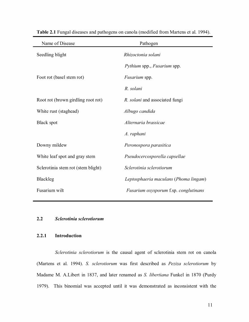
Table 2.1 Fungal diseases and pathogens on canola (modified from Martens et al. 1994).
Name of Disease Pathogen
Seedling blight Rhizoctonia solani
Pythium spp., Fusarium spp.
Foot rot (basel stem rot) Fusarium spp.
R. solani
Root rot (brown girdling root rot) R. solani and associated fungi
White rust (staghead) Albugo candida
Black spot Alternaria brassicae
A. raphani
Downy mildew Peronospora parasitica
White leaf spot and gray stem Pseudocercosporella capsellae
Sclerotinia stem rot (stem blight) Sclerotinia sclerotiorum
Blackleg Leptosphaeria maculans (Phoma lingam)
Fusarium wilt Fusarium oxysporum f.sp. conglutinans
2.2 Sclerotinia sclerotiorum
2.2.1 Introduction
Sclerotinia sclerotiorum is the causal agent of sclerotinia stem rot on canola
(Martens et al. 1994). S. sclerotiorum was first described as Peziza sclerotiorum by
Madame M. A.Libert in 1837, and later renamed as S. libertiana Funkel in 1870 (Purdy
1979). This binomial was accepted until it was demonstrated as inconsistent with the
12
international Rules of Botanical Nomenclature, so the name S. libertiana was changed to S.
sclerotiorum (Lib.) Massee (Duncan 2003b; Wakefield 1924). However, it was later found
that de Bary had used this Latin name earlier, so the proper name was established as S.
sclerotiorum (Lib.) de Bary (Purdy 1979).
2.2.2 Taxonomy
Sclerotinia sclerotiorum, S. trifoliorum, and S.minor belong to the Sclerotiniaceae
(Whetzel 1945 cited in Duncan 2003b) a family of the Class Ascomycotina. The early
taxonomy of the three species was based on the size and general characteristics of the
sclerotium, host range, and dimensions of ascospores and asci (Willetts and Wong 1980).
However, several studies (Purdy 1955; Price and Colhoun 1975; Grogan 1979) showed that
this system was inadequate and that a number of species, originally thought to be unique,
were actually all members of the S. sclerotiorum species.
2.2.3 Host range
S. sclerotiorum is an ubiquitous pathogen, which has an extremely wide host
range. The earliest record of the S. sclerotiorum host range is from a thesis by Dickson
(1930), of 172 species, in 118 genera and 37 families (cited in Willetts and Wong 1980). It
was updated by Boland and Hall (1994) to 75 plant families, 278 genera, 408 species, and
42 subspecies, with most of them present in the Dicotyledonae subclass of Angiospermae.
The wide host range limits the crop rotations (Boland and Hall 1994). Flax (Linum
usitatissimum L.) used to be regarded as a non-susceptible host crop that could be used in
13
rotation, but a recent study by Rashid (2000) found that S. sclerotiorum could successfully
infect flax in Manitoba and Saskatchewan (Duncan 2003b).
2.2.4 Economic importance
Sclerotinia stem rot is one of the two most devastating diseases of canola in
Western Canada, losses ranging from 5-100% in individual fields (Manitoba Agriculture
2002). In central Manitoba, 52% and 76% of canola fields were affected by Sclerotinia in
2002 and 2001 respectively (Duncan 2003a). It was reported that yield loss from
sclerotinia stem rot in Minnesota and North Dakota was $16,768,955 in total in 2001
(Lamey et al. 2001).
S. sclerotiorum also cause damage to various vegetables (Willetts and Wong
1980). Losses on many crops caused by S. sclerotiorum in North America, Europe, and
Asia were reported (Willetts and Wong 1980).
2.2.5 Disease cycle, infection and symptomology
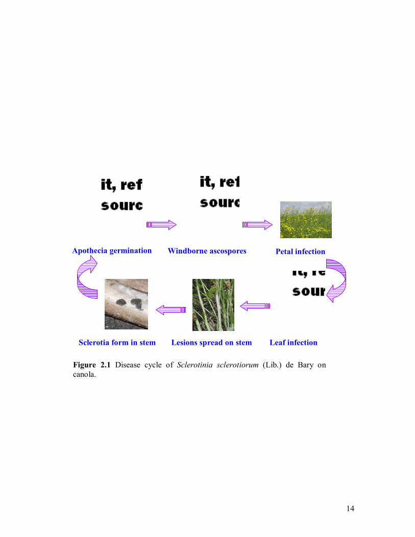
A general life cycle for S. sclerotiorum on canola is shown in Figure 2.1. Ervio,
Halkilahti and Pohjakallio (1964) found that sclerotia, the overwintered structure, can
survive in the soil for up to 4 years (Willetts and Wong 1980). However, Duncan (2003b)
suggested that sclerotial longevity was often overestimated, since his study showed that the
sclerotinia viability reduced to 57.5%, 12.5% and only 2.5% at the 0 cm (soil surface), 5
cm and 10 cm depth, respectively, after buried for 12 months. When conditions are
appropriate, sclerotia can germinate either carpogenically (sexual stage) or
myceliogenically (asexual stage) (Willetts and Wong 1980). A cold conditioning period at
14
Figure 2.1 Disease cycle of Sclerotinia sclerotiorum (Lib.) de Bary on canola.
Apothecia germination Windborne ascospores
Lesions spread on stem Leaf infection
Petal infection
Sclerotia form in stem

15
10 ûC (Huang and Kozub 1989; Huang and Kozub 1991b) or 4 ûC (Smith and Boland 1989)
is usually required for carpogenic germination when sclerotia are produced at temperatures
higher than 20 ûC, except for when sclerotia produced on potato dextrose agar (Huang and
Kozub 1993).
The infection occurrs mainly by ascospores, which are discharged from apothecia
into the air. The sclerotia germinate in the summer, producing mushroom-like structures.
These release wind-borne spores that travel up to 1 km (Manitoba Agriculture 2002) or
several miles (Venette 1998).
Interestingly, senescing blossoms are the most important nutrient source for
ascospore germination (Turkington and Morrall 1993). Sclerotinia ascospores can remain
viable for years under laboratory conditions, but spores in the field can generally only
survive for limited periods (Willetts and Wong 1980), although it was reported that they
could survive up to several months under dry conditions (Willetts and Wong 1980). It takes
a few hours for ascospores to germinate on the petals and at least 24 hours for petals to
infect canola plants, providing the petals were wet (McCartney et al. 2001). The ascospore
germination is optimum at temperatures of 20-25 ºC (Venette 1998). Chupp and Shef
(1960) stated that Sclerotinia spp. could infect susceptible hosts over a range of
temperatures from 0-25 ºC, with optimum at 15 to 20 ºC . However, McCartney et al.
(2001) stated that under close saturated humid conditions, lesions were initiated in 2-4 days
at temperatures 20-25 ºC and continuous high humidity was needed between 24-48 hours to
initiate lesion formation from ascospores on petals placed on leaves. Several studies
showed that humidity and leaf wetness played an important role in oilseed rape/canola

16
infection via petals (Venette 1998; McCartney et al. 2001). S. sclerotiorum can also spread
through plant-to-plant contact by mycelium, but this form of infection is rare (Venette
1998).
Mechanical pressure seems to be of major importance in initial penetration of the
host plant, but subsequent colonization occurs mainly by enzymic tissue dissolution
(Willetts and Wong 1980). Echandi and Walker (1957) showed that S. sclerotiorum
produced pectin methylesterase and polygalacturonase on wheat bran, and Hancock
(Hancock 1966) showed that polygalacturonase could break down pectic substances in
sunflower and tomato tissues. Hemicellulolytic enzymes (Hancock 1967) and cellulolytic
enzymes (Lumsden 1969) produced by S. sclerotiorum were found to be capable of
degrading araban and galactan in sunflower hypocotyls and degrading native cellulose
from bean cell wall, respectively. Maxwell and Lumsden (1970) found that oxalic acid
(OA) was produced by S. sclerotiorum and it appeared to act synergistically with pectic
and cellulolytic enzymes for destruction of host tissues. OA deficient mutants of S.
sclerotiorum were found non-pathogenic in bioassays using bean plants though harbouring
a full complement of degradative enzymes (Godoy et al. 1990a); therefore, OA is an
essential pathogenicity determinant (Dickman and Chet 1998). The accumulation of oxalic
acid offers a lower pH environment, which is a better condition for the activities of other
enzymes (Dutton and Evans 1996).
The sclerotinia stem rot symptoms begin on canola usually as a soft, watery rot on
leaves or stems. The plants then develop pale-grey to white lesions, at or above the soil line
and on upper branches and pods. When a stem is completely girdled, the plant wilts and
17
dies. The diseased stems usually appear to shred and hard black sclerotia form in the
hollow centre of the stem. Thomas and Evans (1981) in Alberta demonstrated that there
was a close relationship between Sclerotinia incidence (percent infected stems) and disease
loss (Lamey 1998).
2.2.6 Epidemiology
S. sclerotiorum is currently found across Canada (Bardin and Huang 2001) as
well as in countries such as U.S.A. (Koike 1999), Norway, Australia (Kohli and Kohn
1996) and China (Zhao and Meng 2003). The pathogen is mainly transported and
initially infected canola as ascospores, and ascopores were reported to be able to travel up
to several miles by wind (Venette 1998). Although mycelial infection by plant contact is
limited, but it could be greatly favoured when plant lodging happens. Stelfox et al. (1978)
reported that S. sclerotiorum-contaminated rapeseed pollen grains transported by honey
bees were involved in the spread of sclerotinia pod rot of rapeseed (Bardin and Huang
2001).
Sclerotinia stem rot is favoured by cool temperatures and prolonged periods of
precipitation. In dry weather, disease progression can be slowed or stopped, but resumes
when extended periods of plant wetness favour fungal growth (Venette 1998). Several
factors may contribute towards plant wetness, such as closed canopies, narrow rows,
restricted wind patterns, irrigation, prolonged dew, and frequent rains.
18
2.3 Management of S. sclerotiorum on canola
2.3.1 Host resistance
Canola cultivars currently registered in Canada have little or no resistance to S.
sclerotiorum (Kharbanda and Tewari 1996). Plant resistance is very difficult to attain for
this pathogen, as it is controlled by additive gene action as demonstrated in bean (Fuller et
al. 1984). Krűger and Stoltenberg (1983) have developed cultivars less susceptible to S.
sclerotiorum, but lower yield was found in these cultivars in sclerotinia-free conditions
(cited in Kharbanda and Tewari 1996). Recently, a few partial resistant cultivars were
reported in China, including Zhongyou 821 and Zhongshuang No.9. Zhongshuang No. 9
was found to be resistant to S. sclerotiorum and lodging, and contains only 0.23% erucic
aicd and 22.69 µmol/g glucosinolates in commercial seeds and also has high yield and
extensive adaptability (Wang et al. 2003).
2.3.2 Cultural management
Several cultural methods can be used to help control sclerotinia stem rot on
canola. Seed without sclerotia contamination should be used. Studies by William and
Stelfox (1980) and Morral and Dueck (1982) showed a 3- to 4-year rotation did not
reduce the incidence of the disease on canola. Nelson (1998) showed that even when the
sclerotia population is as low as 1 sclerotium/800cm of soil, 5 to 6 years rotation were
needed and even such a long rotation did not guarantee the elimination of S. sclerotiorum.
Since S. sclerotiorum has an extremely wide host range, it attacks crops such as beans,
sunflowers, mustard, sweet clover, and potatoes; therefore, it also limits the crops that
can be used in a rotation to manage the disease.
19
Tillage can be regarded as an effective method of reducing the disease by
burying the sclerotia (Gulya et al. 1997). However, Kurle et al. (2001) found carpogenic
germination is more probable within the upper 5 cm soil profile, and this result is
consistent with the study from Duncan (2003b). A significant negative relationship was
found between sclerotial viability and depth of burial, and between sclerotial viability and
populations of colonizing bacteria under zero-tillage condition (Duncan 2003b). These
results suggest proper tillage might help reduce the disease, but what kind of tillage
should be employed has not been clearly answered.
Larger plant spacing will help prevent pathogen from spreading through plant-to-
plant contact, and also decrease the relative humidity, which will slow down the disease
progression. Using lodging resistant cultivars will also help reduce the disease.
2.3.3 Disease forecasting
The current sclerotinia forecasting is mainly based on weather forecasting and
petal test that is available by using a commercial test kit. To help canola growers predict
outbreaks of sclerotinia stem rot, several disease forecasting systems have been developed
in Canada, such as Alberta�s online forecasting system (Government of Alberta 2004) and
Agrometeorological Centre of Excellence in Carman, Manitoba (ACE 2001).
The petal test was developed to predict the disease based on a significant
relationship between disease incidence and the level of infestation of rapeseed petals by S.
sclerotiorum ascospores at early bloom stage (Turkington et al. 1991a). This method
suggested petals should be sampled in the afternoon, due to the diurnal variation of the
20
petal infestation level (Turkington et al. 1991b). Several factors, such as canopy density,
rain fall and sample size, were shown to influence the relationship between disease
incidence and petal infestation (Turkington et al. 1991b; Turkington and Morrall 1993;
Turkington et al. 1988).
2.3.4 Chemical control
The main method of control of Sclerotinia diseases is achieved by applying
fungicides, like Benlate® (benomyl), Ronilan® (vinclozolin), Rovral® (iprodione) and
Quadris® (azoxystrobin) (Minister of Agriculture 2001; Morrall et al. 1985). The
production of Benlate® has already been ceased in 2001 due to the public heath concerns
and the claiming of crop damage by farmers (Gilmour 2001). Fungicides should be applied
before symptoms of stem rot are visible, at the 20-30% bloom stage of the crop.
2.3.5 Biological control
Biological control is a promising method of control of Sclerotinia diseases
(Bardin and Huang 2001). The concept of biological control, biocontrol mechanisms and
application in control of Sclerotinia will be discussed in the following chapter, as it is the
focus of this thesis.
2.4 Biological control
2.4.1 Introduction
Biological control is the reduction of the amount of inoculum or disease-
producing activity of a pathogen accomplished by or through one or more organisms other
21
than man (Cook and Baker 1983a). The first direct application of biocontrol antagonists to
control plant pathogen was made by C. Hartley (1921) inoculating soil with 13 antagonistic
fungi in an attempt to control damping-off of pine seedlings (Cook and Baker 1983d).
There has been a remarkable increase of interest and research on biological control since
1960s, because of the concern about environmental pollution, development of pesticide
resistant in pathogens, lake of adequate or reliable resistance in crops to many important
pathogens, and the trend towards more intensive farming with less crop rotation (Cook and
Baker 1983d)
Antagonists are biological agents with the potential to interfere in the life
processes of plant pathogens, such as fungi, bacteria, nematodes, protozoa, viruses,
viroids, and seed plants (e.g., trap plants) (Cook and Baker 1983b). Biological
mechanisms involved in disease suppression are diverse and include parasitism,
competition, antibiosis and plant induced resistance (Handelsman and Stabb 1996).
2.4.2 Biocontrol mechanisms
2.4.2.1 Antibiosis
2.4.2.1.1 Introduction
W. Roberts (1894) was the first to note antibiotic action in cultures and
introduced the term antagonism into microbiology (Cook and Baker 1983c). One
remarkable example of application of antibiosis in the early study of S.Y.Yin et al. (1957)
is that a Streptomyces spp. was selected from among 4000 isolates of actinomycetes from
roots of cotton and alfalfa based on in vitro antibiosis to Rhizoctonia solani and
22
Verticillium albo-atrum, and that the strain was used on 6 million hectares of cotton over
30 years, giving increased crop growth in China (cited in Cook and Baker 1983c). There
are numerous reports on the evidence of antibiosis in biocontrol of plant pathogens. Some
of the recent studies include the antagonistic effects of Streptomyces violaceusniger strain
G10 on Fusarium oxysporum f. sp. cubense race (Getha and Vikineswary 2002), the
biocontrol of fire blight which caused by Erwinia amylovora by Pantoea agglomerans
strain Eh252 in orchards (Stockwell et al. 2002) and the biocontrol of Pythium damping-
off of pea by Burkholderia cepacia (Heungens and Parke 2001).
There are numerous reports that antifungal metabolites produced by bacteria in
vitro are involved in disease suppression in vivo. These antifungal metabolites include
phenazine-1-carboxylic acid (PCA) (Mavrodi et al. 1998; Delaney et al. 2001), 2, 4-
diacetylphloroglucinol (2,4-DAPG) (Nowak Thompson et al. 1994; Mavrodi et al. 2001),
pyoluteorin (Plt) (de Souza and Raaijmakers 2003; Nowak Thompson et al. 1999;
Brodhagen et al. 2004), pyrrolnitrin (Prn) (de Souza and Raaijmakers 2003; Chernin et al.
1996), zwittermicin A (Raffel et al. 1996), butyrolactones (Gamard et al. 1997), hydrogen
cyanide (Flaishman et al. 1996), kanosamine (Milner et al. 1996a), oligomycin A (Kim et
al. 1999), oomycin A (Howie and Suslow 1991), etc.
2.4.2.1.2 Major bacterial antibiotics in biological control
The ability to produce phenazines is limited almost exclusively to bacteria and
has been reported in Pseudomonas, Streptomyces, Nocardia, Sorangium, Brevibacterium,
and Burkholderia spp. (Turner and Messenger 1986). Phenazines are produced by a
considerable number of Pseudomonas biocontrol strains (Chin-A-Woeng et al. 1998;
23
Anjaiah et al. 1998; Tambong and Hofte 2001; Thomashow et al. 1990; Thomashow and
Weller 1988). Delaney et al (2001) stated that despite the phenazine biosynthetic locus
being highly conserved among fluorescent Psedomonas spp., individual strains differ in
the range of phenazine compounds they produce. Phenazine-1, 6-dicarboxylic acid is the
first phenazine formed, and it is thought to be converted to PCA, a key intermediate in
the synthesis of other phenazines by fluoresent pseudomonads (Delaney et al. 2001). The
mode of action of phenazines has been studied extensively and several strains have
already been developed into commercial biocontrol products (Chin-A-Woeng et al.
2003).
The antibiotic 2, 4-DAPG is a major determinant in the biocontrol activity of
the plant growth-promoting rhizobacteria (McSpadden Gardener et al. 2001). Numerous
studies have demonstrated that 2, 4-DAPG producing Pseudomonas spp. can suppress a
wide variety of plant pathogens including fungi, bacteria and nematodes (Cronin et al.
1997a; Cronin et al. 1997b; Fenton et al. 1992; Duffy and Defago 1997; Keel et al. 1992).
Antifungal action of P. fluorescens CHA0, P. fluorescens Q2-87 and P. fluorescens F113
against Thielaviopsis basicola, Gaeumannomyces graminis var. tritici and Pythium
ultimum is due to the production of 2, 4-DAPG ( Fenton et al. 1992; Harrison et al. 1993;
Keel et al. 1992; Shanahan et al. 1992)
Plt and Prn were reported to be isolated from several Pseudomonas and
Burkholderia spp., and both of them play an important role in the suppression of multiple
plant diseases (de Souza and Raaijmakers 2003). Plt production has also been
24
documented for strains of Enterobacter agglomerans, Myxococcus fulvus, Corallococcus
exiguous, Cystobacter ferrugineus and Serratia spp. (Hammer et al. 1999).
Zwittermicin A is an aminopolyol antibiotic produced by B. cereus and B.
thuringiensis (Stabb et al. 1994; He et al. 1994; Raffel et al. 1996; Stohl et al. 1999b). A
worldwide survey of Bacillus spp. soil insolates showed that there were 104 cfu of
zwittermicin A producers in every gram of soil tested (Stabb et al. 1994; Raffel et al.
1996). Zwittermicin A does not only inhibit diverse eukaryotes and prokaryotes, but also
interact synergistically with Bt toxin to enhance the insecticidal activity of B.
thuringiensis (Broderick et al. 2000; Silo Suh et al. 1994).
Interestingly, some bacteria can produce more than one antibiotic. Several P.
fluorescens strains produce both Prn and Plt (de Souza and Raaijmakers 2003).
Pseudomonas spp. strain PHZ48 produces both phenazine and pyrrolnitrin (de Souza and
Raaijmakers 2003). P. cepacia strain 5.5B, a biocontrol agent of Rhizoctonia solani was
also reported to produce both phenazine and pyrrolnitrin (Cartwright et al. 1995). P.
fluorescens Pf-5 was reported to produce 2,4-DAPG, pyoluteorin and pyrrolnitrin
((Nowak Thompson et al. 1994;de Souza and Raaijmakers 2003). B. cereus UW 85 was
reported to produce both zwittermin A and kanosamine (Milner et al. 1996a; Silo Suh et
al. 1994). Biocontrol agents with ability of producing multi-antibiotics might have greater
potential for broad-spectrum disease suppression.
25
2.4.2.1.3 Identification and characterization of antibiotic-related genes/gene clusters
The application of molecular methods expands our knowledge of antibiotic
production. Several genes or gene clusters responsible for major antibiotics, such as
phenazine, 2, 4-DAPG, plt, prn and zwittermicin A have been isolated and characterized.
Phenazine biosynthetic locus was found to contain a seven-gene core operon, designated
phzABCDEFG (Mavrodi et al. 1998) from Pseudomonas fluorescens 2-79, and
phzXYFABCD, from P. aureofaciens 30-84 (Mavrodi et al. 1998; Delaney et al. 2001),
respectivey. This biosynthetic operon is responsible only for synthesis of PCA. A gene
phzO which located immediately downstream of the core biosynthetic operon of strain
30-84 was not found accordingly in strain 2-79, and phzO was found to associate with the
biosynthesis of 2-hydroxylated phenazine compounds in strain 30-84 (Delaney et al.
2001). Six open reading frames (PhlEDBCAF) responsible for the biosynthesis of 2,4-
DAPD were identified, and phlD is responsible for the production of
monoacetylphloroglucinol (MAPG). PhlA, PhlC and PhlB are necessary to convert
MAPG to 2,4-DAPG, and they may also function in the synthesis of MAPG (Bangera
and Thomashow 1996; Bangera and Thomashow 1999). The pyoluteorin biosynthetic
gene cluster was identified and characterized for P. fluorescens Pf-5 (Kraus and Loper
1995; Nowak Thompson et al. 1999), and there are ten genes (plt) required for the
biosynthesis of plt. Hammer et al. (1997) found that four genes (prnABCD) are required
for prn production, and the function of each gene product was further described (Kirner et
al. 1998). Hammer et al. (1999) also found that the prnABCD gene cluster was highly
conserved among six pyrrolnitrin-producing strains, including Pseudomonas,
Burkholderia and Myxococcus spp.. Milner et al. (1996a) showed that a 1.2-kb DNA
26
fragment defined an open reading frame referred to as zmaR which encoded a
zwittermicn A resistance determinant. Further study by Stohl et al. (1999b) suggest that
zmaR is necessary for high-level resistance to zwittermicin A, but it is not required by but
temporally associated with zwittermicin A production.
The characterization of these antibiotic-related genes has facilitated the primer
design (Raaijmakers et al. 1997) and the use of the polymerase chain reaction (PCR)
method to rapidly detect these genes. Using PCR method with specific primers to detect
antibiotic-producing bacterial strains has so far been mainly used for screening biocontrol
bacterial agents in soil (Raaijmakers et al. 1997; de Souza and Raaijmakers 2003; Landa
et al. 2002; McSpadden Gardener et al. 2000; Picard et al. 2000). Studies of genotypic
and phenotypic analysis of zwittermicin A-producing strains of B. cereus by Raffel et al
(1996) showed that PCR with specific primers for zmaR is a reliable method to identify
zwittermicin A producers.
2.4.2.2 Plant induced resistance
2.4.2.2.1 Introduction
Induced resistance is a state of enhanced defensive capacity developed by a
plant when appropriately stimulated by microorganisms or environmental stress (van
Loon et al. 1998). Signalling pathways and the defence reactions or responses are the
two main research areas in induced resistance. The defence reactions could happen either
local or both local and in remote. The pathogen-related systemic acquired resistance
(SAR) and rhizobacteria-mediated induced systemic resistance (ISR) are the two major
27
components of plant induced resistance (Pieterse and van Loon 1999; Bakker et al. 2003).
The mechanisms of SAR and ISR has been widely integrated into biological control of
plant pathogens (Bakker et al. 2003; Chin-A-Woeng et al. 2003; van Loon et al. 1998).
Unlike animal immunization, plant induced resistance is generally non-specific against
plant pathogens (van Loon 1997). In cucumber, for example, a primary inoculation with
the fungus Colletotrichum lagenarium, the causal agent of anthracnose, induces SAR
against several diseases caused by different pathogens (Sticher et al. 1997). In addition,
the induced resistance usually persists for a relatively long time in plants, even the level
may change after initial elicitation (Kuc 2001). Therefore protection of plants through
plant induced resistance has a great potential in biocontrol of a wide spectrum of plant
pathogens.
2.4.2.2.2 Induced systemic resistance
The induced systemic resistance (ISR) was described as the mode of action of
disease suppression by non-pathogenic rhizosphere bacteria (Peer et al. 1991; Wei et al.
1991). Bacterial determinants of ISR include salicylic acid (SA), siderophores,
antibiotics, and lipopolysaccharides (LPS) (van Loon et al. 1998). The involvement of
ISR in disease suppression has been found in a wide range of biological control
microorganisms and is a powerful mode of action in the biocontrol of both soilborne and
aerial plant diseases (Bakker et al. 2003). These plant-defence-inducing bacteria also
commonly enhance plant growth and are referred to as plant growth-promoting
rhizobacteria (PGPR). The role of ISR in the biocontrol of plant diseases is focused on
non-pathogenic rhizosphere-colonizing Bacillus and Pseudomonas spp. (Whipps 2001).
28
ISR of cucumber to Colletotrichum orbiculare by some PGPR strains indicate that some
PGPR strains can induce systemic resistance to foliar pathogens when used as a seed
treatment (Wei et al. 1991). Ongena et al. (1999) found the induced resistance by
flurorecent pseudomonads can protect cucumber against Pythium root rot, and two of the
tested strains were found to increase cucumber growth.
2.4.2.2.3 Systemic acquired resistance
Systemic acquired resistance (SAR) refers to the plant resistance triggered by
necrotizing pathogens (Bakker et al. 2003). SAR requires accumulation of salicylic acid
(SA) in the plant (Sticher et al. 1997), while ISR is dependent on intact responses to
ethylene and jasmonic acid (JA) (Pieterse et al. 1998). SAR is also associated with the
pathogenesis-related proteins (PRs) (van Loon 1997), but it was suggested that SA is not
the only signal involved in the induction of PRs, but ethylene and JA might also be
involved (Pieterse and van Loon 1999).
2.4.2.2.4 Plant defence-related secondary metabolites
Plant secondary metabolites are now being revealed as essential components of
defence reactions in determining the fate of many host-pathogen interactions (Grayer and
Kokubun 2001; Goodman et al. 1986; Bennett and Wallsgrove 1994; Nicholson and
Hammerschmidt 1992). These compounds could be constitutive in plants and/or induced
by �attack� or �stress� (Grayer and Kokubun 2001). Plant secondary metabolites include
phytoalexins, plant phenolics, plant terpenes, sesquiterpenoids and sterols, cyanogenic
29
glucosides, non-protein amino acids and glucosinolates, etc. (Bennett and Wallsgrove
1994).
Pytoalexins are low molecular weight, antimicrobial compounds that are both
synthesized by and accumulate in plants after exposure to microorganisms and stress
(Goodman et al. 1986). Chemically, phytoalexins may belong to polyacetylenes,
isoprenes and phenolics, such as flavonoids (Goodman et al. 1986). More than 350
phytoalexins have been chemically characterized from approximately 30 plant families
(Kuc 1995). Most of phytoalexins are from dicotyledons, they were found in all plant
organs, and some of them have structural specificity (Kuc 1995). The term �elicitor� has
been commonly used to refer to compounds that induce phytoalexin synthesis in plants.
There are biotic elicitors, such as bacteria, and abiotic elicitors, such as AgNO3 (Kuc
2001; Mert-Turk et al. 2003).
The evidence of phytoalexins as having a role in defence has been studied in
several aspects: one approach is to show that they accumulate to inhibitory concentrations
at the site of pathogen development (Nicholson and Hammerschmidt 1992). Timing and
cellular localization studies have also provided as evidence that supports a role for
phytoalexins in the resistance of cotton to Xanthomonas campestris (Essenberg et al.
1992; Pierce et al. 1996) and Verticillium dahliae (Mace et al. 1989), oats to Puccinia
coronata (Mayama and Tani 1982), soybean to Phytophthora megasperma (Hahn et al.
1985), and carnation to Fusarium oxysporum f. sp. Dianthi (Niemann et al. 1990). The
modes of action of phytoalexins include disruption of membranes, especially plasma
30
membranes, inhibitory effects on protein synthesis, nucleic acid synthesis and respiration
(Smith 1996).
Several bacterial strains have already been reported to induce plants to produce
phytoalexins. For example, induced resistance of carnation to fusarium wilt by WCS417
involved phytoalexin production (van Peer et al. 1991). A P. aureofaciens strain induced
hypersensitive response (HR) on bean cotyledons and induced defence proteins
resembling the plant�s response to pathogens inducing SAR (Zdor and Anderson 1992).
2.4.2.2.5 Plant phenolics
Phenolic compounds are a group of chemicals composed of one or more
aromatic benzene rings with one or more hydroxyl groups (C-OH) (Armstrong 2003).
There are three main biosynthesis pathways of plant phenolics: Shikimate, Acetate-
melonate, Acetate-mevalonate, which are the same as phytoalexins precursors (Kuc 2001;
Goodman et al. 1986). Lots of phenolics are found to be associated with plant defence
mechanisms, and the modes of action of these compounds include direct toxic effects
(phytoalexin and free radicals formed from lignin precursors) and the active and rapid
deposition of barriers such as lignin (Bennett and Wallsgrove 1994). Therefore, lots of
plant phenolics also are phytoalexins. The accumulation of phenolics is observed in
different cases of disease suppression (Prats et al. 2003; Benhamou and Belanger 1998;
Ongena et al. 2000; Daayf et al. 2003; Paul and Sharma 2002; Benhamou et al. 2000).
The study by Daayf (2000) showed the accumulation of several phenolic compounds in
both susceptible and resistant cultivars of cucumber against powdery mildew was
induced by leaf extract of giant knot weed Reynoutria sachalinensis. A recent study by

31
Singh et al. (2003) showed that resistance in chickpea plants induced by Pseudomonas
strains involed the increased induction of phenolic compounds as well as induced
systemic resistance via SA-dependent pathway. In association with phenol biosynthesis,
the activity of phenylalanine ammonia-lyase (PAL) and other biosynthetic enzymes
might be enhanced, such as tyrosine ammonia-lyase (TAL) (Goodman et al. 1986),
cinnamic acid (Shiraishi et al. 1989) and peroxidases (Southerton and Deverall 1990).
2.4.2.3 Plant growth promoting rhizobacteria
As one kind of the biocontrol agents, plant growth promoting rhizobacteria
(PGPR) increase plant growth either by indirect suppression of diseases caused by
pathogens or by reducing the deleterious effects of minor pathogens (Whipps 2001).
Siddiqui and Shaukat (2002) showed three PGPR strains P. fluorescens CHA0, P.
aeruginosa IE-6 S+ and Bradyrhizobium japonicum 569Sm(r) not only suppressed root-
infecting fungi and root-knot nematodes but also enhanced growth of tomato plants both
under glasshouse and field conditions.
PGPR may also increase plant growth in some other ways, for example, by
solubilization of nutrients such as P (Whitelaw 2000), releasing phytohormones (Beyeler
et al. 1999) and decreasing heavy metal toxicity (Burd et al. 1998).
2.4.2.4 Competition
Competition for niche and/or nutrients is another kind of biocontrol mechanism.
A classical example of niche exclusion is the control of leaf frost injury caused by P.
syringae, which has an ice-nucleation protein on its cell surface. Application of an ice-
32
nucleation-minus mutant could prevent damage caused by pathogenic wild type strains
by competing for the niche with the wild type strains (Chin-A-Woeng et al. 2003;
Lindow et al. 1983).
Competition for nutrients, such as carbon, nitrogen and iron, has been
demonstrated in several studies to reduce the pathogens� capacity to propagate in the soil
(Buyer and Leong 1986; Buyer and Leong 1986; Neilands and Leong 1986; Fernando et
al. 1996). Iron is essential for growth for all organisms, but the availability of solubilized
Fe3+ in soils is limiting at neutral and alkaline pH, leading to Fe3+ limitatation (Chin-A-
Woeng et al. 2003). Under Fe3+ limiting conditions, most organisms will take up ferric
iron through high-affinity iron chelators, designated as siderophores. The ability to
produce efficient siderophores is sometimes associated with the ability to take up related
siderophores from other organisms (Raaijmakers et al. 1995; Koster et al. 1995).
2.4.2.5 Parasitism and predation
Parasitism and predation is a fairly common phenomenon among
microorganisms (Cook and Baker 1983b). The study of Fahima et al. (1992) suggested
that mycoparasitism of Verticillium dahliae microsclerotia by Talaromyces flavus hyphae
may be involved in the biological control of verticillium wilt disease. The parasitism of
macroconidia of Fusarium culmorum by several Pythium spp. was shown to be involved
in disease suppression on barley seedings (Davanlou et al. 1999).
Predation is a phenomenon which appeares more common in microorganisms or
mites that feed on insets or grasses (Boulter et al. 2000; Roy and Pell 2000) other than on
33
plants. One interesting example of predation is Allothrombium mitchelli Davis, a large
red velvet mite, was found to feed on Cryptococcus fagisuga Lindinger, an insect species
and a major component of beech scale disease on American beech gaps in the southern
Appalachian mountains (Wiggins et al. 2001).
2.4.2.6. Other mechanisms
A variety of other mechanisms of biological control have been studied, some of
which include disease reduction by lytic enzymes produced by bacteria and fungi such as
β-(1,3)-glucanases (Ruiz Duenas and Martinez 1996), cellulases (Chatterjee et al. 1995),
and proteases (Szekeres et al. 2004), and some of which are involved in the breakdown of
fungal cell wall (Chin-A-Woeng et al. 2003).
Using oxalic oxidase-producing bacteria in controlling S. sclerotiorum is a
novel biocontrol method. Oxalic acid (OA) is known to play a critical role in the success
of infections caused by S. sclerotiorum (Maxwell and Lumsden 1970; Noyes and
Hancock 1981; Godoy et al. 1990b). Less susceptible cultivars were found to be able to
withstand a higher dose of the compound than those that were known to be more
susceptible to S. sclerotiorum in white bean (Tu 1985) and sunflower (Noyes and
Hancock 1981). Dickman and Mitra (1992) developed a selective plating technique to
identify bacterial strains capable of degrading OA. This method was then used to rapidly
identify potential biocontrol bacterial strains in several subsequent studies (Dickman and
Chet 1998; Savchuck 2002).
34
2.4.3 Biocontrol of S. sclerotiorum
There has been a strong interest in biocontrol of Sclerotinia diseases among
Canadian researchers in the last few decades, due to the increasing concern over the use
of chemical pesticides (Bardin and Huang 2001). Parasitic fungi have been widely
studied as biocontrol agents for S. sclerotiorum, such as Coniothyrium minitans Campbell
(Huang 1980), Talaromyces flavus (Huang and Erickson 2000), and Trichoderma spp.
(Huang and Kozub 1991a; Huang and Kokko 1993; Huang 1980). These biocontrol
agents were applied to the soil and inhibit the sclerotia carpogenic germination. Recent
research on C. minitans (Vrije et al. 2001) has led to the development of a commercial
biopesticide named �Contans�. Hedke et al. (1999) showed 60% disease suppression on
oilseed rape by using �Contans� in a 2-year trial, but their experimental design was based
on macroplots surrounded by guard areas to prevent major influences of invading
external ascopores. Earlier tests in microplots of oilseed rape proved a reduction of soil
inoculum; however, this neither led to disease control nor to a yield improvement
(McQuilken et al. 1995). The reasons for this are generally attributed to the fact that S.
sclerotiorum ascospores dispersal occurs over long distances (Venette 1998) and even a
reduced number of sclerotia in a field can cause significant infection and yield loss
(Davies 1986) thus strongly affecting adjacent plots (Hedke et al. 1999). Therefore
studies on foliar applied biocontrol agents are worthwhile (Boyetchko 1999).
Bardin and Huang (Bardin and Huang 2001) suggested the effective
colonization of bean flowers by C. minitans appears to be an effective mechanism to
prevent Sclerotinia ascospore infection. However, Huang et al. (2000) showed that the
35
efficacy of disease suppression on dry bean was not as consistent as fungicide benomyl in
the filed when C. minitans was applied as a spray. Earlier studies using fungal antagonists
screened from growth-room trials did not control white mold of bean consistently in the
field (Inglis and Boland 1992).
Bacterial biocontrol agents against S. sclerotiorum are rarely studied
(Boyetchko 1999). Godoy et al. (1990c) showed the Erwinia herbicola and Bacillus
polymyxa strains could inhibit the S. sclerotiorum growth in vitro. Bean plants pre-treated
by E. herbicola in growth chambers were found to have significantly (P<0.05) less
disease severity than that of control plants. The effective control of white mold of bean
under the field conditions by E. herbicola was not repeatable in a two-year trial (Yuen et
al. 1992). Lyth et al (1993) reported that Bacillus spp. could reduce apothecium
formation by applying it into the soil. B. cereus Frankland and Frankland, strain alf-87A
was reported to reduce incidence of basal pod rot of pea caused by ascospore infection of
S. sclerotiorum, when applied as a spray (Huang et al. 1993). Foliar spray of B. subtilis
(Ehrenberg) Cohn revealed a reduction in white mold in white bean in a two-year field
trial (Tu 1997). However, inconsistent results were found by Boland (Boland 1997) using
B. subtilis to control white mold of bean between fields. Savchuck (2002) screened
several bacterial antagonists against S. sclerotiorum in the lab and greenhouse, and P.
chlororaphis PA-23 had significant disease suppression of sclerotinia stem rot on canola
under field conditions in 2001. P. chlororaphis PA-23 was shown to produce antibiotic
phenazine-1-carboxylic acid in plate assays (W. G. D. Fernando, unpublished).
Pseudomonas spp. #41 was found to inhibit Sclerotinia ascospore germination and germ
tube growth using microscopic techniques. These results suggest that antibiosis might
36
play a vital role in the Sclerotinia control on canola. Boyetchko (1999) suggest that it was
worthwhile to investigate foliar-applied bacteria to control S. sclerotiorum through
various mechanisms such as antibiosis.
Very few studies have been done on induced resistance to S. sclerotiorum in
canola/oilseed rape. Systemic resistance to S. sclerotiorum by oxalic acid in oilseed rape
was the first report of systemic resistance against S. sclerotiorum in oilseed rape (Toal
and Jones 1999). Oxalic acid is the major pathogenesis factor of S. sclerotiorum. Plants
treated with oxalic acid were reported to have significantly smaller leaf lesions than the
control plants after being challenged by the pathogen (Toal and Jones 1999). No study
has been reported on ISR on canola/oilseed rape elicitated by bacterial agents.
Some related studies on plant induced resistance against S. sclerotiorum were
conducted on sunflower (Bazzalo et al. 1985; Prats et al. 2003) and kiwifruit (Reglinski
et al. 1997; Reglinski et al. 2001). Coumarin phytoalexins were found in sunflower to
inhibit the growth of S. sclerotiorum (Urdangarin et al. 1999). Prats et al. (2003) showed
the amount of phenolic compounds accumulated after inoculation with S. sclerotiorum
correlates with the sunflower line, the time after inoculation and the tissue. Higher
constitutive and induced phenolic content as well as phenylalanine ammonia-lyase (PAL)
activity were found in the most resistant lines, and these differences correlated with the
absence/presence of disease symptoms. Earlier studies also showed that Sclerotinia-
sunflower interaction directed towards soluble phenolics in infected- and non-infected
crop lines and wild relatives, and general lower phenolic accumulation was found in the
Sclerotinia tolerant varieties (Bazzalo et al. 1985).
37
Very few studies have been done in induced resistance on canola/oilseed rape.
Brassinin, methoxybrassinin, and cyclobrassinin were the first phytoalexins reported
from Brassica spp. induced by Leptosphaeria maculans (Soledade et al. 1992). Study in
local and systemic changes in glucosinolates in Chinese and European cultivars of oilseed
rape treated with S. sclerotiorum suggests that glucosinolates induction may be an
important marker of resistance to S. sclerotiorum (Li et al. 1999). Pathogen infection,
insect feeding and mechanic wounding can also induce the changes in glucosinolates
(Doughty et al. 1991; Koritsas et al. 1991; Bodnaryk 1992). These studies indicated that
secondary metabolites might play an important role in canola against pathogen S.
sclerotiorum. However, the current studies on phenolic compounds of rapeseed/canola
are mainly for the agronomic purposes other than phytopathological purposes (Naczk et
al. 1998). No study has been reported on the relationship between phenolic compounds
and induced resistance on canola/oilseed rape against S. sclerotiorum.

38
3.0 EVALUATION OF BIOLOGICAL CONTROL OF
SCLEROTINIA SCLEROTIORUM ON CANOLA (BRASSICA
NAPUS) BY BACTERIA THROUGH IN VITRO SCREENINGS,
GREENHOUSE EXPERIMENTS, AND A FIELD STUDY
3.1 Abstract
Sclerotinia stem rot caused by Sclerotinia sclerotiorum (Lib.) de bary is found
worldwide on canola and rapeseed. Eight Gram-positive bacterial strains isolated from
canola and wheat plants were inhibitory against mycelial growth of S. sclerotiorum on
both PDA and LBA plates. Three Bacillus spp., one Staphylococcus spp. and one
Pseudomonas spp. (control) were tested in whole plant assays. Canola petals were pre-
treated with bacteria, and then inoculated with S. sclerotiorum ascospores (105 spores/ml)
24 h later. Seventeen days after inoculation, bacteria and ascospores-treated (BAT) plants
had only 0.0-1.0 disease severity (DS) compared to 4.5 in ascospores-treated (AT) plants.
Disease incidence (DI) on stems was 0.0-12.5% for BAT plants and 75% for AT plants.
DI on leaves was 0.0-50% and 100% for BAT and AT plants, respectively. The BAT
plants also had healthy seeds and significantly higher yields than those of AT plants. The
time of application experiment in the greenhouse suggested the highest disease
suppression was achieved when B. amyloliquefaciens BS6 was inoculated at the same
time as pathogen. In the 2003 field experiment (Trial two), DI was 5.8-12.5% on stems of
bacteria-treated plots and 6.7% in the fungicide-treated (Rovral Flo) plots, which were
significantly different (P < 0.05) from ascospore-treated control plots (20.4%). The DS
on stems was 0.121-0.283 on bacteria-treated plots and 0.167 on fungicide-treated plots
39
compared to 0.426 in ascospore-treated control plots. The bacterial strains were effective
antagonists against S. sclerotiorum under laboratory, growthroom, and field conditions.
3.2 Introduction
Sclerotinia sclerotiorum (Lib.) de Bary is a pathogen on more than 400 plant
hosts, including canola, on which it causes stem rot (Nelson 1998). It is one of the most
important diseases of canola in Western Canada (Martens et al. 1994), causing yield
losses of up to 70% in Manitoba, Canada (Duncan 2003a). The resistance to S.
sclerotiorum is very hard to built up on canola, because it is governed by multiple genes
(Fuller et al. 1984). Crop rotation is also unrealistic due to the long survival ability of
over-wintered structures (sclerotia) in the soil and the vast host range of this pathogen
(Nelson 1998). These limitations necessitate the use of fungicides, which have adverse
effects on non-target organisms (Campbell 1989) and allow the pathogen to develop
resistance. There is also an increasing concern that pesticide residues in soil and water
may be harmful to the environment (Huang et al. 2000). Due to the relatively unreliable
control attained using cultural methods and due to the public concern over pesticide use,
alternative methods of disease control, such as biological control, should be considered.
Research on biocontrol of S. sclerotiorum has been primarily focused on the
control of carpogenic germination or apothecia production by using hyperparasitic fungal
antagonists (Huang 1980; Huang and Erickson 2000; Huang et al. 1997). Recent research
on the Coniothyrium minitans Campbell (Vrije et al. 2001) has led to the development of
a commercial biopesticide named �Contans�. However, the studies by Hedke et al. (1999)
and (McQuilken et al. 1995) suggest that the reduction of the number of sclerotia or
40
apothecia present in the field by C. minitans may not preclude infection caused by
ascospores from neighbouring fields. In addition, Davies (1986) found that the presence
of only a few apothecia in the field might still result in relatively high disease incidence
levels. Hence, research on limiting petal infection by ascospores in biocontrol of S.
sclerotiorum is needed.
Bacterial biocontrol agents against S. sclerotiorum are rarely studied
(Boyetchko 1999). Early studies by Godoy et al. (1990c) and Yuen (1992) showed that
bacterial strains could be used to control white mold of bean caused by S. sclerotiorum.
Savchuck and Fernando (2004) isolated and screened two Gram-negative bacterial strains
(Pseudomonas cholororaphis PA-23 and Psedomonas spp. #41) against S. sclerotiorum
in the lab and greenhouse. The field trials in 2001 to evaluate these two strains showed P.
chlororaphis PA-23 double application significantly reduced sclerotinia stem rot on
canola (Savchuck 2002). However, the bacterial survival ability in the field was not clear.
During the replication of the field trial in 2002, no disease was caused by inoculating the
soil surface with sclerotia. Further replication of the field study, the concerns of using
ascospores as disease inoculation and investigating bacterial survival ability are
necessary. Bacillus spp. are well known for having a better surviving ability under field
conditions due to the production of endospores (Boyetchko 1999; Collins and Jacobsen
2003). Therefore screening Bacillus spp. to use as phyllosphere biocontrol agents is
needed. In addition, using genotypically different bacteria is essential in the screening
process of biocontrol agents (Roberts and Lohrke 2003); therefore, other Gram-positive
bacteria should also be considered.
41
The objectives of this study were (i) to isolate Bacillus spp. (or other gram-
positive bacteria) antagonists against S. sclerotiorum by preliminary screening for
production of antibiotics, oxalate oxidase and/or volatiles in plate assays, and (ii) to
evaluate a select group of potential antagonists from these assays as to the degree of
control attainable in the greenhouse and in the field (including strains PA-23 and #41). In
the greenhouse study, bacterial agents will be screened by a general biocontrol test and a
selected strain will be evaluated on the effect of the time of application on disease
control. Two field trials will be conduced using different disease inoculation methods and
conditions.
3.3 Materials and methods
3.3.1 Bacillus spp. isolation and storage
Bacterial antagonists used in this study were isolated from canola leaves.
Samples were collected from a field (Carman Research Centre, University of Manitoba,
Carman) that had four years of tillage and rotation practices, and also from a collection
from Saskachwan in 2002. Plant material was randomly collected from each field, bagged
and then stored in the lab at 4 °C until the isolations were carried out (within one week).
A modified method (Kim et al. 1997) to isolate Bacillus spp. was employed. One gram of
canola leaves was cut into pieces and put into a tube containing 10 ml sterile, distilled
water, and the tube was vortexed briefly. After incubating at 80 °C for 20 min, 100 µl
solution was spread on both NA (Nutrient Agar, Difco Laboratories, Detroit, Mich.,
U.S.A.) and PYDA [2 g peptone, 2 g yeast extract, 5 g dextrose, 15 g agar (Difco
Laboratories, Detroit, Mich., U.S.A.) L-1] (Brown 2001). Plates were incubated at 30 °C
42
for 48 h. Typical white colonies were picked up individually and re-streaked on fresh NA
plates. Bacteria was stored in Luria Bertani broth [LB broth contains 10.0 g tryptone
peptone, 5.0 g yeast extract (Difco Laboratories, Detroit, Mich., U.S.A.), 5.0 g NaCl L-1]
amended with 20% glycerol (v:v) at �80 °C. Isolates used in the plate assays, greenhouse
and field trials were all sub-cultured onto Luria Bertani Agar [LBA contains 15.0 g agar
technical, 10.0 g tryptone peptone, 5.0 g yeast extract (Difco Laboratories, Detroit, Mich.,
U.S.A.), 5.0 g NaCl L-1)] plates from storage and assessed for viability and purity before
use.
3.3.2 Evaluation of bacterial antagonism through general plate inhibition assays
Inhibition of S. sclerotiorum by bacterial strains in vitro was assayed on both
LBA and PDA (39g Potato Dextrose Agar, Difco Laboratories, Detroit, Mich., U.S.A.)
media. One loop of bacteria taken from fresh sub-cultures grown on LBA plates was
inoculated into 3ml LB broth and placed on a shaker at 30 °C for 16 hours, or until mid-
log phase of growth. Five microliters of the bacterial suspension was pipetted onto either
LBA or PDA media in 15×100 mm petri plates at one side of the plate near the periphery,
and the bacterial solution was then streaked to a line. The plates were sealed with
parafilm and incubated at 30 °C for 24 h to allow sufficient growth of the colonies.
Mycelial plugs, 8 mm in diameter, were taken from actively growing cultures of S.
sclerotiorum (Canadian isolate Clone 33 from L.M. Kohn, University of Toronto,
Canada, was used in all of the biocontrol studies) and placed at the opposite side of each
petri plate, and plates were incubated at room temperature. Measurements of radial
mycelial growth were taken after 72 h on both bacterial and control plates. Twenty
43
isolates were assayed for the initial screening on both LBA and PDA using 2 replicates
per bacterial treatment for each medium type. Eight gram-positive strains available in the
lab, which showed antagonism against other pathogens, were also tested in a similar way.
Strains causing inhibition on either PDA or LBA were stored at �80 °C for further study.
Strains which showed inhibition zones on both LBA and PDA were re-assayed with 5
replicates, and results were compared by Least Significant Difference (LSD) using
Analysis of Variance (ANOVA) in SAS software (Crow 2002). For all general screenings
and for the volatile assays, controls were fungal cultures grown without antagonists.
3.3.3 Antagonism through production of volatile compounds
Bacterial inhibition through volatile production was accessed using a modified
method used by Fernando and Linderman (1994). All 16 bacterial isolates were streaked
onto one half of a divided plate containing tryptic soy agar (TSA, Difco Laboratories,
Detroit, Mich., U.S.A.) and the plates were immediately wrapped in parafilm® to seal in
the volatiles. Following a 72 h incubation period at 30 °C, 8 mm plugs taken from the
periphery of an actively growing culture of S. sclerotiorum were placed onto the other
side of the plate (containing PDA) and the plates were re-sealed immediately. Strain PA-
23 was used as positive control (Savchuck 2002). Measurements of radial mycelial
growth were taken after 72 hours on both bacterial and control plates. Three replicates
per isolate was conducted for the initial screening, and isolates demonstrating average >
25% inhibition or 100% inhibition in some of plates were retested with 5 replicates, and
bacteria were allowed to grow 5 days before the S. sclerotiorum inoculation.
44
3.3.4 Testing for the presence of the oxalate oxidase enzyme
Oxalic acid is known to be the major pathogenesis factor of S. sclerotiorum
infection (Tu 1985; Noyes and Hancock 1981; Godoy et al. 1990a; Godoy et al. 1990b;
Maxwell and Lumsden 1970). To detect the ability of producing oxalate oxidase enzyme,
all 16 bacterial isolates were screened twice using the method of Dickman and Mitra
(1992) and Savchuck (2002). Bacteria were streaked onto plates containing oxalic acid
degradation selective media (1.0 g potassium oxalate, 1.0 g ammonium phosphate, 0.5 g
potassium phosphate, 0.2 g magnesium sulphate, 1.0 g sodium chloride, 1.5% agar L-1
medium) and incubated at 30 °C in the dark. Plates were scored for the presence or
absence of the enzyme (presence of the enzyme being indicated by a clearing in the
media around the bacterial growth) following an incubation period of 7 days.
3.3.5 Bacterial identification
The 16 bacteria isolates in the lab which had inhibition against S. sclerotiorum
on either LBA or PDA plates were Gram-stained (Gram Stain Set, Protocol ,
Biochemical Sciences, Inc. Swedesboro, NJ, U.S.A.) and checked for their morphology
under the light microscope using the oil lens.
Strains BS6, BS8, H and E16, which were used in the greenhouse study, were
further identified using the Biolog Microplates (Microlog, Hayward, CA, U.S.A.)
with the accompanying software. Isolates were assayed using GP (Gram positive)
Microplates .

45
3.3.6 Production of rifampicin-resistant strains and establishing of growth curves
In order to harvest bacteria from the canola petals after inoculation in the
greenhouse and field experiments, rifampicin-resistant isolates of #41, BS6, BS8, H and
E16 were generated by selective growth of these isolates on LBA amended with
rifampicin. Bacteria were grown in LB broth at 30 °C (28 °C for strain #41) for 16 h. One
hundred millilitres of bacterial culture was spread on LBA amended with 150 ppm
rifampicin (Sigma , St. Louis, MO, U.S.A.), and wrapped in aluminium foil to avoid
light. Plates were incubated at 30 °C (28 °C for strain #41) for 5 days, and individual
colonies were picked out and re-streaked on LBA amended with 150 ppm rifampicin.
Rinfampicin-resistant strains were stored at �80 °C as previously described and marked
as #41-R, BS6-R, H-R, and E16-R. Strain PA-23, used in field experiment was
rifampicin-resistant, and the culture is available in the lab. All the bacterial strains used in
greenhouse and field study were rifampicin-resistant strains.
The relationship between bacterial growth rate and OD value was established
for all the rifampicin-resistant strains of PA-23, #41, BS6, BS8, H and E16 by growing
them at 28 ûC (PA-23 and #41) or 30 ûC (BS6, BS8, H, and E16) with a rotation speed of
180rpm.
The growth of strain BS6-R on media containing nystatin and rifampicin was
also determined. Strain BS6-R was grown in LB broth with or without 100 ppm
rifampicin. After incubating at 30 °C for 16 h, a serial dilution was conducted on half
NA, half NA amended with both 6.7 ppm nystatin (Sigma , St. Louis, MO, U.S.A.) and
100 ppm rifampicin, and half NA amended with 100 ppm rifampicin. The plates were
46
covered by aluminium foil and incubated 30 °C for 1-2 d. The colonies of 20-200 on the
plates were counted, and the concentrations of bacterial solution were estimated in each
media type.
3.3.7 Evaluation of biocontol agents against sclerotinia on canola in the
greenhouse
Canola plants (var. Westar) were grown in the growthroom at 16/ 21 °C and 16
h photoperiod. Plants were inoculated with bacteria and ascospores using a dipping petal
method. Plants inoculated with ascospores without bacteria were used as control. Four
bacterial isolates BS6, BS8, H and E16 were tested in this experiment, and strain #41
which showed significant (P<0.05) disease suppression in the growthroom (Savchuck
2002) was used as the positive control. Eight plants were used in each treatment.
Bacteria were grown at 30 °C for 16 h (strain #41 was grown at 28 ûC) and
concentrations were adjusted to log8 cfu/ml according to the OD values. Petals were
sampled and immediately put onto ice. Petals were dipped into bacterial solutions (108
cfu/ml), and briefly dried on water agar for 1 h. Petals were then dipped into S.
sclerotiorum ascospores (105 spores/ml), and briefly dried on water agar for another 1 h
before the petals were placed onto plant leaves. Plants were kept in the humidity chamber
for 3 days (24/16 °C, 16 photoperiod and RH=87%) before being moved to growth
cabinet (25/19 °C, 16 photoperiod). 0.02% Tween 20 was used in all inoculum. DI and
DS were recorded every 3 days in the following two weeks in the growth cabinet. DI was
recorded as 0 (no symptom) and 1 (visual symptom). DS was recorded as 0-7: 0, no
47
symptom; 1, leaf symptom with no stem symptom; 2-7, stem lesions: 2, 0-20 cm; 3, 21-
40 cm; 4, 41-60 cm; 5, 61-80 cm; 6, 81-100 cm, 7, >100 cm or plant death.
The ascospores used in the greenhouse and field studies were purchased from
Dr. M.G. Boosalis, Department of Plant Pathology, University of Nebraska, Lincoln, NE,
U.S.A..
3.3. 8 Evaluation of the effect of time of application of Bacillus spp. BS6 against
sclerotinia on canola and bacterial survival in the greenhouse
Plants were grown to 30% bloom stage, and inoculated with S. sclerotiorum
ascospores (104 spores/ml) with/without bacteria by spraying the plants at different times.
The ascospore and bacterial solutions were prepared as previously described. Plants were
inoculated with ascospores only, ascospores 1 d and 2 d prior to bacteria, ascopores and
bacteria at the same time, and ascopores 1 d and 2d after bacteria. Plants were sprayed
with the inoculum until run-off, after being kept in humidity chamber for 4.5 days (24/16
°C, 16 photoperiod and RH=87%), plants were then moved to the growthroom (24/16 °C,
16 photoperiod). DI was taken 1 week after pathogen inoculation, and DS was taken 2
weeks after pathogen inoculation. Data was analysed by LSD test using SAS software
(Crow 2002). The DI and DS were recorded as previously described. The experiment had
been conducted earlier by keeping plants in the humidity chamber for 2 days after
inoculation, and then moving to 21/16 °C growthroom.
Ten petals per treatment were sampled from the treatments that ascospores were
inoculated 1 d and 2 d after bacteria, as well as that ascospores and bacteria were
48
inoculated at the same day. After 10 seconds of sonication, a serial dilution was
conducted using half NA mended with 100 ppm rifampicin and plates were incubated at
30 °C for 1 to 2 days. Bacterial colonies were counted from the plates.
3.3.9 Evaluation of biocontrol agents under field conditions
Biocontrol of S. sclerotiorum by bacterial strains #41, PA-23, E16 and BS6 was
evaluated under field conditions at the University of Manitoba Carman Research Station
in Carman, Manitoba in 2003. The experiment was conducted in two individual
completely randomized design (CRD) (Savchuck 2002) trials each with eight treatments
and four replications. In Trial One (southern plots), the pathogen was inoculated only by
ascopores at 50% bloom stage. Breeder tents (courtesy, Advanta Seeds Inc. Winnipeg,
MB) were set up over the plots before flowering in Trial One to help pathogen establish
the infection. In Trial Two (northern plots), pathogen was inoculated by both S.
Sclerotiorum sclerotia and ascospores. Sclerotia were inoculated on the surface of the soil
in the previous fall (2002), and the ascospores were inoculated at the 50% bloom stage.
Prior to seeding, a shallow tillage was conducted in northern plots. Treatments were as
follows: (1) ascospore inoculation (control); (2) Fungicide spray (Rovral Flo 240 g/L
iprodione, Bayer CropScience, Cargary, Alberta, Canada) at the recommended rate of
0.85 L/acre; (3) #41 and (4) PA-23, at one spray application; (5) PA-23 at two spray
applications; (6) E16 and (7) BS6, at one spray application; (8) BS6 at two spray
applications. Bacteria were sprayed at a concentration of log 8 cfu/ml. Ascospores were
sprayed at a concentration of log 4 cfu/ml. All plots received an ascospore application.
49
Each trial consisted of 32 4×4 m plots on a 60×35m block with 3.0 m or 4.0 m
alleyways between plots and 3.5 m around the entire trial. A 6.0 m alleyway was set up
between the two trials. The block was situated on a well-drained, clay loam soil (Denham
clay loam). Plots were seeded with Roundup Ready canola (LG3235) on May 28 using a
seeding rate of 10 kg/ha. Fertilizer (23-24-0) was applied with the seed at a rate of 80 kg/
ha. The northern plots were reseeded on June 13 with a seeding rate of 18kg/ha, due to
the uneven earlier germination. Fall rye was seeded between the plots on June 20. The
canola seeds were pre-treated with HELIX, which contains insecticides (thiamethoxam)
and the fungicides (difenoconazole, metalaxyl-M and fludioxonil). The plots were
treated with Roundup Transorb (480 g/L glyphosate, Monsanto, Missisauga, Ont.,
Canada) at a rate of 0.33 L/acre, and DECIS® (27.5 g/L deltamethrin, 811g/L liquid
hydrocarbons, Bayer CropScience) at a rate of 0.3 L/acre.
Plots were sprayed at 30% bloom with the bacterial and fungicide treatments on
July 7 (Trial One) and July 22 (Trial Two), with the second bacterial spray applied at
50% bloom on July 10 (Trial One) and July 25 (Trial Two). Bacteria were grown until
early stationary phase of growth and diluted to log 8 cfu/ml in phosphate buffer (pH =
7.0) prior to spraying. Ascospores were washed off from aluminium foil and diluted to
log 4 cfu/ml prior to spraying on July 8 (Trial One) and July 26 (Trial Two). Two drops
per litre Tween 20 were added as a surfactant for all inoculums. Each plot was sprayed
with a uniform 4.0 L volume using a backpack sprayer. A field misting system (Lamari
and Smith, University of Manitoba, unpublished) was employed. It consisted of 64
nozzles in each trial with two in the opposite corners of each plot. The misting system
was activated for 3 min. every 2 h for 10 days after ascospore inoculation in the Trial
50
One, and then ran for 6 min. every 1.5 h for two weeks in the Trial Two. Each nozzle
(SP16-340 Micro-Bird spinner-type) delivered water at a rate of 1.526 L /min.
Petal infestation levels were assayed by sampling 20 petals from each of two
replicates of each treatment in Trial One on July 12 (3.5 days after ascospore
inoculation). Petals were taken randomly from the plot and put onto ice immediately.
Petals were placed onto PDA media amended with 35 ppm streptomycin sulphate
(Sigma , St. Louis, MO, U.S.A.) and potential S. sclerotiorum colonies identified
visually within five days and affirmed through sub-culturing procedures.
Bacterial survival ability under the field condition was investigated for strains
PA-23 and BS6. Twenty petals were sampled from three out of four plots for PA-23 and
BS 6 double application treatments in Trial Two. Bacterial concentrations on blossom
were studied as previously described in greenhouse experiments.
DI on stem was assessed on August 2 (in half of the plots) and August 22 in
Trial One, and DI and DS on stem were investigated on August 25 in Trial Two, in a 1×1
m quadrate in the centre of each plot. Forty (August 2) or sixty plants (August 22 and
25) per plot were visually rated. DI was recorded as 0 (no symptom) and 1 (visual
symptom). DS was recorded as 0-3: 0, no stem lesion; 1, 1-10 cm stem lesion; 2, upper
branch was infected (half of plant was infected); 3, whole plant was infected. Results of
DI and DS in the field were analyzed separately for each date. The results of petal
infestaion level, DI and DS were analyzed using LSD test between different treatments.
All statistical analyses were conducted using ANOVA by SAS software (Crow 2002).
51
3.4 Results
3.4.1 Bacterial isolation and identification
Nineteen bacterial strains were isolated from canola leaves (Table 3.1). They
were all white colour and wavy looking when growing on NA, PYDA and LBA media.
Eight strains that have antifungal activity to S. sclerotiorum in the general plate assay
were Gram stained and shown to be Gram-positive (Table 3.1). Strains BS6, BS8 and H
were identified using Biolog® as Bacillus spp. The identifications and sources of bacteria
which were used in greenhouse and field study are shown in Table 3.2.
3.4.2 Evaluation of bacterial antagonism through general plate inhibition assays,
inhibitory volatile production and oxidase enzyme presence
Sixteen strains were found to have antifungal activity to S. sclerotiorum in at
least one kind of media (PDA and/or LBA) in the initial general plate assay (eight strains
were isolated from this study and the others were avalaible in the lab). Eight strains
showed inhibition of S. sclerotiorum hyphal growth on both PDA and LBA plates. S.
sclerotiorum hyphal growth was significantly inhibited by these strains, and a clear
inhibition zone was observed between bacterial colonies and fungal hyphae (Figure 3.1).
In volatile test, all sixteen strains were observed to produce inhibitory volatile
compounds against S. sclerotiorum. However, the variance was very high between
replicates. Complete inhibition and no inhibition of fungal growth were sometimes
observed using the same bacterial strain. Strains BS8, H, E12 and E16 may be the most
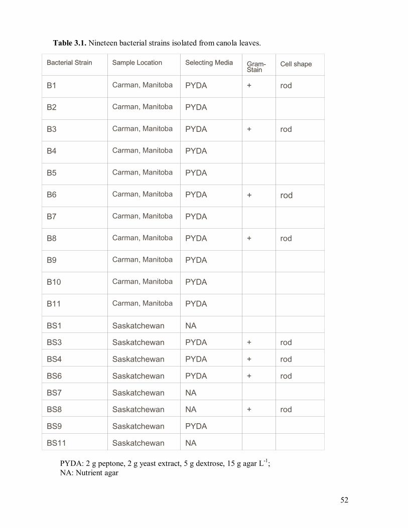
52
Bacterial Strain Sample Location Selecting Media Gram- Stain
Cell shape
B1 Carman, Manitoba PYDA + rod
B2 Carman, Manitoba PYDA
B3 Carman, Manitoba PYDA + rod
B4 Carman, Manitoba PYDA
B5 Carman, Manitoba PYDA
B6 Carman, Manitoba PYDA + rod
B7 Carman, Manitoba PYDA
B8 Carman, Manitoba PYDA + rod
B9 Carman, Manitoba PYDA
B10 Carman, Manitoba PYDA
B11 Carman, Manitoba PYDA
BS1 Saskatchewan NA
BS3 Saskatchewan PYDA + rod
BS4 Saskatchewan PYDA + rod
BS6 Saskatchewan PYDA + rod
BS7 Saskatchewan NA
BS8 Saskatchewan NA + rod
BS9 Saskatchewan PYDA
BS11 Saskatchewan NA
PYDA: 2 g peptone, 2 g yeast extract, 5 g dextrose, 15 g agar L-1; NA: Nutrient agar
Table 3.1. Nineteen bacterial strains isolated from canola leaves.
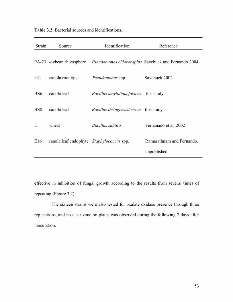
53
Table 3.2. Bacterial sources and identifications.
Strain Source Identification Reference
PA-23 soybean rhizosphere Pseudomonas chlororaphis Savchuck and Fernando 2004
#41 canola root tips Pseudomonas spp. Savchuck 2002
BS6 canola leaf Bacillus amyloliquefaciens this study
BS8 canola leaf Bacillus thringensis/cereus this study
H wheat Bacillus subtilis Fernanado et al. 2002
E16 canola leaf endophyte Staphylococcus spp. Ramarathnam and Fernando,
unpublished
effective in inhibition of fungal growth according to the results from several times of
repeating (Figure 3.2).
The sixteen strains were also tested for oxalate oxidase presence through three
replications, and no clear zone on plates was observed during the following 7 days after
inoculation.
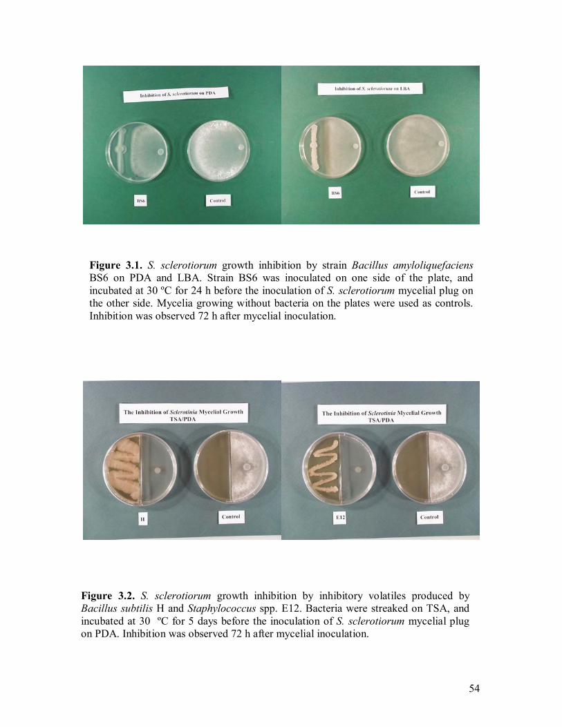
54
Figure 3.1. S. sclerotiorum growth inhibition by strain Bacillus amyloliquefaciens BS6 on PDA and LBA. Strain BS6 was inoculated on one side of the plate, andincubated at 30 ºC for 24 h before the inoculation of S. sclerotiorum mycelial plug on the other side. Mycelia growing without bacteria on the plates were used as controls. Inhibition was observed 72 h after mycelial inoculation.
Figure 3.2. S. sclerotiorum growth inhibition by inhibitory volatiles produced by Bacillus subtilis H and Staphylococcus spp. E12. Bacteria were streaked on TSA, and incubated at 30 ºC for 5 days before the inoculation of S. sclerotiorum mycelial plug on PDA. Inhibition was observed 72 h after mycelial inoculation.
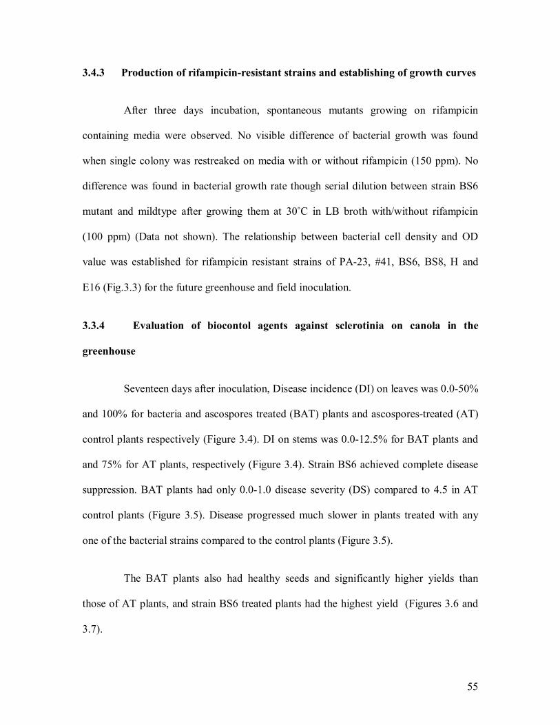
55
3.4.3 Production of rifampicin-resistant strains and establishing of growth curves
After three days incubation, spontaneous mutants growing on rifampicin
containing media were observed. No visible difference of bacterial growth was found
when single colony was restreaked on media with or without rifampicin (150 ppm). No
difference was found in bacterial growth rate though serial dilution between strain BS6
mutant and mildtype after growing them at 30ûC in LB broth with/without rifampicin
(100 ppm) (Data not shown). The relationship between bacterial cell density and OD
value was established for rifampicin resistant strains of PA-23, #41, BS6, BS8, H and
E16 (Fig.3.3) for the future greenhouse and field inoculation.
3.3.4 Evaluation of biocontol agents against sclerotinia on canola in the
greenhouse
Seventeen days after inoculation, Disease incidence (DI) on leaves was 0.0-50%
and 100% for bacteria and ascospores treated (BAT) plants and ascospores-treated (AT)
control plants respectively (Figure 3.4). DI on stems was 0.0-12.5% for BAT plants and
and 75% for AT plants, respectively (Figure 3.4). Strain BS6 achieved complete disease
suppression. BAT plants had only 0.0-1.0 disease severity (DS) compared to 4.5 in AT
control plants (Figure 3.5). Disease progressed much slower in plants treated with any
one of the bacterial strains compared to the control plants (Figure 3.5).
The BAT plants also had healthy seeds and significantly higher yields than
those of AT plants, and strain BS6 treated plants had the highest yield (Figures 3.6 and
3.7).
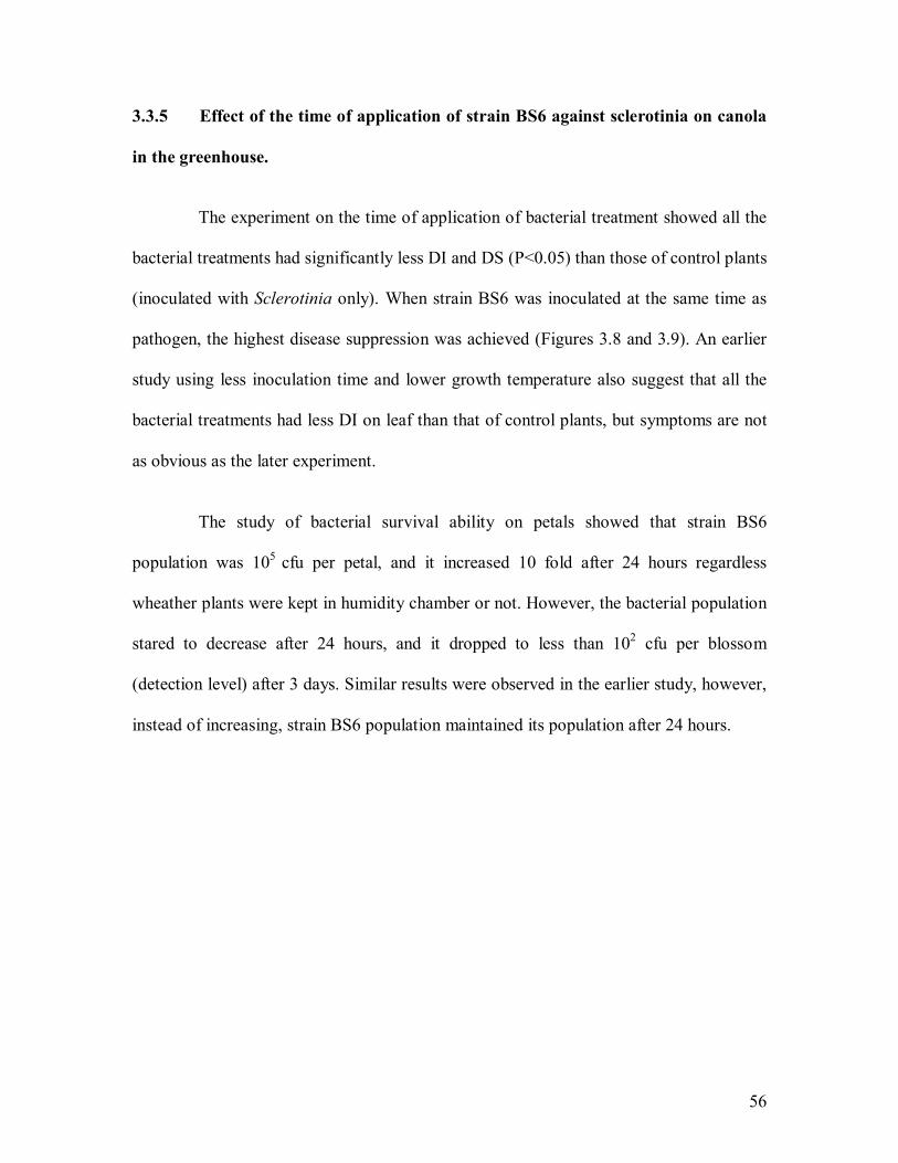
56
3.3.5 Effect of the time of application of strain BS6 against sclerotinia on canola
in the greenhouse.
The experiment on the time of application of bacterial treatment showed all the
bacterial treatments had significantly less DI and DS (P<0.05) than those of control plants
(inoculated with Sclerotinia only). When strain BS6 was inoculated at the same time as
pathogen, the highest disease suppression was achieved (Figures 3.8 and 3.9). An earlier
study using less inoculation time and lower growth temperature also suggest that all the
bacterial treatments had less DI on leaf than that of control plants, but symptoms are not
as obvious as the later experiment.
The study of bacterial survival ability on petals showed that strain BS6
population was 105 cfu per petal, and it increased 10 fold after 24 hours regardless
wheather plants were kept in humidity chamber or not. However, the bacterial population
stared to decrease after 24 hours, and it dropped to less than 102 cfu per blossom
(detection level) after 3 days. Similar results were observed in the earlier study, however,
instead of increasing, strain BS6 population maintained its population after 24 hours.
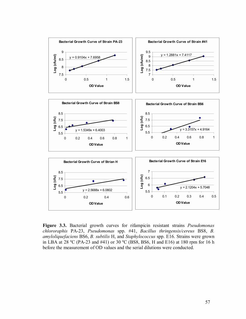
57
Figure 3.3. Bacterial growth curves for rifampicin resistant strains Pseudomonas chlororaphis PA-23, Pseudomonas spp. #41, Bacillus thringensis/cereus BS8, B. amyloliquefaciens BS6, B. subtilis H, and Staphylococcus spp. E16. Strains were grown in LBA at 28 ºC (PA-23 and #41) or 30 ºC (BS8, BS6, H and E16) at 180 rpm for 16 hbefore the measurement of OD values and the serial dilutions were conducted.
Bacterial Growth Curve of Strain BS8
y = 1.5349x + 6.40035.5
6.5
7.5
8.5
0 0.2 0.4 0.6 0.8 1
OD Value
Log
(cfu
)
Bacterial Growth Curve of Strain BS6
y = 3.3157x + 4.91645.5
6.5
7.5
8.5
0 0.2 0.4 0.6 0.8 1
OD ValueLo
g (c
fu)
Bacterial Growth Curve of Strain E16
y = 2.1204x + 5.70485.5
6
6.5
7
0 0.1 0.2 0.3 0.4 0.5
OD Value
Log
(cfu
)
Bacterial Growth Curve of Strian H
y = 2.5688x + 6.08025.5
6.5
7.5
8.5
0 0.2 0.4 0.6
OD Value
Log
(cfu
)
Bacterial Growth Curve of Strain #41
y = 1.2881x + 7.4117
77.5
88.5
99.5
0 0.5 1 1.5
OD Value
Log
(cfu
/ml)
Bacterial Growth Curve of Strain PA-23
y = 0.9104x + 7.6958
7.5
8
8.5
9
0 0.5 1 1.5
OD Value
Log
(cfu
/ml)
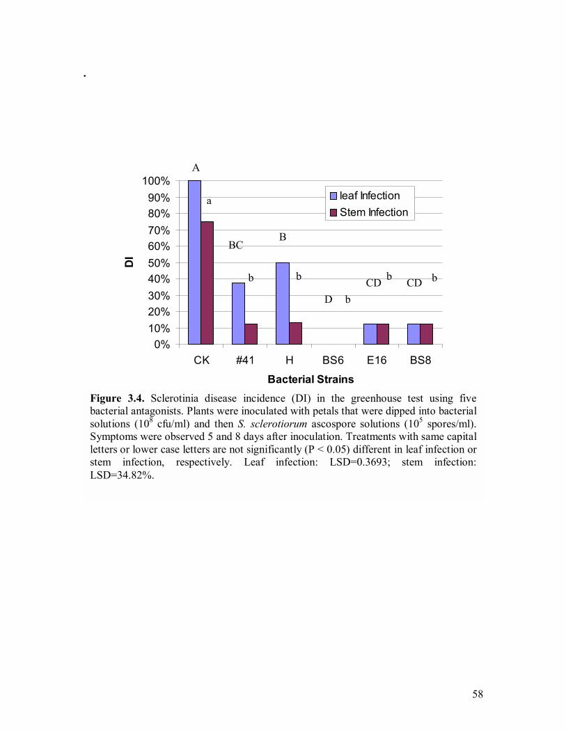
58
.
Figure 3.4. Sclerotinia disease incidence (DI) in the greenhouse test using fivebacterial antagonists. Plants were inoculated with petals that were dipped into bacterialsolutions (108 cfu/ml) and then S. sclerotiorum ascospore solutions (105 spores/ml). Symptoms were observed 5 and 8 days after inoculation. Treatments with same capital letters or lower case letters are not significantly (P < 0.05) different in leaf infection orstem infection, respectively. Leaf infection: LSD=0.3693; stem infection: LSD=34.82%.
0%10%20%30%40%50%60%70%80%90%
100%
CK #41 H BS6 E16 BS8
Bacterial Strains
DI
leaf InfectionStem Infection
b b b
b
b
A
BBC
D CD CD
a
59
Figure 3.5. Inhibition of sclerotinia disease progression in the greenhouse by using five bacterial antagonists. Plants were inoculated with petals which were dipped into bacterial solutions (108 cfu/ml) and then S. sclerotiorum ascospore solutions (105
spores/ml). Symptoms were observed every 3 days after inoculation (Day 0). Bars represent standard deviation.
-2
-1
0
1
2
3
4
5
6
7
2 5 8 11 14 17
Days after Inoculation
DS
on le
af a
nd s
tem
CK#41BS6HE16 BS8
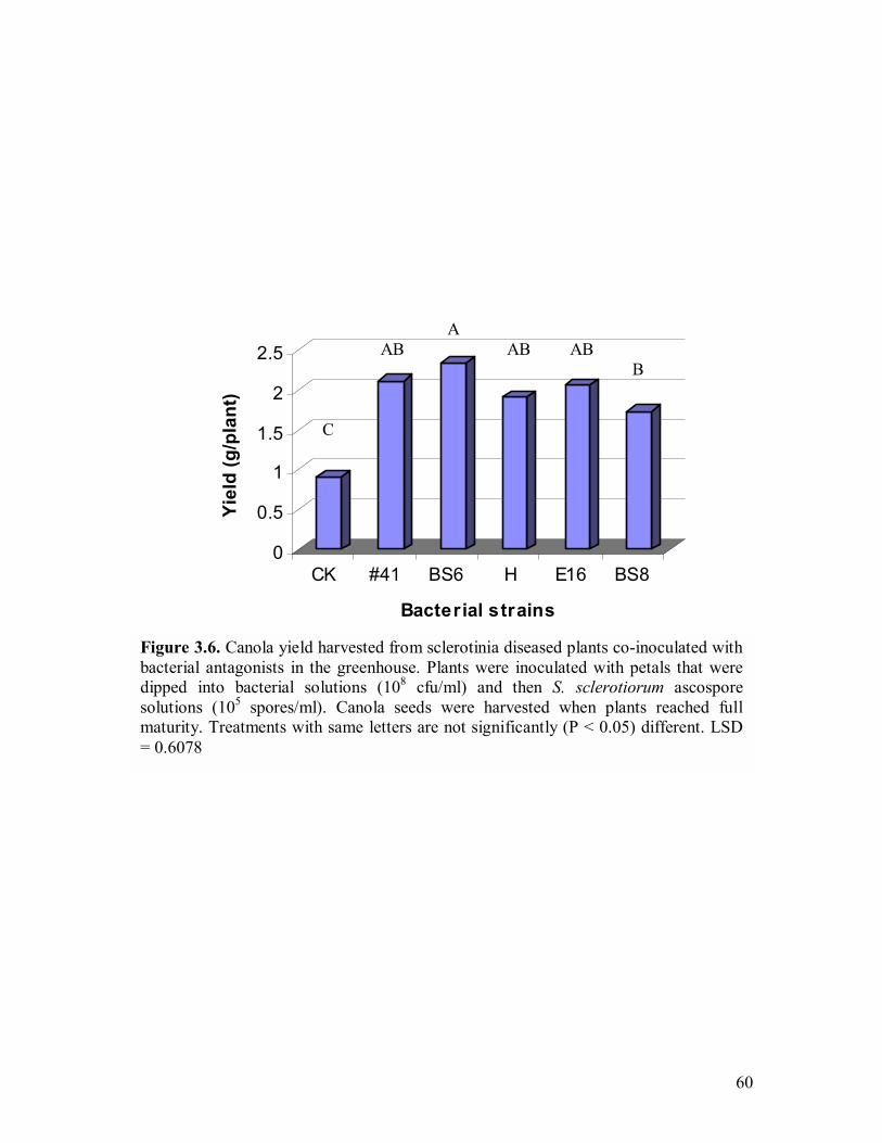
60
Figure 3.6. Canola yield harvested from sclerotinia diseased plants co-inoculated with bacterial antagonists in the greenhouse. Plants were inoculated with petals that were dipped into bacterial solutions (108 cfu/ml) and then S. sclerotiorum ascospore solutions (105 spores/ml). Canola seeds were harvested when plants reached fullmaturity. Treatments with same letters are not significantly (P < 0.05) different. LSD = 0.6078
0
0.5
1
1.5
2
2.5
Yiel
d (g
/pla
nt)
CK #41 BS6 H E16 BS8
Bacterial strains
A AB AB AB
B
C

61
Figure 3.7. Canola seeds harvested from sclerotinia diseased plants inoculated with/without B. amyloliquefaciens BS6. Plants were inoculated with petals that were dipped into bacterial solutions (108 cfu/ml) and then S. sclerotiorum ascospore solutions (105 spores/ml). Canola seeds were harvested when plants reached fullmaturity.
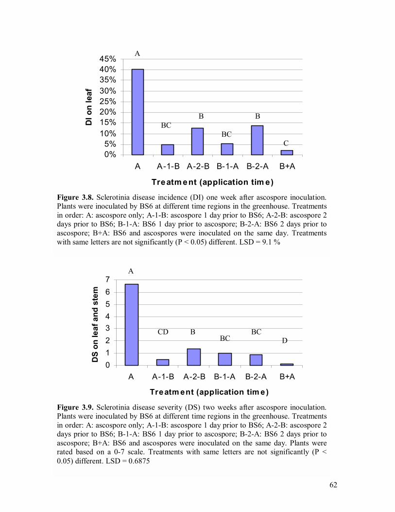
62
Figure 3.9. Sclerotinia disease severity (DS) two weeks after ascospore inoculation. Plants were inoculated by BS6 at different time regions in the greenhouse. Treatmentsin order: A: ascospore only; A-1-B: ascospore 1 day prior to BS6; A-2-B: ascospore 2 days prior to BS6; B-1-A: BS6 1 day prior to ascospore; B-2-A: BS6 2 days prior to ascospore; B+A: BS6 and ascospores were inoculated on the same day. Plants wererated based on a 0-7 scale. Treatments with same letters are not significantly (P <0.05) different. LSD = 0.6875
BC
A
CD B BC D
01234567
A A-1-B A-2-B B-1-A B-2-A B+A
Treatm ent (application tim e)
DS
on le
af a
nd s
tem
Figure 3.8. Sclerotinia disease incidence (DI) one week after ascospore inoculation.Plants were inoculated by BS6 at different time regions in the greenhouse. Treatmentsin order: A: ascospore only; A-1-B: ascospore 1 day prior to BS6; A-2-B: ascospore 2 days prior to BS6; B-1-A: BS6 1 day prior to ascospore; B-2-A: BS6 2 days prior to ascospore; B+A: BS6 and ascospores were inoculated on the same day. Treatments with same letters are not significantly (P < 0.05) different. LSD = 9.1 %
0%5%
10%15%20%25%30%35%40%45%
A A-1-B A-2-B B-1-A B-2-A B+A
Treatm ent (application tim e)
DI o
n le
af
A
BC B
BC
B
C

63
3.4.6 Evaluation of biocontrol agents under field conditions
The field experiment of both Trial One and Trial Two showed significant
disease suppression when using bacterial antagonists and achieved similar results as the
fungicide Rovral Flo® (Figures 3.11-3.14). Petal infestation level during Trial One
(within tents) is shown in Figure 3.10. All bacterial and fungicide treatments had less
petal infestation levels, but only #41, E16 BS6 single application and PA-23 double
application had significant (P<0.05) reduction of petals compared to that of the control
plot.
In Trial One, DI on stem of plants pre-treated wit bacteria were 5-23.8% after
flowering, compared to 10% in fungicide Rovral Flo® treatment and 33.8% in control
plots (Figure 3.11). The DI reduction by #41, E16 and PA-23 single application were not
significant (P<0.05) compared to that in control plots. Other bacterial treatments had
significantly (P<0.05) less DI than that in control plots, with BS6 double application
achieving the highest disease reduction. However, BS6 double application and #41 did
not significantly (P<0.05) reduce the DI after plants reached full maturity, while all the
other bacterial treatments achieved significant disease suppression (P<0.05) (Figure3.12).
In Trial Two, when plants just reached maturity, 5.84-12.5% DI on stems was observed
in bacteria-pre-treated plots, compared to 6.7% and 20.4% in the fungicide treatment and
control plots, respectively (Figure 3.13). The DS on stems was 0.121-0.283 on bacteria-
pre-treated plots and 0.167 in fungicide treatment compared to 0.426 in control plots
(Figure 3.14). The results of DI in Trial One (Figure 3.12) and DS in Trial Two (Figure
3.14) showed that all bacterial treatments were not significantly (P<0.05) different from

64
each other, which indicated the four tested bacterial agents might have similar disease
suppression abilities. The single application and double application did not have
significantly effect on disease suppression by strains PA-23 and BS6 (Figures 3.11-3.14).
Both populations of strain PA-23 and BS6 on petals in the field Trial Two were <102 cfu
per blossom 24 h after first inoculation; however, a significant population increase to
>103 cfu per blossom of strain BS6 was detected 24 h after the second spray. No
significant change in population levels of strain PA-23 was found after the second time
spray.
3.5 Discussion
Results from the field study showed that all the bacterial treatments have almost
similar disease suppression ability as the commercially used fungicide (Rovral Flo®).
Since strains P. chlororaphis PA-23 had shown significant (P < 0.05) disease suppression
in the field in 2001 (Savchuck 2002) and now in 2003, it has been consistently effective
in controlling sclerotinia stem rot in two years and three station years (two identical
experiments in 2003). Although the four bacterial strains tested in the field achieved
generally similar disease suppression in the two field trials in this study, whether the
disease suppression is through similar or different mechanisms is unknown.
The study on population dynamics is important in screening biocontrol bacteria
applied on the phyllosphere. The population dynamics of B. amyloliquefaciens BS6 in the
greenhouse is similar to those of strain #41 tested by Savchuck and Fernando (2004),
despite that BS6 is a Bacillus spp. and #41 is a Pseudomonas spp.
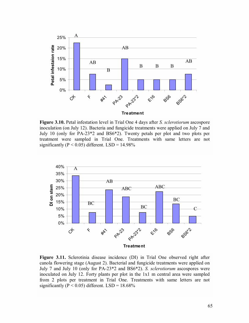
65
Figure 3.10. Petal infestation level in Trial One 4 days after S. sclerotiorum ascospore inoculation (on July 12). Bacteria and fungicide treatments were applied on July 7 andJuly 10 (only for PA-23*2 and BS6*2). Twenty petals per plot and two plots pertreatment were sampled in Trial One. Treatments with same letters are not significantly (P < 0.05) different. LSD = 14.98%
A
AB B
AB
B B B AB
0%
5%
10%
15%
20%
25%
CK F#4
1PA-23
PA-23*2 E16 BS6
BS6*2
Treatment
Peta
l inf
esta
ion
rate
Figure 3.11. Sclerotinia disease incidence (DI) in Trial One observed right aftercanola flowering stage (August 2). Bacterial and fungicide treatments were applied onJuly 7 and July 10 (only for PA-23*2 and BS6*2). S. sclerotiorum ascospores were inoculated on July 12. Forty plants per plot in the 1x1 m central area were sampledfrom 2 plots per treatment in Trial One. Treatments with same letters are not significantly (P < 0.05) different. LSD = 18.68%
0%5%
10%15%20%25%30%35%40%
CK F#4
1PA-23
PA-23*2 E16 BS6
BS6*2
Treatment
DI o
n st
em
A
BC
AB ABC
BC
ABC
BC
C
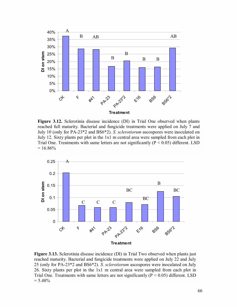
66
Figure 3.12. Sclerotinia disease incidence (DI) in Trial One observed when plantsreached full maturity. Bacterial and fungicide treatments were applied on July 7 andJuly 10 (only for PA-23*2 and BS6*2). S. sclerotiorum ascospores were inoculated on July 12. Sixty plants per plot in the 1x1 m central area were sampled from each plot inTrial One. Treatments with same letters are not significantly (P < 0.05) different. LSD = 16.86%
A B AB
B B
B B
AB
0%
5%
10%
15%
20%
25%
30%
35%
40%
CK F#4
1PA-23
PA-23*2 E16 BS6
BS6*2
Treatment
DI o
n st
em
Figure 3.13. Sclerotinia disease incidence (DI) in Trial Two observed when plants justreached maturity. Bacterial and fungicide treatments were applied on July 22 and July25 (only for PA-23*2 and BS6*2). S. sclerotiorum ascospores were inoculated on July 26. Sixty plants per plot in the 1x1 m central area were sampled from each plot inTrial One. Treatments with same letters are not significantly (P < 0.05) different. LSD = 5.48%
A
C C C
BC
BC
B BC
0
0.05
0.1
0.15
0.2
0.25
CK F#4
1PA-23
PA-23*2 E16 BS6
BS6*2
Treatment
DI o
n st
em
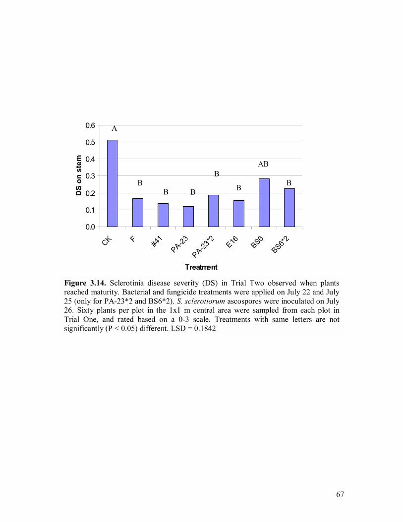
67
Figure 3.14. Sclerotinia disease severity (DS) in Trial Two observed when plantsreached maturity. Bacterial and fungicide treatments were applied on July 22 and July25 (only for PA-23*2 and BS6*2). S. sclerotiorum ascospores were inoculated on July 26. Sixty plants per plot in the 1x1 m central area were sampled from each plot inTrial One, and rated based on a 0-3 scale. Treatments with same letters are not significantly (P < 0.05) different. LSD = 0.1842
A
B B
B
B
B
AB
B
0.0
0.1
0.2
0.3
0.4
0.5
0.6
CK F #41
PA-23
PA-23*2 E16 BS6
BS6*2
Treatment
DS
on s
tem
68
The multiplication of the bacterial population on blossom after inoculation was also
observed by Yuen et al. (1994). They found that populations of three Erwinia herbicola
strains, which were used to control white mold on dry bean caused by S. sclerotiorum,
multiplied from < 102 cfu per blossom at initial level to >107 cfu per blossom within 16 h
at 25 °C. However, it is unlikely that the Pseudomonas and Bacillus strains used in this
study would reach such a high population level on blossom. They also found that the
bacterial population level was lower in the field than in the greenhouse, which agrees
with the result from this study. The results of population dynamics under field conditions
suggested that B. amyloliquefaciens BS6 has longer survival potential than P.
chlororaphis PA-23. This could be explained by the fact that B. amyloliquefaciens BS6
could probably form endospores when it encountered adverse environmental conditions.
However, when comparing the DI and DS from the Trial Two, BS6 single application
was not as effective as PA-23 single application in disease suppression; it achieved
significantly (P < 0.05) less DI but not DS compared to that of control plots. However,
both double application of strains BS6 and PA-23 have achieved similar disease
suppression. This suggested that disease suppression ability of B. amyloliquefaciens BS6
might be more dependent on its population level compared to P. chlororaphis PA-23. P.
chlororaphis PA-23 can achieve significant (P < 0.05) disease suppression even when the
population on blossom is very low, and this may suggest that plant induced resistance
might play a role in disease suppression. Since P. chlororaphis PA-23 may also produces
antibiotic phenazine (Fernando, unpublished), further study on multiple mechanisms
involved in disease suppression is needed. However, the DI in Trial One (Figure 3.12)
and DS in Trial Two (Figure 3.14) of strains PA-23 and BS6 single applications
69
(population levels were both very low when encountering ascospore inoculation) are not
significantly (P < 0.05) different from each other and it suggests that sclerotinia disease
suppression by B. amyloliquefaciens BS6 may also involve plant induced resistance.
However, strain #41 single application and BS6 double application in Trial One
did not significantly (P < 0.05) reduce DI compared to that of control plots when plants
were mature. This might be due to plant logging within the tents. Several plots were
observed with severe plant logging during and after canola flowering, and probably
because of the high humidity within tents. Logging offered more plant-to-plant contact
and allowed pathogen spread by mycelial infection. An intense misting system (at least
run 6 min per 1.5 h after inoculation) without tents is recommended for future biocontrol
tests in the field to control sclerotinia stem rot on canola.
Petal infestation level has been used for forecasting sclerotinia on canola
(Turkington et al. 1991a). In this study, petal infestation level vigorously reflected the DI
in Trial One in the field. Lack of accuracy could be because of disease epidemics
consisting of several factors, including weather changes, relative humidity in plots and
host variety etc. It could also be because of a relatively low sampling size in this study (2
sites per treatment, 20 petals per site). Sampling sizes were discussed by Turkington et al.
(1988) for forecasting sclerotinia stem rot of canola using the petal test. They pointed out
the minimum sample size required to estimate DI at specified levels of precision varied
widely among the experiments. Increasing sampling sites or petals sampled per site
might be needed in similar plot design in a future study. The study by Savchuck (2002)
has shown un-uniform petal infestation by S. sclerotiorum according to the data collected
70
from 7 sampling days within 13 days. This might be explained by the random landing of
ascospores in the natural system, for the plots were not inoculated with ascospores. No
significant (P < 0.05) reduction of leaf infection was observed in 2001 and only P.
chlororaphis PA-23 double application was able to significantly (P < 0.05) reduce the
stem infection (Savchuck 2002). This might be explained by the low disease pressure,
which indicated earlier by the petal infestation level. The application of ascospores in this
study offered a more uniform and higher disease pressure compared to that in the study of
Savchuk (2002). In 2002, a similar study was carried at Carman Research Centre,
Manitoba, and the pathogen was inoculated by spreading over-wintered sclerotia on the
surface of soil in the beginning of spring. Sclerotinia disease was rarely found after
canola flowering. Even in the present study, no apothecia germination was found in Trial
Two before or at the early flowering stage, based on the visual investigation of half of the
plots. An ascospore inoculation (104 cfu/ml) is recommended for further S. sclerotiorum
inoculation in field experiment, and misting system during the flowering stage is needed
(could be activated two days before inoculation and run for at least 2 weeks after
inoculation) to offer enough relative humidity for pathogen infection and disease
development. Setting up tents for helping the inoculation in field is labour consuming,
especially when large tents are used. The combination of tents and misting system are
effective to help Sclerotinia ascospore inoculation in the field (Clinton Jurke, personal
communication). But when both of them are used, crop lodging might happen easily.
Unfortunately the yield data from Trial Two was lost due to losing of the
harvested canola samples in a big wind at the end of the season. However, the results
from greenhouse experiments showed that all tested strains (BS6, BS8, H, E16 and #41)
71
have significantly reduced disease and increased the canola yield. Comparing yield from
this study with non-treated healthy plants and plants treated only with B.
amyloliquefaciens BS6 could help understand whether this strain has the plant growth
promoting effects or not.
At the beginning of this study, several Gram-positive strains that were isolated
from canola leaves were found to have antagonistic effects against S. sclerotiorum in
vitro. The inhibition of fungal mycelial growth on LBA and PDA with zones of inhibition
suggested that antibiosis might play a vital role in disease suppression in vivo. Bacterial
antibiotics involved in fungal disease suppression have been reported in numerous
studies, including phenazine-1-carboxylic acid (PCA) (Mavrodi et al. 1998; Delaney et
al. 2001), 2,4-diacetylphloroglucinol (2,4-DAPG) (Nowak Thompson et al. 1994;
Mavrodi et al. 2001), pyoluteorin (Plt) (de Souza and Raaijmakers 2003; Nowak
Thompson et al. 1999), pyrrolnitrin (Prn) (Chernin et al. 1996; Hammer et al. 1999;
Kirner et al. 1998), zwittermycin A (Raffel et al. 1996), hydrogen cyanide (Flaishman et
al. 1996), kanosamine (Milner et al. 1996a), butyrolactones (Gamard et al. 1997),
oligomycin A (Kim et al. 1999), Oomycin A (Howie and Suslow 1991), etc. Further
studies focusing on antibiotics produced by these bacterial biocontrol agents are needed.
Since there is no inhibition zone observed in the oxalic acid degradation
selective media, the bacteria tested in this study might not have the ability to degrade
oxalic acid, which is the major pathogenesis factor of S. sclerotiorum. Savchuck (2002)
also found strains PA-23 and #41 do not have the ability to produce oxalate oxidase.
72
Inhibitory organic volatiles seem to be commonly produced in gram-positive
bacterial antagonists. All the strains tested in this study showed the inhibition of S.
sclerotiorum hyphal growth by bacterial organic volatiles in the divided plates, but the
results between different strains were hardly compared due to the high variation among
replicates (data not shown). The theory of testing the inhibitory volatile is to spatially
separate the bacteria and target pathogen, but allow them to share the same headspace.
High variation among replicates in the results might be due to the limitations of the test
method (Fernando and Linderman 1994) which would allow certain amounts of volatiles
to be released out of the plate when the inoculation of fungal mycelia was being
conducted a few days after bacterial growth. A recent study by Fernando et al. (2004)
used several methods to test the bacterial inhibitory volatile production, including similar
divided plate assay as previously described, a method with three-compartment divide
plates with/ without charcoal, and sealed plated method which combined two bottom
plates with bacteria and fungi on each of them. The latter two methods might give better
results in the inhibitory volatile test, since no opening of the plate is needed after initial
inoculation. Since this study is to look for biocontrol agents being sprayed at canola
flowering stage, the screening of bacterial candidates was not based on inhibitory volatile
production. However, these strains could be tested to control other soilborne diseases or
reducing sclerotia germination by using as soil drainage.
Bacterial biocontrol agents have been studied in several host-pathogen systems.
Bacterial biocontrol products have a great potential to be used in disease control, since
their mass production is much easier than that of fungi (Shoda 2000). Pseudomonas spp.
have been extensively studied as biocontrol agents. The use of Bacillus spp. as biocontrol
73
agents are relatively rare compared to Gram-negative bacteria, however, as Bacillus spp.
have the characteristics of high thermal tolerance and ready formulation of endospores,
their potential to be developed to commercial products is high (Shoda 2000). Bacillus
subtilis strain GB03 has been successfully registered as a commercial product Kodiak®
(Gustafson, Inc, Plano, TX, USA). It has shown a mixture of cotton growth promotion
(increased root mass) and disease suppression of Rhizoctonia and Fusarium spp., when it
was used as a cotton seed treatment (Brannen and Kenney 1997). Bacterial agents to
control S. sclerotiorum have only been investigated in the recent decade. The four strains
tested in the field have great potential to be developed to commercial products. Based on
the present literature, this is the first extensive study undertaken to collect and screen
Bacillus spp. (including other Gram-positive strains) for the control of S. sclerotiorum on
the canola phyllosphere.
74
4.0 DETECTION OF ANTIBIOTIC-RELATED GENES FROM
BACTERIAL BIOCONTROL AGENTS USING
POLYMERASE CHAIN REACTION
4.1 Abstract
Eight bacterial isolates were screened against Sclerotinia sclerotiorum in vitro
and/or in vivo. The presence of biosynthetic or self-resistant genes of bacterial antibiotics
was investigated using polymerase chain reaction (PCR) method. Thirty-two specific
primers were used to amplify the specific antibiotic target genes. Pseudomonas
fluorescens 2-79, P. fluorescens Q2-87, P. fluorescens Pf-5 and Bacillus cereus UW85
were used as positive controls in some of the PCR reactions to detect phenazine-1-
carboxylic acid (PCA), 2, 4-diacetylphloroglucinol (2,4-DAPG), pyoluterorin (Plt),
pyrrolnitrin (Prn), and zwittermicin A, respectively. PCR products were sequenced and
BLAST searched in the Genebank. Results showed the fungal antagonists Pseudomonas
chlororaphis PA-23 to contain biosynthetic genes for PCA, Prn and probably 2,4-DAPG,
and Bacillus thuringiensis/cereus BS8, B. cereus L, and B. mycoides S contain
zwittermicin A self-resistant gene. The results also suggeted that P. chlororaphis PA-23
may not contain Plt biosynthetic gene and zwittermicin A self-resistant gene, and
Pseudomonas spp. #41 and B. amyloliquefaciens BS6 may not contain PCA, Plt, Prn, 2,4-
DAPG biosunthetic genes and zwittermicin A self-resistant gene. These results suggested
that antibiosis might be involved in the bacterial antifungal activity and disease
suppression.

75
4.2 Introduction
Antibiosis has been widely studied as one of the most important biocontrol
mechanisms in controlling plant pathogens. Biocontrol bacteria Pseudomonas fluoresens
2-79 and P. fluoresens Q2-87 were reported to produce phenazine-1-carboxylic acid
(PCA) and 2,4-diacetylphloroglucinol (2,4-DAPG), respectively (Thomashow and Weller
1988; Harrison et al. 1993). P. fluorescens Pf-5 was reported to produce 2,4-DAPG,
pyoluterorin (Plt) and pyrrolnitrin (Prn) (Nowak Thompson et al. 1994; de Souza and
Raaijmakers 2003). B. cereus UW 85 was reported to produce both zwittermin A and
kanosamine (Silo Suh et al. 1994; Milner et al. 1996a). The production of antibiotics by
these strains has been found relating to the suppression of several different plant
pathogens (Thomashow et al. 1990; Chin-A-Woeng et al. 1998; Chernin et al. 1996;
Cronin et al. 1997a; Howell and Stipanovic 1979; Homma et al. 1989; Keel et al. 1992).
Extensive genetic studies have also been conducted in antibiotic-related genes in these
biocontrol agents (Delaney et al. 2001; Pierson, III et al. 1995; Bangera and Thomashow
1996; Bangera and Thomashow 1999; Kraus and Loper 1995; Nowak Thompson et al.
1999; Hammer et al. 1997; Kirner et al. 1998; Stohl et al. 1999b; Stohl et al. 1999a;
Milner et al. 1996b), and these strains have become model strains in bacterial antibiosis
study.
The biosynthetic genes for PCA, 2,4-DAPG, Prn and Plt, and zwittermicin A
self-resistant gene have been fully sequenced (Hammer et al. 1997; Nowak Thompson et
al. 1999; Stohl et al. 1999b; Mavrodi et al. 1998; Bangera and Thomashow 1999). The
availability of sequenced antibiotic-biosynthetic genes and self-resistant gene facilitated
76
the development of specific primers that can be used to detect naturally occurring
antibiotic-producing bacteria (Raaijmakers et al. 1997). Several specific primers have
been developed from the conserved regions of antibiotic biosynthetic genes or self-
resistant gene ( Raaijmakers et al. 1997; McSpadden Gardener et al. 2001; de Souza and
Raaijmakers 2003; Delaney et al. 2001; Mavrodi et al. 2001; Milner et al. 1996b). For
example, primers PHZ1and PHZ2 were used to detect phenazine biosynthetic genes,
which were designed to amplify a 1.4 kb DNA fragment containing phzF and phzA genes
from P. aureofaciens 30-84 or phzC and phzD in Pseudomonas fluorescens 2-79
(Delaney et al. 2001). Several primers were developed from a 745 bp DNA fragment
amplified from 1,001 bp phlD gene sequence of P. fluorescens Q2-87 by primers Phl2a
and Phl2b; phlD is responsible for the production of monoacetylphloroglucinol (MAPG),
the precusor of 2,4-DAPG (McSpadden Gardener et al. 2001).
These specific primers for PCA, 2,4-DAPG, Prn, Plt, and zwittermicin A have
been used to detect antibiotic-producing bacterial strains by polymerase chain reaction
(PCR) method (Bangera and Thomashow 1999; de Souza and Raaijmakers 2003;
Raaijmakers et al. 1997; McSpadden Gardener et al. 2001; Raffel et al. 1996; Picard et al.
2000). Though this method has mainly been used to detect biocontrol agents in soil, as
PCR is a rapid method to screen antibiotic-producing bacteria among a large quantity of
microorganisms, some studies also suggested that this method could be used to identify
individual bacterial strains with specific antibiotic-producing ability (Raffel et al. 1996;
Hammer et al. 1999).
77
In an earlier study (see Chapter 3), eight bacterial biocontrol agents were
screened against Sclerotinia sclerotiorum de Bary in vivo and/or in vitro. The direct
fungal growth inhibition on PDA/LBA plates suggests that antibiosis might be one of the
most important mechanisms involved in the antifungal activity or disease suppression in
the lab, the greenhouse and the field. The objective of this study is to identify antibiotic
biosynthetic or self-resistant genes from these potential biocontrol bacterial agents in
order to better understand the involvement of bacterial antibiotics in pathogen
suppression by making Tn5 mutants in the future.
4.3 Materials and methods
Eight potential bacterial biocontrol strains were tested: P. chlororaphis PA-23,
Pseudomonas spp. #41, Bacillus amyloliquefaciens BS6, B. thuringiensis BS8, B. subtilis
H, B. cereus L, B. mycoides S and Bacillus spp. B70 (Savchuck and Fernando 2004;
Fernando et al. 2002; Rob Duncan, personal communication). Pseudomonas fluorescens
2-79, P. fluorescens Q2-87, P. fluorescens Pf-5 and Bacillus cereus UW85 (provided by
Dr. Linda Thomashow, Dr. Joyce Loper, Dr. Carolyn Press, Dr. Jo Handelsman) were
used as positive controls in some of the PCR reactions to detect phenazine-1-carboxylic
acid (PCA), 2, 4-diacetylphloroglucinol, pyoluteorin, prrolnitrin and zwittermicin A,
respectively. Thirty primers (InvitrogenTM, Carlsbad, CA, U.S.A.) were used in the PCR
reactions to amplify the specific target genes (Table 4.1).
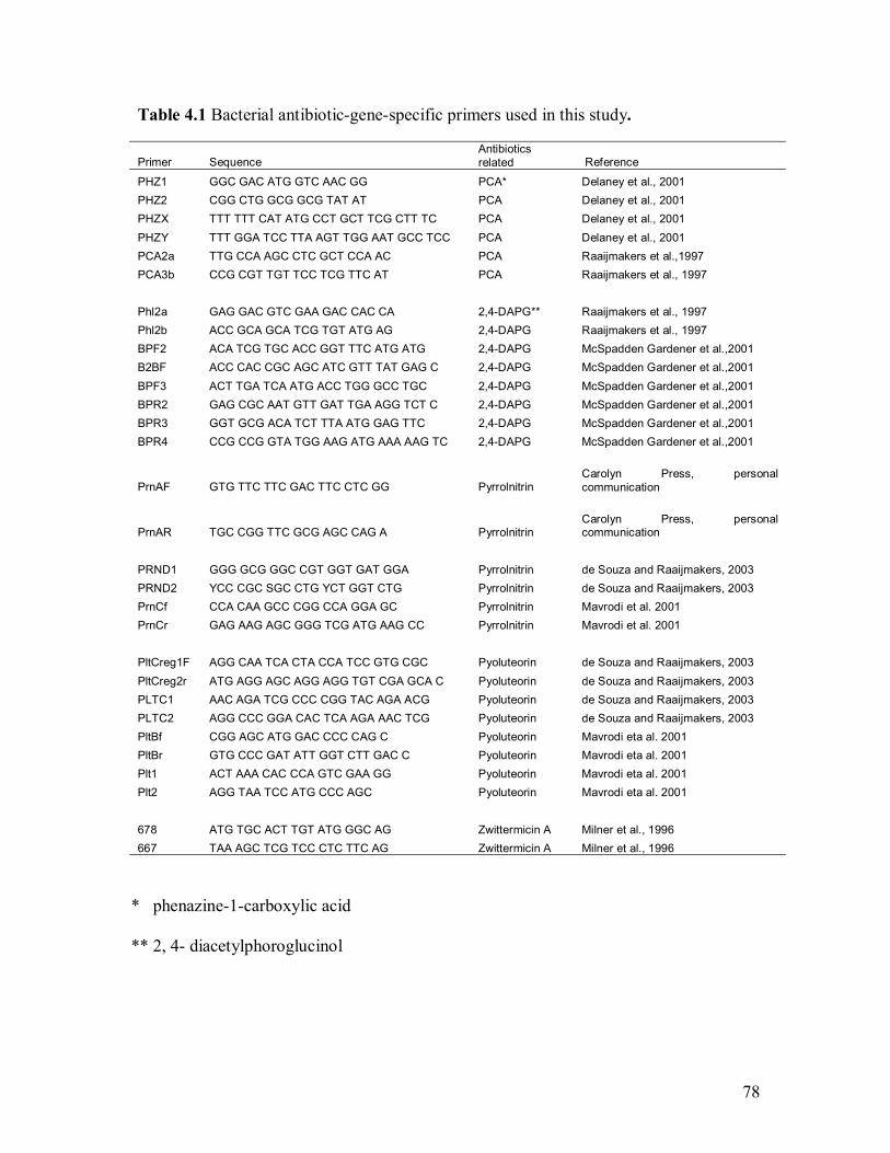
78
Table 4.1 Bacterial antibiotic-gene-specific primers used in this study.
Primer Sequence Antibiotics related Reference
PHZ1 GGC GAC ATG GTC AAC GG PCA* Delaney et al., 2001 PHZ2 CGG CTG GCG GCG TAT AT PCA Delaney et al., 2001 PHZX TTT TTT CAT ATG CCT GCT TCG CTT TC PCA Delaney et al., 2001 PHZY TTT GGA TCC TTA AGT TGG AAT GCC TCC PCA Delaney et al., 2001 PCA2a TTG CCA AGC CTC GCT CCA AC PCA Raaijmakers et al.,1997 PCA3b CCG CGT TGT TCC TCG TTC AT PCA Raaijmakers et al., 1997 Phl2a GAG GAC GTC GAA GAC CAC CA 2,4-DAPG** Raaijmakers et al., 1997 Phl2b ACC GCA GCA TCG TGT ATG AG 2,4-DAPG Raaijmakers et al., 1997 BPF2 ACA TCG TGC ACC GGT TTC ATG ATG 2,4-DAPG McSpadden Gardener et al.,2001 B2BF ACC CAC CGC AGC ATC GTT TAT GAG C 2,4-DAPG McSpadden Gardener et al.,2001 BPF3 ACT TGA TCA ATG ACC TGG GCC TGC 2,4-DAPG McSpadden Gardener et al.,2001 BPR2 GAG CGC AAT GTT GAT TGA AGG TCT C 2,4-DAPG McSpadden Gardener et al.,2001 BPR3 GGT GCG ACA TCT TTA ATG GAG TTC 2,4-DAPG McSpadden Gardener et al.,2001 BPR4 CCG CCG GTA TGG AAG ATG AAA AAG TC 2,4-DAPG McSpadden Gardener et al.,2001
PrnAF GTG TTC TTC GAC TTC CTC GG Pyrrolnitrin Carolyn Press, personal communication
PrnAR TGC CGG TTC GCG AGC CAG A Pyrrolnitrin Carolyn Press, personal communication
PRND1 GGG GCG GGC CGT GGT GAT GGA Pyrrolnitrin de Souza and Raaijmakers, 2003 PRND2 YCC CGC SGC CTG YCT GGT CTG Pyrrolnitrin de Souza and Raaijmakers, 2003 PrnCf CCA CAA GCC CGG CCA GGA GC Pyrrolnitrin Mavrodi et al. 2001 PrnCr GAG AAG AGC GGG TCG ATG AAG CC Pyrrolnitrin Mavrodi et al. 2001 PltCreg1F AGG CAA TCA CTA CCA TCC GTG CGC Pyoluteorin de Souza and Raaijmakers, 2003 PltCreg2r ATG AGG AGC AGG AGG TGT CGA GCA C Pyoluteorin de Souza and Raaijmakers, 2003 PLTC1 AAC AGA TCG CCC CGG TAC AGA ACG Pyoluteorin de Souza and Raaijmakers, 2003 PLTC2 AGG CCC GGA CAC TCA AGA AAC TCG Pyoluteorin de Souza and Raaijmakers, 2003 PltBf CGG AGC ATG GAC CCC CAG C Pyoluteorin Mavrodi eta al. 2001 PltBr GTG CCC GAT ATT GGT CTT GAC C Pyoluteorin Mavrodi eta al. 2001 Plt1 ACT AAA CAC CCA GTC GAA GG Pyoluteorin Mavrodi eta al. 2001 Plt2 AGG TAA TCC ATG CCC AGC Pyoluteorin Mavrodi eta al. 2001 678 ATG TGC ACT TGT ATG GGC AG Zwittermicin A Milner et al., 1996 667 TAA AGC TCG TCC CTC TTC AG Zwittermicin A Milner et al., 1996
* phenazine-1-carboxylic acid
** 2, 4- diacetylphoroglucinol
79
4.3.1 Genomic DNA extraction
A revised protocol (Ausubel et al. 1995) was used for DNA isolation. Bacteria
were grown in 5 ml LB broth 18-20 h at 28 °C at a rotation speed of 200 rpm. 1.5 ml
culture was centrifuged for 2 min in an eppendorf tube (Fisherbrand , Fisher
ScientificTM, U.S.A.). After discarding the supernatant, the pellet was resuspended in 576
µl TE buffer (10 mM Tris.Cl, 1 mM EDTA, pH = 8). Ten percent SDS and 20 mg/ml
Proteinase K (Sigma , St. Louis, MO, U.S.A) were added to make final concentrations
of 5% and 0.5%, respectively. Samples were mixed thothroughly, and incubated at 37 °C
for 1 h. After adding 5 M NaCl to 714 mM and CTAB/NaCl (CTAB: hexadecyltrimethl-
ammonium bromide, Sigma®, Sigma Chemical Co., St. Louis, MO., U.S.A) to 10%, the
samples were incubated at 65 °C for 10 min. Samples were extracted with equal volume
of 24:1 chloroform /isoamyl alcohol (chloroform: Fisher ScentificTM, Fair Lawn, New
Jersey, U.S.A.) and then 25:24:1 phenol /chloroform /isoamyl alcohol (Phenol: Sigma ,
St. Louis, MO, U.S.A.; chloroform and isoamylalcohol: Fisher ScentificTM, Fair Lawn,
New Jersey, U.S.A.), and centrifuged at 13.000 rpm for 10 min. The supernatant was
transferred to a fresh eppendorf tube after each centrifugation. DNA was precipitated by
adding 0.6 vol. cold isopropanol (Sigma , St. Louis, MO, U.S.A). After 5 min the
samples were centrifuged again at 13,000 rpm for 10 min. The supernatant was discarded
and the pellet was washed by 70% ethanol. The eppendorf tubes were placed upside
down to let them air dry for about 30min. The pellet was resuspended in 100 µl of warm
TE buffer (10 mM Tris.Cl, 1 mM EDTA, pH = 8). The DNA extracts were stored at 4 °C.
80
In order to catalyze the hydrolysis of RNA, Ribonuclease A (Sigma , St. Louis,
MO, U.S.A) were added to 0.02 µg, and then incubated at 37 °C for 30 min. DNA was
precipitated again with isopropanol and resuspended in 100 µl of TE buffer as previously
described. Samples were stored at �20 °C.
4.3.2 DNA quantification
DNA quantification was determined by gel electrophoresis and the
spectrophotometer (GeneQuant , BiochromTM, distributed by Fisher ScientificTM,
Nepean, Ontario, Canada). Agarose gel (Agrarose DNA Grade, Electrophoresis Grade,
Fisher ScientificTM, Fair Lawn, New Jersey, U.S.A.) of 1.2% with ethidium bromide
(0.5µg/ml) and 1x TBE buffer (89 mM Tris base, 89 mM boric acid, 2 mM EDTA, pH
8.0) was used in the electrophoresis. The 1 kb DNA size marker was used to estimate the
size of the extracted DNA. The gel was run at 150 volts for approximately 40min. A
photograph was taken under the UV transluminator (Gel Doc 2000, Bio-Rad Laboratories
Inc., Hercules, CA, U.S.A.).
OD values of DNA were read at 260 nm and 280 nm using the
spectrophotometer. The ratio of OD260 to OD280 was calculated to assess the DNA purity.
The dilution of the DNA was calculated based on 1 OD260 = 50 µg/ml and the OD260.
4.3.3 PCR reactions
Each 25µl PCR reaction mixture contained 25 pmol of each of the two primers,
1x PCR buffer, 800 µM dNTP�s, 1.5 mM MgCl2, 1.0-2 units Taq DNA polymerase
81
(InvitrogenTM, Carlsbad, CA, U.S.A.). The PCR cycling conditions were employed
according to the original reference publications (Raaijmakers et al. 1997; McSpadden
Gardener et al. 2001; de Souza and Raaijmakers 2003; Raffel et al. 1996; Giacomodonato
et al. 2001; Delaney et al. 2001; Mavrodi et al. 2001). PCR reactions were carried by
(Delaney et al. 2001) using 0.2 ml PCR tubes (Rose Sclientific Ltd., Edmonton, Alberta,
Canada) with TECHNE Genius PCR machine (Techne Ltd. and Techne Inc.,
Cambridge, UK and New Jeysey, U.S.A.) or 0.6 ml PCR tubes (Rose Sclientific Ltd.,
Edmonton, Alberta, Canada) with Programmable Thermal Controller (PTC-100TM, MJ
Research, Inc., Watertown, Mass, U.S.A.). The amplification products were separated on
1.2-1.5 % agarose gel (Agrarose DNA Grade, Electrophoresis Grade, Fisher ScientificTM,
Fair Lawn, New Jersey, U.S.A.) with ethium bromide (0.5 µg/ml) and 1x TBE buffer (89
mM Tris base, 89 mM boric acid, 2 mM EDTA, pH 8.0). The 100 bp size marker was
used to estimate the size of PCR products. The gel was run at 80 volts for approximately
1.5 h. A photograph was taken under UV transilluminator (Gel Doc 2000, Bio-Rad
Laboratories Inc., Hercules, CA, U.S.A.). All reactions were conducted at least twice.
In order to confirm the positive results in the previous amplifications, the
reactions were conducted again with positive control strains. Strain 2-79, Q2-87 and Pf-5
were used as positive controls and amplified with strain PA-23 using primers
PHZ1/PHZ2, BPF3/BPR2 and PltBf/PltBr respectively. Strain UW85 was used as
positive control using primers 677/678. PCR reactions amplified with water instead of
genomic DNA were used as negative controls.
82
4.3.4 Sequencing and BLAST search
To further confirm the positive results from PCR reactions, the desired PCR
products were sent for sequencing (University Core DNA & Protein Services, University
of Calgary, Calgary, AB). Fifty or 100 µl PCR reaction was conducted and purified by
High Pure PCR Product Purification Kit (Roche , Roche Applied ScienceTM, Mannheim,
Germany). For the reaction with primers BPF3/BPR2 and strain PA-23, the desired band
(470 bp) was cut from the agorase gel, and then the DNA was purified with 5M NaCl and
95% ethanol. After re-dissolution in water, the purified DNA was used as template to
amplify the desired PCR product again with primers BPF3/BPR2. The PCR product was
checked and purified as described previously. The DNA was quantified after gel
electrophoresis, by comparing with 1650 bp band of 1 kb DNA ladder. Each 12 µl
premixed sample for sequencing contained 0.01 µg of template DNA per 100 bp, and 3.2
pmol primer. The results were aligned on the BLAST search at National Centre for
Biotechnology Information (NCBI) using nucleotide-nucleotide BLAST (blastn) and
align two sequences (bl2seq).
4.4 Results
4.4.1 Genomic DNA
Genomic DNA was visualised as white pellets after isopropanol precipitation,
and re-dissolved in TE buffer. The gel electrophoresis suggested the genomic DNA
extracts were intact. No smear was found after adding the RNase A, which suggested that
the RNA was cleaned out. The OD260/OD280 value (1.6 - 1.8) suggested that the purity
83
of genomic DNA should be enough for PCR reaction. The genomic DNA concentrations
and the amount of genomic DNA needed for the PCR reactions were calculated based on
the OD260.
Genomic DNA was stored at �20 ºC, and diluted to 20 ng/µl by water and stored
at 4ºC for a short time prior to PCR reaction.
4.4.2 PCR reactions
P. chlororaphis PA-23 showed a 1400 bp (Figure 4.1), and an 850 bp (Figure
4.5) band in the gel electrophoresis when amplified with primers PHZ1/PHZ2 and
PltBf/PltBr, respectively. The results were confirmed by amplification along with the
positive control strains P. fluorescens 2-79 (Figure 2) and P. fluorescens Pf-5 (Figure 5),
respectively. Identical bands were found from P. chlororaphis PA-23 and positive
control P. fluorescens Pf-5 using primers PrnAF /PrnAR, PRND1/PRND2 and
PrnCf/PrnCr (Figure 3), and the bands are approximately 1050 bp, 786 bp and 719 bp,
respectively. P. chlororaphis PA-23 and positive control P. fluorescens Q2-87 also
showed a 470 bp band and a few other weak bands when amplified with primers
BPF3/BPR2 (Figure 4). B. thuringiensis BS8, B. cereus L and B. mycoides S amplified a
950 bp PCR product, respectively, using zwittermicin A self-resistant gene-specific
primers 678/677, along with positive control B. cereus UW 85 (Fig.5). A very weak band
was observed from B. amyloliquefaciens BS6 when amplified for the first time with
primers 678/677, but the result was not repeatable. No PCR product was amplified from
Pseudomonas spp. #41.
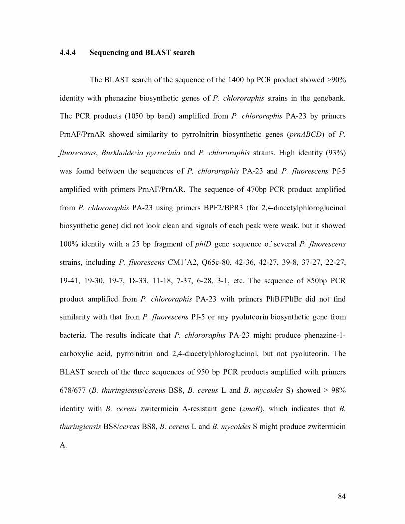
84
4.4.4 Sequencing and BLAST search
The BLAST search of the sequence of the 1400 bp PCR product showed >90%
identity with phenazine biosynthetic genes of P. chlororaphis strains in the genebank.
The PCR products (1050 bp band) amplified from P. chlororaphis PA-23 by primers
PrnAF/PrnAR showed similarity to pyrrolnitrin biosynthetic genes (prnABCD) of P.
fluorescens, Burkholderia pyrrocinia and P. chlororaphis strains. High identity (93%)
was found between the sequences of P. chlororaphis PA-23 and P. fluorescens Pf-5
amplified with primers PrnAF/PrnAR. The sequence of 470bp PCR product amplified
from P. chlororaphis PA-23 using primers BPF2/BPR3 (for 2,4-diacetylphloroglucinol
biosynthetic gene) did not look clean and signals of each peak were weak, but it showed
100% identity with a 25 bp fragment of phlD gene sequence of several P. fluorescens
strains, including P. fluorescens CM1�A2, Q65c-80, 42-36, 42-27, 39-8, 37-27, 22-27,
19-41, 19-30, 19-7, 18-33, 11-18, 7-37, 6-28, 3-1, etc. The sequence of 850bp PCR
product amplified from P. chlororaphis PA-23 with primers PltBf/PltBr did not find
similarity with that from P. fluorescens Pf-5 or any pyoluteorin biosynthetic gene from
bacteria. The results indicate that P. chlororaphis PA-23 might produce phenazine-1-
carboxylic acid, pyrrolnitrin and 2,4-diacetylphloroglucinol, but not pyoluteorin. The
BLAST search of the three sequences of 950 bp PCR products amplified with primers
678/677 (B. thuringiensis/cereus BS8, B. cereus L and B. mycoides S) showed > 98%
identity with B. cereus zwitermicin A-resistant gene (zmaR), which indicates that B.
thuringiensis BS8/cereus BS8, B. cereus L and B. mycoides S might produce zwitermicin
A.
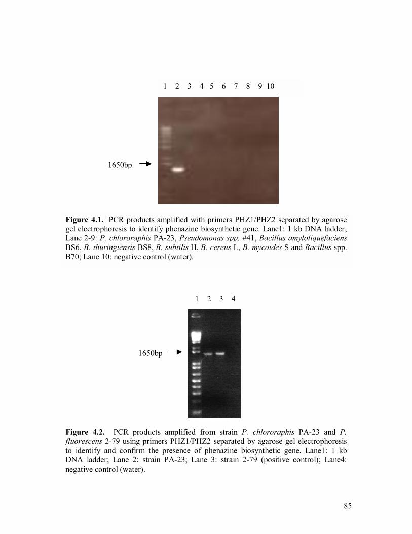
85
Figure 4.1. PCR products amplified with primers PHZ1/PHZ2 separated by agarose gel electrophoresis to identify phenazine biosynthetic gene. Lane1: 1 kb DNA ladder;Lane 2-9: P. chlororaphis PA-23, Pseudomonas spp. #41, Bacillus amyloliquefaciensBS6, B. thuringiensis BS8, B. subtilis H, B. cereus L, B. mycoides S and Bacillus spp. B70; Lane 10: negative control (water).
1650bp
1 2 3 4 5 6 7 8 9 10
Figure 4.2. PCR products amplified from strain P. chlororaphis PA-23 and P. fluorescens 2-79 using primers PHZ1/PHZ2 separated by agarose gel electrophoresisto identify and confirm the presence of phenazine biosynthetic gene. Lane1: 1 kbDNA ladder; Lane 2: strain PA-23; Lane 3: strain 2-79 (positive control); Lane4: negative control (water).
1 2 3 4
1650bp

86
Figure 4.4. PCR products amplified from strain P. chlororaphis PA-23 and P.fluorescens Q2-97 using primers BPF3/BPR2 separated by agarose gel electrophoresisto identify 2,4-DAPG biosynthetic gene. Lane1: 100 bp DNA ladder; Lane 2: strain PA-23; Lane 3: strain Q2-87 (positive control); Lane4: negative control (water).
1 2 3 4
600bp
Figure 4.3. PCR products amplified from P. chlororaphis PA-23 and P. fluorescens Pf-5 with primers PrnAF/RF, PRND1/PRND2 and PrnCf/PrnCr separated by agarose gelelectrophoresis to identify pyrrolnitrin biosynthetic gene. Lane1: 1 kb DNA ladder; Lane2: strain PA-23, primers PrnAF/RF; Lane 3: strain Pf-5, primers PrnAF/RF (positivecontrol); Lane 4: strain PA-23, PRND1/PRND2; Lane 5: strain Pf-5, PRND1/PRND2(positive control); Lane 6: strain PA-23, PrnCf/PrnCr; Lane 7: strain Pf-5, PrnCf/PrnCr(positive control) ; Lane 8: negative control (water).
1650bp
1 2 3 4 5 6 7 8

87
1 2 3 4 5 6 7 8 9 10 11
600bp
Figure 4.6. PCR products amplified with primers 678/677 separated by agarose gelelectrophoresis to identify zwittermicin A self-resistant gene. Lane1: 100 bp DNAladder; Lane 2-9: P. chlororaphis PA-23, Pseudomonas spp. #41, Bacillusamyloliquefaciens BS6, B. thuringiensis BS8, B. subtilis H, B. cereus L, B. mycoides Sand Bacillus spp. B70; Lane 10: B. cereus UW85 (positive control); Lane 11: negativecontrol (water).
Figure 4.5. PCR products amplified from strain P. chlororaphis PA-23 and P.fluorescens Pf-5 using primers PLT BF/PLT BR separated by agarose gelelectrophoresis to identify pyoluterorin biosynthetic gene. Lane1: 100 bp DNA ladder;Lane 2: strain PA-23; Lane 3: strain Pf-5 (positive control); Lane 4: negative control(water).
1 2 3 4
600bp

88
4.5 Discussion
The PCR and sequencing results showed the high possibility of the presence of
phenazine-1-carbocylic acid (PCA) and pyrrolnitrin, but not pyoluterorin biosynthetic
genes in strain PA-23. Sequence analysis of PCR products showed similarity to PCA and
pyrrolnitrin genes of several P. fluorescens and Burkholderia spp. strains deposited in the
GenBank. The presence of pyrrolnitrin biosynthetic gene was also further confirmed by
southern hybridization (Chrystal Berry, personal communication). The presence of 2,4-
DAPG biosynthetic gene phlD was not very clear based on PCR and sequencing results.
In addition, further confirmation using southern hybridization also failed to give a clear
answer, but mainly due to technical difficulties (Chrystal Berry, personal
communication). However, the 2,4-DAPG bacterial extraction from strain PA-23 by the
method of Rosales et al. (1995) showed the same RF (retention factor) value as the 2,4-
DAPG commercial product (2,4-diacetylphloroglucinol, Toronto Rereach Chemicals Inc.,
North York, ON) (Nakkeeran Sevugapperumal, personal communication). Further
confirmation of 2,4-DAPG-producing ability of strain PA-23 is necessary, and analysis
with high performance liquid chromatography followed by mass spectroscopy is probably
a good choice ( Raaijmakers et al. 1999; Picard et al. 2000; Bonsall et al. 1997; Mavrodi
et al. 2001).
Pseudomonas spp. #41 has not shown any specific amplification using all thirty
primers in this study, which was consistent with PCR results carried by Chrystal Berry
(unpblished) when the primers PrnAF/PrnAR, PRND1/PRND2, PrnCf/PrnCr (for
pyrrolnitrin biosynthetic gene PrnA) and Phl2a/Phl2b (for 2,4-DAPG biosynthetic gene
89
phlD) were used. However, southern hybridization showed that Pseudomonas spp. #41
genomic DNA was hydridazied at low strigency (50°C) with the probes of pyrrolnitrin
biosynthetic gene PrnA of P. fluorescens Pf-5 and 2,4-DAPG biosynthetic gene phlD of
P. fluorescens Q2-87, respectively (Chrystal Berry, personal communication). The
presence of biosynthetic genes of pyrrolnitin and 2,4-DAPG in Pseudomonas spp. #41 is
not clear.
The PCR results also suggested that B. amyloliquefaciens BS6 may not contain
PCA, Plt, Prn, 2,4-DAPG biosunthetic genes and zwittermicin A self-resistant gene.
Since B. amyloliquefaciens BS6 amplified a weak band of 950 bp using primers 678/677
and the result was unrepeatable, further confirmation was conducted by using two novel
primers (Ramarathnam and Fernando, unpublished) designed for zwittermicin A
biosynthetic gene. No product was amplified from strain BS6, while a bright band of 800
bp was observed from the positive strain UW85 and several other Bacillus strains (Rajesh
Ramarathnam, personal communication).
All the tested strains in this study had showed antifungal activity in the plate
assay, and those tested in the greenhouse and field as well, but whether these identified
antibiotic-related genes were expressed and whether these antibiotics were responsible
for sclerotinia disease suppression in vivo need to be investigated. Tn5 mutagenesis is a
common method used to evaluate the biosynthetic gene expression and study the role of
antibiosis involved in disease suppression in biocontrol (Thomashow and Weller 1988;
Hill et al. 1994; Salcher and Lingens 1980; Bangera et al. 1994; Heungens and Parke
2001). A Tn5 mutant of P. fluorescens 2-79, which was deficient in PCA production, was

90
less suppressive to take-all on wheat caused by Gaeumannomyces graminis var. tritici
than the parental strain (Thomashow and Weller 1988). A Tn5 mutant of P. fluorescens
CHA0, which is defective in the production of 2,4-DAPG, showed reduced suppression
of black root rot on tobacco caused by Thielaviopsis basicola (Keel et al. 1992). Results
of Tn5 mutant of biocontrol agent Burkholderia cepacia AMMDR1 suggest no other
mechanism other than antibiosis could be identified in suppression of oomycete
pathogens of pea (Heungens and Parke 2001). Earlier studies by Fernando showed an
orange colour compound produced by P. chlororaphis PA-23 on LBA and PDA
(Fernando, unpublished), which indicates that this strain is probably a PCA producer;
however, whether this antibiotic is involved in sclerotinia disease suppression has not
been clear. Since P. chlororaphis PA-23 possibly produces three antibiotics (PCA, 2,4-
DAPG and Prn), Tn5 mutagenesis and subsequent testing of mutants and the wild type
strains on plants will help understand which antibiotic is responsible for sclerotinia
disease suppression.
Inhibition of sclerotial and/or spore germination by 2,4-DAPG extracted from
P. chlororaphis PA-23 has also been observed against several plant pathogens, including
�S. sclerotiorum, Macrophomina phaseolina, Rhizoctonia solani, Sclerotium rolfsii,
Fusarium oxysporum, Alternaria solani, Botryodiplodia theobromae (Nakkeeran,
personal communication). Antibiotics PCA, pyrrolnitrin and 2,4-DAPG are major
determinants of biological control of plant pathogens. The ability of producing multiple
antibiotics by strain PA-23 might explain its wide-spectrum disease suppression of plant
pathogens. Further work on Tn5 mutagenesis will help understand which antibiotic is
responsible for disease suppression in different host-pathogen systems.

91
5.0 PLANT INDUCED RESISTANCE MEDIATED BY
SCLEROTINIA SCLEROTIORUM AND BACTERIAL BIOCONTROL
AGENT BACILLUS AMYLOLIQUEFACIENS BS6
5.1 Abstract
An earlier study (chapter 3) has shown that bacterial strain Bacillus
amyloliquefaciens BS6 gave excellent protection to the canola plant against Sclerotinia
sclerotiorum ascospore infection under lab, greenhouse and field conditions. The
significant disease suppression and the low level of survival of strain BS6 on petals
indicated that plant-induced resistance might be one of the most important mechanisms
triggered by the bacterium. To test this hypothesis, plants were pre-treated with water or
strain BS6 (108 cfu/ml) by spraying the inoculum on to petals at the 30% bloom stage,
and then inoculated two days later with petals which were treated either with only
sclerotinia ascospores (105 spores/ml) (S petals) or with both strain BS6 (108 cfu/ml) and
sclerotinia ascospores (105 spores/ml) (B+S petals), by placing petals on three leaves per
plant. Disease incidence on canola stems was reduced by 60% on plants pre-treated with
strain BS6 and inoculated with S petals compared to plants pre-treated with water and
also inoculated with S petals. Leaves of plants pre-treated with water and then inoculated
with B+S petals had localized water-soaked lesions compared to plants pre-treated with
BS6 and also inoculated with B+S petals, which had no disease symptom (0%) on either
leaves or stems. Disease severity (DS) progressed rapidly in the absence of BS6 and
reached a rate of > 5 (on a 0-7 scale) after 19 d; however, the BS6-treated plants had no
disease (DS=0). Phenolic compounds of canola leaves were extracted using 80%
methanol and fractioned using ethyl acetate. The HPLC results suggest that both B.
92
amyloliquefaciens BS6, and S. sclerotiorum result in the induction of phenolic
compounds in canola leaves, but additional experiments are required to clearly
demonstrate the concentration changes over time.
5.2 Introduction
Sclerotinia stem rot caused by Sclerotinia sclerotiorum is one of the most
economically important diseases of canola in Canada. Usually cultural control methods
do not work due to the extremely wide host range of this pathogen and the long survival
time of sclerotia in soil. We have isolated a bacterial strain Bacillus amyloliquefaciens
BS6 that gave excellent protection to the canola plant against ascospore infection under
lab, greenhouse and field conditions (see Chapter 3). Although there was significant
disease suppression in the field, the low level of survival of strain BS6 on petals indicated
that plant-induced resistance might be one of the most important mechanisms triggered
by the bacterium.
Plant induced resistance is a process which is analogous to immunization on the
plant to trigger the natural defence machinery and results in the prevention of disease
caused by a variety of plant pathogens (van Loon 1997; Boyetchko 1999). Plant induced
resistance was often categorized as systemic acquired resistance (SAR) and induced
systemic resistance (ISR). SAR refers to the plant resistance triggered by necrotizing
pathogens (Bakker et al. 2003). SAR could express locally at the site of primary
inoculation but also systemically in tissues remotely located from the initial treatment.
ISR was described as the mode of action of disease suppression by non-pathogenic
rhizosphere bacteria (van Peer et al. 1991; Wei et al. 1991).
93
Very little work has been done in induced resistance to S. sclerotiorum on
canola/oilseed rape. Systemic resistance against S. sclerotiorum in oilseed rape was first
reported by Toal and Jones (1999). Plants treated by oxalic acid were reported to have
significantly smaller leaf lesions than the control plants after being challenged by the
pathogen. Phenolic compounds, which associate with induced plant resistance
against S. sclerotiorum, have also rarely been studied in this system. Studies by Li
(1999) and Bodnaryk (1992) on changes of glucosinolates treated with S. sclerotiorum or
by wounding suggest that glucosinolates� induction might be related to the induced
resistance to S. sclerotiorum in oilseed rape. Brassinin, methoxybrassinin, and
cyclobrassinin were the first phytoalexins reported from Brassica spp. induced by
Leptosphaeria maculans (Soledade et al. 1992). However, no study has been reported on
the relationship between phenolic compounds and induced resistance on canola/ oilseed
rape against S. sclerotiorum.
The purpose of this study was (i) to investigate induced resistance mediated by
S. sclerotiorum and bacterial strain BS6 in canola in the greenhouse, (ii) to isolate
phenolic compounds from canola leaves, and investigate their role in induced resistance
in the canola plant by monitoring their changes over time.
5.3 Materials and methods
5.3.1 Greenhouse experiment
Canola plants (var. Westar) were grown in the growthroom at 16/ 21 °C and 16
h photoperiod. Plants were pre-treated with water (CK) or strain BS6 (108 cfu/ml) by
spraying the inoculum onto petals at the 30% bloom stage, and then inoculated two days
94
later with petals which were treated either with only Sclerotinia ascospores or with both
stain BS6 and Sclerotinia ascospores by placing them on three leaves per plant (Figure
5.1). Eleven plants were treated in each of the five treatments. Five plants in each
treatment were used to record the disease incidence (DI) and disease severity (DS), and
the other six plants were used for leaf sampling.
One loop of fresh bacteria grown on Nutrient Agar (Difco Laboratories, Detroit,
Mich., U.S.A.) was put into 150ml Luria Bertani broth [LB broth contains 10.0 g tryptone
peptone, 5.0 g yeast extract (Difco Laboratories, Detroit, Mich., U.S.A.), 5.0 g NaCl L-1],
and incubated at 30 ºC and rotated at 180 rpm for 16-18 h. Ascospores of S. sclerotiorum
(University of Nebraska) were washed with a brush and distilled water to release them
from the aluminium foil. The concentrations of bacterial (108 cfu/ml) and ascospore (105
spores/ml) solution were checked using a spectrophotometer and a hemacytometer
(Bright-Line®, Reichert, Buffalo, N.Y., U.S.A.), respectively. Petals were sampled from
other untreated plants and dipped into bacterial and/or ascospore solution, and placed on
water agar for 1-2 h before placing them onto the leaves. Tween 20 (0.02%) was used in
all inoculum. After inoculated with petals, plants were kept in the humidity chamber for
3.5 days (24/16 °C, 16 photoperiod and RH = 87%) before being moved to a growth
cabinet (25/19 °C, 16 photoperiod). DI and DS were recorded every 3 days in the
following two weeks in the growth cabinet. DI of each plant was calculated as percentage
of infected leaves, and data of each treatment were averaged from 5 plants. DS was rated
the same as previous greenhouse experiments, except that the water-soaked lesion was
recorded as 0.5 (Figure 5.2 D) to distinguish with more obviously necrotizing leaf lesion.
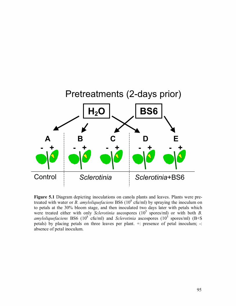
95
Figure 5.1 Diagram depicting inoculations on canola plants and leaves. Plants were pre-treated with water or B. amyloliquefaciens BS6 (108 cfu/ml) by spraying the inoculum on to petals at the 30% bloom stage, and then inoculated two days later with petals whichwere treated either with only Sclerotinia ascospores (105 spores/ml) or with both B. amyloliquefaciens BS6 (108 cfu/ml) and Sclerotinia ascospores (105 spores/ml) (B+S petals) by placing petals on three leaves per plant. +: presence of petal inoculum; -: absence of petal inoculum.
H2O BS6
A - +
B - +
C- +
D- +
E - +
Control Sclerotinia Sclerotinia+BS6
Pretreatments (2-days prior)
96
DI and DS were compared between different treatments using LSD test in ANOVA by
SAS software (Crow 2002).
Canola leaves with petal inoculation (+) and without petal inoculation (-)
(Figure 5.1) in each treatment were sampled separately �2, 0, 3, 5, and 7 days after
pathogen inoculation (DAI), and recorded as A, B-, B+, C-, C+, D-, D+, E- and E+ for
each treatment. Leaves were put onto ice immediately after sampling to bring back to the
lab, weighed and then stored at �80 °C.
5.3.2 Phenolics extraction and fractionation
Free phenolics were extracted using a modified method of Daayf et al. (1997). Leaves
stored at �80 °C were grounded in liquid N2, and immediately put into 80% methanol
(Fisher ScientificTM, Fair Lawn, New Jersey, U.S.A) in 50 ml or 150 ml flasks with the
ratio of 10ml 80% methanol /1 g fresh leaf weight. The flasks were wrapped with
aluminium foil and incubated at room temperature for 24 h at 200 rpm. Each sample was
then vacuum filtrated using Waterman No. 1 filter paper, and was collected to a 50 ml
centrifuge tube (Fisherbrand, Ficher Scientific, Pittsburgh, PA, U.S.A.). Methanol
(100%) was used to wash each flask. Samples were evaporated using N2 at 40 ºC in the
water bath, until the volume left in each tube was approximately 20% of original volume.
Petroleum ether (Fisher ScientificTM, Fair Lawn, New Jersey, U.S.A) (vol. 1:1) was used
twice to exclude the chlorophyll, carotenoids, lipids, and waxes. After petroleum ether
was added, the tube was thoroughly mixed and then let stand for 10 min. The acquous
fraction (lower part), which contains the phenolic compounds, was then partitioned with
ethyl acetate (Fisher ScientificTM, Fair Lawn, New Jersey, U.S.A) (vol. 1:1). The ethyl
acetate fraction was evaporated using N2 as previously described, and was then re-

97
dissolved in 100% methanol and the concentrations were adjusted to 8 g of fresh material
per millilitre of methanol. Samples were stored at �20ºC for further analysis. The samples
from 5DAI were extracted with petroleum ether after the ethyl acetate extraction, and
then extracted again with H2O and ethyl acetate.
5.3.3 HPLC analysis
Samples were analysed by high performance liquid chromatography (HPLC)
with two separate runs: one injection the first time, and another two injections the second
time. Fifteen micro litres of each sample were used in each injection (25 µl and 35 µl
were prepared in the test tube, respectively). The HPLC was equipped with a Waters 996
photodiode array detector, a Waters 2690 separations module, and fitted with a C18
reverse-phase column (Lichrosorb® RP-18 (7 µm), Kartuschenhalter Cartridge holder, E.
Merck, Darmstadt, F.R. Germany). Results were analyzed with the Millennium32
Software. The column was eluted with a gradient of acetonitrile - 0.1% phosphoric acid
(acetonitrile, Fisher ScientificTM, Fair Lawn, New Jersey, U.S.A; 1% phosphoric acid,
Fisher ScientificTM, Fair Lawn, New Jersey, U.S.A) as follows: [time (minutes)
/acetonitrile (percent) /1% phosphoric acid (percent)] c [5 /5 /95, 10 /5 /95, 14 /10 /90, 20
/20 /80, 23 /20 /80, 30 /35 /65, 35 /35 /65, 43 /50 /50, 48 /75 /25, 55 /100 /0, 60 /100 /0,
62 /0 /100, 65 /0 /100]. Samples were monitored at the wavelength between 220.0 nm to
370.0 nm.

98
5.4 Results
5.4.1 Greenhouse experiment
The difference in disease symptoms between each treatment was visualised 5-6
days after pathogen inoculation (DAI) (Figure 5.2). No infection was visualized on either
leaf or stem in treatments A (water-treated control) and E (pre-treated by strain BS6 and
inoculated with B+S petals), which means that plants pre-treated by B. amyloliquefaciens
BS6 and then co-inoculated with both B. amyloliquefaciens BS6 and S. sclerotiorum
ascospores have achieved complete disease suppression (Figure 5.3 and 5.4). DI on leaf
of treatments B (pre-treated by water and inoculated with S petals) and C (pre-treated by
strain BS6 and inoculated with S petals) was observed as 73.3% and 66.7% 4DAI,
respectively, which are not significantly different. However, DI on stem was reduced by
60% in treatment C comparing to treatment B 7DAI (Figure 5.4). In treatment C, disease
lesions are mainly restricted on the inoculated leaves instead of developing onto the
stems (Figure 5. 2 C). A significant (P<0.05) reduction of DI on canola leaf 4DAI was
also observed in treatment E (pre-treated with strain BS6 and inolulated with B+S petals),
compared with the treatment D (pre-treated with water and inoculated with B+S petals)
(Figure 5.3), but no such significant reduction was found on canola stem 7DAI (Figure
5.4).
Disease severity (DS) progressed rapidly in the absence of BS6 and reached a
rate of >5 (on a 0-7 scale) in treatment B after 19 d; however, the BS6-treated plants had
DS<2 (treatment C, DS<2; treatment D, DS<1; treatment E, DS=0) (Figure 5.5). Only
water-soaked lesions were found in plants pre-treated by water and inoculated with B+S
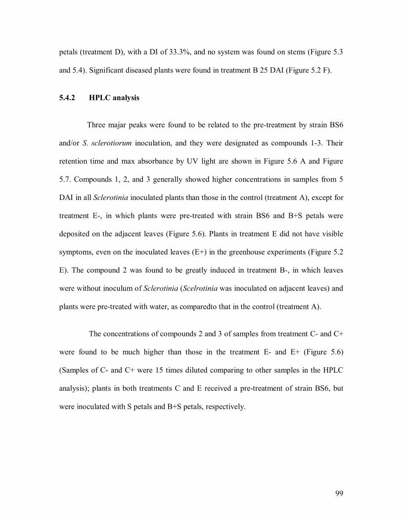
99
petals (treatment D), with a DI of 33.3%, and no system was found on stems (Figure 5.3
and 5.4). Significant diseased plants were found in treatment B 25 DAI (Figure 5.2 F).
5.4.2 HPLC analysis
Three majar peaks were found to be related to the pre-treatment by strain BS6
and/or S. sclerotiorum inoculation, and they were designated as compounds 1-3. Their
retention time and max absorbance by UV light are shown in Figure 5.6 A and Figure
5.7. Compounds 1, 2, and 3 generally showed higher concentrations in samples from 5
DAI in all Sclerotinia inoculated plants than those in the control (treatment A), except for
treatment E-, in which plants were pre-treated with strain BS6 and B+S petals were
deposited on the adjacent leaves (Figure 5.6). Plants in treatment E did not have visible
symptoms, even on the inoculated leaves (E+) in the greenhouse experiments (Figure 5.2
E). The compound 2 was found to be greatly induced in treatment B-, in which leaves
were without inoculum of Sclerotinia (Scelrotinia was inoculated on adjacent leaves) and
plants were pre-treated with water, as comparedto that in the control (treatment A).
The concentrations of compounds 2 and 3 of samples from treatment C- and C+
were found to be much higher than those in the treatment E- and E+ (Figure 5.6)
(Samples of C- and C+ were 15 times diluted comparing to other samples in the HPLC
analysis); plants in both treatments C and E received a pre-treatment of strain BS6, but
were inoculated with S petals and B+S petals, respectively.

100
A B
C D
E F
Figure 5.2 A-E: Typical disease symptoms on canola of Treatment A, B, C, D, andE, respectively, observed 6 Days after disease inoculation (DAI); F: Diseasesymptoms of treatment A-E observed 25 DAI. Treatments C and E were pre-treated with B. amyloliquefaciens BS6 (108 cfu/ml) by spraying; Treatments A (control), B, D were pre-treated with water. Treatment B and C were inoculated with Sclerotiniaascospores (105 spores/ml); Treatments D and E were inoculated with both B. amyloliquefaciens BS6 (108 cfu/ml) and Sclerotinia ascospores (105 spores/ml). Three leaves per plant were inoculated with infested petals.
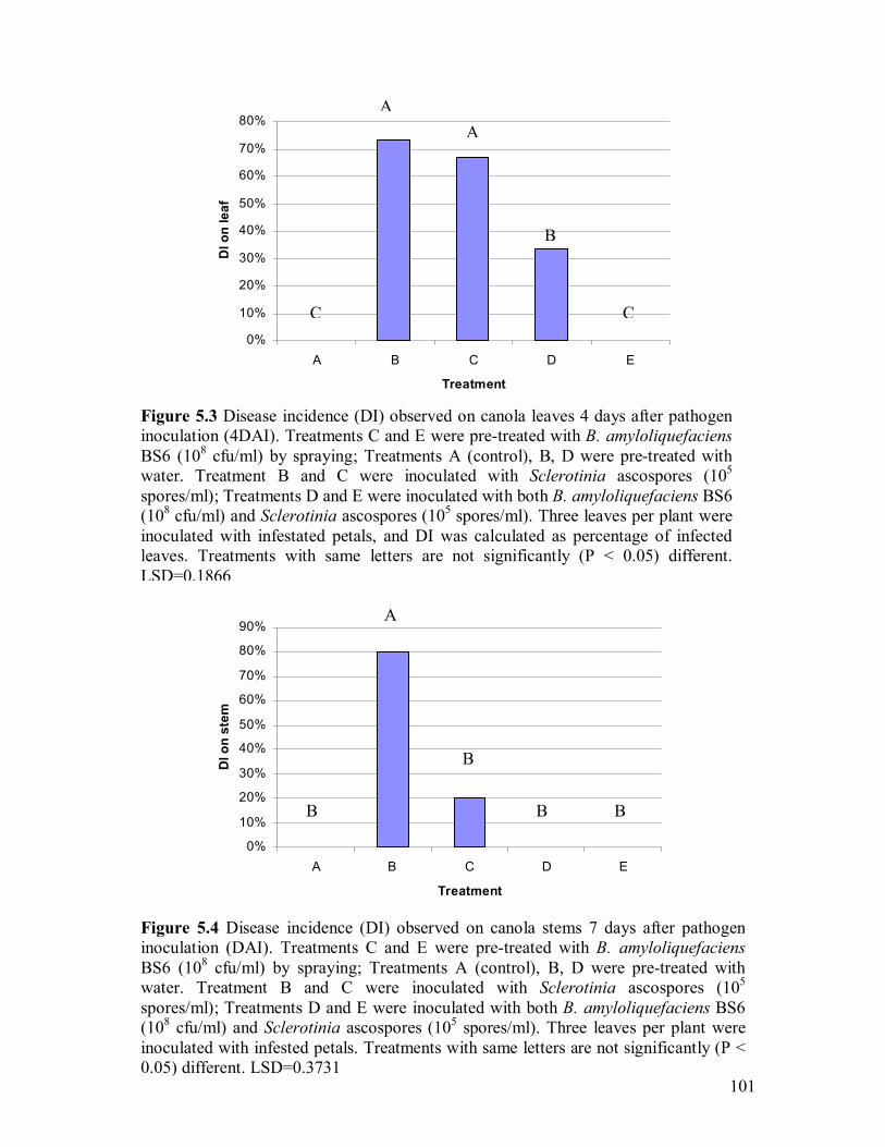
101
Figure 5.3 Disease incidence (DI) observed on canola leaves 4 days after pathogeninoculation (4DAI). Treatments C and E were pre-treated with B. amyloliquefaciensBS6 (108 cfu/ml) by spraying; Treatments A (control), B, D were pre-treated withwater. Treatment B and C were inoculated with Sclerotinia ascospores (105
spores/ml); Treatments D and E were inoculated with both B. amyloliquefaciens BS6(108 cfu/ml) and Sclerotinia ascospores (105 spores/ml). Three leaves per plant wereinoculated with infestated petals, and DI was calculated as percentage of infectedleaves. Treatments with same letters are not significantly (P < 0.05) different.LSD=0.1866
0%
10%
20%
30%
40%
50%
60%
70%
80%
A B C D E
Treatment
DI o
n le
af
A
C
A
B
C
0%
10%
20%
30%
40%
50%
60%
70%
80%
90%
A B C D E
Treatment
DI o
n st
em
A
B
B
B B
Figure 5.4 Disease incidence (DI) observed on canola stems 7 days after pathogeninoculation (DAI). Treatments C and E were pre-treated with B. amyloliquefaciensBS6 (108 cfu/ml) by spraying; Treatments A (control), B, D were pre-treated with water. Treatment B and C were inoculated with Sclerotinia ascospores (105
spores/ml); Treatments D and E were inoculated with both B. amyloliquefaciens BS6 (108 cfu/ml) and Sclerotinia ascospores (105 spores/ml). Three leaves per plant were inoculated with infested petals. Treatments with same letters are not significantly (P < 0.05) different. LSD=0.3731
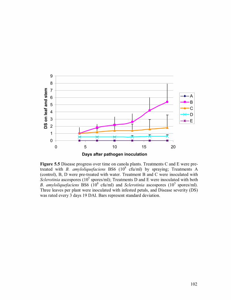
102
Figure 5.5 Disease progress over time on canola plants. Treatments C and E were pre-treated with B. amyloliquefaciens BS6 (108 cfu/ml) by spraying; Treatments A (control), B, D were pre-treated with water. Treatment B and C were inoculated withSclerotinia ascospores (105 spores/ml); Treatments D and E were inoculated with bothB. amyloliquefaciens BS6 (108 cfu/ml) and Sclerotinia ascospores (105 spores/ml). Three leaves per plant were inoculated with infested petals, and Disease severity (DS)was rated every 3 days 19 DAI. Bars represent standard deviation.
0
1
23
4
5
67
8
9
0 5 10 15 20
Days after pathogen inoculation
DS
on le
af a
nd s
tem
ABCDE
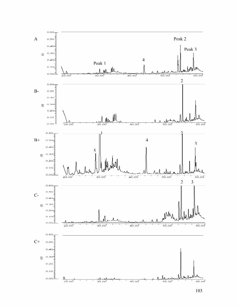
103
A
B-
C+
C-
B+
Peak 1
Peak 2
Peak 3
1 2
34
4
5
2 3
2
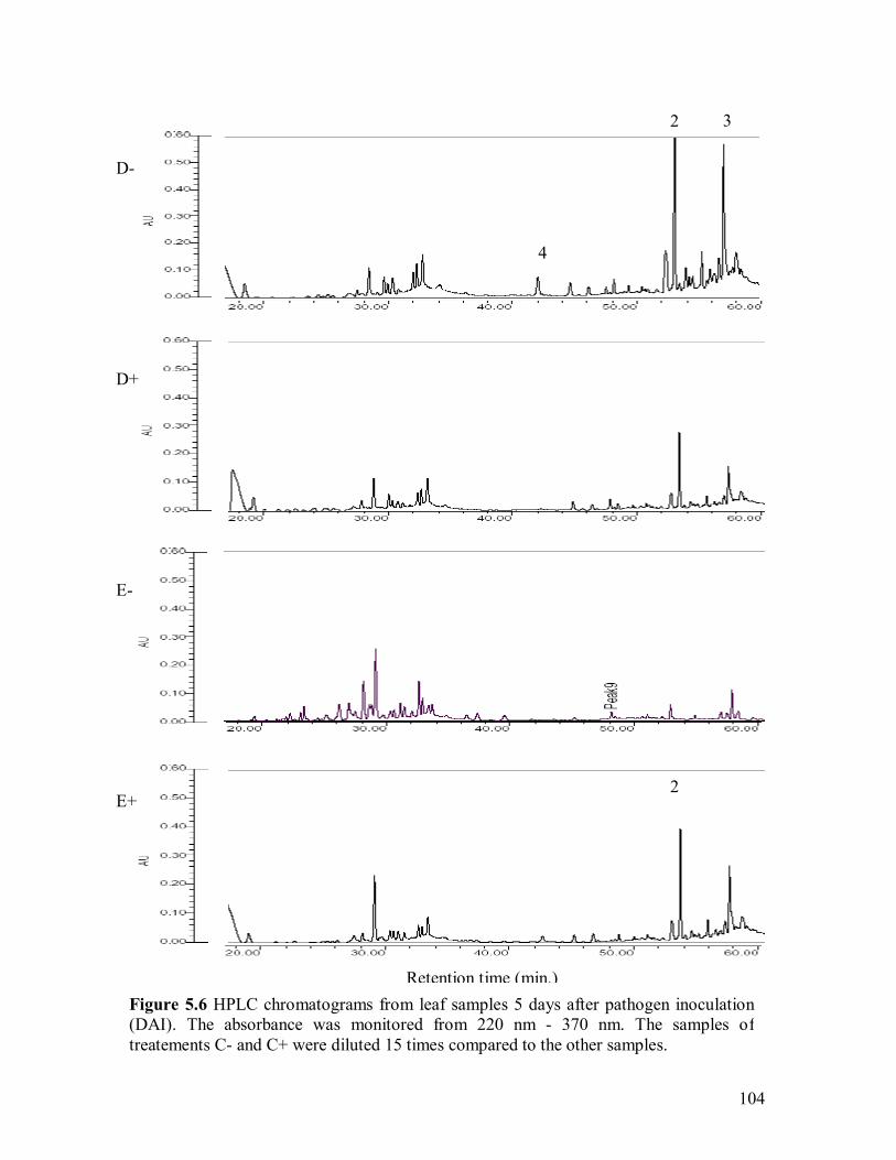
104
Figure 5.6 HPLC chromatograms from leaf samples 5 days after pathogen inoculation (DAI). The absorbance was monitored from 220 nm - 370 nm. The samples of treatements C- and C+ were diluted 15 times compared to the other samples.
Retention time (min.)
D-
E+
E-
D+
4
2
2 3
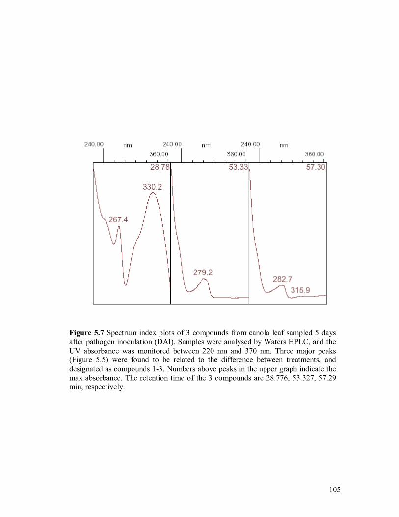
105
Figure 5.7 Spectrum index plots of 3 compounds from canola leaf sampled 5 days after pathogen inoculation (DAI). Samples were analysed by Waters HPLC, and theUV absorbance was monitored between 220 nm and 370 nm. Three major peaks(Figure 5.5) were found to be related to the difference between treatments, and designated as compounds 1-3. Numbers above peaks in the upper graph indicate themax absorbance. The retention time of the 3 compounds are 28.776, 53.327, 57.29min, respectively.
106
The samples from 3 DAI and 7 DAI (data not shown) showed similar results but
are not as clear as those from 5 DPI. The compound 1 seems to reach the highest
concentration 3 DAI, and the compound 2 and 3 seems to reach the highest concentration
5 DAI.
5.5 Discussion
Disease incidence (DI) on canola stems was reduced by 60% on plants pre-
treated with B. amyloliquefaciens BS6 and inoculated with Sclerotinia ascospores
(treatment C) compared to plants pre-treated with water and also inoculated with
Sclerotinia ascospores (treatment B). Plants pre-treated with B. amyloliquefaciens BS6
and inoculated with both B. amyloliquefaciens BS6 and Sclerotinia ascospores (treatment
E), had no disease symptom (0%) on either leaf or stem, compared to plants pre-treated
with water and then inoculated with both B. amyloliquefaciens BS6 and Sclerotinia
ascospores (treatment D) with localized water-soaked lesions. The two cases of
comparison suggest that pre-treatment with B. amyloliquefaciens BS6 did reduce the
sclerotinia disease. However, the pretreatment of B. amyloliquefaciens BS6 only
achieved a significant (P < 0.05) reduction of DI on canola leaf 4DAI, but not on canola
stem 7DAI, by comparing the treatments D and E (Figure 5.3 and 5.4). Therefore, even
though induced resistance by B. amyloliquefaciens BS6 on canola plants may involve be
in the disease suppression, the latter seems to be mainly due to a direct bacteria-fungi
interaction.
In this experiment, treatment E where plants were pre-treated with B.
amyloliquefaciens BS6 and then inoculated with both B. amyloliquefaciens BS6 and
107
Sclerotinia ascospores achieved complete disease suppression. In an earlier study about
the effect of time of application of strain BS6 (see chapter 3), treatment B+A (strain BS6
and ascospores inoculated at the same day) had the best disease control among all the
treatments, including treatment B-2-A (BS6 inoculated 2 days prior to ascospore) (Figure
3.9) that was equivalent to treatment C (pre-treated with water and inoculated with B-S+
petals) in this experiment. The results from the two experiments are not contradictory;
however, the combination of the results demonstrates that when B. amyloliquefaciens
BS6 inoculated with Sclerotinia ascospores at the same day (Figure 3.9) or when plants
were pre-treated with strain BS6 2 days prior to ascospore inoculation, strain BS6 had
significant disease suppression but did not achieve complete disease suppression;
However, only when B. amyloliquefaciens BS6 was used as both pre-treatment and co-
inoculum with Sclerotinia, the complete disease suppression was achieved.
The question here might be the disease suppression by B. amyloliquefaciens
BS6 pre-treatment is due to the bacterial residue on the plant leaf (even bacterial
population level might be low), which might be toxic to ascospores, or to the induced
resistance by the plant itself. The HPLC results of treatments C (inoculated with
ascospores) and E (inoculated with B+S petals), where plants were both pre-treated with
strain BS6, demonstrate that the induction of compounds 2 and 3 are from plants other
than from bacteria, since treatment C has much higher concentration of the two
compounds than treatment E. Interestingly, the elicitation of compounds 2 and 3 by B.
amyloliquefaciens BS6 seems to happen only when the plants are under high disease
pressure, such as Sclerotinia ascospores are active or in high concentration in infecting
plants. This might be the reason why even treatments C and E are both pre-treated with
108
strain BS6, but the two compounds are found at high concentration only in treatment C,
but not in E, in which Sclerotinia ascosproes might be greatly inhibited by the bacteria.
The study by Collins et al. (2003) showed that the bacterial cell density of B.
subtilis BacB isolated from sugarbeet phyllosphere was not strongly correlated to the
disease suppression of sugarbeet cercospora leaf spot, caused by Cercospora beticola
under field conditions. Their greenhouse study also showed that plants that were pre-
treated by B. subtilis BacB 3 days prior to C. beticola inoculation have significant better
control than those treated with bacteria right before the pathogen, which may indicate that
the foliar-applied B. subtilis BacB had induced the sugarbeet to build up its resistance to
C. beticola. Similar results were found by Liu et al. (1995), Hoffland et al. (1995) and
Meena et al. (1999) using foliar-applied plant growth-promoting rhizobacteria (PGPR).
Therefore, the ISR might not be only a common phenomenon of PGPR when bacteria are
applied on rhizosphere, but also common in foliar-applied biocontrol bacteria agents.
Since the foliar-applied bacterial agents and the S. sclerotinia both target plant
phyllosphere (or blossoms), the inoculation method of studying induced resistance needs
to be especially concerned. The inoculation could either be done by spatially separating
the inoculations of bacteria and S. sclerotinia, or by adding a bacterial treatment (without
S. sclerotinia) to the experimental design of this study. A better design will help clarify
the role of the pathogen and the bacteria in the process of induced resistance in canola.
The induction of phenolic compounds in treatment B+ and B- (plants were pre-
treated with B. amyloliquefaciens BS6, and then Sclerotinia was inoculated on B+ leaves)
compared to control (treatment A) is an indication of systemic acquired resistance
109
mediated by the pathogen S. sclerotiorum. However, whether this form of induced
resistance will deviate the further pathogen attacking is unknown. Systemic resistance to
S. sclerotiorum in oilseed rape was first reported by Toal and Jones (1999) by pre-treated
oilseed rape with oxalic acid. Oxalic acid is the major pathogenesis factor of S.
sclerotiorum. Plants treated with oxalic acid were found to have significantly smaller leaf
lesions than the control plants after being challenged by the pathogen. Whether the
phenolic induction in this experiment was due to the pathogenesis factor oxalic acid or
other determinants of S. sclerotiorum is unkonwn.
Since induced resistance is generally expressed against a broad spectrum of
organisms, it usually results in preventing a variety of plant diseases (Kuc 2001). The
study by Bonnet et al. (1996) showed that elicitins, which are a family of proteins
excreted by Phytophthora spp., not only induced resistance to the Phytophthora
parasitica var. nicotianae on tobacco, but also to S. sclerotiorum on oilseed rape.
Therefore it is also valuable to investigate the induced resistance elicited by B.
amyloliquefaciens BS6 against other canola pathogens, such as Leptosphaeria maculans.
Integrating plant induced resistance as one of the strategies or mechanisms in disease
control under field conditions will be promising, since there are different pathogens
existing in the natural farm system.
110
6.0 GENERAL DISCUSSION AND CONCLUSIONS
Antibiosis and plant induced resistance appear to be two separate mechanisms
of disease suppression, and the their combination may lead to improved efficacy of
biological control. Bakker et al. (2003) stated that antibiosis would first weaken or even
partly kill the population of the pathogen and, subsequently, this weakened population of
the pathogen would be confronted on a plant that is in a state of enhanced defensive
capacity, thus resulting in much less disease development. Therefore, integrating the two
mechanisms in biocontrol of Sclerotinia on canola should be a promising prospect.
Study of bacterial population dynamics is important in biocontrol of plant
pathogens. The understanding of bacterial population and the time of application is
essential to help understand biocontrol mechanisms. In this study, the bacterial
population dynamics were investigated with B. amyloliquefaciens BS6 and Pseudomonas
chlororaphis PA-23, and the effect of time of application and plant induced resistance
were tested with B. amyloliquefaciens BS6; however, whether plant induced resistance is
involved in the disease suppression by other bacteria that were used in the field study is
unknown.
Most studies on induced systemic resistance (ISR) are on plant growth
promoting rhizobacteria (PGPR) (van Loon et al. 1998), and studies on ISR mediated by
foliar applied bacteria are comparatively rare. Leaf application of PGPR strains which
induce resistance on plants were reported by Liu et al. (1995), Hoffland et al. (1995) and
Meena et al. (1999). Bacillus subtilis BacB was isolated from phyllosphere and has been
found to induce resistance on sugar beet against Cercospora leaf spot when used as a
111
foliar application (Collins and Jacobsen 2003). The preliminary study of induced
resistance mediated by B. amyloliquefaciens BS6 (chapter 5) has already strongly
suggested that induced resistance by foliar applied bacteria may be involved in sclerotinia
disease control on canola. Therefore, the induced resistance mediated by P. chlororaphis
PA-23 and other bacteria should be evaluated. Investigating plant induced resistance
mediated by foliar applied bacteria will help us to establish a better system to control
Sclerotinia sclerotiorum by combining different mechanisms in a single biocontrol agent.
P. chlororaphis PA-23 has shown successful disease control in three field trials
in two years (one in 2001 and then in 2003), even though the population density was
quite low in some of the cases. The high disease suppression of sclerotinia stem rot by P.
chlororaphis PA-23 seems to involve multiple mechanisms. It was found by Kavitha et
al. (2003) that P. chlororaphis PA-23 had significant increase in plant growth as well as
disease control of damping-off in chilli. It was also found that plants pre-treated with P.
chlororaphis PA-23 and challenged with Pythium aphanidermatum in tomato had higher
levels of phenylalanine ammonia lyase, peroxidase, polyphenol oxidase and β-(1,3)-
glucanase, which indicated it may have triggered induced systemic resistance in plants.
This study has shown that P. chlororaphis PA-23 could probably produce two or three
antibiotics (chapter 4). The future work on Tn5 mutagenesis of the wild-type strain, and
testing antibiotic deficient mutants and wild type strians against S. sclerotiorum on
canola, will help understand the role of phenazine, pyrrolnitrin and perhaps 2,4-
diacetylphloroglucinol in controlling Sclerotinia on canola.

112
Bacillus spp. are well known to have good survival under field conditions due to
the production of endospores (Boyetchko 1999; Collins and Jacobsen 2003). Bacillus
amyloliquefaciens BS6 was observed to survive longer than P. chlororaphis PA-23 in the
field study (chapter 3), but single application of P. chlororaphis PA-23 had better disease
control than that of B. amyloliquefaciens BS6 in Trial Two (without tents). Therefore, the
disease suppression under field conditions does not necessarily correlate with bacterial
survival ability and this should be taken into account when biocontrol agents are screened
and developed. It was suggested that effective screening of biocontrol bacteria could be
conducted by screening on media plates first and following it with a field experiment that
includes as much genotypically-diverse bacterial strains as possible (Roberts and Lohrke
2003). Bacillus spp. have an advantage to commercialization due to their endospore
forming ability (Collins and Jacobsen 2003). The commercialization of Pseudomonas
spp. as biocontrol agents seems to be much more difficult than that of Bacillus spp.
(Scherm et al. 2004). Slininger et al. (1996) stated, �Barriers to the commercial use of
phenazine-producing pseudomonads, such as strain 2-79 include the lack of liquid-culture
and formulation technologies needed to optimize cost-effective mass production and
application.� However, the study by Janisiewics and Jeffers (1997) have shown that the
formulation of P. syringae (ESC-11) as a biofungicide (Bio-Save 11), did not have
adverse effect on disease control of blue mold and gray mold of apples, compared with
fresh bacterial cells. Therefore, the study on formulation of P. chlororaphis PA-23
towards commercialization is needed, since it might have greater disease suppression
than Bacillus spp. when appropriately applied on canola plants. In addition, as different
bacterial strains may be involved in different mechanisms, it is useful to develop
113
formulations to combine the bacterial strains, which are capable of producing antibiotics,
inducing plant resistance and promoting plant growth.
S. sclerotiorum is comparatively easy to handle in vitro, but its inoculation on
canola plants is difficult, especially under field conditions. In the greenhouse studies
(chapter 3 and chapter 5), different inoculation conditions were employed, and this was
mainly due to lack of standard protocol. In the study by Jurke (2003), several Sclerotinia
inoculation methods were compared for selecting lines of sclerotinia stem rot resistance
in B. napus , and mycelium-infested petal technique (Mypetal) appeared to be the most
accurate, reliable and efficient disease inoculation method. In his study, ascospore-
infested petal technique (Ascpetal), which is similar to the dipping petals method in this
study, was also regarded as the most accurate measure of physiological resistance since it
mimics the natural infection process; however, it was found that Ascpetal technique had
low efficiency and high variability in growthroom trials, so it was not recommended for
disease inoculation in growthroom for screening resistance against Sclerotinia. However,
S. sclerotiorum infects canola plants mainly through ascospore-infected petals, and petals
offer a critical nutrient factor for ascospores to germinate. So many of the other methods,
such as Mypetal technique are more artificial than the inoculation described in this study.
Mypetal technique is not recommended for screening biocontrol agents in the
greenhouse, for it might mask the real biocontrol ability of potential biocontrol agents.
Ascospore inoculation was used in both greenhouse and field in this study, and they both
appeared effective and reliable if proper conditions such as a misting system were
provided. The dipping petal method used in this study was modified from the work of
Savchuck (2002), by placing petals on water agar for 1 h instead of 24 h before placing

114
onto plants. The dipping petal method described in chapter 3 and following with an
incubation period of 3-4 days with daytime temperatures of 23-24 °C in humidity
chamber is recommended for Sclerotinia ascospore inoculation in the greenhouse. After
the incubation period, plants should be kept no less than 23 °C (25 ° C is probably the
best) to allow the disease to progress.
There is also no generally used standard for Sclerotinia disease evaluation on
canola so far. The disease evaluation used in both greenhouse study (chapter 3 and
chapter 5) and field study (chapter 5) is recommended for future disease rating of
Sclerotinia stem rot on canola, accompanied by the recommended inoculation methods.
In summary, several potential biocontrol agents against S. sclerotiorum were
screened in the lab and greenhouse in this study. The two field trials showed that P.
chlororaphis PA-23, Pseudomonas spp. #41, B. amyloliquefaciens BS6, and
Staphylococcus spp. E16 achieved significant disease suppression, and their disease
suppression ability may not depend on bacterial survival. The presence of antibiotic
biosynthetic or self-resistance genes in several potential biocontrol bacteria and the
evidence of induced resistance mediated by B. amyloliquefaciens BS6 have strongly
suggested that antibiosis and plant induced resistance might be two important
mechanisms responsible for the effective sclerotinia disease control on canola.
115
Literature Cited
ACE. 2001. Agrometeorological Centre of Excellence (ACE), Carman, Manitoba. Available from http://www.aceweather.ca/moreinfo.html: Accessed June 25, 2004.
Altenbach, S. B., C. C. Kuo, L. C. Staraci, K. W. Pearson, C. Wainwright, A. Georgescu, and J. Townsend. 1992. Accumulation of a Brazil nut albumin in seeds of transgenic canola results in enhanced levels of seed protein methionine. Plant Mol. Biol. 18: 235-245.
Andow, D. A. 2003. UK farm-scale evaluations of transgenic herbicide-tolerant crops. Nature Biotechnology 21; 1453-1454.
Anjaiah, V., P. Cornelis, N. Koedam, M. Hoefte, and J. Tabi Tambong. 1998. Biocontrol of Fusarium wilt and Pythium damping-off by the metabolises of P. aeruginosa PNA1 and its TN5-derivatives: Phenazines and anthranilate. Mededelingen. Faculteit. Landbouwkundige. en. Toegepaste. Biologische. Wetenschappen. Universiteit. Gent. 63: 1671-1678.
Anoop, V. M., U. Basu, M. T. McMcammon, L. McAlister-Henn, and G. J. Taylor. 2003. Modulation of citrate metabolism alters aluminum tolerance in yeast and transgenic canola overexpressing a mitochondrial citrate synthase. Plant Physiology (Rockville) 132: 2205-2217.
Armstrong, W. P. 2003. Phenolic compounds, glycosides & Alkaloids in Major Types of Chemical Compounds in plants & animals. available from http://waynesword.palomar.edu/chemid2.htm#phenolics: Accessed on July 20, 2004.
Arnoldo, M., C. L. Baszczynski, G. Belemare, G. Brown, J. Carlson, B. Gillespie, B. Huang, N. Maclean, W. D. MacRae, and G. Rayner. 1992. Evaluation of transgenic canola plants under field conditions. Genome 35: 58-63.
Arshad, M. A., Y. K. Soon, and R. H. Azooz. 2002. Modified no-till and crop sequence effects on spring wheat production in northern Alberta, Canada. Soil Tillage Res. 65: 29-36.
Ausubel, F. M., R. Brent, R. E. Kingstons, D. D. Moore, J. G. Seidman, J. A. Smith, and K. e. Struhl. 1995. Short protocols in molecular biology: a compendium of methods from current protocols in molecular biology. New York: John Wiley & Sons, Inc.
Baker, J. A. R. A. and C. Preston. 2003. Predicting the spread of herbicide resistance in Australian canola fields. Transgenic Research 12: 731-737.
Bakker, P. A. H. M., L. X. Ran, C. M. J. Pieterse, and L. C. Van Loon. 2003. Understanding the involvement of rhizobacteria-mediated induction of systemic resistance in biocontrol of plant diseases. Can. J. Plant Pathol. 25: 5-9.
116
Bangera, M. G. and L. S. Thomashow. 1996. Characterization of a genomic locus required for synthesis of the antibiotic 2,4-diacetylphloroglucinol by the biological control agent Pseudomonas fluorescens Q2-87. Mol. Plant Microb. Interac. 9: 83-90.
Bangera, M. G. and L. S. Thomashow. 1999. Identification and characterization of a gene cluster for synthesis of the polyketide antibiotic 2,4-diacetylphloroglucinol from Pseudomonas fluorescens Q2-87. J. Bacteriol. 181: 3155-3163.
Bangera, M. G., D. M. Weller, and L. S. Thomashow. 1994. Genetic analysis of the 2,4-diacetylphloroglucinol biosynthetic locus from Pseudomonas fluorescens Q2-87. Curr. Plant Sci. Biotechnol. Agric. 21: 383-386.
Bardin, S. D. and H. C. Huang. 2001. Research on biology and control of Sclerotinia diseases in Canada. Can. J. Plant Pathol. 23: 88-98.
Bazzalo, M. E., E. M. Heber, M. A. Pero Martinez, and O. H. Caso. 1985. Phenolic compounds in stems of sunflower plants inoculated with Sclerotinia sclerotiorum and their inhibitory effects on the fungus. Phytopathol. Z. 112: 322-332.
Bell, J. M. 1982. From rapeseed to canola: a brief history of research for superior meal and edible oil Production in Canada, feeding trials. Poult. Sci. 61: 613-622.
Benhamou, N. and R. R. Belanger. 1998. Induction of systemic resistance to Pythium damping-off in cucumber plants by benzothiadiazole: Ultrastructure and cytochemistry of the host response. Plant Journal 14: 13-21.
Benhamou, N., S. Gagne, D. Le Quere, and L. Dehbi. 2000. Bacterial-mediated induced resistance in cucumber: Beneficial effect of the endophytic bacterium Serratia plymuthica on the protection against infection by Pythium ultimum. Phytopathology 90: 45-56.
Bennett, R. N. and R. M. Wallsgrove. 1994. Secondary metabolites in plant defence mechanisms. New Phytol. 127: 617-633.
Beyeler, M., C. Keel, P. Michaux, and D. Haas. 1999. Enhanced production of indole-3-acetic acid by a genetically modified strain of Pseudomonas fluorescens CHA0 affects root growth of cucumber, but does not improve protection of the plant against Pythium root rot. FEMS Microbiol. Ecol. 28: 225-233.
Blackshaw, R. E., F. J. Larney, C. W. Lindwall, P. R. Watson, and D. A. Derksen. 2001. Tillage intensity and crop rotation affect weed community dynamics in a winter wheat cropping system. Can. J. Plant Sci. 81: 805-813.
Bodnaryk, R. P. 1992. Effects of wounding on glucosinolates in the cotyledons of oilseed rape and mustard. Phytochemistry 31: 2671-2677.
117
Boland, G. J. 1997. Stability analysis for evaluating the influence of environment on chemical and biological control of white mold (Sclerotinia sclerotiorum) of bean. Biol. Control 9: 7-14.
Boland, G. J. and R. Hall. 1994. Index of plant hosts of Sclerotinia sclerotiorum. Can. J. Plant Pathol. 16: 93-100.
Bonnet, P., E. Bourdon, M. Ponchet, J. P. Blein, and P. Ricci. 1996. Acquired resistance triggered by elicitins in tobacco and other plants. Eur. J. Plant Pathol. 181-192.
Bonsall, R. F., D. M. Weller, and L. S. Thomashow. 1997. Quantification of 2,4-diacetylphloroglucinol produced by fluorescent Pseudomonas spp. in vitro and in the rhizosphere of wheat. Appl. Environ. Microbiol. 63: 951-955.
Boulter, J. I., G. J. Boland, and J. T. Trevors. 2000. Compost: A study of the development process and end-product potential for suppression of turfgrass disease. World J. Microbiol. and Biotechnol. 16: 115-134.
Boyetchko, S. M. 1999. Biological control agents of canola and rapeseed diseases-status and practical approaches. In K. Mukerji, B. Chamola, and R. Upadhyay, eds. Biotechnological Approaches in Biocontrol of Plant Pathogens. New York: Kluwer Acadimic / Plenum Publishers. pp. 51-71.
Brannen, P. M. and D. S. Kenney. 1997. Kodiak--a successful biological-control product for suppression of soil-borne plant pathogens of cotton. J. Ind. Microbiol. Biotechnol. 19: 169-171.
Broderick, N. A., R. M. Goodman, K. F. Raffa, and J. Handelsman. 2000. Synergy between zwittermicin A and Bacillus thuringiensis subsp. kurstaki against gypsy moth (Lepidoptera: Lymantriidae). Environmental Entomology 29: 101-107.
Brodhagen, M., M. D. Henkels, and J. E. A. R. A. Loper. 2004. Positive autoregulation and signaling properties of pyoluteorin, an antibiotic produced by the biological control organism Pseudomonas fluorescens Pf-5. Appl. Environ. Microbiol. 70: 1758-1766.
Brown, J. W. 2001. Enrichment & isolation of Bacillus. Lecture MB409 Microbial diversity, Department of Microbiology, North Carolina State University. Available from http://www.mbio.ncsu.edu/jwb/mb409/lab/bacillus/isolation.html: Accessed July 2, 2004.
Burd, G. I., D. G. Dixon, and B. R. Glick. 1998. A plant growth-promoting bacterium that decreases nickel toxicity in seedlings. Appl. Environ. Microbiol. 64: 3663-3668.
Busch, L. B., V. Gunter, T. Mentele, M. Tachikawa, and K. Tanaka. 1994. Socializing nature: technoscience and the transformation of rapeseed into canola. Crop Sci. 34: 607-614.

118
Buyer, J. S. and J. Leong. 1986. Iron transport-mediated antagonism between plant growth-promoting and plant-deleterious Pseudomonas strains. Journal of Biological Chemistry 261: 791-794.
Campbell, R. E. 1989. Biological control of microbial plant pathogens. Great Britain: Cambridge University Press. pp. 576 .
Canola Coucil of Canada. 2001. Origin & history. Canola. Available from http://www.canola-council.org/pubs/origin.html: Accessed June 18, 2004.
Canola Council of Canada. 2001a. An agronomic and economic assessment of transgenic canola. Available from http://www.canola-council.org/production/gmo_toc.html: Accessed June 25, 2004.
Canola Council of Canada. 2001b. Canola meal in pig diets. Council publications. Available from www.canola-council.org/pubs/meal6.html: Accessed June 18, 2004.
Canola Council of Canada. 2001c. Effects of moisture on canola growth. The growers manual. Available from http://www.canola-council.org/production/moisteff.html: Accessed June 25, 2004.
Canola Council of Canada. 2001d. Effects of temperature, frost and hail on canola growth. The growers manual. Available from http://www.canola-council.org/production/growdegr.html: Accessed June 25, 2004.
Canola Council of Canada. 2001e. Origin & history. Canola. Available from http://www.canola-council.org/pubs/origin.html: Accessed June 18, 2004.
Canola Council of Canada. 2003. Canola varieties. The growers manual. Available from http://www.canola-council.org/: Accessed June 18, 2004.
Canola Council of Canada. 2004. Markets and statistics. Available from http://www.canola-council.org/markets/acreageproductionandyield.html: Accessed June 25, 2004.
Cartwright, D. K., W. S. Chilton, and D. M. Benson. 1995. Pyrrolnitrin and phenazine production by Pseudomonas cepacia, strain 5.5B, a biocontrol agent of Rhizoctonia solani. Appl. Microbiol. Biotechnol. 43: 211-216.
Chatterjee, A., Y. Cui, Y. Liu, C. Korsi Dumenyo, and A. K. Chatterjee. 1995. Inactivation of rsmA leads to overproduction of extracellular pectinases, cellulases, and proteases in Erwinia carotovora subsp. carotovora in the absence of the starvation/cell density-sensing signal, N-(3-oxohexanoyl)-L-homoserine lactone. Appl. Environ. Microbiol. 61: 1959-1967.
Chernin, L., A. Brandis, Z. Ismailov, and I. Chet. 1996. Pyrrolnitrin production by an Enterobacter agglomerans strain with a broad spectrum of antagonistic activity towards fungal and bacterial phytopathogens. Curr. Microbiol. 32: 208-212.
119
Chin-A-Woeng, T. F. C., G. V. Bloemberg, A. J. Bij, K. M. G. M. Drift, J. Schripsema, B. Kroon, R. J. Scheffer, C. Keel, P. A. H. M. Bakker, and H. V. Tichy. 1998. Biocontrol by phenazine-1-carboxamide-producing Pseudomonas chlororaphis PCL1391 of tomato root rot caused by Fusarium oxysporum f.sp. radicis-lycopersici. Mol. Plant Microb. Interac. 11: 1069-1077.
Chin-A-Woeng, T. F. C., G. V. Bloemberg, and B. J. J. Lugtenberg. 2003. Phenazines and their role in biocontrol by Pseudomonas bacteria. New Phytol. 157: 503-523.
Chupp, C. and A. F. Sherf. 1960. Vegetable diseases and their control. New York: Ronald Press. pp. 43-51.
Collins, D. P. and B. J. Jacobsen. 2003. Optimizing a Bacillus subtilis isolate for biological control of sugar beet cercospora leaf spot. Biol. Control 26: 153-161.
Cook, R. J. and K. F. Baker. 1983a. Approaches to biological control. in The natural and practice of biological control of plant pathogens. St. Paul, Minnesota, U.S.A.: the American Phytopathological Society. pp. 84-131.
Cook, R. J. and K. F. Baker. 1983b. Components of biological control. in The natural and practice of biological control of plant pathogens. St. Paul, Minnesota, U.S.A.: the American Phytopathological Society. pp. 57-82.
Cook, R. J. and K. F. Baker. 1983c. Developmental history of biological control of plant pathogens. in The natural and practice of biological control of plant pathogens. St. Paul, Minnesota, U.S.A.: the American Phytopathological Society. pp. 30-54.
Cook, R. J. and K. F. Baker. 1983d. Why biological control? in The nature and practice of biological control of plant pathogens. St. Paul, Minnesota, U.S.A.: the American Phytopathological Society. pp. 1-28.
Cronin, D., Y. Moenne Loccoz, A. Fenton, C. Dunne, D. N. Dowling, and F. O'Gara. 1997a. Ecological interaction of a biocontrol Pseudomonas fluorescens strain producing 2,4-diacetylphloroglucinol with the soft rot potato pathogen Erwinia carotovora subsp. atroseptica. FEMS Microbiol. Ecol. 23: 95-106.
Cronin, D., Y. Moenne Loccoz, A. Fenton, C. Dunne, D. N. Dowling, and F. O'Gara. 1997b. Role of 2,4-diacetylphloroglucinol in the interactions of the biocontrol pseudomonad strain F113 with the potato cyst nematode Globodera rostochiensis. Appl. Environ. Microbiol. 63: 1357-1361.
Crow, G. H. 2002. Using SAS analyst in Research - A manual for the course, 035.750 Methodology in Agricultural and Food Sciences. Department of Animal Sicence, University of Manitoba.
Daayf, F., M. El Bellaj, M. El Hassni, F. J'Aiti, and I. El Hadrami. 2003. Elicitation of soluble phenolics in date palm (Phoenix dactylifera) callus by Fusarium

120
oxysporum f. sp. albedinis culture medium. Environmental and Experimental Botany 49: 41-47.
Daayf, F., A. Schmitt, and R. R. Belanger. 1997. Evidence of phytoalexins in cucumber leaves infected with powdery mildew following treatment with leaf extracts of Reynoutria sachalinensis. Plant Physiol. 113: 719-727.
Daayf, F., M. Ongena, R. Boulanger, I. El Hadrami, and R. R. Belanger. 2000. Induction of phenolic compounds in two cultivars of cucumber by treatment of healthy and powdery mildew-infected plants with extracts of Reynoutria sachalinensis. Journal of Chemical. Ecology. 26: 1579-1593.
Davanlou, M., A. M. Madsen, C. H. Madsen, and J. Hockenhull. 1999. Parasitism of macroconidia, chlamydospores and hyphae of Fusarium culmorum by mycoparasitic Pythium species. Plant Pathology 48: 352-359.
Davies, J. 1986. Diseases of oilseed rape. In D. H. Scarisbrick and R. W. Daniels, eds. in Oilseed rape. Great Britain: Mackays of Chantham. pp. 195-236.
de Souza, J. T. and J. M. Raaijmakers. 2003. Polymorphisms within the prnD and pltC genes from pyrrolnitrin and pyoluteorin-producing Pseudomonas and Burkholderia spp. FEMS Microbiol. Ecol. 43: 21-34.
Delaney, S. M., D. Mavrodi, V, R. F. Bonsall, and L. S. Thomashow. 2001. phzO, a gene for biosynthesis of 2-hydroxylated phenazine compounds in Pseudomonas aureofaciens 30-84. J. Bacteriol. 183: 318-327.
Dickman, M. B. and I. Chet. 1998. Biodegradation of oxalic acid: a potential new approach to biological control. Soil Biol. Biochem. 30: 1195-1197.
Dickman, M. B. and A. Mitra. 1992. Arabidopsis thaliana as a model for studying Sclerotinia sclerotiorum pathogenesis. Physiol Mol. Plant Pathol. 41: 255-263.
Doughty, K. J., A. J. R. Porter, A. M. Morton, G. Kiddle, C. H. Bock, and R. Wallsgrove. 1991. Variation in the glucosinolate content of oilseed rape (Brassica napus L.) leaves. II. Response to infection by Alternaria brassicae (Berk.) Sacc. Ann. Appl. Biol. 118: 469-477.
Duffy, B. K. and G. Defago. 1997. Zinc improves biocontrol of Fusarium crown and root rot of tomato by Pseudomonas fluorescens and represses the production of pathogen metabolites inhibitory to bacterial antibiotic biosynthesis. Phytopathology 87: 1250-1257.
Duncan, R. W. 2003a. Don't impact your neighbours' profits. Available from http://www.umanitoba.ca/afs/fiw/031127.html: Accessed June 25, 2004.
121
Duncan, R. W. 2003b. Evaluation of host tolerance, biological, chemical, and cultural control of Sclerotinia sclerotiorum in sunflower (Helianthus annuus L.). Msc. thesis, University of Manitoba. pp. 7-40, 88-114.
Dutton, M. V. and C. S. Evans. 1996. Oxalate production by fungi: its role in pathogenicity and ecology in the soil environment. Can. J. Microbiol. 42: 881-895.
Echandi, E. and J. C. Walker. 1957. Pectolytic enzymes produced by Sclerotinia sclerotiorum. Phytopathology 47: 306.
Ervio, L. R., A. M. Halkilahti, and O. Pohjakallio. 1964. The survival in the soil of sclerotia of two Sclerotinia species and their ability to form mycelia. Adv. Front. Plant Sci. 8: 121-133.
Essenberg, M., M. L. Pierce, E. C. Cover, B. Hamilton, P. E. A. Richardson, and V. E. A. Scholes. 1992. A method for determining phytoalexin concentrations in fluorescent, hypersensitively necrotic cells in cotton leaves. Physiological and Molecular Plant Pathology 4: 101-109.
Fahima, T., L. Madi, and Y. Henis. 1992. Ultrastructure and germinability of Verticillium dahliae microsclerotia parasitized by Talaromyces flavus on agar medium and in treated soil. Biocontrol. Sci. Technol. 2: 69-78.
Falco, S. C., T. Guide, T. Guida, M. Locke, J. Mauvais, C. Sanders, R. T. Ward, and P. Webber. 1995. Transgenic canola and soybean seeds with increased lysine. Bio. Technology 13: 577-582.
Fenton, A. M., P. M. Stephens, J. Crowley, M. O'Callaghan, and F. O'Gara. 1992. Exploitation of gene(s) involved in 2,4-diacetylphloroglucinol biosynthesis to confer a new biocontrol capability to a Pseudomonas strain. Appl. Environ. Microbiol. 58: 3873-3878.
Fernando, W. G. D. and Y. Chen. 2003. First report of the prescence of Leptosphaeria maculans pathogenicity group-3, the causal agent of blackleg of canola in Manitoba. Plant Dis.
Fernando, W. G. D., Y. Chen, and P. Parks. 2002. Effect of three Bacillus sp. from wheat on FHB production. in National Fusarium Head Blight Forum Proceedings-Chemical and Biological Control. Erlanger, KY, U. S. A. ,Dec. 7-9, 2002: 73-75.
Fernando, W. G. D. and R. G. Linderman. 1994. Chemical control of stem and root rot of cowpea caused by Phytophthora vignae. Plant Dis. 78: 967-971.
Fernando, W. G. D., R. Ramarathnam, A. S. Krishnamoorthy, and S. C. Savchuck. 2004. Indentification and use of potential bacterial organic antifungal volatiles in biocontrol. In press of Soil Biology & Biochemistry 36: xxx-xxx.
122
Fernando, W. G. D., A. K. Watson, and T. C. Paulitz. 1996. The role of Pseudomonas spp. and competition for carbon, nitrogen and iron in the enhancement of appressorium formation by Colletotrichum coccodes on velvetleaf. Eur. J. Plant Pathol. 102: 1-7.
Flaishman, M. A., Z. Eyal, A. Zilberstein, C. Voisard, and D. Haas. 1996. Suppression of Septoria tritici blotch and leaf rust of wheat by recombinant cyanide-producing strains of Pseudomonas putida. Mol. Plant Microb. Interat. 9: 642-645.
Fuller, P. A., D. P. Coyne, and J. R. Steadman. 1984. Inheritance of resistance to white mold disease in a diallel cross of dry beans [Sclerotinia sclerotiorum]. Crop Sci. 24: 929-933.
Gamard, P., F. Sauriol, N. Benhamou, R. R. Belanger, and T. C. Paulitz. 1997. Novel butyrolactones with antifungal activity produced by Pseudomonas aureofaciens strain 63-28. Journal of Antibiotics 50: 742-749.
Getha, K. and S. Vikineswary. 2002. Antagonistic effects of Streptomyces violaceusniger strain G10 on Fusarium oxysporum f.sp. cubense race 4: indirect evidence for the role of antibiosis in the antagonistic process. J. Ind. Microbiol. Biotechnol. 28: 303-310.
Giacomodonato, M. N., M. J. Pettinari, G. I. Souto, B. S. Mendez, and N. I. Lopez. 2001. A PCR-based method for the screening of bacterial strains with antifungal activity in suppressive soybean rhizosphere. World J. Microbiol. Biotechnol. 17: 51-55.
Gilmour, G. 2001. Canola growers losing Benlate fungicide. MB Cooperator 58: 1.
Godoy, G., J. R. Steadman, M. Dickman, and R. Dam. 1990a. Proof that oxalic acid is the primary determinant of pathogenicity for Sclerotinia sclerotiorum on bean and implications for white mold disease control. Annu. Rep. Bean. Improv. Coop. 33: 47-48.
Godoy, G., J. R. Steadman, M. B. Dickman, and R. Dam. 1990b. Use of mutants to demonstrate the role of oxalic acid in pathogenicity of Sclerotinia sclerotiorum on Phaseolus vulgaris. Physiol Mol. Plant Pathol. 37: 179-191.
Godoy, G., J. R. Steadman, and G. Yuen. 1990c. Bean blossom bacteria have potential for biological control of white mold disease caused by Sclerotinia sclerotiorum. Annu. Rep. Bean. Improv. Coop. 33: 45-46.
Goodman, R. N., Z. Kiraly, and K. R. Wood. 1986. Secondary metabolites. The biochemistry and physiology of plant disease. Columbia, MO: University of Missouri Press. pp. 211-238.
Gossen, B. D. and D. A. Derksen. 2003. Impact of tillage and crop rotation on ascochyta blight (Ascochyta lentis) of lentil. Canadian Journal of Plant Science 83: 411-415.
123
Government of Alberta. 2004. Disease forecast for sclerotinia white stem rot in canola. Available from http://www1.agric.gov.ab.ca/$department/deptdocs.nsf/all/agdex148?opendocument: Accessed June 25, 2004.
Grayer, R. J. and T. Kokubun. 2001. Plant-fungal interactions: the search for phytoalexins and other antifungal compounds from higher plants. Phytochemistry 56: 253-263.
Griffiths, D. W., A. N. E. Birch, and J. R. Hillman. 1998. Antinutritional compounds in the Brassicaceae: analysis, biosynthesis, chemistry and dietary effects. J. Hortic. Sci. Biotech. 73: 1-18.
Grogan, R. G. 1979. Sclerotinia species: summary and comments on needed research. Phytopathology 69: 908-910.
Gulya, T., K. Y. Rashid, and S. M. Masirevic. 1997. Sunflower Diseases. In A. A. Schneiter, ed. Sunflower Technology and Production. Madison, Wisconsin: American Society of Agronomy, Crop Science Society of America, Soil Society of America. pp. 834.
Hahn, M. G., A. Bonhoff, and H. Grisebach. 1985. Quantitative localization of the phytoalexin glyceollin I in relation to fungal hyphae in soybean glycine-max cultivar harosoy roots infected with phytophthora-megasperma-f-sp-glycinea. Plant Physiology 77: 591-601.
Halfhill, M. D., H. A. Richard, S. A. Mabon, and C. N. Jr. Stewart. 2001. Expression of GFP and Bt transgenes in Brassica napus and hybridization with Brassica rapa. Theor. Appl. Genet. 103: 659-667.
Hall, L., K. Topinka, J. Huffman, L. Davis, and A. Good. 2000. Pollen flow between herbicide-resistant Brassica napus is the cause of multiple-resistant B. napus volunteers. Weed Sci. 48: 688-694.
Hammer, P. E., D. S. Hill, S. T. Lam, K. H. Pee, and J. M. Ligon. 1997. Four genes from Pseudomonas fluorescens that encode the biosynthesis of pyrrolnitrin. Appl. Environ. Microbiol. 63: 2147-2154.
Hammer, P. E. A., W. Burd, D. S. Hill, J. M. Ligon, and K. H. R. a. van Pee. 1999. Conservation of the pyrrolnitrin biosynthetic gene cluster among six pyrrolnitrin-producing strains. FEMS Microbiology Letters. 180: 39-44.
Hancock, J. G. 1966. Degradation of pectic substances associated with pathogenesis by Sclerotinia sclerotiorum in sunflower and tomato stems. Phytopathology 56: 975-979.
Hancock, J. G. 1967. Hemicellulose degradation in sunflower hypocotyls infected wit Sclerotinia sclerotiorum. Phytopathology 57: 203-206.
124
Handelsman, J. and E. V. Stabb. 1996. Biocontrol of soilborne plant pathogens. Plant Cell 8: 1855-1869.
Harrison, L. A., L. Letendre, P. Kovacevich, E. Pierson, and D. Weller. 1993. Purification of an antibiotic effective against Gaeumannomyces graminis var. tritici produced by a biocontrol agent, Pseudomnas aureofaciens. Soil Biol. Biochem. 25: 215-221.
He, H., L. A. Silo Suh, J. Handelsman, and J. Clardy. 1994. Zwittermicin A, an antifungal and plant protection agent from Bacillus cereus. Tetrahedron Lett. 35: 2499-2502.
Hedke, K., P. Luth, and A. von Tiedemann. 1999. Contans - first biocontrol agent against Sclerotinia sclerotiorum in oilseed rape. in the Proceedings of the 10th International Rapeseed Congress. 26-29 September 2001. Canberra, Australia.
Heungens, K. and J. L. Parke. 2001. Postinfection biological control of oomycete pathogens of pea by Burkholderia cepacia AMMDR1. Phytopathology 91: 383-391.
Hill, D. S., J. I. Stein, N. R. Torkewitz, A. M. Morse, C. R. Howell, J. P. Pachlatko, J. O. Becker, and J. M. Ligon. 1994. Cloning of genes involved in the synthesis of pyrrolnitrin from Pseudomonas fluorescens and role of pyrrolnitrin synthesis in biological control of plant disease. Appl. Environ. Microbiol. 60: 78-85.
Hoffland, E., C. M. J. Pieterse, L. Bik, and J. A. Van Pelt. 1995. Induced systemic resistance in radish is not associated with accumulation of pathogenesis-related proteins. Physiological and Molecular Plant Pathology 46: 309-320.
Homma, Y., Z. Sato, F. Hirayama, K. Konno, H. Shirahama, and T. Suzui. 1989. Production of antibiotics by Pseudomonas cepacia as an agent for biological control of soilborne plant pathogens. Soil Biol. Biochem. 21: 723-728.
Howell, C. R. and R. D. Stipanovic. 1979. Control of Rhizoctonia solani on cotton seedlings with Pseudomonas fluorescens and with an antibiotic produced by the bacterium. Phytopathology 69: 480-482.
Howie, W. J. and T. V. Suslow. 1991. Role of antobiotic biosynthesis in the inhibition of Pythium ultimum in the cotton mosphere and rhizosphere by Pseudomonas fluorescens. Mol. Plant Microb. Interac. 4: 393-399.
Huang, H. C. 1980. Control of Sclerotinia wilt of sunflower by hyperparasites. Can. J. Plant Pathol. 2: 26-32.
Huang, H. C., E. Bremer, R. K. Hynes, and R. S. Erickson. 2000. Foliar application of fungal biocontrol agents for the control of white mold of dry bean caused by Sclerotinia sclerotiorum. Biol. Control 18: 270-276.
125
Huang, H. C. and R. S. Erickson. 2000. Biocontrol of apothecial production of Sclerotinia sclerotiorum in pulse and oilseed crops. Annu. Rep. Bean. Improv. Coop. 43: 90-91.
Huang, H. C., J. W. Huang, G. Saindon, and R. S. Erickson. 1997. Effect of allyl alcohol and fermented agricultural wastes on carpogenic germination of sclerotia of Sclerotinia sclerotiorum and colonization by Trichoderma spp. Can. J. Plant Pathol. 19: 43-46.
Huang, H. C. and E. G. Kokko. 1993. Trichothecium roseum, a mycoparasite of Sclerotinia sclerotiorum. Can. J. Bot. 71: 1631-1638.
Huang, H. C., E. G. Kokko, I. J. Yanke, and R. C. Phillippe. 1993. Bacterial suppression of basal pod rot and end rot of dry peas caused by Sclerotinia sclerotiorum. Can. J. Microbiol. 39: 227-233.
Huang, H. C. and G. C. Kozub. 1989. A simple method for production of apothecia from sclerotia of Sclerotinia sclerotiorum. Plant Protection Bulletin (Taishung) 31: 333-344.
Huang, H. C. and G. C. Kozub. 1991a. Monocropping to sunflower and decline of sclerotinia wilt. Botanical Bulletin of Academia Sinica (Taipei) 32: 163-170.
Huang, H. C. and G. C. Kozub. 1991b. Temperature requirements for carpogenic germination of sclerotia of Sclerotinia sclerotiorum isolates of different geographic origin. Botanical Bulletin of Academia Sinica (Taipei) 32: 279-286.
Huang, H. C. and G. C. Kozub. 1993. Influence of inoculum production temperature on carpogenic germination of sclerotia of Sclerotinia sclerotiorum. Can. J. Microbiol. 39: 548-550.
Inglis, G. D. and G. J. Boland. 1992. Evaluation of filamentous fungi isolated from petals of bean and rapeseed for suppression of white mold. Can. J. Microbiol. 38: 124-129.
Janisiewicz, W. J. and S. N. Jeffers. 1997. Efficacy of commercial formulation of two biofungicides for control of blue mold and gray mold of apples in cold storage. Crop Prot. 16: 629-633.
Johnston, A. M., D. L. Tanaka, P. R. Miller, S. A. Brandt, D. C. Nielsen, G. P. Lafond, and N. R. Riveland. 2002. Oilseed crops for semiarid cropping systems in the northern Great Plains. Agron. J. 94: 231-240.
Jurke, C. J. 2003. Evaluation of components of sclerotinia stem rot (Sclerotinia sclerotiorum) in management in canola: seeding rates, avoidance, mechanisms, and physiological resistance screening methodologies. Msc. thesis, University of Manitoba. pp. 95-132.
126
Kavitha, K., S. Nakkeeran, G. Chandrasekar, W. G. D. Fernando, S. Mathiyazhagan, P. Renukadevi, and A. S. Krishnamoorthy. 2003. Exploitation of Pseudomonas chlororaphis and Bacillus subtilis for the management of damping off of chili (Capsicum annuum L). in proceedings of the International PGPR Workshop. 5-10 October 2003. Calicut, India.
Kazan, K., A. A. J. P. Rusa, K. C. Goulter, and J. M. Manners. 2002. Enhanced quantitative resistance to Leptosphaeria maculans conferred by expression of a novel antimicrobial peptide in canola (Brassica napus L.). Mol. Breed. 10: 63-70.
Keel, C., U. Schnider, M. Maurhofer, C. Voisard, J. Laville, U. Burger, P. Wirthner, D. Haas, and G. Defago. 1992. Suppression of root diseases by Pseudomonas fluorescens CHA0: importance of the bacterial secondary metabolite 2,4-diacetylphloroglucinol. Mol. Plant Microb. Interact. 5: 4-13.
Kharbanda, P. D. and J. P. Tewari. 1996. Integrated management of canola diseases using cultural methods. Can. J. Plant Pathol. 18: 168-175.
Kim, B. S., S. S. Moon, B. K. Hwang, B. S. Kim, S. S. Moon, and B. K. R. a. Hwang. 1999. Isolation, identification, and antifungal activity of a macrolide antibiotic, oligomycin A, produced by Streptomyces libani. Can. J. Bot. 77: 850-858.
Kim, D. S., R. J. Cook, and D. M. Weller. 1997. Bacillus sp. L324-92 for biological control of three root diseases of wheat grown with reduced tillage. Phytopathology 87: 551-558.
Kirner, S., P. E. Hammer, D. S. Hill, A. Altmann, I. Fischer, L. J. Weislo, M. Lanahan, K. H. Pee, and J. M. Ligon. 1998. Functions encoded by pyrrolnitrin biosynthetic genes from Pseudomonas fluorescens. J. Bacteriol. 180: 1939-1943.
Kohli, Y. and L. M. Kohn. 1996. Mitochondrial haplotypes in populations of the plant-infecting fungus Sclerotinia sclerotiorum: Wide distribution in agriculture, local distribution in the wild. Molecular Ecology 5: 773-783.
Koritsas, V. M., J. A. Lewis, and G. R. Fenwick. 1991. Glucosinolate responses of oilseed rape, mustard and kale to mechanical wounding and infestation by cabbage stem flea beetle (Psylliodes chrysocephala). Ann. Appl. Biol. 118: 209-221.
Koster, M., W. Ovaa, W. Bitter, and P. Weisbeek. 1995. Multiple outer membrane receptors for uptake of ferric pseudobactins in Pseudomonas putida WCS358. Molecular. and General Genetics 248: 735-743.
Kraus, J. and J. E. Loper. 1995. Characterization of a genomic region required for production of the antibiotic pyoluteorin by the biological control agent Pseudomonas fluorescens Pf-5. Appl. Environ. Microbiol. 61: 849-854.
Kuc, J. 1995. Phytoalexins, stress metabolism, and disease resistance in plants. Ann. Rev. Phytopathol. 33: 275-297.
127
Kuc, J. 2001. Concepts and direction of induced systemic resistance in plants and its application. Eur. J. Plant Pathol. 107: 7-12.
Kurle, J. E., C. R. Grau, E. S. Oplinger, and A. Mengistu. 2001. Tillage, crop sequence, and cultivar effects on sclerotinia stem rot incidence and yield in soybean. Agron. J. 93: 973-982.
Kwon, Y. W. and D. S. Kim. 2001. Herbicide-resistant genetically-modified crop: its risks with an emphasis on gene flow. Weed Biol. Manag. 1: 42-52.
Kwon, Y. W., D. S. Kim, and K. O. Yim. 2001. Herbicide-resistant genetically modified crop: assessment and management of gene flow. Weed Biol. Manag. 1: 96-107.
Lamey, A., J. E. G. Knodel, K. Andol, R. Ashley, D. Barondeau, B. Craig, V. Crary, Z. Fore, N. Johnson, S. Knoke, M. Liane, T. Lykken, R. Melaas, M. Miller, C. Nyegaard, H. Person, N. Peterson, and R. Severson. 2001. 2001 Canola disease survey in Minnesota and North Dakota. Available from http://www.ext.nodak.edu/extpubs/plantsci/crops/er71w.htm: Accessed June 18, 2004.
Lamey, A. 1998. Disease losses. In Proceedings of the Sclerotinia Workshop. Fargo, North Dakota, U. S. A. , Jan. 21, 1998.
Landa, B. B., O. V. Mavrodi, J. M. Raaijmakers, B. B. McSpadden Gardener, L. S. A. Thomashow, and D. M. Weller. 2002. Differential ability of genotypes of 2,4-diacetylphloroglucinol-producing Pseudomonas fluorescens strains to colonize the roots of pea plants. Appl. Environ. Microbiol. 68: 3226-3237.
Li, Y., G. Kiddle, R. N. Bennett, and R. M. Wallsgrove. 1999. Local and systemic changes in glucosinolates in Chinese and European cultivars of oilseed rape (Brassica napus L.) after inoculation with Sclerotinia sclerotiorum (stem rot). Ann. Appl. Biol. 134: 45-58.
Lindow, S. E., D. C. Arny, and C. D. Upper. 1983. Biological control of frost injury: an isolate of Erwinia herbicola antgonistic to ice nucleation active bacteria Effects of Pseudomonas syringae and active isolates of Erwinia herbicola, on maize (Zea mays) seedlings. Phytopathology 73: 1097-1102.
Liu, L., J. W. Kloepper, and S. Tuzun. 1995. Induction of systemic resistance in cucumber by plant growth-promoting rhizobacteria: Duration of protection and effect of host resistance on protection and root colonization. Phytopathology 85: 1064-1068.
Lumsden, R. D. 1969. Sclerotinia sclerotiorum infection of bean and the production of cellulase. Phytopathology 59: 653-657.
Lupwayi, N. Z., M. A. Monreal, G. W. Clayton, C. A. Grant, A. M. Johnston, and W. A. Rice. 2001. Soil microbial biomass and diversity respond to tillage and sulphur fertilizers. Can. J. Soil. Sci. 81: 577-589.
128
Maali, S. H. and G. A. Agenbag. 2003. Effect of soil tillage, crop rotation and nitrogen application rates on soil mineral-N levels in the Swartland wheat producing area of South Africa. South African Journal of Plant and Soil 20: 119-126.
Mace, M. E., R. D. Stipanovic, and A. A. Bell. 1989. Histochemical localization of desoxyhemigossypol, a phytoalexin in Verticillium dahliae-infected cotton stems. New Phytol. 111: 229-232.
Manitoba Agriculture, F. a. R. I. 2002. Pest management - Plant disease. Slcerotinia. Available from http://www.gov.mb.ca/agriculture/crops/diseases/fac07s00.html: Accessed June 25, 2004.
Manitoba Agriculture, F. a. R. I. 2003. Canola sector. Available from http://www.gov.mb.ca/agriculture/statistics/pdf/aac04s06.pdf: Accessed June 25, 2004.
Martens, J. W., W. L. Seaman, and T. G. Atkinson. 1994. Diseases of canola, rapeseed, and mustard. Diseases of field crops in Canada. pp. 116-121.
Mavrodi, D. V., V. N. Ksenzenko, R. F. Bonsall, R. J. Cook, A. M. Boronin, and L. S. Thomashow. 1998. A seven-gene locus for synthesis of phenazine-1-carboxylic acid by Pseudomonas fluorescens 2-79. J. Bacteriol. 180: 2541-2548.
Mavrodi, O. V., B. B. McSpadden Gardener, D. V. Mavrodi, R. F. Bonsall, D. M. Weller, and L. S. Thomashow. 2001. Genetic diversity of phlD from 2,4-diacetylphloroglucinol-producing fluorescent Pseudomonas spp. Phytopathology 91: 35-43.
Maxwell, R. D. and R. D. Lumsden. 1970. Oxalic-acid production by Sclerotina sclerotiorum in infected bean-D and in culture. Phytopathology 60: 1395-1398.
Mayama, S. and T. Tani. 1982. Microspetrophotometeric analysis of the location of avenalumn accumulation in oat avena-sativa cultivar shokan-1 leaves in response to fungal infection. Physiological Plant Pathology 21: 141-150.
McCartney, A., A. Heran, and Q. Li. 2001. Infection of oilseed rape (Brassica napus) by petals containing ascospores of Sclerotinia slcerotiorum. Sclerotinia 2001 - York 8-12 July The XI th International Sclerotinia Workshop: 181-182.
McDonald, B. E. 2001. Canola oil: nutritional properties. Available from http://www.canola-council.org/pubs/Nutritionalprop.pdf: Accessed June 25, 2004.
McQuilken, M. P., S. J. Mitchell, S. P. Budge, J. M. Whipps, J. S. Fenlon, and S. A. Archer. 1995. Effect of Coniothyrium minitans on sclerotial survival and apothecial production of Sclerotinia sclerotiorum in field-grown oilseed rape. Plant Pathology 44: 883-896.
129
McSpadden Gardener, B. B., D. V. Mavrodi, L. S. Thomashow, and D. M. Weller. 2001. A rapid polymerase chain reaction-based assay characterizing rhizosphere populations of 2,4-diacetylphloroglucinol-producing bacteria. Phytopathology 91: 44-54.
McSpadden Gardener, B. B., K. L. Schroeder, S. E. Kalloger, J. M. Raaijmakers, L. S. Thomashow, and D. M. Weller. 2000. Genotypic and phenotypic diversity of phlD-containing Pseudomonas strains isolated from the rhizosphere of wheat. Appl. Environ. Microbiol. 66: 1939-1946.
Meena, B., R. Radhajeyalakshmi, P. Vidhyasekaran, and R. Velazhahan. 1999. Effect of foliar application of Pseudomonas fluorescens on activities of phenylalanine ammonia-lyase, chitinase and beta-1,3-glucanase and accumulation of phenolics in rice. Acta Phytopathologica et Entomologica Hungarica 34: 307-315.
Mert-Turk, F. A. R. A., M. H. Bennett, J. W. Mansfield, and E. B. Holub. 2003. Camalexin accumulation in Arabidopsis thaliana following abiotic elicitation or inoculation with virulent or avirulent Hyaloperonospora parasitica. Physiological and Molecular Plant Pathology 62: 137-145.
Milner, J. L., L. Silo Suh, J. C. Lee, H. He, J. Clardy, and J. Handelsman. 1996a. Production of kanosamine by Bacillus cereus UW85. Appl. Environ. Microbiol. 62: 3061-3065.
Milner, J. L., E. A. Stohl, and J. Handelsman. 1996b. Zwittermicin A resistance gene from Bacillus cereus. J. Bacteriol. 178: 4266-4272.
Minister of Agriculture, F. a. F. G. o. B. C. 2001. Canola Diseases in British Columbia - Sclerotinia stem rot. Available from http://www.agf.gov.bc.ca/cropprot/sclerot.htm: Accessed June 25, 2004.
Morrall, R. A. A. and J. Dueck. 1982. Epidemiology of sclerotinia stem rot of rapeseed in Saskatchewan Sclerotinia sclerotiorum, Brassica napus, Brassica campestris. Can. J. Plant Pathol. 4: 161-168.
Morrall, R. A. A., P. R. Verma, and J. Dueck. 1985. Recent progress in chemical control of Sclerotinia stem rot of rape in western Canada. Meded. Fac. Landbouwwet. Rijksuniv. 50: 1189-1194.
Morris, D. H. 2002. Canola oil protects blood vessel linings to help reduce heart disease and stroke. Available from http://www.canola-council.org/pubs/oilprotectseng.pdf: Accessed June 25, 2004.
Naczk, M., R. Amarowicz, A. Sullivan, and F. Shahidi. 1998. Current research developments on polyphenolics of rapeseed/canola: a review. Food Chem. 62: 489-502.
Neilands, J. B. and S. A. Leong. 1986. Siderophores in relation to plant growth and disease. Annu. Rev. Plant Physiol 37: 187-208.
130
Nelson, B. 1998. Biology of Sclerotinia. in Proceedings of the Sclerotinia Workshop. 21 Janurary 1998. Fargo, North Dakota, U. S. A.
Nicholson, R. L. and R. Hammerschmidt. 1992. Phenolic compounds and their role in disease resistance. Ann. Rev. Phytopathol. 30: 369-389.
Nie, L., S. Shah, A. Rashid, G. Burd, D. G. Dixon, and B. R. Glick. 2002. Phytoremediation of arsenate contaminated soil by transgenic canola and the plant growth-promoting bacterium Enterobacter cloacae CAL2. Plant Physiology and Biochemistry 40: 355-361.
Nielsen, E. L., D. D. Baltensperger, E. D. Kerr, and C. L. Rife. 2003. Host suitability of rapeseed for Heterodera schachtii. Journal of Nematology. 35: 35-38.
Niemann, G. J., R. P. Baayen, and J. J. Boon. 1990. Localization of phytoalexin accumulation and determination of changes in lignin and carbohydrate composition in carnation dianthus-carylphyllus L. xylem as a consequence of infection with Fusarium-oxysporium-f-sp-dianthi by pyrolysis-mass spectrometry. Netherlands Journal of Plant Pathology 96: 133-154.
Nowak Thompson, B., N. Chaney, J. S. Wing, S. J. Gould, and J. E. Loper. 1999. Characterization of the pyoluteorin biosynthetic gene cluster of Pseudomonas fluorescens Pf-5. J. Bacteriol. 181: 2166-2174.
Nowak Thompson, B., S. J. Gould, J. Kraus, and J. E. Loper. 1994. Production of 2,4-diacetylphloroglucinol by the biocontrol agent Pseudomonas fluorescens Pf-5. Can. J. Microbiol. 40: 1064-1066.
Noyes, R. D. and J. G. Hancock. 1981. Role of oxalic acid in the Sclerotinia sclerotiorum wilt of sunflower Helianthus annuus. Physiol Plant Pathol. 18: 123-132.
Ongena, M., F. Daayf, P. Jacques, P. Thonart, N. Benhamou, T. C. Paulitz, and R. R. Belanger. 2000. Systemic induction of phytoalexins in cucumber in response to treatments with fluorescent pseudomonads. Plant Pathology 49: 523-530.
Ongena, M., F. Daayf, P. Jacques, P. Thonart, N. Benhamou, T. C. Paulitz, P. Cornelis, N. Koedam, and R. R. Belanger. 1999. Protection of cucumber against Pythium root rot by fluorescent pseudomonads: Predominant role of induced resistance over siderophores and antibiosis. Plant Pathology 48: 66-76.
Paul, P. K. and P. D. Sharma. 2002. Azadirachta indica leaf extract induces resistance in barley against leaf stripe disease. Physiological and Molecular. Plant Pathology 61: 3-13.
Peer, R. v., G. J. Niemann, and B. Schippers. 1991. Induced resistance and phytoalexin accumulation in biological control of Fusarium wilt of carnation by Pseudomonas sp. strain WCS417r. Phytopathology 81: 728-734.
131
Picard, C., F. Di Cello, M. Ventura, R. Fani, and A. Guckert. 2000. Frequency and biodiversity of 2,4-diacetylphloroglucinol-producing bacteria isolated from the maize rhizosphere at different stages of plant growth. Appl. Environ. Microbiol. 66: 948-955.
Pierce, M. L., E. C. Cover, P. E. Richardson, V. E. Scholes, and M. Essenberg. 1996. Adequacy of cellular phytoalexin concentrations in hypersensitively responding cotton leaves. Physiol Mol. Plant Pathol. 305-324.
Pierson, L. S., III, T. Gaffney, S. Lam, and F. Gong. 1995. Molecular analysis of genes encoding phenazine biosynthesis in the biological control bacterium Pseudomonas aureofaciens 30-84. FEMS Microbiology Lett. 134: 299-307.
Pieterse, C. M. J. and L. C. van Loon. 1999. Salicylic acid-independent plant defence pathways. Trends Plant Sci. 4: 52-58.
Pieterse, C. M. J., S. C. M. Wees, J. A. Pelt, M. Knoester, R. Laan, H. Gerrits, P. J. Weisbeek, and L. C. Loon. 1998. A novel signalling pathway controlling induced systemic resistance in Arabidopsis. Plant Cell 10: 1571-1580.
Ponstein, A. S., J. B. Bade, T. C. Verwoerd, L. Molendijk, J. Storms, R. F. Beudeker, and J. Pen. 2002. Stable expression of Phytase (phyA) in canola (Brassica napus) seeds: towards a commercial product. Mol. Breed. 10: 31-44.
Prats, E., M. E. Bazzalo, A. Leon, and J. V. Jorrin. 2003. Accumulation of soluble phenolic compounds in sunflower capitula correlates with resistance to Sclerotinia sclerotiorum. Euphytica 132: 321-329.
Price, K. and J. Colhoun. 1975. Pathogenicity of Isolates of Sclerotinia scleroiorum (Lib.) de Bary to several hosts. Phytopathology 83: 232-238.
Pullins, E. E. and R. L. Myers. 1998. Agronomic and economic performance of wheat and canola-based double-crop systems. Am. J. Altern. Agric. 13: 124-131.
Purdy, L. H. 1955. A broad concept of the species Sclerotinia sclerotiorum based on variability. Phytopathology 45: 421-427.
Purdy, L. H. 1979. Sclerotinia sclerotiorum: history, diseases and symptomatology, host range, geographic distribution, and impact. Phytopathology 8: 875-880.
Raaijmakers, J. M., D. M. Weller, and L. S. Thomashow. 1997. Frequency of antibiotic-producing Pseudomonas spp. in natural environments. Appl. Environ. Microbiol. 63: 881-887.
Raaijmakers, J. M., R. F. Bonsall, and D. M. Weller. 1999. Effect of population density of Pseudomonas fluorescens on production of 2,4-diacetylphloroglucinol in the rhizosphere of wheat. Phytopathology 89: 470-475.
132
Raaijmakers, J. M., I. A. Van Der Sluis, M. Koster, P. A. H. M. Bakker, P. J. Weisbeek, and B. Schippers. 1995. Utilization of heterologous siderophores and rhizosphere competence of fluorescent Pseudomonas spp. Can. J. of Microbiol. 41: 126-135.
Raffel, S. J., E. V. Stabb, J. L. Milner, and J. Handelsman. 1996. Genotypic and phenotypic analysis of zwittermicin A-producing strains of Bacillus cereus. Microbiol. 142: 3425-3436.
Ramachandran, S., G. D. Buntin, J. N. All, P. L. Raymer, and C. N. Jr. Stewart. 1998a. Movement and survival of diamondback moth (Lepidoptera: Plutellidae) larvae in mixtures of nontransgenic and transgenic canola containing a cryIA(c) gene of Bacillus thuringiensis. Environ. Entomol. 27: 649-656.
Ramachandran, S., G. D. Buntin, J. N. All, B. E. Tabashnik, P. L. Raymer, M. J. Adang, D. A. Pulliam, and C. N. Jr. Stewart. 1998b. Survival, development, and oviposition of resistant diamondback moth (Lepidoptera: Plutellidae) on transgenic canola producing a Bacillus thuringiensis toxin. J. Evon. Entomol. 91: 1239-1244.
Rashid, K. Y. 2000. Sclerotinia on flax in Western Canada - warning for a potential disease problem. Can. J. Plant Pathol. 21: 175.
Raymer, P. L. 2002. Canola: an emerging oilseed crop. In J. Janick and A. Whipkey, eds. Trends in new crops and new uses. Alexandria, VA: ASHS Press. pp. 122-126.
Reglinski, T., P. R. Poole, G. Whitaker, and S. M. Hoyte. 1997. Induced resistance against Sclerotinia sclerotiorum in kiwifruit leaves. Plant Pathol. 46: 716-721.
Reglinski, T., G. Whitaker, J. M. Cooney, J. T. Taylor, P. R. Poole, P. B. Roberts, and K. K. Kim. 2001. Systemic acquired resistance to Sclerotinia sclerotiorum in kiwifruit vines. Physiol. Mol. Plant Pathol. 58: 111-118.
Rieger, M. A., M. Lamond, C. Preston, S. B. Powles, and R. T. Roush. 2002. Pollen-mediated movement of herbicide resistance between commercial canola fields. Science 296: 2386-2388.
Roberts, D. P. and S. M. Lohrke. 2003. United States Department of Agriculture-Agricultural Research Service research programs in biological control of plant diseases. Pest Management Science 59: 654-664.
Rosales, A. M., L. Thomashow, R. J. Cook, and T. W. Mew. 1995. Isolation and identification of antifungal metabolites produced by rice-associated antagonistic Pseudomonas spp. Phytopathology 85: 1028-1032.
Roy, H. E. R. a. and J. K. Pell. 2000. Interactions between entomopathogenic fungi and other natural enemies: Implications for biological control. Biocontrol. Science and Technology 10: 737-752.
133
Ruiz Duenas, F. J. and M. J. Martinez. 1996. Enzymatic activities of Trametes versicolor and Pleurotus eryngii implicated in biocontrol of Fusarium oxysporum f. sp. lycopersici. Curr. Microbiol. 32: 151-155.
Salcher, O. and R. Lingens. 1980. Isolation and characterization of a mutant of Pseudomonas aureofaciens ATCC 15926 with an increased capacity for synthesis of pyrrolnitrin. Journal of General Microbiology 118: 509-513.
Savchuck, S. C. 2002. Evaluation of biological control control of Sclerotinia scleroiorum on Canola (Brassica napus) in the laboratoty, in the greenhouse, and in the field. Msc. thesis, University of Manitoba. pp. 49-83.
Savchuck, S. and W. G. D. Fernando. 2004. Effect of timing of application and population dynamics on the degree of biological control of Sclerotinia sclerotiorum by bacterial antagonists. In press of FEMS Microbiol. Ecol. 49: 376-388.
Scherm, H., H. K. Ngugi, A. T. Savelle, and J. R. Edwards. 2004. Biological control of infection of blueberry flowers caused by Monilinia vaccinii-corymbosi. Biological Control 29: 199-206.
Shahidi, F. 1990. Rape seed and canola: Global production and distribution. In F. Shahidi, ed. Canola and rapeseed: Production, chemistry, nutrition, and processing technology. New York: Van Nostrand Reinhold. pp. 3-13.
Shanahan, P., D. J. O'Sullivan, P. Simpson, J. D. Glennon, and F. O'Gara. 1992. Isolation of 2,4-diacetylphloroglucinol from a fluorescent Pseudomonad and investigation of physiological parameters influencing its production. Appl. Environ. Microbiol. 58: 353-358.
Shiraishi, T., N. Yamaoka, and H. Kunoh. 1989. Association between increased phenylalanine ammonia-lyase activity and cinnamic acid synthesis and the induction of temporary inaccessibility caused by Erysiphe graminis primary germ tube penetration of the barley leaf. Physiol. Mol. Plant Pathol. 34: 75-83.
Shoda, M. 2000. Bacterial control of plant diseases. Journal of Bioscience and Bioengineering. 89: 515-521.
Siddiqui, I. A. and S. S. Shaukat. 2002. Mixtures of plant disease suppressive bacteria enhance biological control of multiple tomato pathogens. Biol. Fertil. Soils. 36: 260-268.
Silo Suh, L. A., B. J. Lethbridge, S. J. Raffel, H. He, J. Clardy, and J. Handelsman. 1994. Biological activities of two fungistatic antibiotics produced by Bacillus cereus UW85. Appl. Environ. Microbiol. 60: 2023-2030.
Singh, U. P., B. K. Sarma, and D. P. Singh. 2003. Effect of plant growth-promoting rhizobacteria and culture filtrate of Sclerotium rolfsii on phenolic and salicylic acid contents in chickpea (Cicer arietinum). Curr. Microbiol. 46: 131-140.

134
Slininger, P. J., J. E. Van Cauwenberge, R. J. Bothast, D. M. Weller, L. S. Thomashow, and R. J. Cook. 1996. Effect of growth culture physiological state, metabolites, and formulation on the viability, phytotoxicity, and efficacy of the take-all biocontrol agent Pseudomonas fluorescens 2-79 stored encapsulated on wheat seeds. Appl. Microbiol. Biotechnol. 45: 391-398.
Smith, C. J. 1996. Tansley Review No. 86: Accumulation of phytoalexins: Defence mechanism and stimulus response systems. New Phytol. 132: 1-45.
Soledade, M., C. Pedras, and G. S. Seguin Swartz. 1992. The blackleg fungus: phytotoxins and phytoalexins. Can. J. plant pathol. 14: 67-75.
Soon, Y. K. and M. A. Arshad. 2002. Comparison of the decomposition and N and P mineralization of canola, pea and wheat residues. Can. J. Soil. Sci. 36: 10-17.
Soon, Y. K. and G. W. Clayton. 2002. Eight years of crop rotation and tillage effects on crop production and N fertilizer use. Boil. Fertil. Soils. 82: 165-172.
Southerton, S. G. and B. J. Deverall. 1990. Changes in phenylalanine ammonia-lyase and peroxidase activities in wheat cultivars expressing resistance to the leaf-rust fungus. Plant Pathol. 39: 223-230.
Stabb, E. V., L. M. Jacobson, and J. Handelsman. 1994. Zwittermicin A-producing strains of Bacillus cereus from diverse soils. Appl. Environ. Microbiol. 60: 4404-4412.
Stewart, C. N. Jr., M. J. Adang, J. N. All, P. L. Raymer, S. Ramachandran, and W. A. Parrott. 1996. Insect control and dosage effects in transgenic canola containing a synthetic Bacillus thuringiensis crIAc gene. Plant Physiology 112: 115-120.
Sticher, L., B. Mauch Mani, and J. P. Metraux. 1997. Systemic acquired resistance. Annu. Rev. Phytopathol. 35: 235-270.
Stockwell, V. O., K. B. Johnson, D. Sugar, and J. E. Loper. 2002. Antibiosis contributes to biological control of fire blight by Pantoea agglomerans strain Eh252 in orchards. Phytopathology 92: 1202-1209.
Stohl, E. A., S. F. Brady, J. Clardy, and J. Handelsman. 1999a. ZmaR, a novel and widespread antibiotic resistance determinant that acetylates zwittermicin A. J. Bacteriol. 5455-5460.
Stohl, E. A., J. L. Milner, and J. Handelsman. 1999b. Zwittermicin A biosynthetic cluster. Gene 237: 403-411.
Stringam, G. R., V. L. Ripley, H. K. Love, and A. Mitchell. 2003. Transgenic herbicide tolerant canola: The Canadian experience. Crop Science 43: 1590-1593.
135
Szekeres, A., L. Kredics, Z. Antal, F. Kevei, and L. Manczinger. 2004. Isolation and characterization of protease overproducing mutants of Trichoderma harzianum. FEMS Microbiology Lett. 233: 215-222.
Tambong, J. T. and M. Hofte. 2001. Phenazines are involved in biocontrol of Pythium myriotylum on cocoyam by Pseudomonas aeruginosa PNA1. Eur. J. Plant Pathol. 107: 511-521.
Thomashow, L. S. and D. M. Weller. 1988. Role of a phenazine antibiotic from Pseudomonas fluorescens in biological control of Gaeumannomyces graminis var. tritici. J. Bacteriol. 170: 3499-3508.
Thomashow, L. S., D. M. Weller, R. F. Bonsall, and L. S. I. Pierson. 1990. Production of the antibiotic phenazine-1-carboxylic acid by fluorescent Pseudomonas species in the rhizosphere of wheat. Appl. Environ. Microbiol. 56: 908-912.
Toal, E. S. and P. W. Jones. 1999. Induction of systemic resistance to Sclerotinia sclerotiorum by oxalic acid in oilseed rape. Plant Pathology 48: 759-767.
Tu, J. C. 1985. Tolerance of white bean (Phaseolus vulgaris) to white mold (Sclerotinia sclerotiorum) associated with tolerance to oxalic acid. Physiol Plant Pathol. 26: 111-117.
Tu, J. C. 1997. Biological control of white mould in white bean using Trichoderma viride, Gliocladium roseum and Bacillus subtilis as protective foliar spray. in proceedings of 49th International Symposium on Crop Protection. 6 May 1997, Gent. Part IV. Meded. Fac. Landbouwkd. Toegep. Biol. Wet. Univ. Gent, 62: 979-986.
Turkington, T. K., G. W. Clayton, H. Klein-Bebbinck, and D. L. Woods. 2000. Residue decomposition and blackleg of canola: influence of tillage practices. Can. J. Plant Pathol. 22: 150-154.
Turkington, T. K. and R. A. A. Morrall. 1993. Use of petal infestation to forecast Sclerotinia stem rot of canola: the influence of inoculum variation over the flowering period and canopy density. Phytopathology 83: 682-689.
Turkington, T. K., R. A. A. Morrall, and R. J. Baker. 1988. Sample sizes in relation to forecasting sclerotinia stem rot of canola. Can. J. Plant Pathol. 10: 159-165.
Turkington, T. K., R. A. A. Morrall, and R. K. Gugel. 1991a. Use of petal infestation to forecast sclerotinia stem rot of canola: evaluation of early bloom sampling, 1985-90. Can. J. Plant Pathol. 13: 50-59.
Turkington, T. K., R. A. A. Morrall, and S. V. Rude. 1991b. Use of petal infestation to forecast sclerotinia stem rot of canola: the impact of diurnal and weather-related inoculum fluctuations. Can. J. Plant Pathol. 13: 347-355.
136
Turner, J. M. and A. J. Messenger. 1986. Occurrence, biochemistry and physiology of phenazine pigment production. Adv. Microb. Physiol. 27: 211-275.
Urdangarin, C., M. C. Regente, J. Jorrin, and L. de la Canal. 1999. Sunflower coumarin phytoalexins inhibit the growth of the virulent pathogen Sclerotinia sclerotiorum. Journal of Phytopathology 147: 441-443.
van Loon, L. C. 1997. Induced resistance in plants and the role of pathogenesis-related proteins. Eur. J. Plant Pathol. 103: 753-765.
van Loon, L. C., P. A. H. M. Bakker, and C. M. J. Pieterse. 1998. Systemic resistance induced by rhizosphere bacteria. Ann. Rev. Phytopathol. 36: 453-483.
van Peer, R., G. J. Niemann, and B. Schippers. 1991. Induced resistance and phytoalexin accumulation in biological control of Fusarium wilt of carnation by Pseudomonas sp. strain WCS417r. Phytopathology 81: 728-734.
Venette, J. 1998. Sclerotinia spore formation, transport, and infection. in Proceedings of the Sclerotinia Workshop. 21 January 1998. Fargo, North Dakota, U. S. A. .
Vrije, T. d., N. Antoine, R. M. Buitelaar, S. Bruckner, M. Dissevelt, A. Durand, M. Gerlagh, E. E. Jones, P. Luth, and J. Oostra. 2001. The fungal biocontrol agent Coniothyrium minitans: production by solid-state fermentation, application and marketing. Appl. Microbiol. Biotechnol. 56: 58-68.
Wakefield, I. N. 1924. On the names of Sclerotinia sclerotiorum (Lib.) Massee, and S. libertiana Funkel. Phytopathology 14: 126-127.
Wang, H., G. Liu, Y. Zheng, X. Wang, and Q. Yang. 2003. Breeding of Brassica napus cultivar Zhongshuang No. 9 with high resistance to Sclerotinia sclerotiorum. in proceedings of 11th International Rapeseed Congress. 6-10 July 2003. Copenhagen, Denmark. pp. 238 238.
Wei, G., J. W. Kloepper, and S. Tuzun. 1991. Induction of systemic resistance of cucumber to Collectotrichum orbiculare by select strains of plant growth-promoting rhizobacteria. Phytopathology 81: 1508-1512.
Whipps, J. M. 2001. Microbial interactions and biocontrol in the rhizosphere. J. Exp. Bot. 52: 487-511.
Whitelaw, M. A. 2000. Growth promotion of plants inoculated with phosphate-solubilizing fungi. Adv. Agron. 69: 99-151.
Wiggins, G. J., J. F. Grant, and W. C. Welbourn. 2001. Allothrombium mitchelli (Acari: Trombidiidae) in the Great Smoky Mountains National Park: incidence, seasonality, and predation on beech scale (Homoptera: Eriococcidae). Ann. Entomol. Soc. Am. 94: 896-901.
137
Willetts, H. J. and J. A. L. Wong. 1980. The biology of Sclerotinia sclerotiorum, S. trifoliorum, and S. minor with emphasis on specific nomenclature. The Botanical Review 46: 101-165.
Williams, J. R. and D. Stelfox. 1980. Influence of farming practices in Alberta on germination and apothecium production of sclerotia of Sclerotinia sclerotiorum causal organism of sclerotinia stem rot and head blight of rapeseed. Can. J. Plant Pathol. 2: 169-172.
Wilson, J. P. and D. V. Phillips. 1987. Double-cropping and crop rotation effects on diseases and grain yield of pearl millet. J. Prod. Agric. 12: 198-202.
Yan, Z. 1990. Overview of rapeseed production and research in China. in Proceedings of International Canola Conference. April 1990. Atlanta, GA, U. S. A. . pp. 29-35.
Yuen, G. Y., M. L. Craig, E. D. Kerr, and J. R. Steadman. 1994. Influences of antagonist population levels, blossom development stage, and canopy temperature on the inhibition of Sclerotinia sclerotiorum on dry edible bean by Erwinia herbicola. Phytopathology 84: 495-501.
Yuen, G. Y., E. D. Kerr, J. R. Steadman, and M. L. Craig. 1992. Bacterial biocontrol of white mold disease (Sclerotinia sclerotiorum). Annu. Rep. Bean Improv. Coop. 35: 54-55.
Zdor, R. E. and A. J. Anderson. 1992. Influence of root colonizing bacteria on the defense responses of bean. Plant and Soil. 140: 99-107.
Zhao, J. and J. Meng. 2003. Genetic analysis of loci associated with partial resistance to Sclerotinia sclerotiorum in rapeseed (Brassica napus L.). Theor. Appl. Genet. 106: 759-764.