Embed Size (px)
Citation preview

International Journal of Agricultural Technology 2017 Vol. 13(1): 19-30
Available online http://www.ijat-aatsea.com ISSN 2630-0192 (Online)
Biocontrol of Virulent Ralstonia solanacearum isolates by an
Indigenous Bacillus cereus
Mandal, H.1, Chakraborty, P. S.
1, Saha, D. A.
1, Sarkar, T.
1, Saha, D.
2
and Saha, A.1*
1Department of Botany, University of North Bengal, Siliguri- 734013 India;
2Department of
Biotechnology, University of North Bengal, Siliguri- 734013 India.
Mandal H., Chakraborty, P. S., Saha, D. A., Sarkar, T., Saha, D. and Saha, A. (2017).
Biocontrol of virulent Ralstonia solanacearum isolates by an indigenous Bacillus cereus.
International Journal of Agricultural Technology 13(1):19-30.
Abstract In Sub-Himalayan West Bengal tomato is grown round the year. Substantial loss
of the crop has been experienced by the farmers’ of the area due to wilt disease caused by
Ralstonia solanacearum. R. solanacearum is a devastating, soil borne bacterial pathogen of
tomato. The pathogen is non-motile in plant but highly motile in culture. In the present
study sixteen strains of R. solanacearum were isolated, purified and identified on the basis
of physiological and biochemical characteristics. Pathogenicity of all the sixteen isolates
was tested and all the isolates were found to be pathogenic.Three of them were virulent and
coded as RSG01, RSG02 and RSG03. Identification of most virulent bacterium (RSG01)
was also done by 16S rRNA studies. Finally the most virulent pathogen was controlled by
an indigenous antagonistic soil bacteria Bacillus cereus.
Keywords: Ralstonia solanacearum, Wilt, Tomato, Bacillus cereus, Antagonism
Introduction
Tomato (Lycopersicon esculentum Mill., family Solanaceae) is one
of the commercially important vegetable crops. It is rich in minerals,
vitamins, organic acids, essential amino acids, dietary fibers, lycopene, beta-
carotene etc. and therefore known as protective food. Cultivation of tomato
has been increased over the years due to its popularity and economic
importance (Elphinstone et al., 1996). During 2012-13, the production of
tomato in India was 18,226.6 thousand metric tonnes with a production area
of 879.6 thousand hectors. (Anonymous, 2013). The disease problem of
tomato especially with Ralstonia solanacearum has great importance due to
its substantial economic loss making capacity. R. solanacearum is a soil
born bacterium originated from the tropics, subtropics and warm temperate
regions (Hayward, 1991). Ralstonia solanacearum (Smith) Yabuuchi et al.
have been reported to be a wilt disease causing pathogen of solanaceous
plants in India (Tans-Kersten et al., 2001). Bacterial wilt is a disease where
loss of turgor in plant or plant parts occurs (Windham and Windham, 2004).
*Corresponding author: Saha, A.; Email: [email protected]

20
In sub-Himalayan West Bengal (the present study area) tomato is grown
round the year and substantial loss of crops has been experienced by the
farmers’ mainly due to R. solanacearum. Considerable efforts have been
made to manage the disease in various crops and in different places with the
use of host resistance, changed cultural practices, bio agents and chemicals
(Dubey et al., 1996; Kisore et al., 1996; Ciampi et al., 1997; Adhikari and
Basnyat, 1998; Momol et al., 2000). Chemical and cultural control of this
disease in infested soils is a hard task (Grimault et al., 1993). Biocontrol has
been proposed to prevent Ralstonia solanacearum in many cases (Thongwai
and Kunopakarn, 2007).
Hence, in the present study, it was thought to isolate virulent R.
solanacearum from different wilted tomato plants cultivated in different
parts of sub-Himalayan west Bengal, adjoining parts of Assam and Bihar.
Isolations were also done from the rhizosphere of tomato plants. In addition
it was also thought to find out some indigenous antagonistic organism, if
any, to control R. solanacearum.
Materials and methods
Isolation of Ralstonia solanacearum: On the basis of bacterial wilt
symptoms (observed in the field condition) diseasedplants were selected
from the different tomato growing fields of sub-Himalayan West Bengal.
Diseased plant samples along with rhizospheric soil were collected for
isolation of bacterial pathogens.Bacterial ooze characteristic of bacterial wilt
was collected aseptically in sterile distilled water (Fig-1). Ooze solution (1
ml) was mixedwith20 ml of nutrient broth and finally poured into a Petri
plate of 90mm in diameter. Bacteria were also isolated from rhizospheric
soil by using soil dilution techniqueand agar plate method as suggested by
Dhingra and Sinclair (1995). Pseudomonas solanacearum specific agar
medium was routinely used for maintaining the isolates.
Pathogenicity test: Pathogenicity test of the bacterial pathogens
were performed following the method of Hoque and Mansfield, 2005.
Healthy (10 cm long) tomato plants [Var. PKM1] weretransplanted into
plastic pots (size: 15 cm diameter × 15 cm in length) containing sterilized
garden soil. All the bacterial strains to be tested were freshly cultured in
nutrient broth medium. Twenty milliliter of half diluted culture was mixed
with the soil surrounding the test plant. In case of control 20 ml of sterilized
water was added instead of half diluted culture. Percent disease symptom of
wilt was recorded after 3rd
day ofinoculation till 12th
day.

International Journal of Agricultural Technology 2017 Vol. 13(1): 19-30
21
Figure 1. Bacterial Ooze coming out in clear water from cut end of a wilt
infected tomato stem.
Screening and isolation of antagonistic microorganism: Screening
and isolation of antagonistic microorganisms were done following the
method of Lwin and Ranamukhaarachchi (2006). Rhizospheric soil sample
(10g) was mixed with 100ml of sterilized water. A serial dilution was made
upto 104 dilution factor.Sterilized nutrient agar platesware inoculated by the
0.1 ml soil solution of 10-4
dilutions. After, 72 hours of incubation at 30ºC, a
large bacterial colony intermingled with several other colonies was found to
inhibit the growth of the other bacterial colonies. On the basis of
preliminary antagonism shown, the bacteria was isolated carefully and used
for tests against the pathogenic bacteria. Theantagonistic bacterium was sub-
cultured in nutrient agar medium. All such bacteria isolated were observed
under light microscope to confirm the shape of each bacterium.
Biochemical characterization: The classical approach to bacterial
identification involves preliminarily microscopic examination following
Gram reaction which divides bacteriainto two broad groups (Gram + ve and
Gram – ve). In addition several biochemical tests were performed to identify
the bacteria up to the genus level as suggested by Trigiano et al. (2004). The

22
major biochemical experiments performed were Gram staining, suitable
growth condition (aerobic/anaerobic) on common laboratory media
(Nutrient agar & nutrient broth), Yellow pigmentation on YDC medium,
Growth on D1M medium, Growth above 40ºC, Growth bellow 4ºC, Oxidase
test and finally different carbohydrate utilization test with KB009
HiCarbohydrateTM
Kit.
Scanning Electron Microscopy: One sterile cover glass was placed
in a sterile Petriplate and a bacterial smear was prepared on that coverglass.
The cover glass was subjected to series of treatment prior to observation
under scanning electron microscope (SEM). The process of Samaranayake
et al. (2005) was followed for treating the bacterial smear on cover glass.
The bacteria were fixed with 2.5% glutaraldehyde solution for one hour.
Glutaraldehyde was removed by decanting. Then dehydration of the
material was done by an ascending series of ethanol. After dehydration the
samples were coated with gold (IB2-ion coater, Japan) and observed under
scanning electron microscope [Model: Hitachi S-530(Japan) 1986].
DNA extraction and PCR amplification: The method of Ausubel et
al. (1992) was followed for the isolation of genomic DNA. Polymerase
chain reaction (PCR) was performed using three sets of 16S rDNA primers
(Table-1) as suggested by Boudazin et al. (1999) for identification of
pathogenic Pseudomonas and antagonistic Bacillus sp. Amplicons were
analyzed by electrophoresis in 0.8% agarose gel. Finally the expected
amplicons were sent for sequencing to Xcelris Genomics Ltd., annotation
and BLAST (http://blast.ncbi.nlm.nih.gov/blast.cgi) analysis and submitted
to GenBank.
Table 1. Primers used in the study
Primer 1 Forward 5´-GAGTTTGATCATCATCGCTCAG-3´
Reverse 5´-GGCGGGACTTAACCAACAT-3´
Primer 2 Forward 5´-GTCCGGAAAGAAATCGCTTC-3´
Reverse 5´-CCAGTCATGAACCCTACGTG-3´
Primer 3 Forward 5´-AGAGTTTGATCCTGGCTCAG-3´
Reverse 5´-TACGGTTACCTTGTTACGACTT-3´
Results
Sixteen different bacteria (Ralstonia solanacearum) were isolated
from either wilted tomato plants or from rhizospheric soil of the infected
tomato plants of sub-Himalayan West Bengal. All the sixteen isolates were
subjected to identification by morphological and biochemical
characterization along with ooze test (Windham and Windham, 2004). On
the basis of biochemical characterization it was found that all the sixteen
bacteria were Gram negative, aerobic and oxidase positive. But they were
negative in all other biochemical tests performed (anaerobic growth,
pigmentation on YDC medium, growth bellow 4°C and above 40°C and
International Journal of Agricultural Technology 2017 Vol. 13(1): 19-30
23
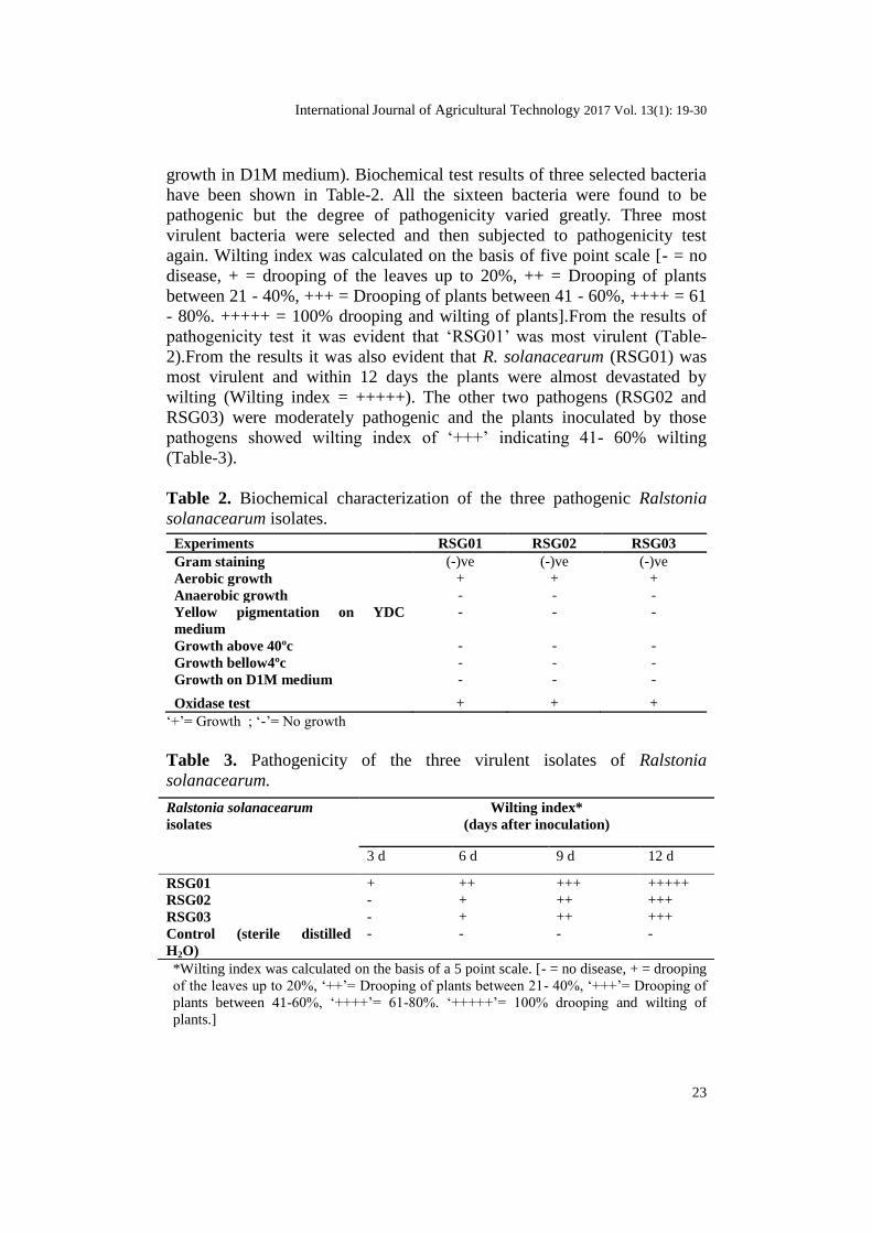
growth in D1M medium). Biochemical test results of three selected bacteria
have been shown in Table-2. All the sixteen bacteria were found to be
pathogenic but the degree of pathogenicity varied greatly. Three most
virulent bacteria were selected and then subjected to pathogenicity test
again. Wilting index was calculated on the basis of five point scale [- = no
disease, + = drooping of the leaves up to 20%, ++ = Drooping of plants
between 21 - 40%, +++ = Drooping of plants between 41 - 60%, ++++ = 61
- 80%. +++++ = 100% drooping and wilting of plants].From the results of
pathogenicity test it was evident that ‘RSG01’ was most virulent (Table-
2).From the results it was also evident that R. solanacearum (RSG01) was
most virulent and within 12 days the plants were almost devastated by
wilting (Wilting index = +++++). The other two pathogens (RSG02 and
RSG03) were moderately pathogenic and the plants inoculated by those
pathogens showed wilting index of ‘+++’ indicating 41- 60% wilting
(Table-3).
Table 2. Biochemical characterization of the three pathogenic Ralstonia
solanacearum isolates.
Experiments RSG01 RSG02 RSG03
Gram staining (-)ve (-)ve (-)ve
Aerobic growth + + +
Anaerobic growth - - -
Yellow pigmentation on YDC
medium
- - -
Growth above 40ºc - - -
Growth bellow4ºc - - -
Growth on D1M medium - - -
Oxidase test + + +
‘+’= Growth ; ‘-’= No growth
Table 3. Pathogenicity of the three virulent isolates of Ralstonia
solanacearum.
Ralstonia solanacearum
isolates
Wilting index*
(days after inoculation)
3 d 6 d 9 d 12 d
RSG01 + ++ +++ +++++
RSG02 - + ++ +++
RSG03 - + ++ +++
Control (sterile distilled
H2O)
- - - -
*Wilting index was calculated on the basis of a 5 point scale. [- = no disease, + = drooping
of the leaves up to 20%, ‘++’= Drooping of plants between 21- 40%, ‘+++’= Drooping of
plants between 41-60%, ‘++++’= 61-80%. ‘+++++’= 100% drooping and wilting of
plants.]

24
All the bacteria were observed in light microscope. Shape of all the
bacteria was found to be rod. The most virulent R. solanacearum
(RSG01),was also observed under scanning electron microscope to
understand the surface topography of the bacterium (Fig-1).Tomato plants
[Var. PKM1] were artificially infected by R. solanacearum (RSG01) and
after six days ooze test were performed. From the ooze test it was evident
that substantial amount of ooze was coming out from the cut end of the stem
(Fig-2). This indicated that R. solanacearum (RSG01) was a virulent
pathogen.
Figure 2. Scanning Electron Microscopic image of RSG01.
R. solanacearum (RSG01) and the other two virulent bacteria
(RSG02 and RSG03) were selected and were subjected to carbohydrate
utilization tests. Altogether, 33 carbohydrates and their derivatives were
used for the test. All the three virulent isolates could use only thirteen
carbohydrates (Ramnose, Cellobiose, Xylitol, ONPG, D-Arabinose, Citrate,
Malonate, Xylose, Fructose, Dextrose, Galactose, Malibiose and L-
Arabinose) among the tested carbohydrates. Other twenty carbohydrates
(Melizitose, Methyl-D-Manoside, Esculin, Solbitol, Lactose, Raffinose,
Maltose, Trehalose, Sucrose, Inuline, Sodium glucanate, Glycerol, Salicin,
Dulcitol, Inocitol, Sorbitol, Mannitol, Adonitol, Arbitol and Erythritol)
tested could not be used by any of the three bacteria. Identification of the
bacteria was performed by comparing with the characters of the present
study with that of stated in the Bergey’s manual of systematic bacteriology,
9th
edition.
Host range test: In order to check infectivity of the three bacteria,
potato, another solanaceous plant, was taken into consideration. Results of
the infectivity of the bacterium in potato in comparison to tomato were
observed. The results indicated that all the three isolates (showed virulence
to tomato) were capable to infect potato but they were less virulent in potato
than in tomato as evidenced by the results presented in the form of wilting

International Journal of Agricultural Technology 2017 Vol. 13(1): 19-30
25
index (Table-4).The RSG01 strain was found to be more virulent than the
other two strains both in potato and tomato plants.
Table 4. Host infectivity of the bacteria on two solanaceous plants.
Host RSG01 RSG02 RSG03
Tomato plant +++ ++ ++
Potato plant ++ + +
*Wilting index was calculated on the basis of a 5 point scale. [‘+’= drooping of the leaves
up to 30%, ‘++’= Drooping of plants between 31- 60%, ‘+++’= Drooping of plants between
61-90%.]
Evaluation of antagonism: One bacterial isolate (HS01) was found
from the rhizosphere soil of wilted tomato field. The bacterium showed
antagonism in mixed soil bacterial culture in Petriplate. The bacterium was
isolated in pure form and was subjected to cross culture following the
method of Dhingra and Sinclair (1995) in nutrient agar medium. Result
showed that the most virulent isolate could not grow at the cross point but
the antagonist could grow at that point (Fig-3). From the result it was
evident that the bacterium (HS01) was antagonistic against virulent
Ralstonia solanacearum (RSG01).
Fig-3: a) Dual culture of Bacillus cereus and Ralstonia solanacearum
isolate RSG01 and b) Dual culture of Bacillus cereus and Ralstonia
solanacearum isolate RSG02 .
16SrRNA studies for identification of bacteria: One virulent and
one antagonistic bacterium (RSG01 and HS01) were subjected to molecular
identification following 16S rRNA studies. Initially, expected amplicons
raised through PCR using suitable primer pairs were subjected to agarose
gel electrophoresis (Fig-4). The size of the amplicons was 1016 bp long for
R. solanacearum (virulent isolate RSG01) and 789 bp long for Bacillus
cereus (antagonistic isolate HS01) (Fig-5). The 16S rRNA sequences of

26
both the bacteria were submitted in the GenBank for accession. The
sequences received from the GenBank were analyzed by BLAST. The
virulent bacterium was identified as Ralstonia solanacearum while the
antagonist was identified as Bacillus cereus. The GenBank accession
numbers of the two bacteria are KC237236 and KC959841 respectively.
Figure 4. Expected amplicons (raised through PCR using suitable 16s
rRNA primer pairs) of R. solanacearum isolates on agarose gel. M= 500bp
ladder; L1, L2, L3= RSG01, RSG02, RSG03 isolates respectively.
Figure 5. Expected amplicons (raised through PCR using suitable 16s
rRNA primer pairs) of Bacillus cereus isolate on agarose gel. M= 100bp
DNA ladder; L1, L2 and L3= HS01 isolate
Discussion
Bacterial wilt by Ralstonia solanacearum is one of the devastating
diseases of tomato in the world (Chandrashekara et al., 2012). Sixteen
different bacteria causing wilt disease in tomato in sub-Himalayan West

International Journal of Agricultural Technology 2017 Vol. 13(1): 19-30
27
Bengal were isolated. All such bacteria were found to produce bacterial
ooze. A cross section of the stem of a plant showing bacterial wilt may
produce white, milky strands of bacterial cells in clear water. This ooze
distinguishes the wilt caused by bacterium from that caused by fungal
pathogens (Leppla et al., 2004; Hernandez-Romano et al., 2012).Tomato is
a major horticultural crop of sub-Himalayan West Bengal. The area is
popularly known as North Bengal. It has been reported that the incidence of
bacterial wilt in tomato crops in India ranges from 15 to 55% and the
disease causes 25 to 75% yield loss of solanaceous vegetables (Rao and
Sohi 1977). In our study, also it has been experienced that about 20% of the
plants in the fields, where proper control measures were not taken, were
attacked by pathogenic bacteria Ralstonia solanacearum. According to
Chandrashekara et al. (2012) the virulence of R. solanacearum may be of
great use for managing the pathogen.
Bacterial wilt pathogens of the infected samples were isolated and
purified. Some other Ralstonia solanacearum were also isolated from the
rhizospheres of the tomato plants which were severely affected by bacterial
wilt. Sixteen different Ralstonia solanacearum were isolated, purified and
identified on the basis of physiological and biochemical characteristics.
Identification of the bacteria on the basis of physiological and biochemical
characteristics were performed following the flowchart identification
scheme of Trigiano et al. (2004). Study of the bacteriological properties of
the isolates confirmed that the isolates were aerobic and gram-negative. The
notions of our study of bacterial properties were similar with that of Ozaki
and Watabe (2009).
All the sixteen isolates from infected tomato plants or tomato
rhizosphere were subjected to pathogenicity test, which showed that all the
sixteen isolates were differentially pathogenic to tomato plants but three of
them were virulent and highly pathogenic. Those three isolates were coded
as RSG01, RSG02 and RSG03. Pathogenicity of the three isolates was also
studied in potato plants to know their capability of infection in a related crop
of the same family solanaceae. All the three isolates could infect both potato
and tomato. Pathogenicity test of R. solanacearum isolates of geranium and
portulaca were also studied by Ozaki and Watabe (2009) in different hosts
to know their infectious capacity.
Identification of most virulent bacterium (RSG01), of the present
study, was also done by 16S rRNA studies following BLAST analysis. A
number of studies have supported the molecular identification of Ralstonia
solanacearum following 16S rRNA studies (Fouche-Weich et al., 2006).The
pathogen (RSG01) was also controlled by an indigenous antagonistic
bacterium isolated from the soil and identified as Bacillus cereus HS01 by
16S rRNA study and BLAST analysis. The antagonism of the bacterium
was studied following dual culture technique as suggested by several
workers (Yoshida et al., 2001; Romero et al., 2004; Thongwai and

28
Kunopakarn, 2007). Antagonism of other Bacillus sp. and several other
bacterial antagonists towards phytopathogens has been demonstrated by
many scientists. (Manjula and Podile, 2005; Seleim et al., 2011; Singh et
al., 2012; Chen et al., 2013; Maji and Chakrabartty, 2014; Singh and
Siddiqui, 2015 ).
Thus, from the present study it may be concluded that the virulent R.
solanacearum isolates of the present study area are pathogenic to tomato
and potato. One antagonistic bacterium against virulent pathogenic R.
solanacearum isolates are available in the fields of the present study area
and may be exploited for development of suitable formulations for use in
the fields to control R. solanacearum to get rid of bacterial wilt of tomato.
Acknowledgement
Financial assistance received in the form of fellowship (Rajiv Gandhi National
Fellowship) to H.M. from University Grants Commission (UGC), New Delhi is greatly
acknowledged.
References
Adhikari, T. B. and Basnyat, R. C. (1998). Effect of crop rotation and cultivar resistance on
bacterial wilt of tomato in Nepal. Canadian Journal of Plant Pathology 20: 283-
287.
Ausubel, F. M., Brent, R., Kingston, R. E., Moore, D. D., Seidman, J. G., Smith, J, A. and
Struhl, K. (1992). Current protocols in molecular biology, v.1. Greene Publishing
Association, New York.
Boudazin, G., Leroux, A. C., Josi, K., Labarre, P. and Jouan, B. (1999). Design of divisions
specific primars of Ralstonia solanacearum and application to the identification of
European isolate. European Journal of Plant Pathology 105:373-380.
Chandrashekara, K. N., Prasanna Kumar, M. K. and Saroja, S. (2012). Aggressiveness of
Ralstonia solanacearum isolates on tomato. Journal of Experimental Sciences 3:
5-9.
Chen, Y., Yan, F., Chai, Y., Liu, H., Kolter, R., Losick, R. and Guo, J. H. (2013).
Biocontrol of tomato wilt disease by Bacillus subtilis isolates from natural
environments depends on conserved genes mediating biofilm formation.
Environmental Microbiology 15:848-864.
Ciampi, L., Burzio, L. O. and Burzio, L. A. (1997). Carriers for Pseudomonas (Ralstonia)
solanacearum, causal agent of bacterial wilt. Fitopathologia 32:64-70.
Dhingra, O. D. and Sinclair, J. B. (1995). Basic Plant Pathology Methods, 2nd
ed. CRC
Press, London.
Dubey, L. N., Das, K. K. and Hazarika, D. K. (1996). Evaluation of some chemicals against
bacterial wilt of Sesamum. Journal of Mycological Plant Pathology. 26:94-95.
Elphinstone, J., Hennessy, J., Wilson, J. and Stead, D. E. (1996). Sensitivity of different
methods for the detection of Pseudomonus solanacearum (Smith) Smith in potato
tuber extracts. EPPO Bulletin 26:663-678.
Fouche-Weich, J., Poussier, S., Trigalet-Demery, D., Berger, D. and Coutinho, T. (2006).
Molecular identification of some African strains of Ralstonia solanacearum from
eucalypt and potato. Journal of General Plant Pathology 72:369-373.

International Journal of Agricultural Technology 2017 Vol. 13(1): 19-30
29
Grimault, V., Schmit, J. and Prior, P. (1993). Some characteristics involved in bacterial wilt
(Pseudomonas solanacearum) resistance in tomato. In: Hartman GL, Hayward
(eds). Bacterial wilt. ACIAR Proceedings 112-119.
Hayward, A. C. (1991). Biology and epidemiology of bacterial wilt, caused by Ralstonia
solanacearum. Annu. Rev. Phytopathol 29: 65-87.
Hernandez-Romano, J., Ramirez-Rojas, S. and Ydrac-Morales, C, J. (2012). First report of
Ralstonia solanacearum causing tomato bacterial wilt in Mexico. New Disease
Reports 26:22.
Hoque, M. E. and Mansfield, J. W. (2005). A sample and reliable method for pathogenicity
tests of bacterial blight disease of rice. Bangladesh Journal of Botany 34:11-16.
Kisore, V., Shekhawat, G. S. and Sunaina, V. (1996). Cultural practices to reduce
Pseudomonas solanacearum in the infested soil. Journal of Indian Potato. 23:130-
133.
Leppla, N., Momol, T., Nesheim, N. and Dusky, J. (2004). Plant, animal and human
protection, FAS 2 Focus Area. University of Florida / IFAS. Available at:
http://edis.ifas.ufl.edu/.
Lwin, M. and Ranamukhaarachchi, S. L. (2006). Development of biological control of
Ralstonia solanacearum through antagonistic microbial populations. International
Journal of Agriculture Biology 5:657-660.
Maji, S. and Chakrabartty, P. K. (2014). Biocontrol of bacterial wilt of tomato caused by
Ralstonia solanacearum by isolates of plant growth promoting rhizobacteria.
Australian Journal of Crop Science 8:208-214.
Manjula, K. and Podile, A. R. (2005). Increase in seedling emergence and dry weight of
pigeon pea in the field with chitin-supplemented formulations of Bacillus subtilis
AF1. World Journal Microbiological Biotechnology 21:1057-1062.
Momol, M. T., Mitchell, D. J., Rayside, P. A., Olson, S, M. and Momol, E. A. (2000). Plant
essential oils as potential biofumigants for the management of soilborne pathogens
of tomato. Phytopathology 90:S127.
Anonymous (2013). Indian Horticulture Database 2013. National Horticulture Board, New
Delhi, India.
Ozaki, K. and Watabe, H. (2009). Bacterial wilt of geranium and portulaca caused by
Ralstonia solanacearum in Japan. Bull. Minamikyushu Univ. 39:67-71.
Rao, M. V. B. and Sohi, H. S. (1977). Control of bacterial wilt of brinjal. Indian
Horticulture 22:11-13.
Romero, D., Peerez-Garcia, A., Rivera, M, E., Cazorla, F. M. and de Vicente, A. (2004).
Isolation and evaluation of antagonistic bacteria towards the curcurbit powdery
mildew fungus Podosphaera fusca. Applied Microbiological Biotechnology
64:263-269.
Samaranayake, Y, H., Ye, J., Yau, J. Y., Cheung, B. P. and Samaranayake, L. P. (2005). In
vitro method to study antifungal perfusion in Candida biofilms. Journal Clinical
Microbiology 43:818-825.
Seleim, M. A. A., Saead, F. A., Abd-El-Moneem and Abo-Elyousr, K. A. M. (2011).
Biological control of bacterial wilt of tomato by plant growth promoting
rhizobacteria. Plant Pathology Journal 10:146-153.
Singh, D., Yadav, D. K., Sinha, S. and Upadhyay, B. K. (2012). Utilization of plant growth
promoting Bacillus subtilis isolates for the management of bacterial wilt incidence
in tomato caused by Ralstonia solanacearum race 1 biovar 3. Indian
Phytopathology 65:18-24.
Singh, N. and Siddiqui, Z. A. (2015). Effects of Bacillus subtilis, Pseudomonas fluorescens
and Aspergillus awamori on the wilt-leaf spot disease complex of tomato.
Phytoparasitica 43:61-75.
Tans-Kersten, J., Huang, H. and Allen, C. (2001). Ralstonia solanacearum needs motility
for invasive virulence on tomato. Journal of Bacteriology 183:3597-3605.
30
Thongwai, N. and Kunopakarn, J. (2007). Growth Inhibition of Ralstonia solanacearum
PT1J by antagonistic bacteria isolated from soils in the northern part of Thailand.
Chiang Mai Journal of Science 34:345-354.
Trigiano, R. N., Windham, M. T. and Windham, A. S. (2004). Plant Pathology: Concepts
and Laboratory Exercises. 5th ed. CRC Press, Boca Raton, FL, USA.
Windham, M. T. and Windham, A. S. (2004). Plant Pathology and Historical Perspectives.
In: Trigiano RN, Windham MT, Windham A. S. (eds.) in Plant Pathology:
Concepts and Laboratory Exercises. 5th ed. CRC Press, Boca Raton, FL, USA, pp.
14-15.
Yoshida, S., Hiradate, S., Tsukamato, T., Hatakeda, K. and Shirata, A. (2001).
Antimicrobial activity of culture filtrate Bacillus amyloliquefaciens RC-2 isolated
from mulberry leaves. Phytopathology 91:181-187.
(Received: 15 November 2016, accepted: 8 January 2017)