Embed Size (px)
Citation preview

Biodegradation of Dental Resin Composites and Adhesives by Streptococcus mutans: An in vitro Study
By
Maher Bourbia
A thesis submitted in conformity with the requirements for the degree
Of
Masters of Applied Science
Biomaterials Department
Faculty of Dentistry
University of Toronto
Maher Bourbia, 2013

ii
Biodegradation of Dental Resin Composites and Adhesives by Streptococcus mutans: An in vitro Study
Masters of Applied Science, 2013
Maher Bourbia
Biomaterials Department, Faculty of Dentistry, University of Toronto
ABSTRACT
A major cause for dental resin composite restoration replacement is secondary caries
attributed to Streptococcus mutans. Salivary esterases were shown to degrade resin
composites. Hypothesis: S. mutans contain esterase activities that degrade dental resin
composites and adhesives. Esterase activities of S. mutans were measured using synthetic
substrates. Standardized specimens of resin composite (Z250), total-etch (Scotchbond-
Multipurpose, SB), and self-etch (Easybond, EB) adhesives were incubated with S.
mutans UA159 for up to 30 days. Quantification of a bisphenol-glycidyl-dimethacrylate
(BisGMA)-derived biodegradation by-product, bishydroxy-propoxy-phenyl-propane
(BisHPPP) was performed using high performance liquid chromatography. Results: S.
mutans were shown to contain esterase activities in levels comparable to human saliva. A
trend of increasing BisHPPP release throughout the incubation period was observed for
all materials and was elevated in the presence of bacteria vs. control for EB and Z250

iii
(p<0.05) but not SB. Conclusion: biodegradation by cariogenic bacteria could
compromise the resin-dentin interface and reduce the longevity of the restoration.

iv
Acknowledgements:
I would like to thank my supervisor Dr. Yoav Finer for his unlimited support, guidance,
and encouragement throughout my Master’s program. I would also like to thank my co-
supervisor Dr. J.P Santerre, and my committee member Dr. Dennis Cvitkovitch for their
support, motivation, insightful comments, and hard questions.
My sincere thanks to my parents, as my supervisor always says “you would not be here
without them”.
I also would like to thank Dr. Dilani Senadheera, Ms. Martha Cordova, and Ms. Kirsten
Krastel for their assistance with bacterial cultures, and Drs. Meilin Yang and Dengbo Ma
for their technical support. Also I would like to thank Jian Wang for his help with SEM.
Many thanks to all the students and post-docs in Dr. Yoav Finer’s, Dr. JP Santerre’s, and
Dr. Dennis Cvitkovitch’s labs for making this journey an enjoyable and unforgettable
one.

v
Table of Contents
Acknowledgements: ............................................................................................................... iv
List of Abbreviations: ........................................................................................................... vii
List of Figures: ............................................................................................................................ x
List of Tables: ............................................................................................................................ xi
Chapter 1 – Introduction: ....................................................................................................... 1 Hypothesis: ............................................................................................................................................ 3 Objectives: ............................................................................................................................................. 3 References: ............................................................................................................................................ 4
Chapter 2 – Literature review: ............................................................................................. 6 2.1 Preamble: ........................................................................................................................................ 6 2.2 Resin composites: ........................................................................................................................ 6 2.2.1 The matrix: ................................................................................................................................. 7 2.2.2 Filler systems: ............................................................................................................................ 9 2.2.3 Polymerization systems: .................................................................................................... 12 2.3 Dental resin adhesives: ........................................................................................................... 12 2.3.1 Total-etch systems: ............................................................................................................... 13 2.3.2 Self-etch systems: ........................................................................................................... 14 2.4 Degradation of dental resin composites and adhesives: ............................................ 15 2.4.1 Degradation by human salivary esterase activity: .................................................... 18 2.4.2 Degradation by model esterases (cholesterol esterase and pseudocholinesterase): ................................................................................................................. 21 2.4.3 Degradation of resin-dentin interfacial margins: ..................................................... 23 2.5 Interactions between bacteria and dental resin composites and adhesives: ..... 27 2.6 Summary: ..................................................................................................................................... 31 2.7 References: .................................................................................................................................. 32
Chapter 3 – Cariogenic Bacteria Degrades Dental Resin Composite and Adhesives .................................................................................................................................. 36
3.1 Introduction: .............................................................................................................................. 36 3.2 Materials and methods: .......................................................................................................... 37 3.2.1 Bacterial esterase activity assay:..................................................................................... 37 3.2.2 Preparation of composite and adhesive resin specimens: ..................................... 38 3.2.3 Degree of vinyl group conversion at the surface: ...................................................... 38 3.2.4 X-ray photoelectron spectroscopy: ................................................................................. 38 3.2.5 Contact angle measurements:........................................................................................... 38 3.2.6 Biodegradation experiments: ........................................................................................... 39 3.2.7 Scanning electron microscopy: ........................................................................................ 39 3.2.8 Statistical analysis: ............................................................................................................... 39 3.3 Results: ......................................................................................................................................... 39

vi
3.3.1 Bacterial esterase activity assay:..................................................................................... 39 3.3.2 Material characterization: ................................................................................................. 40 3.3.3 Biodegradation: ..................................................................................................................... 40 3.4 Discussion: .................................................................................................................................. 41 3.5 Acknowledgements: ................................................................................................................. 45 3.5 References: .................................................................................................................................. 46 3.6 Figures: ......................................................................................................................................... 49
Chapter 4 – Cariogenic bacteria studies: ....................................................................... 53 4.1 Extended materials and methods: ...................................................................................... 53 4.1.1 Bacterial strains: ................................................................................................................... 53 4.1.2 Bacterial CE-like activity assay: ....................................................................................... 53 4.1.3 Bacterial PCE-like activity assay: ..................................................................................... 54 4.1.4 Bacterial esterase stability assays: ................................................................................. 56 4.1.5 Construction of ΔUA159_SMU.118c: ............................................................................... 56 4.1.6 Construction of UA159_SMU.118c+: ............................................................................... 58 4.1.7 Bacterial esterase-like activity profile assay: ............................................................. 60 4.1.8 Monomer degradation: ....................................................................................................... 60 4.1.9 High performance liquid chromatography (HPLC): ................................................. 61 4.2 Extended results and discussion: ........................................................................................ 63 4.2.1 CE-like and PCE-like activity assays: .............................................................................. 63 4.2.2 Bacterial esterase stability assays: ................................................................................. 65 4.2.3 Bacterial esterase-like activity profile assay: ............................................................. 67 4.2.4 Monomer degradation: ....................................................................................................... 68 4.3 References: .................................................................................................................................. 71
Chapter 5 – Conclusions and recommendations: ....................................................... 73 5.1 Conclusions: ................................................................................................................................ 73 5.2 Recommendations: ................................................................................................................... 74 5.3 References: .................................................................................................................................. 76

vii
List of Abbreviations: BHI Brain heart infusion
BisEMA Bisphenol A polyethylene glycol diether dimethacrylate
BisGMA Bisphenol glycidyl dimethacrylate
BisHPPP Bishydroxypropoxyphenylpropane
BTC Butyrylthiocholine iodide
CE Cholesterol esterase
CSP Competence stimulating peptide
DEGDMA Diethylene glycol dimethacrylate
DTNB 5,5-dithio-bis (2-nitrobenzoic acid)
EB Easybond
E-BPA Ethoxylated Bisphenol A
EGDMA Ethylene glycol dimethacrylate
Erm Erythromycin
gtfb glucosyltransferase B
HEMA Hydroxyethyl methacrylate
HPLC High performance liquid chromatography
HSDEA Human salivary derived esterases
viii
LB Luria-Bertani
LTA Lipoteichoic acid
MA Methacrylic acid
MS Mass spectrometry
PCE Pseudocholinesterase
o-NPA o-nitrophenolacetate
o-NPB o-nitrophenolbutyrate
p-NPA p-nitrophenolacetate
p-NPB p-nitrophenolbutyrate
PCR Polymerase chain reaction
SB Scotchbond
SEM Scanning electron microscopy
SHSE Simulated human saliva esterases
TEG Triethylene glycol
TEGDMA Triethylene glycol dimethacrylate
TEM Transmission electron microscopy
THYE Todd-Hewitt supplemented with 0.3% yeast extract

ix
TYEG Tryptone yeast extract supplement with 0.2% glucose broth
UDMA Urethane dimethacrylate
UV Ultra violet
XPS X-ray photoelectron spectroscopy

x
List of Figures: Chapter 2 – Literature Review Fig 2.1: Common dimethacrylate monomers
Fig 2.2: Classification of resin-based filling composites
Fig 2.3: Structural formula of γ-methacryloxpropyltrimethoxy silane
Fig 2.4: The bonding mechanism of a dentin-composite interface based on the formation of a resin-infiltrated interface zone (hybrid layer) (10).
Fig 2.5: Biodegradation of BisGMA and TEGDMA
Fig 2.6: HPLC chromatographic profile of biodegradation products following incubation of Z250 and TPH with HSDEA and PBS Fig 2.7: SEM analysis of Z250 samples prior to and following 16 days of incubation with HSDEA and PBS Fig 2.8: Z-stack image series captured at interfacial regions of interest of 2 90-day PCE+CE incubated resin-dentin specimens
Chapter 3 – Biodegradation of Dental Resin Composites and Adhesives by Streptococcus mutans: An in vitro Study
Fig 3.1: Activity profile for S. mutans strains
Fig 3.2: Amount of BisHPPP production after 30 days of incubation of Z250, SB, and EB with BHI + S. mutans UA159 or with BHI alone
Fig 3.3: Scanning electron micrographs of Z250, SB, and EB at day 0 and following 30 days of incubation with BHI, and with S. mutans UA159
Chapter 4 – Cariogenic bacteria studies
Fig 4.1: CE-like activity of S. mutans UA159 at lag, log and stationary phases
Fig 4.2: Relative CE-like activity of S. mutans incubated in chemically defined media, and with the addition of BisGMA or TEGDMA monomers
Fig 4.3: Activity profile for S. mutans strains
Fig 4.4: Relative % of TEGDMA and BisGMA remaining in solution after 72 hours of incubation with BHIS + S. mutans, or with BHIS alone

xi
List of Tables: Chapter 2 – Literature Review
Table 2.1: Typical composition of dental composites
Table 2.2: Type of fillers and filler size used in dental composites
Table 2.3: Degradation products, their retention times and chemical formula
Chapter 3 – Biodegradation of Dental Resin Composites and Adhesives by Streptococcus mutans: An in vitro Study
Table 3.1: Surface properties of composite resin (Z250), total-etch (SB) and self-etch (EB) adhesives. Also, the composition (% by weight) of the materials according to manufacturing company (3M Canada Inc., Material Safety Data Sheet). Chapter 4 – Cariogenic bacteria studies Table 4.1: Primers for PCR ligation mutagenesis to delete S. mutans SMU.118c gene
Table 4.2: HPLC gradient method for separation of biodegradation products
1
Chapter 1 – Introduction:
In the United States, 166 million dental restorations were placed in 2005 (1), and clinical
studies suggest that nearly 70% were replacements for failed restorations (2).
Replacement dentistry costs $5 billion/year in the US alone (3). Studies have shown that
dental resin composites have an average replacement time of 5.7 years, mainly due to
secondary caries and fracture of the restoration (4). Recurrent or secondary caries is one
of the primary causes (31-70%) for composite restorative replacement and occurs at the
compromised restoration-tooth interface margin (5,6).
The choice between resin composite and amalgam restorations has been widely driven by
aesthetic and health concerns. Over the past decades, concerns with respect to the
possibility of adverse health effects from exposure to mercury in dental amalgams, and
the desire for improved esthetic dental restorations have lead to the steady and rapid
increase of the use of composite resin restorations (7). However, concern over higher
fracture rates, reduced longevity, prevalence of secondary caries, and bacterial
proliferation associated with biodegradation of resin composites, have been an issue and
a focus of research for several years (8,9).
Most research on resin materials has focused on physical process (wear, mechanical
studies and effect of diet) that lead to degradation. These physical processes are classified
either; under material loss and uptake (sorption, extraction, dissolution and
mineralization) or physical changes (softening, stress cracking, fatigue fracture, etc…)
(10). On the other hand, biochemical processes leading to degradation have seldom been
2
discussed in literature; however, attention to the issue has increased in the last decade
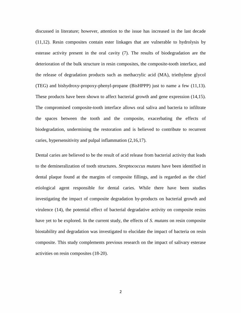
(11,12). Resin composites contain ester linkages that are vulnerable to hydrolysis by
esterase activity present in the oral cavity (7). The results of biodegradation are the
deterioration of the bulk structure in resin composites, the composite-tooth interface, and
the release of degradation products such as methacrylic acid (MA), triethylene glycol
(TEG) and bishydroxy-propoxy-phenyl-propane (BisHPPP) just to name a few (11,13).
These products have been shown to affect bacterial growth and gene expression (14,15).
The compromised composite-tooth interface allows oral saliva and bacteria to infiltrate
the spaces between the tooth and the composite, exacerbating the effects of
biodegradation, undermining the restoration and is believed to contribute to recurrent
caries, hypersensitivity and pulpal inflammation (2,16,17).
Dental caries are believed to be the result of acid release from bacterial activity that leads
to the demineralization of tooth structures. Streptococcus mutans have been identified in
dental plaque found at the margins of composite fillings, and is regarded as the chief
etiological agent responsible for dental caries. While there have been studies
investigating the impact of composite degradation by-products on bacterial growth and
virulence (14), the potential effect of bacterial degradative activity on composite resins
have yet to be explored. In the current study, the effects of S. mutans on resin composite
biostability and degradation was investigated to elucidate the impact of bacteria on resin
composite. This study complements previous research on the impact of salivary esterase
activities on resin composites (18-20).

3
Hypothesis: In addition to acid production, cariogenic bacteria are hypothesized to contain esterase
activities that degrade dental resin composites and adhesives.
Objectives: 1) To measure esterase activities from different strains of S. mutans.
2) To measure the hydrolytic-mediated degradation of dental resin monomers by S.
mutans.
3) To measure the hydrolytic-mediated degradation of cured dental resin composites and
adhesives by S. mutans.

4
References:
(1) Beazoglou T, Eklund S, Heffley D, Meiers J, Brown LJ, Bailit H. Economic impact of regulating the use of amalgam restorations. Public Health Rep 2007 SEP-OCT;122(5):657-663.
(2) Murray PE, Windsor LJ, Smyth TW, Hafez AA, Cox CF. Analysis of pulpal reactions to restorative procedures, materials, pulp capping, and future therapies. Critical Reviews in Oral Biology & Medicine 2002;13(6):509-520.
(3) Jokstad A, Bayne S, Blunck U, Tyas M, Wilson N. Quality of dental restorations - FDI Commission Project 2-95. Int Dent J 2001 JUN;51(3):117-158.
(4) Burke F, Wilson N, Cheung S, Mjor I. Influence of patient factors on age of restorations at failure and reasons for their placement and replacement. J Dent 2001 JUL;29(5):317-324.
(5) Mjor IA, Dahl JE, Moorhead JE. Age of restorations at replacement in permanent teeth in general dental practice. Acta Odontol Scand 2000 JUN;58(3):97-101.
(6) Browning WD, Dennison JB. A survey of failure modes in composite resin restorations. Oper Dent 1996 JUL-AUG;21(4):160-166.
(7) Jaffer F, Finer Y, Santerre JP. Interactions between resin monomers and commercial composite resins with human saliva derived esterases. Biomaterials 2002 APR;23(7):1707-1719.
(8) Khalichi P, Singh J, Cvitkovitch DG, Santerre JP. The influence of triethylene glycol derived from dental composite resins on the regulation of Streptococcus mutans gene expression. Biomaterials 2009 FEB;30(4):452-459.
(9) Santerre JP, Shajii L, Tsang H. Biodegradation of commercial dental composites by cholesterol esterase. J Dent Res 1999 AUG;78(8):1459-1468.
(10) Santerre JP, Shajii L, Leung BW. Relation of dental composite formulations to their degradation and the release of hydrolyzed polymeric-resin-derived products. Critical Reviews in Oral Biology & Medicine 2001;12(2):136-151.
(11) Y. Finer. The Influence of Resin Chemistry on a Composite's Inherent Biochemical StabilityUniversity of Toronto; 2000.
(12) Finer Y, Jaffer F, Santerre JP. Mutual influence of cholesterol esterase and pseudocholinesterase on the biodegradation of dental composites. Biomaterials 2004 MAY;25(10):1787-1793.

5
(13) Kermanshahi S, Santerre JP, Cvitkovitch DG, Finer Y. Biodegradation of Resin-Dentin Interfaces Increases Bacterial Microleakage. J Dent Res 2010 SEP;89(9):996-1001.
(14) Khalichi P, Cvitkovitch DG, Santerre JP. Effect of composite resin biodegradation products on oral streptococcal growth. Biomaterials 2004 NOV;25(24):5467-5472.
(15) Singh J, Khalichi P, Cvitkovitch DG, Santerre JP. Composite resin degradation products from BisGMA monomer modulate the expression of genes associated with biofilm formation and other virulence factors in Streptococcus mutans. Journal of Biomedical Materials Research Part a 2009 FEB;88A(2):551-560.
(16) Brannstrom M. Communication between the Oral Cavity and the Dental-Pulp Associated with Restorative Treatment. Oper Dent 1984;9(2):57-68.
(17) van Meerbeek B, van Landuyt K, de Munck J, Hashimoto M, Peumans M, Lambrechts P, et al. Technique-sensitivity of contemporary adhesives. Dent Mater J 2005 MAR;24(1):1-13.
(18) Lin BA, Jaffer F, Duff MD, Tang YW, Santerre JP. Identifying enzyme activities within human saliva which are relevant to dental resin composite biodegradation. Biomaterials 2005 JUL;26(20):4259-4264.
(19) Shokati B, Tam LE, Santerre JP, Finer Y. Effect of salivary esterase on the integrity and fracture toughness of the dentin-resin interface. Journal of Biomedical Materials Research Part B-Applied Biomaterials 2010 JUL;94B(1):230-237.
(20) Finer Y, Santerre JP. Salivary esterase activity and its association with the biodegradation of dental composites. J Dent Res 2004 JAN;83(1):22-26.

6
Chapter 2 – Literature review:
2.1 Preamble: The concerns over the negative effects of biodegradation on dental resin composites and
adhesives have raised suspicion over the biocompatibility of resin composites and
adhesives, and led to a host of studies on resin composite and adhesive stability using
human saliva or model esterases, cholesterol esterase (CE) and pseudocholinesterase
(PCE). A review of these studies was conducted, the information summarized and
organized as follows. First, composite and adhesive resins are introduced and their
properties discussed, followed by an analysis of human saliva’s hydrolytic activity on
resin composites and adhesives, as well as CE and PCE’s hydrolytic activity and their
suitability to be used as a model for human saliva. Lastly, the effects of biodegradation
and its associated products on bacteria and oral health will be discussed.
2.2 Resin composites: The constituents of dental restorative composites are its polymeric matrix (usually
methacrylate based), filler particles (usually glass, quartz, or ceramic oxide such as
alumina or silica), and coupling agents, which are used to improve bonding at the
filler/polymer-matrix, in addition to a photoinitiator system or in some cases other curing
systems and further additives (Table 2.1) (1-4).

7
Table 2.1: Typical composition of dental composites (4).
Dental Composites
Inorganic fillers 75-85% w/w - Radiopaque silicate glasses
- Fumed oxides/mixed oxides
- etc.
Organic matrix 15-25% w/w - Polymerizable monomers
- Initiator system
- Stabilizers, mixers
2.2.1 The matrix: Dental restorative resin monomers have been primarily based on the coupling of chemical
moieties via ester linkages (Figure 2.1). The predominant resin monomers consist of
complexed methacrylate resins. Some of the resin monomers used in dental restorative
materials, as shown in Figure 2.1, are bisphenol-glycidyl-dimethacrylate (BisGMA),
ethylene glycol dimethacrylate (EGDMA), diethylene glycol dimethacrylate
(DEGDMA), triethylene glycol dimethacrylate (TEGDMA), urethane dimethacrylate
(UDMA), and bisphenol A polyethylene glycol diether dimethacrylate (BisEMA) (2,3).
C O
O
CH2C
CH3
CH2CHCH2O
OH
C
CH3
CH3
OCH2CHCH2O
OH
C
O
C CH2
H3C
bisphenol-glycidyl-dimethacrylate (BisGMA)

8
n
n=1: Ethylene glycol dimethacrylate (EGDMA), n=2: Diethylene glycol
dimethacrylate (DEGDMA), n=3: Triethylene glycol dimethacrylate (TEGDMA)
Urethane dimethacrylate (UDMA)
Figure 2.1: Common dimethacrylate monomers. Each of the structures has a common vinyl monomer group coupled to different organic molecules via an ester bond (3).
BisGMA is a very common monomer because it is relatively non-volatile, exhibits low
polymerization shrinkage, hardens rapidly under oral conditions, and is compatible with
current inorganic filler systems. A disadvantage of BisGMA is its high viscosity, which
results from the hydrogen bonds between hydroxyl groups in the alkyl chains and the
rigid aromatic ring structure. To facilitate handling and manipulation, various diluent
monomers are used in conjunction with BisGMA, most commonly TEGDMA, but other
monomers such as UDMA are also used. The ratios and compositions of monomers
constituting resin composites vary depending on the application, location (anterior vs.
H2C C
CH3
C
O
O (CH2CH2O)n C
C
O
H3C
CH2
H2C C
CH3
C
O
O CH2CH2O C N
O H
CH2CHCH2
CH3
C
CH3
CH3
CH2CH2 N
H
C
O
O CH2CH2O C
O
C
H3C
CH2

9
posterior) of the restoration, and on the manufacturer. Therefore, resin composites vary in
characteristics and properties depending on the monomers, fillers and ratios used (2,3,5).
2.2.2 Filler systems: The reinforcing fillers have been the major constituents of resin composites by weight
and volume. Fillers provide the composite with improved physical properties such as
increased strength and modulus of elasticity, as well as reduced polymerization
shrinkage, coefficient of thermal expansion and water sorption. Composite restorations
have been classified according to the type of filler used (Figure 2.2) (4). Fillers are
characterized by different chemical composition, average particle size, and
manufacturing techniques. Macrofilled particles are inorganic particles that are produced
by grinding larger particles of glass, quartz, or ceramics into smaller ones and are usually
splinter shaped. Macrofilled composites have an average particle size of 0.2–5 μm. On
the other hand, microfilled paricles such as pyrogenic silica are usually spherical with an
average particle size of 5-100 nm. They are also referred to as nanoparticles because of
the small particle sizes. Agglomerates are often formed from these particles and the
formed agglomerates may influence the transparency of the composite. A significant
thickening effect can be observed because of the large surface area of microfiller or
nanofiller particles. These particles have been used in order to increase the microfiller
loading in heterogeneous microfilled composites. This can be achieved by incorporating
pyrogenic silica into a resin matrix, curing the mixture, and then milling the obtained
microfilled composite into splinter shaped particles, with a particle size of 10-100 μm.
Traditionally, the inorganic component of these hybrid composites consist of 70-80%
w/w of glass fillers and 20-30% w/w of microfillers (4). Microfilled composites contain

10
silica microfine particles with filler concentrations approximately 38% by weight.
Because of the greater percentage of resin, microfilled materials exhibit increased water
sorption and a higher coefficient of thermal expansion when compared to microhybrid
composites that contain a filler concentration of 74-84% w/w (6). Many contemporary
dental composites use the fillers listed in Table 2.2.
Figure 2.2: Classification of resin-based filling composites (4).
Table 2.2: Type of fillers and filler size used in dental composites (4).
Filler composition Particle size
Highly dispersed SiO2 10-40 nm
Radiopaque, finely ground Ba or Sr silicate glasses 0.7 µm, 1.0 µm, 1.5 µm, or larger
Radiopaque, finely ground Ba/Sr fluoro silicate glasses
1.0 µm, 1.5 µm, or larger
Macrofiller
(ground glasses)
Microfiller
(pyrogenic silica)
Microfiller-
Based complexes
Macrofilled Composite
Hybrid Composite
Homogeneous Microfilled Composite
Heterogeneous Microfilled Composite

11
Ground quartz glasses 1.0-1.5 µm
YbF3, YF3 0.10-3.0 µm
Si/Zr mixed oxide 250-500 nm
Ti, Zr, and Al oxide used as opacifier 250-500 nm
Splinter polymerisate mainly based on SiO2 10-100 µm
In order to improve the properties of resin composites, coupling agents have been
developed to make a stronger link between the hydrophobic organic matrix, and the
hydrophilic inorganic fillers. Coupling agents contain the characteristics of both the filler
particles and the organic matrix; they have hydrophilic hydroxyl groups at one end and
hydrophobic methacrylate groups at another end. The most common coupling agents are
organic silicon compounds called silanes (Figure 2.3). The silane coupling agent’s
hydroxyl groups covalently bind to the hydroxyl groups found on the surface of silica
based glasses on one end. Also, the silane coupling agent’s methacrylate group can
covalently link to the resin matrix via carbon double bond at the other end, resulting in a
strong bond between the organic matrix and the inorganic fillers (3).
Figure 2.3: Structural formula of γ-methacryloxpropyltrimethoxy silane
H2C C
CH3
C
O
O CH2CH2CH2 Si
O
CH3
O
CH3
O CH3

12
2.2.3 Polymerization systems: Resin composites are converted from a viscous resin to a rigid solid through free radical
polymerization of the methacrylate monomers via photo-polymerization. During
polymerization, each molecule grows by the addition of a monomer to a terminal free
radical reaction site. Not all of the monomer’s double bonds have reacted once the
polymerization reaction has terminated. In fact, studies suggest that degrees of
conversion of the methacrylate double bonds range from 40 to 85% (1,7,8). This is in part
due to a reduction in the diffusion rates of the propagating free radicals, the unreacted
dimethacrylate molecules and the pendant methacrylate species as the polymerization
reaction progresses (3,8).
2.3 Dental resin adhesives: Resin composite restorations require the application of resin adhesives in order to bond
efficiently to the tooth (dentin and enamel) by forming the resin-dentin interface. Dental
resin adhesives are low viscosity methacrylate based liquids, which spread on the dentin
surface, and solidify to bond the primed dentin and composite substrates (9).
Micromechanical interlocking is believed to be the primary mechanism by which dental
resin adhesives bind to tooth substrates. Micromechanical interlocking is achieved by the
replacement of tooth inorganic material by adhesive resin monomers that become
interlocked upon curing, forming what is known as the “hybrid layer” (Figure 2.4)
(10,11). Currently, there are two types of adhesive systems; total-etch (also known as
etch-and-rinse), and self-etch adhesive systems.

13
Figure 2.4: The bonding mechanism of a dentin-composite interface based on the formation of a resin-infiltrated interface zone (hybrid layer) (10).
2.3.1 Total-etch systems: In total-etch adhesives, the first step is the demineralization
of the dentin surface (1-10 μm) by the application of phosphoric acid (30-40%
phosphoric acid), followed by a rinsing step, and then the application of the primer and
the adhesive agents, which will infiltrate the exposed collagen and polymerize to form the
dentin-composite interface (11,12). Dental adhesive agents and primers contain resin
monomers that are similar to those found in the composite resin matrix. These adhesive
monomers provide the covalent link between the adhesive and the composite. Therefore
they provide structural continuity and thus physical co-mechanical properties such as
strength. These monomers can be classified into two categories: functional monomers
and cross-linkers. Functional monomers have one polymerizable group and a particular
chemical group “functional group” which imparts monomer specific functions. Cross-
linking monomers have two or more polymerizable groups. As suggested by their name,

14
cross-linking monomers form cross-linked polymers upon curing, whereas functional
monomers form linear polymers. Cross-linked polymers exhibit better mechanical
properties, such as strength, and therefore they are important to reinforcing the adhesive
resin. Traditionally, the primers contained hydrophilic functional monomers, whereas the
adhesive agents contained hydrophobic functional cross-linkers. The application of a
primer followed by the adhesive agent successively in separate steps forms the basis for
the three-step etch-and-rinse technique. Simplified two-step etch-and-rinse adhesives
combine the primer and the adhesive agent into one step (bottle) (12).
2.3.2 Self-etch systems: A trend towards developing simpler techniques that are less
practitioner-sensitive and that are more time efficient has resulted in the manufacturing of
what are known as “self-etch adhesives”. Self-etch adhesives function by using non-rinse
acidic monomers that simultaneously condition and prime dentin. Self-etch adhesives can
be divided into two categories: two-step and one-step self-etch adhesives, depending on
whether the hydrophobic adhesive resin is applied in a separate step (two-step) or
combined with the hydrophilic self-etch primer (one-step) (13).
Unlike total-etch adhesives, self-etch adhesives do not completely expose collagen when
forming the hybrid layer at dentin surfaces, resulting in lower levels of micromechanical
interlocking (10,13,14). Clinical studies have shown that three-step total-etch adhesives
are the most effective approach to achieve efficient and stable bonding. The clinical
effectiveness of two-step etch-and-rinse adhesives was less favorable when compared to
its three-step counterpart (12,14). Meanwhile, one-step self-etch adhesives result in an
“inefficient bond” clinically and perform the worst (12,14). Two-step self-etch adhesives
were found to be clinically reliable because the application of a separate hydrophobic

15
adhesive resin that makes the interface more hydrophobic and therefore seals it better
resulting in a better bond durability (10,12,14). Therefore, the drive towards simpler
techniques that are less susceptible to practitioner variability has resulted in a self-etch
approach that needs more development to reach a satisfactory clinical performance.
2.4 Degradation of dental resin composites and adhesives: The degradation of dental resin composites and adhesives in the oral cavity is a result of
many complex interactions, physical, chemical, and biochemical. Most research has
focused on physical processes (food, chewing, etc) that lead to degradation. These
physical processes are classified either under material loss and uptake (sorption,
extraction, dissolution and mineralization) or physical changes (softening, stress
cracking, fatigue fracture, etc…). On the other hand, chemical processes leading to
degradation have seldom been discussed in literature until the last decade. Chemical
processes that lead to degradation are thermolysis, oxidation, solvolysis, photolysis and
radiolysis. Solvolysis, and more specifically hydrolysis, is the most investigated and
relevant chemical degradation process because of the presence of unprotected ester
linkages in the resin monomers, polymer, and coupling agents. By definition, hydrolysis
is a chemical reaction during which water is used to cleave a molecule into two parts.
One fragment of the parent molecule gains a hydrogen ion, and the other a hydroxyl
group from the water molecule. This process can be catalyzed by enzymes present in the
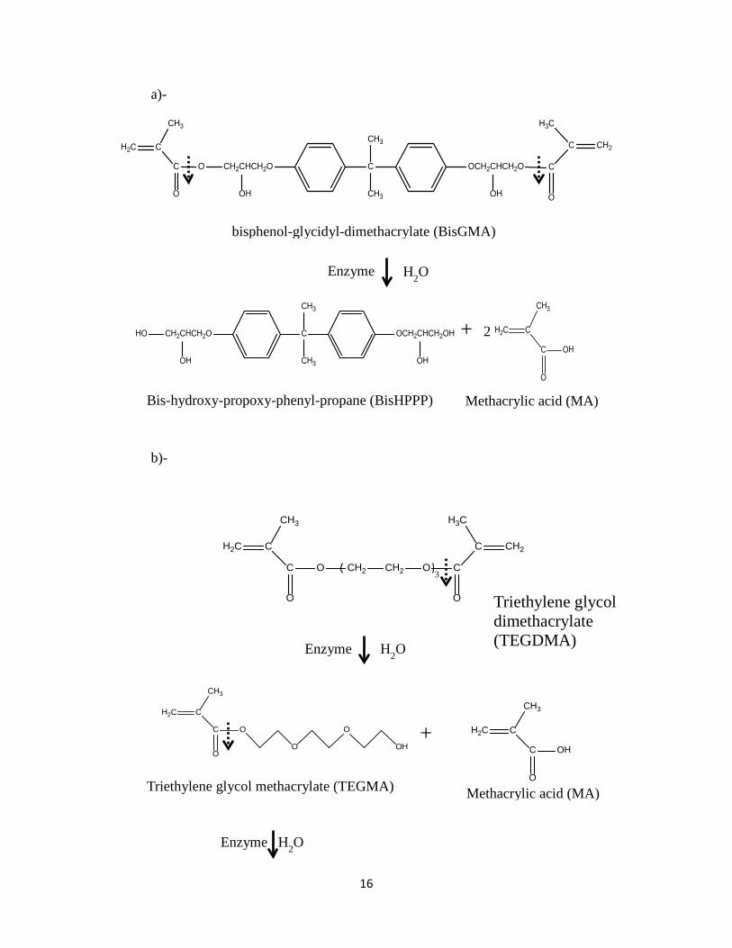
oral cavity and is often referred to as biodegradation. Figure 2.4 illustrates the
biodegradation of dental resin monomers BisGMA and TEGDMA (3).
16
a)-
b)-
C OH
O
CH2C
CH3
C O
O
CH2C
CH3
CH2CHCH2O
OH
C
CH3
CH3
OCH2CHCH2O
OH
C
O
C CH2
H3C
C OH
O
CH2C
CH3
Methacrylic acid (MA)
HO CH2CHCH2O
OH
C
CH3
CH3
OCH2CHCH2OH
OH
Bis-hydroxy-propoxy-phenyl-propane (BisHPPP)
Enzyme H2O
+ 2
bisphenol-glycidyl-dimethacrylate (BisGMA)
C
O
CH2C
CH3
O
O
O
OH
C O
O
CH2 CH2 O C
O
CC CH2H2C
H3CCH3
3
Enzyme H2O
+
Triethylene glycol methacrylate (TEGMA) Methacrylic acid (MA)
Triethylene glycol dimethacrylate (TEGDMA)
Enzyme H2O

17
Figure 2.5: Biodegradation of BisGMA (a) and TEGDMA (b) by salivary esterases resulting in the production of biodegradation by-products BisHPPP, TEGMA, TEG, and MA. Fillers are chemically inert, but their associated coupling agents intended to improve the
link between the inorganic filler and the organic matrix of the composite resins are
vulnerable to hydrolysis via ester linkages within the coupling agents or siloxane links
that are formed with the filler particle (Figure 2.3) (3).
Generally, material discoloration is a sign of chemical changes. Studies by Seung-Heon
et al. (15) and Yong-Keun et al., (16) who incubated resin composite materials with
porcine liver esterase for 9 weeks and compared them to controls incubated in PBS for
the same period, showed that esterase influence on dental resin composite color was
negligible. This does not mean that chemical change is negligible and that biodegradation
should not be looked at as a contributing factor to resin degradation. Rather, another
study conducted by Yong-Keun et al. (17) showed that sequential immersion of
composite resin specimens in porcine liver esterase, organic substances and chemical
agents resulted in various discolorations of resin composite specimens. This latter
experiment is one that parallels the real world scenarios, where resin composites are
subjected to various agents in conjunction with biodegradation. The results of the latter
experiment signal the existence of chemical activity as a factor in resin composite
degradation, since there was no physical loading in the study.
HO
O
O
OHC OH
O
CH2C
CH3
+
Methacrylic acid (MA) Triethylene glycol (TEG)

18
2.4.1 Degradation by human salivary esterase activity: The potential for enzymes to interact with resin composites and adhesives is significant
and accomplished via salivary enzymes, tissue inflammatory responses and bacterial
activity. In the oral cavity, the enzymes most associated for aiding in the hydrolysis of
resin composites and adhesives are esterases. Salivary esterase origins include human
gingival epithelium, salivary glands, inflammatory responses and microorganisms (8).
Studies have shown that human saliva contains esterase activities that hydrolyze resin
monomers such as BisGMA and TEGDMA (8), as well as matrix polymers (2,3,5,8).
Amongst these studies is one conducted by Jaffer and colleagues (8), who showed that
human saliva degrades commercial composite resins (Z250 from 3M Inc, and Spectrum
TPH from L.D. Caulk), which contain BisGMA, TEGDMA and urethane modified
BisGMA. Standardized commercial photopolymerized composites were incubated with
buffer and human saliva under standard conditions (pH 7.0 and 370C) for 2, 8 and 16
days. The incubation solutions revealed that human saliva catalyzed the biodegradation of
both commercial composites. Biodegradation products were identified, isolated and
quantified via high performance liquid chromatography (HPLC) in combination with UV
spectroscopy and mass spectrometry (MS) (3). A typical HPLC chromatographic profile
is shown in Figure 2.5, where retention times of 7 min, 10.5 min, 19 min, 20 min and 21
min are highlighted; each peak represents a biodegradation product (8).

19
Figure 2.6: HPLC chromatographic profile of biodegradation products following 2 days incubation; a)- Z250 incubated in HSDEA, b)- TPH incubated in HSDEA, c)- TPH incubated in D-PBS and d)- HSDEA in the absence of composite samples (3).
The identity of the degradation products associated with the various retention times were
determined (MA, TEGMA, BisHPPP, TEGDMA and Ethoxylated Bisphenol A (E-BPA))
and are summarized in Table 2.3.
Table 2.3: Degradation products, their retention times and chemical formula (8).

20
The nature of degradation products was similar in both composite resins, but differences
existed in the amounts of degradation products released. Both BisHPPP and methacrylic
acid were produced in significantly greater values as a result of the biodegradation of
Z250 composite resin when compared to TPH degradation. However, E-BPA was
produced in greater amounts as a consequence of TPH degradation, albeit in less quantity
than BisHPPP and methacrylic acid. These differences can be explained by the variability
of composite resin composition with respect to monomer types, monomer ratios, filler
content and filler/resin ratios (1,2,18). Also, the surface of the final cured structure can
offer easier access to human salivary esterases and therefore access to more binding sites,
and this can also lead to discrepancies in degradation product profile. Another factor may
be the fact that urethane modified BisGMA resin associated with TPH is more stable and
resilient to hydrolysis and thus results in less degradation products released from TPH
(2). Therefore, degradation product profiles vary in terms of identity and amount of
degradation products released, depending on the identity of the composite resin under
investigation.
Other researchers using similar materials and methods as the ones described above to
purify and quantify the degradation by-products reached similar conclusions on saliva’s
ability to hydrolyze dental resin composites (19,20). Hsu et al. (19) incubated commercial
dental composite resins containing different monomers (BisGMA, TEGDMA, and
UDMA) with human saliva and found that saliva hydrolyzes the restorative materials at
the ester bonds releasing the expected degradation by-products from these monomers
(such as TEGMA and MA). Overall, studies via isolation and identification of
degradation by-products, scanning electron microscopy (SEM) analysis, and fracture

21
toughness tests confirmed that human salivary esterases hydrolyze resin composites
(2,5,8,19-21). The consensus is that human saliva degrades whole matrix and not just
unreacted monomers, as displayed by SEM analysis of Z250 samples prior to incubation
as compared to samples incubated in human saliva (test group) or PBS buffer (control
group), which revealed the appearance of exposed filler particles (Figure 2.6). Analysis
of SEM images of other resin composite products showed similar results.
Figure 2.7: SEM analysis of Z250 samples a)- prior to incubation in HSDEA, b)- following 16 days of incubation in D-PBS and c)- following 16 days of incubation in HSDEA (8).
2.4.2 Degradation by model esterases (cholesterol esterase and pseudocholinesterase): The potential for the spread of infection when handling saliva from different donors, as
well as the variability of esterase activity and the cumbersome protocol involved in
collecting and treating saliva to harvest its esterase activity from different donors, has
necessitated the search for model salivary esterases that are safer and more practical to
use for scientific research. A study by Finer et al. (5) compared human saliva’s hydrolase
activity to that of model esterases, cholesterol esterase (CE) and pseudocholinesterase
(PCE), by recording data for human saliva’s ability to hydrolyze p-nitrophenolacetate (p-

22
NPA), p-nitrophenolbutyrate (p-NPB), o-nitrophenolbutyrate (o-NPB), o-
nitrophenolacetate (o-NPA), or butyrylthiocholine iodide (BTC), and of CE and PCE’s
ability to degrade the same five substrates. The synthetic substrates contain ester
linkages, and the hydrolysis of these ester linkages results in the substrates’ breakdown to
their respective alcohol and carboxylic acid degradation products. The findings showed
that human saliva has PCE character by degrading BTC, and that human saliva also has
CE character by degrading p-NPA, o-NPA, p-NPB, and o-NPB yielding a similar profile
of sensitivity to the different substrates. The average CE-like activity level for human
saliva was determined to be 0.19 ± 0.02 units/ml, and the average PCE-like activity level
for human saliva was 0.011 ± 0.001 units/ml. Therefore, this study (5) showed that
indeed human saliva contains CE- and PCE-like esterase activities that can degrade
composite resin restorative materials. This study also revealed that a combination of CE
and PCE could be used as a model for human salivary esterase activity in biodegradation
studies.
Several studies have been conducted on the hydrolytic activity of CE and PCE (22-25).
Studies showed that CE and PCE are enzymes that can catalyze the hydrolysis of
BisGMA and TEGDMA monomers, as well as composite resins (22,24,25). CE is an
inflammatory cell-derived enzyme (22) and pseudo-cholinesterase belongs to a family of
cholinesterases, a family of esterases that hydrolyze choline esters at a higher rate than
they do other esters (5). Studies revealed that the degradation of resin matrix by CE and
PCE is a concentration dependent process and that CE has a greater specificity towards
catalyzing the hydrolysis of BisGMA and BisEMA components, while PCE has a greater
specificity towards hydrolyzing TEGDMA and TEGMA components (8,22). In another

23
study, Finer et al. (25) showed that CE and PCE act synergistically to increase the
biodegradation of composite resin materials; i.e. each of the two enzymes functions better
in the presence of the other, resulting in higher levels of degradation products released in
a solution of both enzymes as opposed to when they are alone. One proposed explanation
for this was that the activity of one of the enzymes can provide access for the other
enzyme to sites that would have been difficult to engage and bind to otherwise, and vice
versa, leading to an increased efficiency of resin matrix degradation.
It has been suggested that CE and PCE catalyze the hydrolysis of resin composites in a
mechanism that is similar to their common function (i.e. using the same active site) (5).
This conclusion was reached by observing a decrease in CE and PCE activity by
63±0.5% (p<0.05) and 58±4.7% (p<0.05) respectively when incubated with
phenylmethyl sulfonyl fluoride (PMSF). PMSF is a serine esterase inhibitor, which
alkylates the hydroxyl of the active serine site in the esterases. More kinetic studies need
to be conducted to elucidate the exact mechanism by which CE and PCE degrade
composite resins.
2.4.3 Degradation of resin-dentin interfacial margins: In addition to the degradation of the resin composite bulk, the dentin-resin interface also
faces the challenges of biodegradation. Shokati et al. (26) incubated adhesive resin
(Scotchbond Multi Purposes), resin composite (Z250) and mini short-rod specimens in
buffer and human saliva derived esterases (HSDE) for up to 180 days (pH 7.0 and 37°C).
The adhesive resin and resin composite specimens were incubated with either D-PBS or
HSDE for 8 days (pH 7.0, 370C). BisHPPP as a marker of resin matrix and polymerized
adhesive resin degradation was identified and measured using HPLC. BisGMA is a high

24
molecular weight molecule that has rigid phenyl rings, and hydrogen bonding capacity.
When taking this into consideration it is realized that BisGMA is a molecule that has
limited diffusion through the resin matrix and out to the surface to interact with the
enzymes. Therefore BisHPPP, a BisGMA derived degradation product, is a good
indicator of resin matrix degradation (1). This analysis revealed that HSDEA degraded
both adhesive resin and resin composite to produce BisHPPP in comparable amounts.
The mini short-rod specimens were tested for fracture toughness. Results of the fracture
toughness test showed that mini short-rod specimens incubated for 180 days in HSDEA
had the lowest fracture toughness (0.55 ± 0.13 MPa/m1/2), while non-incubated mini
short-rod specimens had the highest fracture toughness values (0.80 ± 0.16 MPa/m1/2).
This result emphasized the effect of biodegradation on the structural integrity of resin
composites. The combination of BisHPPP production, fracture toughness results, and
SEM images showed that alongside surface composite resin degradation, the dentin-resin
interface also faces degradation by HSDEA (26,27). Other researchers also investigated
the effects of biodegradation on the resin-dentin interface (28-31). Jung et al. (32)
evaluated the effect of esterase activity on resin-dentin interface by transmission electron
microscopy (TEM) and found an increased tendency of nanoleakage in the bounded
interface after storage in an esterase solution as compared to a PBS buffer solution after 4
weeks. Also, Sirichan et al. (31) investigated the effect of collagenase and
acetylcholinesterase on the resin dentin interface. The above-mentioned enzymes were
selected to simulate oral salivary enzymes. Sirichan et al. (31) incubated resin composite
(Z350, 3M) bonded to human dentin by four different adhesive systems; a total-etch
adhesive (Single Bond 2, 3M, St. Paul, MN, USA), a two-step self-etch adhesive (Clearfil

25
SE Bond, Kuraray, Tokyo, Japan), and two one-step self-etch adhesives (Clearfil tri-S
Bond, Kuraray, Tokyo, Japan; and G-Bond, GC, Tokyo, Japan). The prepared
restorations were incubated for three months in water, collagenase, or acetylesterase.
Microtensile bond strengths (μTBS) were measured immediately after restorations were
placed, or after 3 months of incubation in either medium. The specimens were also
analyzed by SEM. The researchers found that μTBS were significantly lower in groups
incubated in enzymes when compared to incubation in water or non-incubated specimens.
They also found that the self-etch adhesives exhibited water-tree patterns within the
adhesive layer, and the total-etch adhesive exhibited nanoleakage within the hybrid layer
and the adhesive. These findings further provide evidence of the effects of biodegradation
on the resin-dentin interface caused by salivary enzymes (31).
The effects of biodegradation at the resin-dentin interface are aided and amplified by
polymerization shrinkage, thermal changes, mastication stresses, and chemical attacks by
acids. These effects result in microleakage, and in the creation and expansion of marginal
gaps at the resin-dentin interface that may lead to the colonization and propagation of
bacteria at the interface as illustrated by previous research (27,31,33). Kermanshahi et
al.(27) investigated the idea that biodegradation on resin composite restorations
accelerates marginal microleakage. Kermanshahi and colleagues incubated resin
composite (Z250, 3M) bonded to human dentin (Scotchbond MP, 3M, St. Paul, MN,
USA) in either buffer or dual esterase media (PCE + CE) with activity levels resembling
those of human saliva for 90 days. Analysis of incubation solutions for BisHPPP was
performed, and as anticipated significant amounts of it were produced (BisHPPP is a
marker of resin matrix and polymerized adhesive resin degradation). Post-incubation,

26
specimens were suspended in a chemostat-based biofilm fermentor cultivating
Streptococcus mutans NG8 for 7 days. After, bacterial microleakage was assessed by
confocal laser scanning microscopy. S. mutans is a species that is low G+C Gram-
positive, non-haemolytic, non-spore forming, and non-motile. It is regarded as the main
etiological agent in dental caries (34). Dental caries are the result of acid release from
carbohydrate metabolism that leads to demineralization of tooth structures. Cumulated
data showed that there was greater bacterial surface adherence and penetration along the
resin-dentin marginal interface in CE + PCE incubated specimens. Precisely, CE + PCE
incubated specimens had nearly 4 times more bacteria, and the maximum interfacial
depth penetration was nearly 4 times more in CE + PCE incubated specimens than in
buffer incubated specimens. Confocal laser scanning microscopy Z-stack images, Figure
2.8, showed bacterial invasion and growth along the marginal gap. The bacteria displayed
characteristics indicative of three-dimensional biofilm growth. These findings confirm
that biodegradation is a relevant issue at the resin-dentin interface and they prove that
biodegradation contributes to microleakage and bacterial invasion of the marginal gap.
The significant results obtained by Kermanshahi et al. (27) are for in vitro experiments
that only lasted 90 days, whereas dental restorative composite resins are under the
constant stresses of the oral environment for a period of time that is much longer than 90
days. Therefore, the real life impact of biodegradation and microleakage are expected to
be much more substantial than those observed in in vitro experiments.

27
Figure 2.8: Z-stack image series captured at interfacial regions of interest of 2 90-day PCE+CE incubated resin-dentin specimens. (A) Interfacial void spanning approximately 4-5 μm in height. (B) Interfacial void spanning over 20 μm in height. Specimens were stained by means of a Live/Dead Baclight Viability Kit. The bacteria displayed characteristics indicative of three-dimensional biofilm growth (27).
2.5 Interactions between bacteria and dental resin composites and adhesives: Research exploring the topic of bacterial adhesion and viability on dental resin composite
surfaces has focused on the influences of material hydrophobicity/hydrophilicity, surface
free energy, and surface roughness on bacterial adhesion and survival on these materials
(35-41). Some researchers propose that hydrophobic resin composites lead to resistance
of attacks by water-soluble species (40,41). Other researchers propose that adhesive
forces may arise for hydrophobic materials because water is easily removed from the
areas between cell surface and hydrophobic materials than from the cell surface and

28
hydrophilic materials, enabling a closer approach and therefore stronger adhesion (42-
44). Inconclusive evidence for the effects of surface free energy and surface roughness on
bacterial adhesion to resin composite surfaces has also been reported. A study by Stefan
et al. (35) investigated the adhesion and viability of oral bacteria on the surfaces of resin-
based dental restorative materials. The researchers postulated that modifying resin
composite materials with low-surface tension active agents (hydroxyfunctional
polydimethylsiloxane and polydimethylsiloxane, or silicone polyether acrylate) would
result in lower bacterial count or bacterial viability. The hypothesis was tested by
incorporating the above-mentioned active agents into the composition of standardized
resin composite materials, with a non-modified resin as the control. The total count and
viability of Actinomyces naeslundii, Actinomyces viscosus, Streptococcus mitis,
Streptococcus oralis, and Streptococcus sanguinis on human saliva pellicle-coated
specimens was analyzed using fluorescence microscopy after 8 and 24 h. The researchers
found that all test materials had significantly fewer vital cells after 8 or 24 hours
compared to the control. However, they found no difference in total bacterial count on
test surfaces except in the group modified with silicone polyether acrylate that showed
lower total bacterial count after 8 and 24 h. The researchers also concluded that contact
angle did not influence bacterial adhesion, but no conclusive evidence for the effects of
low total surface free energy resulting in fewer bacteria was found. Therefore, the
researchers concluded that in addition to hydrophobicity/hydrophilicity and surface free
energy, material chemistry (i.e: monomer mixtures) is an important factor that has to be
considered when analyzing for bacterial adhesion and viability on dental resin
composites.

29
When analyzing for the impact of resin composite chemistry on bacterial adhesion and
viability, it is important to understand how the resin monomers and biodegradation by-
products interact and influence bacterial cells. Studies have revealed that biodegradation
products, such as TEG, influence bacterial growth and virulence gene expression (45,46).
Research by Kalichi et al. (47) showed that TEG, at levels found in vivo modulated the
expression levels of glucosyltransferase B (gtfB) (involved in biofilm formation) and yfiV
(a putative transcription regulator). This finding directly links biodegradation to bacterial
proliferation in the oral cavity, which is significant because it implies that resin
composites, in their current form, are not only structurally vulnerable and not suitable for
long term use, moreover they contribute to oral disease. On the other hand, another study
found that BisGMA degradation products (BisHPPP and MA) slightly inhibits S. mutans
growth (45). This suggests that different degradation products have different effects on
oral bacteria. Therefore, to convincingly reach a conclusion on the complete effect of
degradation products on bacterial activity in the oral cavity, research must be conducted
on the effect of cumulative degradation products on bacterial growth.
The mechanism of biodegradation product generation is complex since residual unreacted
monomers, the polymer matrix, and adhesive resin are all undergoing degradation. Also,
the products of the aforementioned degradations are themselves undergoing degradation,
leaving a complex matrix of degradation products such as BisHPPP, TEGMA, TEG, E-
BPA, and MA, whose cumulative effect on bacterial activity has been suggested to be a
harmful one for oral health. Overall, biodegradation is an ongoing clinically relevant
process that affects structural integrity of resin composites and possibly oral health. More
30
research needs to be conducted to determine its precise effects and how to minimize or
eliminate them.
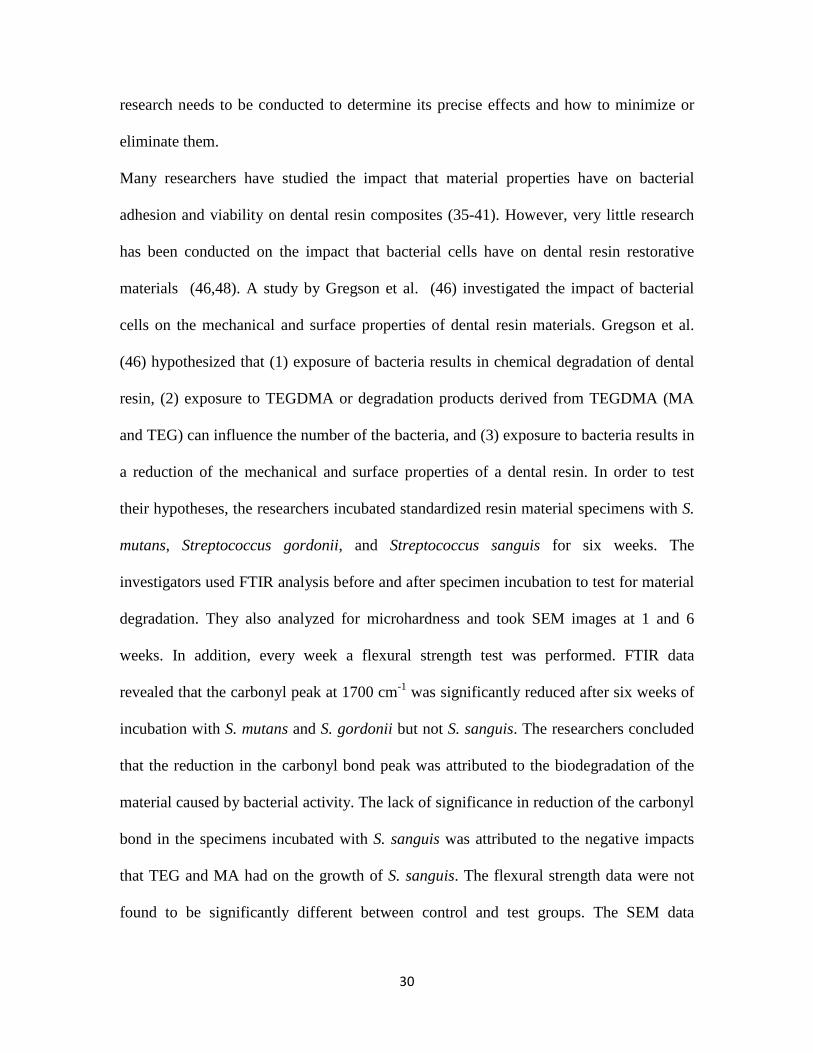
Many researchers have studied the impact that material properties have on bacterial
adhesion and viability on dental resin composites (35-41). However, very little research
has been conducted on the impact that bacterial cells have on dental resin restorative
materials (46,48). A study by Gregson et al. (46) investigated the impact of bacterial
cells on the mechanical and surface properties of dental resin materials. Gregson et al.
(46) hypothesized that (1) exposure of bacteria results in chemical degradation of dental
resin, (2) exposure to TEGDMA or degradation products derived from TEGDMA (MA
and TEG) can influence the number of the bacteria, and (3) exposure to bacteria results in
a reduction of the mechanical and surface properties of a dental resin. In order to test
their hypotheses, the researchers incubated standardized resin material specimens with S.
mutans, Streptococcus gordonii, and Streptococcus sanguis for six weeks. The
investigators used FTIR analysis before and after specimen incubation to test for material
degradation. They also analyzed for microhardness and took SEM images at 1 and 6
weeks. In addition, every week a flexural strength test was performed. FTIR data
revealed that the carbonyl peak at 1700 cm-1 was significantly reduced after six weeks of
incubation with S. mutans and S. gordonii but not S. sanguis. The researchers concluded
that the reduction in the carbonyl bond peak was attributed to the biodegradation of the
material caused by bacterial activity. The lack of significance in reduction of the carbonyl
bond in the specimens incubated with S. sanguis was attributed to the negative impacts
that TEG and MA had on the growth of S. sanguis. The flexural strength data were not
found to be significantly different between control and test groups. The SEM data

31
revealed that the surface of specimens incubated with S. mutans and S. gordonii were
changed, indicating degradation. Another study by Fucio et al. (48) investigated the
effects of a 30-day S. mutans biofilm on resin composite (Filtek Supreme, 3M, St. Paul,
MN, USA) surface roughness, hardness and morphology. The authors found no
statistically significant differences in surface roughness and hardness after 30 days of
incubation. However, the scanning electron micrographs showed an increase in surface
degradation, corroborating the findings of Gregson et al (46). Overall, the results
obtained by Gregson et al. (46) and Fucio et al. (48) point towards the degradative
effects that bacteria have on dental resin composites and highlight the potential of
secondary caries and changes in esthetic properties seen clinically with the use of resin
materials in dental restorations.
2.6 Summary: Although dental resin composite restorative materials have advantages such as esthetic
appeal, and no perceived danger of mercury poisoning, they do seem to be at a
disadvantage when discussing long-term stability, which comes from physical and
chemical influences. The chemical influences, in terms of studies of biodegradation
caused by HSDE alone seem to create conditions for the proliferation of bacteria capable
of causing secondary caries and resin composite failure. In addition, dental resin
composites have been shown to interact with bacteria and influence it’s growth and
virulence gene expression. When taking into consideration that the oral cavity is much
more complex and various influences are acting in concurrence, then it is evident that
more research needs to be done in order to better understand the problems facing resin
composites and develop better biocompatible dental resin composite restorative materials.

32
2.7 References:
(1) Finer Y, Santerre JP. Influence of silanated filler content on the biodegradation of bisGMA/TEGDMA dental composite resins. Journal of Biomedical Materials Research Part a 2007 APR;81A(1):75-84.
(2) Finer Y, Santerre JP. The influence of resin chemistry on a dental composite's biodegradation. Journal of Biomedical Materials Research Part a 2004 MAY 1;69A(2):233-246.
(3) Santerre JP, Shajii L, Leung BW. Relation of dental composite formulations to their degradation and the release of hydrolyzed polymeric-resin-derived products. Critical Reviews in Oral Biology & Medicine 2001;12(2):136-151.
(4) Klapdohr S, Moszner N. New inorganic components for dental filling composites. Mon Chem 2005 JAN;136(1):21-45.
(5) Finer Y, Santerre JP. Salivary esterase activity and its association with the biodegradation of dental composites. J Dent Res 2004 JAN;83(1):22-26.
(6) Iris Daniel. Biodegradation of Polyacid Modified Composite Resins by Human Salivary EsterasesUniversity of Toronto; 2009.
(7) Obicia AC, Sinhoretia MAC, Frollinib E, Sobrinhoa LC, Consania S. Degree of Conversion of Z250 Composite Determined by Fourier Transform Infrared Spectroscopy: Comparison of Techniques, Storage Periods and Photo-activation Methods. Materials Research 2004;7:605-610.
(8) Jaffer F, Finer Y, Santerre JP. Interactions between resin monomers and commercial composite resins with human saliva derived esterases. Biomaterials 2002 APR;23(7):1707-1719.
(9) Van Landuyt KL, Snauwaert J, De Munck J, Peurnans M, Yoshida Y, Poitevin A, et al. Systematic review of the chemical composition of contemporary dental adhesives. Biomaterials 2007 SEP;28(26):3757-3785.
(10) Van Landuyt KL, Snauwaert J, De Munck J, Peurnans M, Yoshida Y, Poitevin A, et al. Systematic review of the chemical composition of contemporary dental adhesives. Biomaterials 2007 SEP;28(26):3757-3785.
(11) Osorio E, Toledano M, Yamauti M, Osorio R. Differential nanofiller cluster formations in dental adhesive systems. Microsc Res Tech 2012 JUN;75(6):749-757.
(12) Peumans M, Kanumilli P, De Munck J, Van Landuyt K, Lambrechts P, Van Meerbeek B. Clinical effectiveness of contemporary adhesives: A systematic review of current clinical trials. Dental Materials 2005 SEP;21(9):864-881.

33
(13) Moszner N, Salz U, Zimmermann J. Chemical aspects of self-etching enamel-dentin adhesives: A systematic review. Dent Mater 2005 OCT;21(10):895-910.
(14) Van Meerbeek B, Peumans M, Poitevin A, Mine A, Van Ende A, Neves A, et al. Relationship between bond-strength tests and clinical outcomes. Dental Materials 2010 FEB;26(2):E100-E121.
(15) Kim SH, Lee YK, Lim BS. Influence of porcine liver esterase on the color of dental resin composites by CIEDE2000 system. Journal of Biomedical Materials Research Part B-Applied Biomaterials 2005 FEB 15;72B(2):276-283.
(16) Lee YK, Lim BS, Powers JM. Color changes of dental resin composites by a salivary enzyme. Journal of Biomedical Materials Research Part B-Applied Biomaterials 2004 JUL 15;70B(1):66-72.
(17) Lee YK, Powers JM. Discoloration of dental resin composites after immersion in a series of organic and chemical solutions. Journal of Biomedical Materials Research Part B-Applied Biomaterials 2005 MAY;73B(2):361-367.
(18) Shajii L, Santerre JP. Effect of filler content on the profile of released biodegradation products in micro-filled bis-GMA/TEGDMA dental composite resins. Biomaterials 1999 OCT;20(20):1897-1908.
(19) Hsu W, Wang V, Lai C, Tsai F. Simultaneous determination of components released from dental composite resins in human saliva by liquid chromatography/multiple-stage ion trap mass spectrometry. Electrophoresis 2012 FEB;33(4):719-725.
(20) Hagio M, Kawaguchi M, Motokawa W, Miyazaki K. Degradation of methacrylate monomers in human saliva. Dent Mater J 2006 JUN;25(2):241-246.
(21) Mirmohammadi H, Kleverlaan CJ, Aboushelib MN, Feilzer AJ. Influence of salivary enzymes and alkaline pH environment on fatigue behavior of resin composites. Am J Dent 2011 FEB;24(1):31-36.
(22) Finer Y, Santerre JP. Biodegradation of a dental composite by esterases: dependence on enzyme concentration and specificity. Journal of Biomaterials Science-Polymer Edition 2003;14(8):837-849.
(23) Lin BA, Jaffer F, Duff MD, Tang YW, Santerre JP. Identifying enzyme activities within human saliva which are relevant to dental resin composite biodegradation. Biomaterials 2005 JUL;26(20):4259-4264.
(24) Santerre JP, Shajii L, Tsang H. Biodegradation of commercial dental composites by cholesterol esterase. J Dent Res 1999 AUG;78(8):1459-1468.

34
(25) Finer Y, Jaffer F, Santerre JP. Mutual influence of cholesterol esterase and pseudocholinesterase on the biodegradation of dental composites. Biomaterials 2004 MAY;25(10):1787-1793.
(26) Shokati B, Tam LE, Santerre JP, Finer Y. Effect of salivary esterase on the integrity and fracture toughness of the dentin-resin interface. Journal of Biomedical Materials Research Part B-Applied Biomaterials 2010 JUL;94B(1):230-237.
(27) Kermanshahi S, Santerre JP, Cvitkovitch DG, Finer Y. Biodegradation of Resin-Dentin Interfaces Increases Bacterial Microleakage. J Dent Res 2010 SEP;89(9):996-1001.
(28) Park J, Ye Q, Topp EM, Spencer P. Enzyme-Catalyzed Hydrolysis of Dentin Adhesives Containing a New Urethane-Based Trimethacrylate Monomer. J Biomed Mater Res Part B 2009 NOV;91B(2):562-571.
(29) Skovron L, Kogeo D, Arana Gordillo LA, Meier MM, Gomes OMM, Reis A, et al. Effects of immersion time and frequency of water exchange on durability of etch-and-rinse adhesive. J Biomed Mater Res Part B 2010 NOV;95B(2):339-346.
(30) Zou Y, Jessop JLP, Armstrong SR. In vitro enzymatic biodegradation of adhesive resin in the hybrid layer. J Biomed Mater Res Part A 2010 JUL;94A(1):187-192.
(31) Chiaraputt S, Roongrujimek P, Sattabanasuk V, Panich N, Harnirattisai C, Senawongse P. Biodegradation of all-in-one self-etch adhesive systems at the resin-dentin interface. Dent Mater J 2011 NOV;30(6):814-826.
(32) Jung Y, Hyun H, Kim Y, Jang K. Effect of collagenase and esterase on resin-dentin interface: A comparative study between a total-etch adhesive and a self-etch adhesive. Am J Dent 2009 OCT;22(5):295-298.
(33) Manuja N, Nagpal R. Resin-tooth interfacial morphology and sealing ability of one-step self-etch adhesives: Microleakage and SEM study. Microsc Res Tech 2012 JUL;75(7):903-909.
(34) Kidd E, Beighton D. Prediction of secondary caries around tooth-colored restorations: A clinical and microbiological study. J Dent Res 1996 DEC;75(12):1942-1946.
(35) Ruettermann S, Bergmann N, Beikler T, Raab WH-, Janda R. Bacterial viability on surface-modified resin-based dental restorative materials. Arch Oral Biol 2012 NOV;57(11):1512-1521.
(36) Busscher HJ, Rinastiti M, Siswomihardjo W, van der Mei HC. Biofilm Formation on Dental Restorative and Implant Materials. J Dent Res 2010 JUL;89(7):657-665.

35
(37) Velazquez-Enriquez U, Scougall-Vilchis RJ, Contreras-Bulnes R, Flores-Estrada J, Uematsu S, Yamaguchi R. Adhesion of Streptococci to various orthodontic composite resins. Aust Dent J 2013 MAR;58(1):101-105.
(38) Buergers R, Rosentritt M, Handel G. Bacterial adhesion of Streptococcus mutans to provisional fixed prosthodontic material. J Prosthet Dent 2007 DEC;98(6):461-469.
(39) Hahnel S, Rosentritt M, Buergers R, Handel G. Surface properties and in vitro Streptococcus mutans adhesion to dental resin polymers. Journal of Materials Science-Materials in Medicine 2008 JUL;19(7):2619-2627.
(40) Weinmann W, Thalacker C, Guggenberger R. Siloranes in dental composites. Dental Materials 2005 JAN;21(1):68-74.
(41) Eick JD, Kotha SP, Chappelow CC, Kilway KV, Giese GJ, Glaros AG, et al. Properties of silorane-based dental resins and composites containing a stress-reducing monomer. Dental Materials 2007 AUG;23(8):1011-1017.
(42) Mei L, Busscher HJ, van der Mei HC, Chen Y, de Vries J, Ren Y. Oral bacterial adhesion forces to biomaterial surfaces constituting the bracket-adhesive-enamel junction in orthodontic treatment. Eur J Oral Sci 2009 AUG;117(4):419-426.
(43) Gyo M, Nikaido T, Okada K, Yamauchi J, Tagami J, Matin K. Surface response of fluorine polymer-incorporated resin composites to cariogenic biofilm adherence. Appl Environ Microbiol 2008 MAR;74(5):1428-1435.
(44) Buergers R, Schneider-Brachert W, Hahnel S, Rosentritt M, Handel G. Streptococcal adhesion to novel low-shrink silorane-based restorative. Dental Materials 2009 FEB;25(2):269-275.
(45) Khalichi P, Cvitkovitch DG, Santerre JP. Effect of composite resin biodegradation products on oral streptococcal growth. Biomaterials 2004 NOV;25(24):5467-5472.
(46) Gregson KS, Shih H, Gregory RL. The impact of three strains of oral bacteria on the surface and mechanical properties of a dental resin material. Clin Oral Investig 2012 AUG;16(4):1095-1103.
(47) Khalichi P, Singh J, Cvitkovitch DG, Santerre JP. The influence of triethylene glycol derived from dental composite resins on the regulation of Streptococcus mutans gene expression. Biomaterials 2009 FEB;30(4):452-459.
(48) Fucio SBP, Carvalho FG, Sobrinho LC, Sinhoreti MAC, Puppin-Rontani RM. The influence of 30-day-old Streptococcus mutans biofilm on the surface of esthetic restorative materials - An in vitro study. J Dent 2008 OCT;36(10):833-839.

36
Chapter 3 – Cariogenic Bacteria Degrades Dental Resin Composite and Adhesives
(Note: The following was submitted to the journal of Dental Research for Publication; 28/04/2013, reviews are pending)
Maher Bourbia1,2, Dengbo Ma1, Dennis G Cvitkovitch1,2, J Paul Santerre1,2, Yoav Finer1,2 1Faculty of Dentistry, University of Toronto
2Institute of Biomaterials and Biomedical Engineering, University of Toronto
3.1 Introduction: Out of the 166 million dental restorations that were placed in the USA in 2005 (1), nearly
70% were replacements for failed restorations (2). Recurrent or secondary caries is one of
the primary reasons given for composite restorative replacement (3).
Resin composite restorations require the application of resin adhesives in order to bond
efficiently to the tooth structure (dentin and enamel). Currently there are two main
adhesive systems, total-etch (etch-and-rinse) and self-etch. In the total-etch adhesive
systems, acid etching and priming/bonding of the dentin are a separate step, whereas self-
etch adhesive systems combine etching and priming/bonding in one step (4). In order for
self-etch adhesive systems to etch and prime simultaneously, they have been designed to
contain hydrophilic and acidic resin monomers (5,6).
Human saliva contains esterase activities, cholesterol esterase-like (CE-like) and
pseudocholinesterase that degrade bis-phenyl glycidyl dimethacrylate (BisGMA)-
containing resin composites and adhesives (7), yielding the degradation product

37
bishydroxypropoxyphenylpropane (BisHPPP) (8). This process compromises the resin–
dentin interface allowing for cariogenic bacterial ingression along the interface (9).
Dental caries is the result of acid production from bacterial carbohydrate metabolism that
leads to the demineralization of tooth structures. S. mutans is regarded as the chief
etiological agent responsible for dental caries (10). In addition, streptococcus species
were shown to contain esterases (11). While there have been studies investigating the
impact of composite degradation products on bacterial growth and virulence gene
expression (12,13), the potential effect of bacterial degradative activity on the release of
degradation products from composite resins and adhesives has yet to be explored.
Therefore, it is hypothesized that in addition to acid production, cariogenic bacteria
contain esterase activities that degrade dental resin composites and adhesives, to release
monomer derived products into the media.
3.2 Materials and methods:
3.2.1 Bacterial esterase activity assay: S. mutans strains UA159, JH1005, LT11, NG8, UA140, BM71, and GS5 were sub-
cultured on Todd-Hewitt agar plates supplemented with 0.3% yeast extract (THYE) (14).
Colonies of S. mutans from THYE plates were cultivated overnight in THYE broth
(37oC, 5 % CO2) and then diluted 1:10 and allowed to grow to mid-log growth phase,
washed and resuspended in phosphate buffer (pH=7.0). Esterase activities, CE-like and
PCE-like were determined by incubating 1ml of the bacterial cell suspension in 0.5ml of
either p-nitrophenolbutyrate (p-NPB), o-nitrophenolbutyrate (o-NPB), p-
nitrophenolacetate (p-NPA), or butyrylthiocholine iodide (BTC) substrates (Sigma, St.
Louis, MO), as described previously (15).

38
3.2.2 Preparation of composite and adhesive resin specimens: Photopolymerized (Sapphire plus, Den-Mat, Santa Maria, USA) cylindrical specimens
(4mm diameter x 4mm height) were made from resin composite (Z250), a total-etch
adhesive (Scotchbond Multipurpose, SB) and a self-etch adhesive (Easybond, EB), (3M
Canada Inc., London, ON) (7).
3.2.3 Degree of vinyl group conversion at the surface: Degree of vinyl group conversion for the different specimens was measured as described
before (16) using a Spectrum Bx FT-IR system (Perkin Elmer, Massachusetts, USA) at
the Analest laboratory, University of Toronto. Analysis of the data was carried out using
the software Spectrum version 5.0.1.
3.2.4 X-ray photoelectron spectroscopy: Elemental composition analysis of all the pre-incubated specimens was performed by X-
ray photoelectron spectroscopy (XPS) at 90° take-off angle relative to the sample surface,
as described previously (16). The specimens were analyzed in both high and low-
resolution mode, and the spectra were obtained on a Thermo Scientific K-Alpha. XPS
system (East Grinstead, UK) located at Surface Interface Ontario, University of Toronto.
3.2.5 Contact angle measurements: Advancing water contact angle measurements were obtained using a goniometer (NRL
C.A. goniometer, Ramé-Hart, Inc., Mountain Lakes, NJ). Briefly, a microsyringe was
used to place a droplet of distilled/deionized water on the materials surfaces (n=3). For
each droplet, the contact angle on either side was measured and the average standard
deviation was reported as a single measurement (17).

39
3.2.6 Biodegradation experiments: Cured specimens (N=3 per group) were incubated in sterile vials containing either 2ml of
brain heart infusion (BHI) (Becton, Dickinson and Co, Spark, MD, USA) (control group),
or a 1:10 dilution in BHI of overnight S. mutans UA159 grown in BHI (test group).
Incubation solutions were collected every 48 hours from each group and replaced with
fresh solutions. Incubation solutions were accumulated, pooled and analyzed for isolation
and quantification of BisHPPP degradation product at 2, 4, 7, 14, and 30 days using high
performance liquid chromatography (HPLC) as previously reported (8). The purity of the
bacterial culture was assessed by gram stain at each media replacement (18).
3.2.7 Scanning electron microscopy: Surface morphology observations for pre- and post-incubation specimens were performed
using scanning electron microscopy Hitachi S 2500 SEM (Hitachi, Mito City, Japan) at
an operating voltage of 10 kV as described before (8). Specimens were sonicated prior to
analysis to remove bacterial cells adhering to the surface therefore allowing for imaging
of the materials.
3.2.8 Statistical analysis: Statistical analysis was performed by analysis of variance (ANOVA) and Tukey’s
multiple comparison analysis, or by an independent sample t-test where appropriate.
Statistical significance was reported for (p<0.05).
3.3 Results:
3.3.1 Bacterial esterase activity assay: All strains of S. mutans had activity towards the nitrophenyl esters (Fig. 3.1). All strains
had preference toward the p-NPA and p-NPB vs. o-NPB (p<0.05), with no significant
difference between the para-isomers. All S. mutans strains showed no activity towards

40
BTC substrate. The highest activity with p-NPB was observed with S. mutans UA159, at
2.07±0.15 units/mg cell dry weight.
3.3.2 Material characterization: The degree of vinyl group conversion ranged from 66.1±4.5 % to 74.9±5.8% with no
significant differences between the materials (Table 3.1).
XPS analyses showed that the initial surface elemental composition was similar for all
materials with virtually pure resin on the surface (Table 3.1). High-resolution spectra of
the C1s peak (Table 3.1) indicated the C1s binding energies at 285.0, 286.5, and 289eV
corresponded respectively to the C-C, C-O, and C=O bonds. All materials had similar
chemical group function, with a higher amount of C-C bonds, followed by C-O and C=O
(p<0.05). The presence of C=O is assigned primarily to the ester bond of the resin and
identified that ester groups were present in similar levels (approximately 10%) within the
surface region of all materials and potentially available for hydrolysis.
Surface wettability of the materials was analyzed by advancing water contact angle
measurements (Table 3.1). The most hydrophilic material was EB, having the lowest
contact angle (55.4±4.0), followed by SB and then Z250 (p<0.05).
3.3.3 Biodegradation: A trend of increasing BisHPPP release with time throughout the incubation period was
observed for all three materials (Fig. 3.2). The amount of BisHPPP released was elevated
in the presence of bacteria vs. control for EB and Z250 but not for SB (p<0.05) after 14
and 30 days of incubation. The amount of BisHPPP released from EB after 30 days of
incubation with S. mutans UA159 (143.15±3.28μg/cm2) was 39 and 82 times higher than

41
that released from Z250 (3.71±0.24μg/cm2) and SB (1.74±0.31μg/cm2), respectively
(p<0.05).
In the specimens incubated with bacteria, the total amount of BisHPPP released
throughout the incubation period was significantly higher in EB (375.4±3.6μg/cm2) as
compared to Z250 (13.1±0.7μg/cm2) and SB (6.1±0.3μg/cm2) (p<0.05).
SEM micrographs (Fig. 3.3) demonstrate that the surface of the specimens incubated with
S. mutans UA159 for 30 days appear rougher than BHI-incubated and non-incubated
specimens.
3.4 Discussion: The results of this study support the hypothesis that cariogenic bacteria (S. mutans)
contain esterase activities at levels capable of hydrolytic-mediated degradation of cured
dental resin composites and adhesives. This represents a significant finding for the field
and identifies a clear vulnerability of current restorative materials to one of the most
prominent bacteria in oral pathology.
Human saliva has been shown to hydrolyze composite resins and adhesives (8). Human
salivary esterases were previously analyzed and shown to have activity toward o- and p-
nitrophenyl esters and BTC (15). In the current study, all tested strains of S. mutans had
activity towards the nitrophenyl esters, but not BTC, in levels that were shown previously
to degrade resin composites and adhesives (8,19). Overall, the activity patterns of S.
mutans suggest that microorganisms are significant contributors to acetate-like dependent
esterase activities of saliva and less to the butyrate-like dependent esterases that are
characteristic of human salivary esterase activity (15).
42
S. mutans UA159 was selected over the other strains for the subsequent biodegradation
study because it had the highest activity with respect to p-NPB, a characteristic activity
previously shown to also affect composites incubated with human saliva (7). BisHPPP, a
BisGMA-derived biodegradation product, was analyzed since it is a good marker of true
resin biodegradation due to the hydrophobic nature of its precursor, and therefore the
results provide a good indication of the biodegradation potential of S. mutans UA159 in
vivo (16).
Many bacterial species express esterases, but despite this knowledge the overall function
of S. mutans esterases, and more specifically their importance in contributing to the
biodegradation process of dental resin composite restorations, are not well-understood. In
other bacteria, esterases have been linked to virulence and pathogenesis. A cell wall-
anchored carboxylesterase has been shown to be essential for the virulence of
Mycobacterium tuberculosis (20). Also, a Streptococcal secreted esterase from Group A
Streptococcus was identified as a virulence factor that contributes to severe invasive
infection (11). In addition, increased bacterial expression of esterases in acidic conditions
has been observed; Lactobacillus reuteri was found to increase the expression of a
putative esterase (Ir1516) in acidic conditions. This enzyme is believed to function by
changing the cell wall structure and therefore increasing the cells tolerance towards acidic
conditions (21). These studies highlight the importance of esterases and point towards the
need to study the potential role of S. mutans esterases in aciduricity, virulence, and
pathogenesis (i.e. dental caries).
The self-etch adhesive (EB) exhibited a higher release of BisHPPP relative to the total-
etch adhesive (SB), and the composite resin (Z250), after incubation with bacteria or

43
bacterial media. The differences ranged between 39 and 82 times higher than that
released from Z250 and SB, respectively after 30 days of incubation with bacteria.
Because there was no difference in the degree of conversion (FTIR) and surface
elemental composition of the specimens (XPS) between the different materials, and since
the amount of BisHPPP production for each material was not correlated with the
BisGMA content for each material, the difference in the amounts of released products
could only arise from the materials’ inherent bulk differences in chemical composition.
The incorporation of acidic monomers (phosphoric acid-6-methacryloxy-hexylesters, and
copolymer of acrylic and itaconic acid) and water as a co-solvent in EB at high
concentrations renders this material more hydrophilic when compared to SB and Z250, as
demonstrated by the advancing water contact angles. The increased hydrophilicity
amplifies water sorption, which in turn leads to greater susceptibility of the ester bonds to
hydrolysis (22,23). Water sorption has also been shown to contribute to hydrolysis,
plasticization of the polymer, and the lowering of mechanical properties (22,23). In
addition to esterase mediated hydrolysis, hydrogen ions from acidic resin monomers, and
hydrogen ions produced by oral biofilms, all have the potential to catalyze the hydrolysis
of the ester bonds present in the polymer matrix, generating degradation products such as
BisHPPP (5,24,25). Our findings corroborate clinical studies that showed the annual
failure rates of cervical bonded restorations to be greater in one-step self-etch adhesives,
as compared to three-step total-etch (etch-and-rinse) adhesives and this appears to result
from the poor combination of chemistry in the material (26,27).
Advancing water contact angle values revealed that SB is more hydrophilic than Z250,
which could be attributed to the inclusion of 30-40 % by weight of HEMA in SB, and the

44
use of different monomers in Z250. A Previous degradation study (8) demonstrated
greater degradation of SB vs. Z250 by human saliva. Yet, in the current study, SB
showed slightly more stability and released less BisHPPP than Z250 in the presence of
bacteria. Therefore, in addition to the material’s hydrophilicity, other factors are affecting
the material’s relative biostability.
The co-existence of both CE-like and PCE-like activities in in vitro systems was shown
to result in a more efficient biodegradation of the resin matrix (28). The lack of activity
for S. mutans toward the PCE-like substrate BTC, as compared with the broader activity
of saliva which contains both CE-like and PCE activity (15) could explain the less
efficient degradation of SB by bacteria, in part because SB contains water-soluble
moieties such as HEMA which may show more susceptibility to PCE-like enzymes. In
previous work it was demonstrated that CE has greater specificity than PCE to hydrolyze
phenol-containing monomers, such as BisGMA and BisEMA, while PCE showed greater
affinity than CE toward short water-soluble monomers such as TEGDMA (15,29).
XPS data and SEM micrographs revealed that the surfaces of all pre-incubated specimens
were composed mainly of resin, precluding any influence of the filler on the initial
amount of resin surface available for degradation. Also, the release of BisHPPP was not
correlated with filler contents for the different materials, which is in agreement with other
work by the investigators (16). Following 30-days incubation, SEM analyses
demonstrated the degradation of all materials, as the surfaces of all the specimens
incubated with bacteria were rougher than the controls. This observation corroborates
previous studies, which also showed that bacteria such as S. mutans, Streptococcus
gordonii, and Streptococcus sanguis could degrade polymeric surfaces (18,30).

45
Kermanshahi et al showed that exposure of dentin-composite restorations to salivary
esterase-like activity resulted in the formation of micro-gaps that were infiltrated and
colonized by biofilms of the cariogenic bacteria such as S. mutans (9). When present
within the confined space of the restoration-tooth marginal interface, S. mutans could
contribute to the deterioration of the resin-dentin interface by producing both acids (24)
and esterases (Fig. 3.1), affecting the hybrid layer, tooth and composite, and ultimately
compromising the integrity of the margins and reducing the longevity of the restoration.
Although esterase-mediated degradation occurred in all materials used in this study, the
extent of degradation was material dependent and material chemistry appeared to be a
critical factor in determining a restoration’s biochemical stability. Manufacturers of
dental resin composites and adhesives should test for the materials biochemical stability
in order to conceive more biostable materials.
3.5 Acknowledgements:
The authors thank Dr. Dilani Senadheera, Ms. Martha Cordova, and Ms. Kirsten Krastel
for their assistance with bacterial cultures, and Dr. Meilin Yang for his technical support.
Grants: The project described was supported in part by the Canadian Institute of Health
Research Operating Grant MOP115113; Award Number R01DE021385 from the
National Institute Of Dental & Craniofacial Research, Canadian Foundation of
Innovation and University of Toronto. The content is solely the responsibility of the
authors and does not necessarily represent the official views of the National Institute Of
Dental & Craniofacial Research and the National Institutes of Health.

46
3.5 References:
(1) Beazoglou T, Eklund S, Heffley D, Meiers J, Brown LJ, Bailit H. Economic impact of regulating the use of amalgam restorations. Public Health Rep 2007 SEP-OCT;122(5):657-663.
(2) Murray PE, Windsor LJ, Smyth TW, Hafez AA, Cox CF. Analysis of pulpal reactions to restorative procedures, materials, pulp capping, and future therapies. Critical Reviews in Oral Biology & Medicine 2002;13(6):509-520.
(3) Mjor IA, Dahl JE, Moorhead JE. Age of restorations at replacement in permanent teeth in general dental practice. Acta Odontol Scand 2000 JUN;58(3):97-101.
(4) Liu Y, Tjaderhane L, Breschi L, Mazzoni A, Li N, Mao J, et al. Limitations in Bonding to Dentin and Experimental Strategies to Prevent Bond Degradation. J Dent Res 2011 AUG;90(8):953-968.
(5) Moszner N, Salz U, Zimmermann J. Chemical aspects of self-etching enamel-dentin adhesives: A systematic review. Dent Mater 2005 OCT;21(10):895-910.
(6) Van Landuyt KL, Snauwaert J, De Munck J, Peurnans M, Yoshida Y, Poitevin A, et al. Systematic review of the chemical composition of contemporary dental adhesives. Biomaterials 2007 SEP;28(26):3757-3785.
(7) Jaffer F, Finer Y, Santerre JP. Interactions between resin monomers and commercial composite resins with human saliva derived esterases. Biomaterials 2002 APR;23(7):1707-1719.
(8) Shokati B, Tam LE, Santerre JP, Finer Y. Effect of salivary esterase on the integrity and fracture toughness of the dentin-resin interface. Journal of Biomedical Materials Research Part B-Applied Biomaterials 2010 JUL;94B(1):230-237.
(9) Kermanshahi S, Santerre JP, Cvitkovitch DG, Finer Y. Biodegradation of Resin-Dentin Interfaces Increases Bacterial Microleakage. J Dent Res 2010 SEP;89(9):996-1001.
(10) Kidd E, Beighton D. Prediction of secondary caries around tooth-colored restorations: A clinical and microbiological study. J Dent Res 1996 DEC;75(12):1942-1946.
(11) Zhu H, Liu M, Sumby P, Lei B. The Secreted Esterase of Group A Streptococcus Is Important for Invasive Skin Infection and Dissemination in Mice. Infect Immun 2009 DEC;77(12):5225-5232.

47
(12) Khalichi P, Singh J, Cvitkovitch DG, Santerre JP. The influence of triethylene glycol derived from dental composite resins on the regulation of Streptococcus mutans gene expression. Biomaterials 2009 FEB;30(4):452-459.
(13) Singh J, Khalichi P, Cvitkovitch DG, Santerre JP. Composite resin degradation products from BisGMA monomer modulate the expression of genes associated with biofilm formation and other virulence factors in Streptococcus mutans. Journal of Biomedical Materials Research Part a 2009 FEB;88A(2):551-560.
(14) Khalichi P, Cvitkovitch DG, Santerre JP. Effect of composite resin biodegradation products on oral streptococcal growth. Biomaterials 2004 NOV;25(24):5467-5472.
(15) Finer Y, Santerre JP. Salivary esterase activity and its association with the biodegradation of dental composites. J Dent Res 2004 JAN;83(1):22-26.
(16) Finer Y, Santerre JP. Influence of silanated filler content on the biodegradation of bisGMA/TEGDMA dental composite resins. Journal of Biomedical Materials Research Part a 2007 APR;81A(1):75-84.
(17) Battiston KG, Labow RS, Santerre JP. Protein binding mediation of biomaterial-dependent monocyte activation on a degradable polar hydrophobic ionic polyurethane. Biomaterials 2012 NOV;33(33):8316-8328.
(18) Fucio SBP, Carvalho FG, Sobrinho LC, Sinhoreti MAC, Puppin-Rontani RM. The influence of 30-day-old Streptococcus mutans biofilm on the surface of esthetic restorative materials - An in vitro study. J Dent 2008 OCT;36(10):833-839.
(19) Lin BA, Jaffer F, Duff MD, Tang YW, Santerre JP. Identifying enzyme activities within human saliva which are relevant to dental resin composite biodegradation. Biomaterials 2005 JUL;26(20):4259-4264.
(20) Lun S, Bishai WR. Characterization of a novel cell wall-anchored protein with carboxylesterase activity required for virulence in Mycobacterium tuberculosis. J Biol Chem 2007 JUN 22;282(25):18348-18356.
(21) Wall T, Bath M, Britton RA, Jonsson H, Versalovic J, Roos S. The early response to acid shock in Lactobacillus reuteri involves the ClpL chaperone and a putative cell wall-altering esterase. Appl Environ Microbiol 2007 JUN;73(12):3924-3935.
(22) Hashimoto M, Fujita S, Kaga M, Yawaka Y. Effect of water on bonding of one-bottle self-etching adhesives. Dent Mater J 2008 MAR;27(2):172-178.
(23) Ito S, Hashimoto M, Wadgaonkar B, Svizero N, Carvalho R, Yiu C, et al. Effects of resin hydrophilicity on water sorption and changes in modulus of elasticity. Biomaterials 2005 NOV;26(33):6449-6459.

48
(24) Borges MAP, Matos IC, Mendes LC, Gomes AS, Miranda MS. Degradation of polymeric restorative materials subjected to a high caries challenge. Dental Materials 2011 MAR;27(3):244-252.
(25) Silva EM, Almeida GS, Poskus LT, Guimaraes JGA. Influence of organic acids present in the oral biofilm on the microtensile bond strength of adhesive systems to human dentin. Journal of Biomedical Materials Research Part B-Applied Biomaterials 2012 APR;100B(3):735-741.
(26) Peumans M, Kanumilli P, De Munck J, Van Landuyt K, Lambrechts P, Van Meerbeek B. Clinical effectiveness of contemporary adhesives: A systematic review of current clinical trials. Dental Materials 2005 SEP;21(9):864-881.
(27) van Meerbeek B. Peumans M. Poitevin A. Mine A. Van Ende A. Neves A. et al. Relationship between bond-strength tests and clinical outcomes. Dent.Mater.J. 2010;26:e100-e121.
(28) Finer Y, Jaffer F, Santerre JP. Mutual influence of cholesterol esterase and pseudocholinesterase on the biodegradation of dental composites. Biomaterials 2004 MAY;25(10):1787-1793.
(29) Yourtee D, Smith R, Russo K, Burmaster S, Cannon J, Eick J, et al. The stability of methacrylate biomaterials when enzyme challenged: Kinetic and systematic evaluations. J Biomed Mater Res 2001 DEC 15;57(4):522-531.
(30) Gregson KS, Shih H, Gregory RL. The impact of three strains of oral bacteria on the surface and mechanical properties of a dental resin material. Clin Oral Investig 2012 AUG;16(4):1095-1103.

49
3.6 Figures:
Figure 3.1: Activity profile for S. mutans UA159, JH1005, LT11, NG8, UA140, BM71, and GS5 with p-nitrophenolacetate (p-NPA), p-nitrophenolbutyrate (p-NPB), o-nitrophenolbutyrate (o-NPB), and butyrylthiocholine iodide (BTC). N=3, Data are the mean ± S.E. *p<0.05.
0
0.5
1
1.5
2
2.5
UA159 JH1005 LT11 NG8 UA140 BM71 GS5
Act
ivity
(uni
ts/m
g dr
y w
eigh
t cel
ls)
Streptococcus mutans strains
pNPA pNPB oNPB BTC
* * * *
*
* *

50
Figure 3.2: A) Cumulative amounts of BisHPPP production after incubation of a composite (Z250) (A), total-etch adhesive (SB) (B), and self-etch adhesive (EB) (C) in BHI with S. mutans UA159 (Black), and with BHI alone (Grey). N=3, Data are the mean ± S.E, t-test or one way ANOVA, Tukey’s post-hoc test (p<0.05). * represents significant differences between the two incubation conditions (groups) for the same time point. Values with the same lower-case letter denote statistically non-significant differences within groups for each material (p>0.05). Values with the same capital letters indicate non-significant differences between materials (p>0.05).
012345
2 4 7 14 30
Bis
HPP
P A
mou
nt (μ
g/cm
2 )
Incubation Time (Days)
Z250 + S. mutans Z250 (control)
00.5
11.5
22.5
2 4 7 14 30Bis
HPP
P A
mou
nt (μ
g/cm
2 )
Incubation Time (Days)
Scotchbond + S. mutans Scotchbond (control)
0
50
100
150
200
2 4 7 14 30Bis
HPP
P A
mou
nt (μ
g/cm
2 )
Incubation Time (Days)
Easybond + S. mutans Easybond (control)
A,a A,a,b A,a
A,a,b,c
A,b,c,d A,b
A,c,d
A,b
A,d
A,a,b *
A,a A,a
A,a A,a
A,a A,a
A,a, A,a
A,b A,a
B,c
B,d
B,e
B,b
B,a A,a
B,b
B,a,b
B,b
B,a,b
* *
A)
B)
C)

51
Figure 3.3: Scanning electron micrographs of Z250 (a-c), SB (d-f), and EB (g-i) at day 0 (a,d,g), and following 30 days of incubation with BHI (b,e,h), and with S. mutans UA159 (c,f,i) (10
4x original magnification). Scale bar applies to all figures and represents 3 µm.
Note the rougher surfaces of bacteria-incubated specimens, with rougher surface for EB vs. SB and Z250.
a) b) c)
d) e) f)
g) h) i)
Z250
SB
EB
Day 0 Day 30 - BHI Day 30 – BHI + S. mutans
3 µm

52
Table 3.1: Composition (based on MSDS 3M) and surface properties of composite resin (Z250), total-etch (SB) and self-etch (EB) adhesives. Data are the mean ± S.D. Statistical analysis was done using one-way ANOVA, Tukey’s post-hoc test. Values with the same letter (a,b or c) denote statistically non-significant differences between materials for the same assay (p>0.05).
Z250 SB EB
Contact angle 89 ± 4.1a 75 ± 5.0b 55.4 ± 4.0c
Degree of conversion 71.7 ± 2.2a 66.1 ± 4.5a 74.9 ± 5.8a
Ele
men
tal
(ato
mic
%
)
C1s 66.4 ± 3.6 72.2 ± 3.0 71.4 ± 2.3 O1s 24.8 ± 1.8 24.8 ± 2.1 25.0 ± 1.2 Si2p 3.6 ± 1.4 1.7 ± 0.7 1.5 ± 0.9 Zr3d 0.4 ± 0.2 0.1 ± 0.1 0.05 ± 0.04
Car
bon
bond
s (a
tom
ic
%)
C-C 66.7 ± 1.5 66.9 ± 2.2 66.4 ± 1.3 C-O 22.4 ± 1.4 21.8 ± 2.7 23.7 ±1.6 C=O 9.4 ± 0.5 10.0 ± 3.1 9.8 ± 0.2
Z250 SB EB
Com
posi
tion
Bisphenol A diglycidyl ether dimethacrylate (BisGMA) 5-10 60-70 15-25
2-Hydroxyethyl methacrylate (HEMA) ── 30-40 15-25
Diurethane dimethacrylate (UDMA) 5-10 ── ──
Bisphenol A polyethylene glycol diether dimethacrylate (BisEMA) 5-10 ── ──
Triethylene glycol dimethacrylate (TEGDMA) 1-5 ── ──
Silane treated ceramic 75-85 ── 8-12 Water 1-2 ── 10-15
Ethanol ── ── 10-15
Phosphoric acid-6-methacryloxy-hexylesters ── ── 10-15 1,6-Hexanediol dimethacrylate ── ── 5-10
Copolymer of acrylic and itaconic acid ── ── 1-5

53
Chapter 4 – Cariogenic bacteria studies:
4.1 Extended materials and methods:
4.1.1 Bacterial strains: S. mutans strains UA159, JH1005, LT11, NG8, UA140, BM71, GS5,
UA159_SMU.118c+, and ΔUA159_SMU.118c were used in this study. All S. mutans
strains were stored in 15% glycerol broths at -80 oC. Bacterial cells were sub-cultured on
Todd-Hewitt agar plates supplemented with 0.3% yeast extract (THYE) (1). E.coli strain
DH5α was grown aerobically at 37 oC in Luria-Bertani (LB) medium. Antibiotics
erythromycin (erm) (10 μg/ml) and chloramphenicol (20 μg/ml) were added to the media
as needed. Bacterial growth was assessed by changes in optical density at 600 nm.
4.1.2 Bacterial CE-like activity assay: CE-like esterase activity from S. mutans UA159 strains was measured using p-NPB
substrate (Sigma, St. Louis, MO) at the lag, log, and stationary phases. The substrate p-
NPB was chosen because it was previously shown to be a good indicator of salivary
esterase activity (8).
Colonies of S. mutans from THYE plates were cultivated overnight in THYE broth (37oC
and 5 % CO2). The following day, a 1:10 dilution in THYE broth was performed.
Bacterial cells were incubated and allowed to grow to lag, mid-log, and stationary phases,
and then they were washed and resuspended in PBS buffer (pH=7.0). Esterase activity
was determined by incubating 1 ml of the bacterial cell suspension and 500 μl of the
prepared p-NPB substrate. Esterase-like activity was normalized to dry weight of
bacterial cells.

54
1 CE unit is defined as the release of 1 nmol of p-nitrophenol per min at 401 nm
measured using an Ultrospec II spectrophotometer unit (LKB Biochrom, Cambridge,
UK) (pH 7.0 and 37 oC).
Optical density (O.D) was recorded every 30 seconds for 300 seconds, with a blank
cuvette containing 1000 μl of PBS buffer and 500 μl of p-NPB as a reference. The rate of
absorbance per minute was graphed and the resulting slope was taken as the average
optical density per minute (O.D/min) (2,3)
CE activity was calculated according to the following equation:
O.D * T.V * 109
CE-like activity (units/ μg dry weight cells ) =
E.C * L.P * D.W
Where:
O.D = change in absorbance per minute at 401 nm
T.V = Total Volume (0.0015L)
E.C = molar absorptivity of p-nitrophenol at 401 nm (16000 M-1
cm-1
)
L.P = Length Path (1cm)
D.W = Dry Weight of bacterial cells
4.1.3 Bacterial PCE-like activity assay: PCE activity was determined using butyrylthiocholine iodide (BTC) and 5,5-dithio-bis
(2-nitrobenzoic acid) (DTNB) as the substrate at the lag, log, and stationary phases.

55
Esterase-like activity was normalized to dry weight of bacterial cells. One unit of PCE
activity is defined as the release of 1mmol of 5-thio-2-nitrobenzoic acid per minute at 405
nm.
An Ultrospec II spectrophotometer unit (LKB Biochrom, Cambridge, England) was used
for PCE activity measurements. PCE activity was measured by adding 500 μl of BTC +
DTNB solution to 1000 μl of bacterial cell suspension in a 1.5 ml plastic cuvette. Optical
density measurements were obtained every 30 seconds for 300 seconds, with a blank
cuvette of 500 μl of BTC + DTNB solution and 1000 μl PBS as a reference. The rate of
absorbance per minute was graphed and the resulting slope was taken as the average
optical density per minute (O.D/min). PCE activity was calculated according to the
following formula (2,3):
O.D * T.V*1000
PCE-like activity (units/ml) =
E.C * L.P * D.W
Where:
O.D = change in absorbance per minute at 405 nm
T.V = Total Volume (0.0015L)
E.C = molar absorptivity of 5-thio-2-nitrobenzoic acid at 405 nm (13600 M-1
cm-1
)
L.P = Length Path (1cm)
D.W = Dry Weight of bacterial cells

56
4.1.4 Bacterial esterase stability assays: CE-like esterase activities from S. mutans UA159 were measured using p-
nitrophenolbutyrate (p-NPB) substrate (Sigma, St. Louis, MO) as described above
(5.1.2). Colonies of S. mutans UA159 from THYE plates were cultivated overnight in
THYE broth (37oC and 5 % CO2). The following day, a 1:10 dilution in tryptone yeast
extract supplement with 0.2% glucose broth (TYEG) was performed. Bacterial cells were
incubated and allowed to grow to mid-log, and then washed and resuspended in a
chemically defined media (4) with or without TEGDMA (0.5 x 10⁻⁴ M) and BisGMA (10-
4 M) (Esschem, Linwood, PA). CE-like activity measurements were carried out at times
0, 30min, 1h, 2h, 4h, and 8h.
4.1.5 Construction of ΔUA159_SMU.118c: S. mutans UA159 knockout mutant for SMU.118c (ΔUA159_SMU.118c) was
constructed through PCR-ligation mutagenesis according to a previously established
protocol (5). Briefly, polymerase chain reaction (PCR) primer pairs SMU.118c-P1/
SMU.118c-P2 and SMU.118c-P3/ SMU.118c-P4 (Table 4.1) were used to amplify the 5’
and 3’ flanking regions of SMU.118c respectively from genomic DNA. The flanking
regions were received from NCBI database. Erm-PA and Erm-PB primers (Table 3.1)
were used to amplify the ermr gene from the erm cassette. SMU.118c-P2 contains AscI
restriction site and SMU.118c-P3 contains FseI restriction site, both attached to the 5’
end. Also, Erm-PA contains AscI site and Erm-PB contains an FseI site, both attached to
the 5’ end.
The contents of the PCR mixtures were 10 x PCR buffer, MgCl2, dNTPs, primers, Taq
DNA polymerase, and the template DNA. Enzymes, deoxynucleotides, buffers and
solutions for PCR were supplied by MBI Fermentas (Burlington, ON, Canada), while

57
primers were purchased from Operon (Toronto, ON, Canada). Thermal cycling was
performed using a T Gradient Thermocycler (Whatman Biometra, Goettinggen,
Germany) with the following parameters: (1) 94 oC for 10 minutes, (2) 30 cycles of 94 oC
for 30 seconds; 55 oC for 30 seconds; 72 oC for 1 minute, (3) 72 oC for 10 minutes, and
(4) pause at 4 oC. PCR products were electrophoresed on a 1% agarose gel for
visualization. PCR products were also purified using the Fermentas PCR product
purification kit (Burlington, ON, Canada) according to manufacturer's instructions. The
purified PCR products of the 5' and 3' flanking fragments were digested with restriction
endonucleases AscI and FseI respectively overnight at 37 oC (New Englan BioLabs,
Beverly, MA, USA). The ermr insert was digested with both enzymes. The digestion
reaction was heat-inactivated at 65 oC for 10 minutes. Directional ligation was then
performed by mixing the three digested PCR products, NEB 10 x buffer for T4 DNA
ligase, ddH2O, and T4 DNA ligase (New England BioLabs) overnight at room
temperature.
DNA from the ligation reaction was incubated with S. mutans UA159 and competence
stimulating peptide (CSP) for 90 minutes. At the end of the incubation, cells were
sonicated for 15-20 seconds and plated on selective THYE-erm agar plates and incubated
for 48 hours at 37 oC and 5% CO2. Clones obtained were confirmed via PCR and DNA
sequencing.

58
Table 4.1: Primers for PCR ligation mutagenesis to delete S. mutans SMU.118c gene
Primer Nucleatide sequence
Erm-PA 5' GGCGCGCCCCGGGCCCAAAATTTGTTTGAT 3'
Erm-PB 5' GGCCGGCCAGTCGGCAGCGACTCATAGAAT 3'
SMU.118c-P1 5' AAGAAGTCTGTTTGCTGGG 3'
SMU.118c-P2 5' GGCGCGCCTTGACGAATGGCTGTAGCG 3'
SMU.118c-P3 5' GGCCGGCCGAACCATAGAAAGTTGAGG 3'
SMU.118c-P4 5' CCCTATTAAAACAGCACCC 3'
4.1.6 Construction of UA159_SMU.118c+: S. mutans UA159 overexpression mutant for SMU.118c (UA159_SMU.118c+) was
constructed according to a previously established method (6). Briefly, PCR primer pairs
SMU.118c-F (5' CAGAGCTCCTTTGTTGAAATTGTTGTCC), and SMU.118c-R (5'
CGCGAATTCGCAGGCATGAAACAACTATC) were used to PCR amplify SMU.118c
from S. mutans UA159 genomic DNA. SMU.118c-F contains SacI restriction site and
SMU.118c-R contains EcoRI restriction site, both attached to the 5’ end. Thermal cycling
was performed using a T Gradient Thermocycler (Whatman Biometra, Goettinggen,
Germany) with the following parameters: (1) 94 oC for 10 minutes, (2) 30 cycles of 94 oC
for 30 seconds; 55 oC for 30 seconds; 72 oC for 1 minute, (3) 72 oC for 10 minutes, and
(4) pause at 4 oC. The SMU.118c PCR product was purified using the Fermentas PCR
product purification kit (Burlington, ON, Canada) and digested with SacI and EcoRI
(New Englan BioLabs, Beverly, MA, USA) overnight at 37oC. The enzymes in the
restriction digestion were heat-inactivated at 65oC for 10 minutes before ligation. The

59
shuttle plasmid pIB166 was also digested with the same restriction enzymes (SacI and
EcoRI). Cloning on the digested plasmid pIB166 and SMU.118c was then performed
overnight at room temperature under the control of the lactococcal promoter P23. The
recombinant plasmid was transferred into and purified from DH5α competent E. coli
cells. The recombinant plasmid was confirmed by sequencing and then transformed in S.
mutans UA159 cells using a protocol that employs CSP as described above.
Overnight cultures of S. mutans UA159 wild type and UA159_SMU.118c+ cells were
diluted 20X in THYE and incubated (37 oC and 5% CO2) to mid-log growth phase. Total
RNA was extracted using Trizol reagent (Invitrogen, Burlington, ON, Canada). After
treating with DNAse, the RNA samples were subjected to reverse transcription using the
First Strand cDNA Synthesis Kit (Pharmacia, Biotech). Controls included reactions with
no RNA template and another in the absence of reverse transcriptase. Finally, the single
stranded cDNAs were incorporated into rtPCR experiments using a Mx3000P QPCR
system (Stratagene, La Jolla, CA) and Quantitect SYBR-Green PCR kit (Qiagen,
Mississauga, Ontario, Canada). The primer pair used for rtPCR were: 5'
CCTGTGATACAAGTTTGCTGCTGTC3' & 5' GTTATGCTGCCATCTCTGCTTTTG
3'. The fold expression change was calculated according to the method of Pfaffl et al.
using the following formula (7):
Fold change = [Eff target gene (CT-control- CT-experimental)] / [Eff 16S rRNA (CT-control- CT-
experimental)]
Where E = (10-1/slope) represents the efficiency of gene amplification and CT values are
the threshold cycle values of the target gene. Results were normalized against S. mutans

60
16SrRNA expression that was invariant under the experimental test conditions.
4.1.7 Bacterial esterase-like activity profile assay: S. mutans strains UA159, JH1005, LT11, NG8, UA140, BM71, GS5,
ΔUA159_SMU.118c, and UA159_SMU.118c+ were sub-cultured on Todd-Hewitt agar
plates supplemented with 0.3% yeast extract (THYE) (1). Colonies of S. mutans from
THYE plates were cultivated overnight in THYE broth (37oC, 5 % CO2) and then diluted
1:10 and allowed to grow to mid-log, washed and resuspended in phosphate buffer
(pH=7.0). Esterase activities, CE-like and PCE-like were determined by incubating 1 ml
of the bacterial cell suspension in 0.5 ml of either p-nitrophenolbutyrate (p-NPB), o-
nitrophenolbutyrate (o-NPB), p-nitrophenolacetate (p-NPA), or butyrylthiocholine iodide
(BTC) substrates (Sigma, St. Louis, MO) (8).
The substrates o-NPB and p-NPA (4 mM for both substrates) were prepared by adding
17.75 μl of o-NPB or 18.11 mg of p-NPA to 5.5 ml of acetonitrile in a 25ml glass tube.
The solutions were vortexed and diluted by 19.5 ml of PBS buffer, and stored at -80°C
until needed.
4.1.8 Monomer degradation: S. mutans UA159 was cultivated overnight in brain heart infusion supplemented with % 1
sucrose broth (BHIS, 37 oC and % 5 CO2). The following day, a 1:10 dilution in BHIS
containing either BisGMA (10-4 M) or TEGDMA (0.5 x 10⁻⁴) monomers was prepared.
This mixture was incubated for 72 hours (test group). The control group was not
inoculated with bacterial cells. At specific time points (0, 12h, 24h, 48h, and 72h), 200μl
samples of the incubation solutions were removed and an equal volume of methanol
added to denature enzyme activity and stop the hydrolysis reaction (9). The sample

61
solutions were then filtered using Amicon Ultra centrifugal filters (3 kDa cut-off
membrane) at 14000 g for 10 minutes and 4°C to remove bacterial cells and high
molecular weight proteins from resin degradation products. The resulting filtered
solutions were refrigerated at 4°C until required for analysis (injection into HPLC) (2,9).
4.1.9 High performance liquid chromatography (HPLC): High performance liquid chromatography (high pressure liquid chromatography) is a
chromatographic method used to separate, isolate, identify and quantify individual
components of mixtures. The HPLC apparatus consists of a reservoir, pump, injector,
column, detector, a computer data station and a waste collector.
Figure 4.1: high performance liquid chromatography system (2).
Reservoir Pump
Injector
Column
Detector
Waste
Computer data station

62
The process of separating degradation products begins with injecting a sample of the
incubation solution through the injector. The injector then introduces the sample to the
continuously flowing solvent (mobile phase), in which the sample dissolves. The pump
provides the high pressure needed to pump the dissolved solution through the column.
The column contains the chromatographic packing material (stationary phase) needed to
separate sample components. The rates by which sample components are eluted are
influenced by the affinity of these components to the mobile and stationary phases
(hydrophobic and hydrophilic interactions). The detector senses the separated materials
as they elute from the HPLC column. The time it takes for compounds to travel through
the column to the detector is known as the retention time (2).
In the current study, reverse phase chromatographic process was performed, where a non-
polar material will be used for the stationary phase and a polar solvent will be employed
for the mobile phase. This means that polar compounds elute faster than non-polar ones
because of their greater affinity to the mobile phase. Whereas non-polar compounds have
a greater affinity to the non polar stationary phase, resulting in longer retention times for
them. Separation of degradation products was achieved using a gradient method set to run
over a period of 30 minutes. The mobile phase consisted of a polar solvent HPLC grade
methanol and a 2mM buffer solution of ammonium acetate (99.99% pure) with pH
adjusted to 3.0 with HCl 6.00 N (8,10). Table 4.2 represents the optimized mobile phase
method that was applied for the separation of biodegradation products.

63
Also in the current study, A WatersTM HPLC system (Waters, Mississauga, ON) was
used, it consisted of a 600E multi-solvent delivery system and a 996 photodiode array
(PDA) detector coupled with a Millennium chromatography manager, version 2.15. A
Phenomenex Luna 5µm C18 4.6 x 250 (Phenomenex, Torrance, CA) column was used to
separate and isolate degradation products (10).
Table 4.2: HPLC gradient method for separation of biodegradation products.
Time (Minutes) Flow Rate (ml/min)
Methanol (%) Buffer (%) Water (%)
0 1.0 40 60 0
8 1.0 60 40 0
16 1.0 100 0 0
30 1.0 100 0 0
The chromatograms were reported at a UV wavelength of 280nm (2,9).
4.2 Extended results and discussion:
4.2.1 CE-like and PCE-like activity assays: S. mutans UA159 had no PCE-like activities at any of the three stages measured; lag
phase, log phase, and stationary phase (no activity towards BTC substrate). CE-like
activities of S. mutans UA159 were measured at the three different stages (activity
towards p-NPB) and illustrated in Figure 4.1.
One-way ANOVA followed by Tukey’s post-hoc analysis demonstrated significant
differences between the CE-like activities of the lag and log phases cells compared to the
stationary phase cells (p<0.01). There was no statistical difference between the CE-like

64
activities of lag and log phases cells (p=0.283) (Fig. 4.1). Higher CE-like activities in the
lag and log phases indicate the presence of more esterase-like activities in these phases
when the bacteria are preparing to multiply (lag) and are multiplying and dividing (log).
Research has shown increased bacterial expression of esterases in acidic conditions;
Lactobacillus reuteri (a gram positive bacterium that naturally inhabits the gut) was
found to increase the expression of a putative esterase (Ir1516) in acidic conditions. This
enzyme is a putative esterase belonging to a class of penicillin-binding proteins (beta-
lactamase family class C) and is believed to function by changing the cell wall structure
and therefore increasing the cells tolerance towards acidic conditions (20). Penicillin-
binding proteins are usually involved in peptidoglycan synthesis. Another study points
towards the importance of esterases in biofilm formation. S. mutans can utilize sucrose-
dependent adhesion mediated by glucosyltransferases (Gtfs), which are enzymes that
produce water soluble and insoluble glucans from sucrose (11). In order to bind to these
glucans, S. mutans produces four glucan binding proteins (GBPs): GbpA, GbpB, GbpC,
and GbpD. GbpD is a glucan binding protein that can be both secreted and cell-associated
(12). Loss of GbpD has been shown to result in extremely fragile biofilms, signifying the
importance of this protein (GbpD) in providing biofilm scaffolding and promoting
cohesiveness between glucan and bacteria in the biofilm (13,14). In addition to binding to
glucan, GbpD has been shown to contain lipase (esterase) activity by binding to a range
of triglycerides in the presence of calcium and releasing free fatty acids (FFA). Because
of the bi-functionality (glucan binding/lipase) of GbpD, it has been suggested that the
natural substrate for this enzyme maybe a surface macromolecule consisting of
carbohydrate linked to lipid. GbpD derived from S. mutans was found to bound to and

65
release FFA from lipoteichoic acid (LTA) of S. sanguinis, but had no effect on LTA from
S. mutans. This result has been linked to the possibility that GbpD may be involved in
direct interspecies competition within the plaque biofilm (15). Overall, when considering
the potential role of esterases in cell wall modifications (20), our results are in line with
expression of these activities when the bacteria are actively multiplying compared to
when they are established (stationary phase).
Figure 4.1: CE-like activity (units/mg dry weight cells) of S. mutans UA159 at lag, log and stationary phases. Mean ± SD, One way ANOVA, N=3. *p<0.05.
4.2.2 Bacterial esterase stability assays: The results for the stability assay are reported as relative CE-like activity (%) versus time
(Figure 4.2). The results for CE-like stability show that S. mutans UA159 undergoes an
initial increase in activity that reaches a maximum after 30 minutes of incubation to
0
0.5
1
1.5
2
2.5
Lag Log Stationary
CE-
like
activ
ity (u
nits
/mg
dry
wei
ght)
Cell growth (Phase)
*

66
approximately 118% of initial activity, and then the activity declines and diminishes after
4 hours of incubation. A similar CE-like activity trend as that of S. mutans UA159 is
observed for S. mutans UA159 incubated with BisGMA and TEGDMA, with the only
noticeable difference being an initial decline in activity of both S. mutans
UA159+BisGMA and S. mutans UA159+TEGDMA. This reduction can possibly be
attributed to the enzymes in S. mutans UA159 undergoing interaction with the monomers.
Also, the presence of biodegradation byproducts in the incubation solution can make it
more difficult for p-NPB to access the enzymes active sites (16).
Figure 4.2: Relative CE-like activity of S. mutans incubated in chemically defined media (370C and 5% CO2) and with the addition of BisGMA or TEGDMA monomers, (mean ± SD, N=3).
0
20
40
60
80
100
120
140
0 2 4 6 8 10
Rel
ativ
e C
E-lik
e ac
tivity
[%]
Time [hours]
UA159
UA159 + BisGMA
UA159 + TEGDMA

67
4.2.3 Bacterial esterase-like activity profile assay: All strains of S. mutans had activity towards the nitrophenyl esters (Figure 4.3). All
strains had preference toward the p-NPA and p-NPB vs. o-NPB (p<0.05) but there was
no difference in the affinity between the 2 butyrate-isomers. All S. mutans strains showed
no activity towards BTC substrate. A slight increase in activity of UA159_SMU.118c+
towards p-NPA and p-NPB was observed, this increase in activity is directly associated
with the increased gene expression of the putative esterase SMU.118c. Also a slight
decrease in ΔUA159_SMU.118c activity towards p-NPA and p-NPB compared to wild
type (UA159) is observed because of the absence of the SMU.118c gene. This result
indicates that SMU.118c contributes to the measured overall esterase activity, but there
are other sources of bacterial esterase activity toward these substrates.
Human saliva has been shown to hydrolyze composite resins and adhesives (17). Human
salivary esterases were previously analyzed to have activity toward o- and p-nitrophenyl
esters and BTC (8). In the current study, all strains of S. mutans had activity towards the
nitrophenyl esters, but not BTC, in levels that were shown previously to degrade resin
composites and adhesives (17,18). Overall, the activity patterns of S. mutans suggest that
microorganisms are a potential contributor but not the sole contributor to esterase
activities of saliva (8).

68
Figure 4.3: a) Activity profile for S. mutans UA159, JH1005, LT11, NG8, UA140, BM71, GS5, UA159_SMU.118c+, and ΔUA159_SMU.118c with p-nitrophenolbutyrate (p-NPB), o-nitrophenolbutyrate (o-NPB), p-nitrophenolacetate (p-NPA), and butyrylthiocholine iodide (BTC). All strains of S. mutans had no activity towards BTC. N=3, Data are the mean ± S.D. *p<0.05.
4.2.4 Monomer degradation: For BisGMA and TEGDMA monomers incubated with S. mutans UA159, no detectable
amounts of biodegradation byproducts were observed. Therefore, the relative amounts of
BisGMA and TEGDMA over time were traced (Figure 4.4). There were no differences
observed between control and test groups in the amount of BisGMA and TEGDMA
remaining in solution throughout the 72 hours incubation period (p > 0.05). After 72
hours of incubation, 67.1 ± 6.3 % and 66.6 ± 8.5 % of BisGMA monomer were detected
0
0.5
1
1.5
2
2.5
3
UA
159
JH1005
LT11
NG
8
UA
140
BM
71
GS5
UA
159_118c+
ΔU
A159_
SMU
.118c
Act
ivity
(uni
ts/m
g dr
y w
eigh
t cel
ls)
Streptococcus mutans strains
pNPA pNPB oNPB
*
*
*
*
*
*
*
*
*

69
in solution for the test and control groups respectively (p > 0.05). Similarly, 89.6 ± 5.8 %
and 90.4 ± 10.8 % of TEGDMA monomer were detected in solution for the test and
control groups respectively (p > 0.05). This result indicates that S. mutans interacts with
the monomers differently than compared to the polymer (Chapter 3). When exposed to
the polymers, the bacteria adheres to the surface of the material (Chapter 3), establishes a
biofilm community and interacts with the material over a long period of time (1 month).
In contrast, in the monomer study, the monomer is dissolved in solution and interacts
with bacteria that is grown in planktonic phase for a shorter period of time (72 hours).
These differences may explain why degradation byproducts are detected and measured in
the polymer study but not the monomer study. Another factor that could explain the
observed differences is the interaction between monomers and bacteria. Previous research
has shown that monomers influence S. mutans growth negatively (1,13-15,19). Both
BisGMA and TEGDMA have been shown to inhibit cariogenic bacterial growth (13-15).
Thus, the effect of monomers on bacterial growth and gene expression must be taken into
account when analyzing the observed results.

70
(a)
(b)
Figure 4.4: Relative % of (a) TEGDMA and (b) BisGMA remaining in solution after 72 hours of incubation with S. mutans and BHIS or with BHIS alone (370C and 5% CO2). mean ± SD, N=3.
0
20
40
60
80
100
120
0 20 40 60 80
Rela
tive
TEGD
MA
(%)
Time (hours)
BHIS+TEGDMA+UA BHIS+TEGDMA
0
20
40
60
80
100
120
0 20 40 60 80
Rela
tive
BisG
MA
(%)
Time (hours)
BHIS+BisGMA+UA BHIS+BisGMA

71
4.3 References:
(1) Khalichi P, Cvitkovitch DG, Santerre JP. Effect of composite resin biodegradation products on oral streptococcal growth. Biomaterials 2004 NOV;25(24):5467-5472.
(2) Iris Daniel. Biodegradation of Polyacid Modified Composite Resins by Human Salivary EsterasesUniversity of Toronto; 2009.
(3) S. Kermanshahi. Biodegradation of Resin-Dentin Interfaces Increases Bacterial MicroleakageUniversity of Toronto; 2009.
(4) FUJIWARA S, KOBAYASHI S, NAKAYAMA H. Development of a Minimal Medium for Streptococcus-Mutans. Arch Oral Biol 1978;23(7):601-602.
(5) Lau P, Sung C, Lee J, Morrison D, Cvitkovitch D. PCR ligation mutagenesis in transformable streptococci: application and efficiency. J Microbiol Methods 2002 APR;49(2):193-205.
(6) Syed MA, Koyanagi S, Sharma E, Jobin M, Yakunin AF, Levesque CM. The Chromosomal mazEF Locus of Streptococcus mutans Encodes a Functional Type II Toxin-Antitoxin Addiction System. J Bacteriol 2011 MAR;193(5):1122-1130.
(7) Pfaffl M. A new mathematical model for relative quantification in real-time RT-PCR. Nucleic Acids Res 2001 MAY 1;29(9):e45.
(8) Finer Y, Santerre JP. Salivary esterase activity and its association with the biodegradation of dental composites. J Dent Res 2004 JAN;83(1):22-26.
(9) Jaffer F, Finer Y, Santerre JP. Interactions between resin monomers and commercial composite resins with human saliva derived esterases. Biomaterials 2002 APR;23(7):1707-1719.
(10) Finer Y, Santerre JP. Biodegradation of a dental composite by esterases: dependence on enzyme concentration and specificity. Journal of Biomaterials Science-Polymer Edition 2003;14(8):837-849.
(11) Banas J. Virulence properties of Streptococcus mutans. Frontiers in Bioscience 2004 MAY;9:1267-1277.
(12) Stipp RN, Goncalves RB, Hofling JF, Smith DJ, Mattos-Graner RO. Transcriptional analysis of gtfB, gtfC, and gbpB and their putative response regulators in several isolates of Streptococcus mutans. Oral Microbiol Immunol 2008 DEC;23(6):466-473.
(13) Banas J, Vickerman M. Glucan-binding proteins of the oral streptococci. Critical Reviews in Oral Biology & Medicine 2003 MAR;14(2):89-99.

72
(14) Lynch DJ, Fountain TL, Mazurkiewicz JE, Banas JA. Glucan-binding proteins are essential for shaping Streptococcus mutans biofilm architecture. FEMS Microbiol Lett 2007 MAR;268(2):158-165.
(15) Shah D, Russell R. A novel glucan-binding protein with lipase activity from the oral pathogen Streptococcus mutans. Microbiology-Sgm 2004 JUN;150:1947-1956.
(16) Y. Finer. The Influence of Resin Chemistry on a Composite's Inherent Biochemical StabilityUniversity of Toronto; 2000.
(17) Shokati B, Tam LE, Santerre JP, Finer Y. Effect of salivary esterase on the integrity and fracture toughness of the dentin-resin interface. Journal of Biomedical Materials Research Part B-Applied Biomaterials 2010 JUL;94B(1):230-237.
(18) Lin BA, Jaffer F, Duff MD, Tang YW, Santerre JP. Identifying enzyme activities within human saliva which are relevant to dental resin composite biodegradation. Biomaterials 2005 JUL;26(20):4259-4264.
(19) Kraigsley AM, Tang K, Lippa KA, Howarter JA, Lin-Gibson S, Lin NJ. Effect of Polymer Degree of Conversion on Streptococcus mutans Biofilms. Macromolecular Bioscience 2012 DEC;12(12):1706-1713.
(20) Wall T, Bath M, Britton RA, Jonsson H, Versalovic J, Roos S. (2007) The early response to acid shock in Lactobacillus reuteri involves the ClpL chaperone and a putative cell wall-altering esterase. Appl Environ Microbiol 73:3924-3935.

73
Chapter 5 – Conclusions and recommendations:
5.1 Conclusions: • Streptococcus mutans contain esterase activities at levels (1.02 ± 0.15 units/mg
dry weight of cells for S. mutans NG8 to 2.07 ± 0.09 units/mg dry weight of cells
for S. mutans UA159) that degrade dental resin composites and adhesives.
• The activity patterns of S. mutans suggest that microorganisms are significant
contributors to acetate-like dependent esterase activities in saliva and less to the
more predominant butyrate-like dependent esterases that is characteristic of
human salivary esterase activity.
• Biodegradation of dental resin composites and adhesives by S. mutans occurred in
all materials used in this study. However, the extent of degradation was material
dependent, and material chemistry was the most important factor in determining
its biochemical stability. The self-etch adhesive (EB) was the most degradable
material followed by resin composite (Z250) and total-etch adhesive (SB).
• S. mutans had CE-like activity but no PCE-like activity. The lack of activity of S.
mutans toward the PCE-like substrate BTC could explain the less efficient
degradation of SB by bacteria, in part because SB contains water-soluble moieties
such as HEMA which may show more susceptibility to PCE-like enzymes (1).
• Measuring esterase activities of wild type, knockout and overexpression strains of
SMU.118c indicated that this gene contributes to overall esterase activities from
S. mutans UA159.

74
5.2 Recommendations: • Future investigation to identify and verify the specific source of esterase activities
in S. mutans that is responsible for dental resin composite and adhesive
degradation (in addition to SMU.118c).
• Future investigations should explore the impacts that cariogenic bacteria may
have on the mechanical properties of dental resin composites and adhesives such
as fracture toughness, and the impacts on the resin-dentin interface (2,3).
• In addition to the four substrates used to measure bacterial esterase activities in
the current study (p-NPA, p-NPB, o-NPB, and BTC). Future investigations should
explore measuring esterase activities produced by bacteria using p-nitrophenyl
palmitate (which is cleaved by lipases) vs. p-NPB (which is cleaved by esterases
and sometimes by lipases also). Furthermore, future investigations should also
explore measuring salivary esterase activities with the same substrate (4).
• Future investigations should explore using co-cultures as a bacterial model
because this better simulates the conditions in the oral cavity and bacterial
genotypic and phenotypic expression are different in a co-culture system
compared to mono-culture system. The choice of species of bacteria, the duration
of incubation, medium of incubation, incubation under static or flow conditions
are challenges that must be taken into account.
• When present within the confined space of the restoration-tooth marginal
interface, S. mutans could contribute to the deterioration of the resin-dentin
interface by producing both acids and esterases, affecting the hybrid layer, tooth

75
and composite, ultimately compromising the integrity of the margins and reducing
the longevity of the restoration.
76
5.3 References:
(1) Lin BA, Jaffer F, Duff MD, Tang YW, Santerre JP. (2005) Identifying enzyme activities within human saliva which are relevant to dental resin composite biodegradation. Biomaterials 26:4259-4264.
(2) Gregson KS, Shih H, Gregory RL. The impact of three strains of oral bacteria on the surface and mechanical properties of a dental resin material. Clin Oral Investig 2012 AUG;16(4):1095-1103.
(3) Silva EM, Almeida GS, Poskus LT, Guimaraes JGA. Influence of organic acids present in the oral biofilm on the microtensile bond strength of adhesive systems to human dentin. Journal of Biomedical Materials Research Part B-Applied Biomaterials 2012 APR;100B(3):735-741.
(4) Bornscheuer U. Microbial carboxyl esterases: classification, properties and application in biocatalysis. FEMS Microbiol Rev 2002 MAR;26(1):73-81.