Embed Size (px)
Citation preview
materials
Review
Biological and Chemo-Physical Featuresof Denture Resins
Gabriele Cervino 1 , Marco Cicciù 1,* , Alan Scott Herford 2, Antonino Germanà 3
and Luca Fiorillo 1
1 Department of Biomedical and Dental Sciences, Morphological and Functional Images,University of Messina, Policlinico G. Martino, Via Consolare Valeria, 98100 Messina, Italy;[email protected] (G.C.); [email protected] (L.F.)
2 Department of Maxillofacial Surgery, Loma Linda University, Loma Linda, CA 92354, USA; [email protected] Department of Veterinary Sciences, University of Messina, 98122 Messina, Italy; [email protected]* Correspondence: [email protected]
Received: 6 June 2020; Accepted: 20 July 2020; Published: 28 July 2020�����������������
Abstract: In the dental field, the study of materials has always been the basis of the clinical practice.Over the years, with the evolution of materials, it has been possible to produce safe and predictableprosthetic devices, with ever better aesthetic features, biocompatibility and patient satisfaction.This review briefly analyzes the features of dental resin materials to underline the biological,microbiological and chemo-physical characteristics. The main aim of prosthodontics is to rehabilitatepatients and therefore improve their quality of life. Dental resins are the main materials used forthe production of dentures. Once solidified, these polymers have different mechanical or surfacecharacteristics. The results of the literature on these characteristics were analyzed and some newbrand dental resins, known as modern resin, were subsequently evaluated. The new materialsare undoubtedly a step forward in the creation of dental prostheses, and also in all subsequentmaintenance phases. This review shows how changing the chemical structure of the resins couldhave microbiological influences on the growth and management of the biofilm, and also physicalinfluences in terms of its mechanical characteristics. The development of new materials is a constantgoal in dentistry in order to obtain increasingly predictable rehabilitations.
Keywords: dental materials; resin; acrylic; biomechanics; dentistry; dentures; biofilm; bacteria;dental technician
1. Introduction
1.1. Background
Mobile dental prostheses aim to rehabilitate the oral functions of patients suffering from partial ortotal edentulism by replacing natural teeth with artificial dental elements and tissues. Some types ofprosthetic interventions could be used to correct functional anomalies as well as aesthetics of shape,color or position of natural teeth [1–4]. For this to happen, dental technology has relied on the field ofdental materials for years to obtain better performing, compatible and long-lasting materials [5–8].
Artificial or synthetic resin generally refers to a viscous material, similar in appearance to vegetableresin and capable of hardening. It is generally a wide class of different and complex polymers, whichcould be obtained with a large variety of methods and raw materials. Among the most commonsynthetic resins are phenolic resins, acrylic resins, epoxy resins, unsaturated polyester resins (UPR),vinyl ester resins (VE), thermoplastic resins, thermosetting resins and elastomers. Acrylic resins(polyacrylates) are obtained by polymerization of acrylic monomers, mainly acrylic acid and acrylic ormethacrylic esters [9–12]. The comonomer mixture is optimized to obtain copolymers with particular
Materials 2020, 13, 3350; doi:10.3390/ma13153350 www.mdpi.com/journal/materials
Materials 2020, 13, 3350 2 of 22
characteristics, such as flame resistance, elasticity, cross-linkability, antistatic behavior etc [13,14].The main applications include paints for construction, coating of metals, adhesives and sealants,coating of paper, fabrics and leather or even in the dental field as an aesthetic material for theconstruction of prostheses [15–17]. Synthetic resins are particular materials with physical, chemicaland aesthetic characteristics that allow them to be widely used in dental technology. They havethe fundamental characteristic of being able to take on the most varied forms in certain conditionsof temperature and pressure. Chemically they originate from well-defined compounds (polymers),which, with the intervention of suitable catalysts (monomers), give rise to the chemical reaction calledpolymerization which allows one to obtain a prosthesis with adequate characteristics [18–22].
1.2. Aim
The aim of this narrative review is to evaluate what the current chemical–physical characteristicsof the dental resins that are used for total prostheses, and therefore consequently their clinical features.Surface outcomes, biocompatibility or ability to give inflammatory reactions, plaque’s ability to adhere,and biomechanical features will certainly be included among the outcomes taken into consideration.This material has been minimally explored in the literature and systematic reviews on it are still few innumber and the objectives are also different, for example, some evaluate only new materials such aspolyamide [23,24].
The main questions of this systematic review are the following:
• Does dental resin material feature influence predictability of the rehabilitation in patients whohave dentures? Are dental resin material characteristics influenced by composition in denture?
2. Materials and Methods
2.1. Protocol and Registration
The following systematic review was conducted in accordance with the PRISMA protocols(preferred reporting items for systematic reviews and meta-analyses) [25–27]. The following systematicreview was also recorded on the PROSPERO (International Prospective Register of Systematic Reviews)website and is accessible with the protocol number 190790 and date 06/06/2020. In addition, the PICOprotocol was used to formulate the main question of this systematic review.
2.2. Eligibility Criteria
The following inclusion and exclusion criteria were used to carry out this systematic review.Inclusion criteria:
• Scientific articles concerning the dental materials of removable prostheses.• Scientific articles containing information on dental resins.• Scientific articles concerning chemical–physical and biological interface information on dental
acrylic resins.
Exclusion criteria:
• Resins used for other purposes or in other areas of medicine.• Items not accessible, with missing or incomplete dates.• Articles not in English.• Short articles, theses, or letters.
2.3. Information Sources
The sources of information used to search for results in this systematic review include differentscientific search engines: Pubmed, Embase, Scopus and MDPI (Multidisciplinary Digital PublishingInstitute). The latest research was carried out in June 2020.
Materials 2020, 13, 3350 3 of 22
2.4. Search
The following keywords were searched in the scientific search sources mentioned in Section 2.3,with the aim of obtaining the highest possible number of results:
“Denture resin” AND “Acrylic”
2.5. Study Selection
After careful electronic selection, manual studies related to the topic were selected. Subjects of greatscientific interest and research of recent publication were selected, according to the eligibility criteria.
2.6. Data Collection Process
The first phase of the research consisted of the selection of titles, which allowed us to make a firstscreening of the manuscript, eliminating those not concerning this research. Finally, the full text of allstudies was obtained and according to the expected inclusion/exclusion criteria, articles were selectedand included in the present review.
2.7. Data Items
Dental resins provide mechanical characteristics and interaction with specific tissues so that theycan be used. Synthetic resin requirements:
• Adequate mechanical and chemical characteristics: they should have high elasticity and resistanceas they should bear the weight of the chewing load or the stresses of the buccal liquids.
• High chemical stability.• Good aesthetic characteristics: the color and translucency should be similar to natural tissue (it is
important that the color is maintained over time).• Insolubility in buccal fluids and absorption of these in the least amount possible.• Low density, particularly total prostheses should be light and reproduce at the same time all the
morphological details.• High softening temperature, such as not to generate deformations of the prosthesis in the
oral cavity.• Absence of taste, smell and of irritative and allergic phenomena [28–31].
Currently, the most used synthetic resins are acrylic resins based on polymethylmethacrylate: thisis an acrylic resin obtained by the polymerization of methyl methacrylate. Methyl methacrylate is themethyl ester of methacrylic acid. Synthetic resins could be subdivided into two groups: heat-cured andcold-cured. The former needs a certain amount of heat to make the polymerization occur and thereforeobtain all the necessary requirements for a correct prosthetic reconstruction. The cold-cured agentsdo not require external heating as the polymerization occurs spontaneously at room temperature(the composition of the powder and the liquid are the same as the heat-cured ones, but the addition of achemical activator gives rise to polymerization even at room temperature, as when present in the liquid,it mixes with the benzoyl peroxide in the powder as an initiator). The most appropriate proportionbetween the polymer (powder) and the monomer (liquid) is three parts to one by volume and twoparts to one by weight. Polymer high percentage tends to lower the reaction time and the tendencyof the resin to contract during polymerization; on the other hand, it is advisable to use an adequatequantity of monomer so that it can completely wet the polymer particles (in fact the polymer–monomerproportions may vary according to the size of the particles of the polymer powder). These resins couldbe used for:
• Synthetic resins for prosthetic bases: They are used in mobile prostheses for their characteristics,in fact their main component is polymethyl-methacrylate.
• Resins for relining of mobile prostheses: The soft tissues underlying the prosthetic bases tend toundergo changes in shape over time due to the slow reabsorption of the underlying bone tissue.
Materials 2020, 13, 3350 4 of 22
It is therefore necessary to change the shape of the surface of the resin prosthesis that comesinto contact with the mucosa to maintain adequate adhesion. For this operation, resins similarto the previous ones need to be used, but they need to be able to perfectly adapt to achieve thedesired purpose.
• Resins for repair of mobile prostheses: Despite the constant stresses that prostheses undergoduring normal chewing functions, relatively few fractures occur in the mouth; this is often due totoo thin bases and too deep or acute frenum measurements.
• Artificial resins: These are similar in composition to those for prosthetic bases but contain a greaterconcentration of substances that increase their wear resistance and the weight of the chewing load(the part of the teeth that is fixed to the resin base, however, contains a lower amount of thesesubstances in order to allow a correct union with the resin of the base itself).
• Resins for fixed prosthesis crowns and bridges: For this use, various types of resins are available thathave a wide range of colors similar to natural teeth, heat-cured acrylic resins, thermopolymerizablevinyl-acrylic copolymers, modified acrylic resins, acrylic resins with reinforcing substances,and composite resins based on the Bowen monomer [32–34].
2.8. Risk of Bias
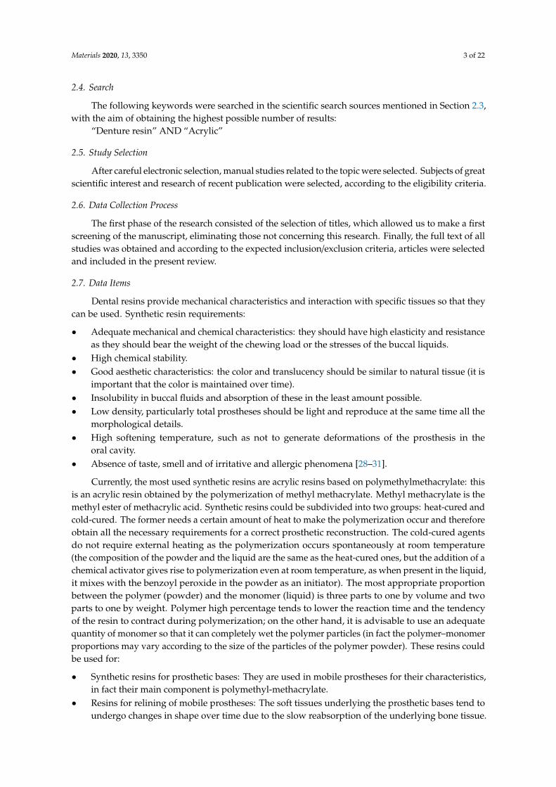
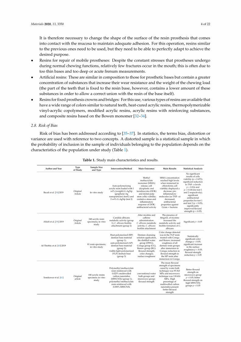
Risk of bias has been addressed according to [35–37]. In statistics, the terms bias, distortion orvariance are used with reference to two concepts. A distorted sample is a statistical sample in whichthe probability of inclusion in the sample of individuals belonging to the population depends on thecharacteristics of the population under study (Table 1).
Table 1. Study main characteristics and results.
Author and Year Typeof Study
Sample Sizeand Type Intervention/Method Main Outcomes Main Results Statistical Analysis
Bacali et al. [38] 2019 OriginalArticle In vitro study
Auto-polymerizingacrylic resin loaded with 1
wt% (weight) G-AgNp(graphene-Ag
nanoparticles) (test1) and2 wt% G-AgNp (test 2)
Methylmethacrylate
monomer (MMA)release; cell
(dysplastic oralkeratinocytes (DOK)
and dental pulpstem cells) viability;oxidative stress and
inflammatoryresponse of DOK;
antibacterial activity
MMA concentrationreached high levelswhen immersed in
chloroform; cellviability displayed a
decrease; proinflammatory
molecules (as TNF- α)decreased;
antibacterialproperties againstGram + bacteria
No significantresults of cells
viability (p = 0.472);significant reduction
in TNF- α levelsp = 0.016 and
p = 0.104 (for test 1and 2 respectively);
significantantibacterial
properties for test 1and test 2 (p < 0.05);
significantlyimproved flexuralstrength (p < 0.05)
Alfaifi et al. [39] 2019 OriginalArticle
240 acrylic resinspecimens; in vitro
study
Candida albicansmetabolic activity (group
1); C. albicans biofilmattachment (group 2)
After nicotine andcaffeine
administration:C. albicans metabolicactivity; C. albicansbiofilm attachment
The presence of8mg/mL of nicotine
increased themetabolic activity andbiofilm formation of C.
albicans
Significant p < 0.05
Al-Thobity et al. [40] 2019 81 resin specimens;in vitro study
Heat-polymerized (HP)denture base material
(group 1);auto-polymerized (AP)denture base material
(group 2);visible-light-polymerized
(VLP) denture base(group 3)
Denture cleansingsolution application,the distilled water
group (DWG),Corega group (CG),Renew group (RG):
flexural strength;color changes;
surface roughness
Color change detectedwas in the VLP resintreated with Corega
and Renew; increasedroughness of all
denture resin groupsafter immersion in
Corega; reduction inflexural strength inthe HP resin after
immersion in Corega.
Statisticallysignificant colorchange p < 0.05;
significant increasein the surface
roughness p < 0.05;flexural strength
reduction p < 0.05
Somkuwar et al. [41] Originalarticle
180 acrylic resinsspecimen; in vitro
study
Polymethyl methacrylateresin reinforced with0.025% multiwalledcarbon nanotubes
(MWCNTs) (group 1);polymethyl methacrylate
resin reinforced with0.050% MWCNTs
conventional waterbath groups and
microwave group:flexural strength
The mean flexuralstrength of specimenscured by water bathtechnique was 95.563MPa and microwave
technique was 118.416MPa. High
percentage ofmultiwalled carbonnanotubes present
better flexuralstrength
Better flexuralstrength on
microwave groupp < 0.05; better
flexural strength onhigh MWCNTsgroups p < 0.05

Materials 2020, 13, 3350 5 of 22
Table 1. Cont.
Author and Year Typeof Study
Sample Sizeand Type Intervention/Method Main Outcomes Main Results Statistical Analysis
Lee et al. [42] 2017 Originalarticle
Six thermoplasticresin materials;in vitro study
Three polyamidematerials (Smile tone, ST;valplast, VP; and Luciton
FRS, LF), two acrylicmaterials (Acrytone, AT;
and Acryshot, AS),and one polypropyleneresin material (Unigum,UG), heat-polymerizedacrylic resin (Vertex RS,
RS) (control)
Extracts and culturewith immortalized
human oralkeratinocytes
(IHOKs) or mousefibroblasts:cytotoxicity
VP at 70◦ extract andAT at 121◦ extract
showed lowercytotoxicity
p < 0.05
Wagner et al. [43] 2017 Originalarticle
20 dentures; in vitrostudy
PMMA (poly(methylmethacrylate)) acrylic
resin
Microwaveirradiation at 700W
and 420W:dimensional
stability
Denture experienceda linear dimensional
change ofapproximately 3%
Significant atp < 0.05
De Sousa Porta et al. [44] 2014 Originalarticle
15 participants;clinical study Acrylic resin dentures
0.5% NaOCl for 3min over 90 days:biofilm formation,
color stability,surface roughness,
patient acceptability
Reduction inmicroorganism and
C. albicans; nodifference in color androughness; increased
level of patientsatisfaction
Significantmicroorganism
reduction p = 0.001;color p = 0.68;
roughness p = 0.47
Wang et al. [45] 2014 OriginalArticle In vitro study
Acrylic resin denturesloaded with 0.5, 1, 2 wt%
multiwalled carbonnanotubes
Flexural strength
2 wt%MWCNT-loaded
dentures showed notbeneficial results
Worst mechanicalproperties on 2 wt%
MWCNT-loadeddentures
Akalin-Evren et al. [46] 2014 OriginalArticle
48 denture baseresins;
in vitro study
Denture base resinreinforced with E-glass
fiber-reinforcedcomposites (FRC)
Treated with salivaor distilled water;
C. albicans adhesion
C. albicans adhesiondid not show
differences
Not significantp = 0.436
Mansour et al. [47] 2013 OriginalArticle
199 denture bases;in vitro study
Wet ground muscovitemica and Lucitone 199original shade denturebase resin: (A) control
group with 0 vol% mica,(B) 10 vol% W200 mica,(C) 20 vol% W200 mica,
(D) 10 vol% P66 mica, (E)20 vol% P66 mica.
The mica was silanetreated in a solution
of3-methacryloxypropyl
trimethoxysilane,ethanol, and water,
and then dried;flexural strength
and microhardness
The flexural strengthof the control group
77–94%. Nosignificant differenceswere found within the
four mica groups.Microhardnesses of
the 20% mica groupswere 33–26%.
Flexural strengthhigher in control
than mica p ≤ 0.05.Microhardness ofthe C group was
higher than control(p ≤ 0.05).
So et al. [48] 2012 OriginalArticle
50 specimens;in vitro study
Cold cured PMMA with0%, 2%, 3%, 5% E-glassfibers with and without
post-curing microwave at800 w for 3 min
Water storage for7,14 and 30 days;Flexural strength,maximum load onthe load-deflection
curve
The group with 3%fiber and microwave
treatment, and thegroups with 5% fiber
increase in theflexural strength
values compared withthe control group
Flexural strength on3% and 5% E-glass
fiber(p = 0.003 and p≤ 0.003)
Monteiro et al. [49] 2011 OriginalArticle
Denture resin;in vitro study
Denture base resincontaining silver colloidalnanoparticles in different
concentration 0.05, 0.5,and 5 vol% silver
colloidal
Specimens werestored in deionizedwater at 37 ◦C for 7,15, 30, 60 and 120
days; silverdistribution and
release
Silver was notdetected in deionized
water; silverdistribution anddispersion was
improved with lowersilver concentration
/
Ladha et al. [50] 2011 OriginalArticle
160 resin specimens;in vitro study
Conventional PMMAdenture resin;
unidirectional stick (S)glass fiber
reinforced-PMMAdenture resin; wovenstick net (SN) glass
fiber-reinforced PMMAdenture resin; nylon
fiber-reinforced PMMAdenture resin
Each group wasstored in dry andwet conditions;
flexural strength
Glass fiberreinforcements
enhanced flexuralstrength of heat cured
PMMA denture
Significantenhanced flexuralstrength in glassfiber-reinforced
group
Fan et al. [51] 2011 Originalarticle In vitro study
Light-cure denture resinswith Ag benzoate of
various concentration (0,0.002, 0.02, 0.1, 0.15 and
0.2%); chemical-curesystems with Ag benzoatevarious concentration (0,0.002, 0.02, 0.1, 0.15 and
0.2%)
Resin hardness,silver release,
antibacterial activity
Hardness wasunaffected by Ag
benzoate, and silverwas released only at aconcentration higher
than 0.1%
/
Zortuk et al. [52] 2008 Originalarticle
48 specimens;in vitro study
Auto-polymerizingacrylic resin (no fiber);
auto-polymerizing acrylicresin with glass fiber
(0.5%); auto-polymerizingacrylic resin with glass
fiber (1%);auto-polymerizing acrylicresin with glass fiber (2%)
Surface specimenspolishing; surface
roughness (Ra)
Difference in resinsurface roughness
with differentconcentrations of fiber
p < 0.001
Materials 2020, 13, 3350 6 of 22
Table 1. Cont.
Author and Year Typeof Study
Sample Sizeand Type Intervention/Method Main Outcomes Main Results Statistical Analysis
Puri et al. [53] 2008 Originalarticle In vitro study
PMMA resin Lucitone199; PMMA resin with
ethylene glycolmethacrylate phosphate
(EGMP) 10%; PMMAresin with ethylene glycolmethacrylate phosphate
(EGMP) 15%; PMMAresin with ethylene glycolmethacrylate phosphate
(EGMP) 15% + crosslinking agent; PMMA
resin with ethylene glycolmethacrylate phosphate
(EGMP) 20%
Impact strength,fracture toughness,wettability, resinbonding ability
Hydrophilicity wasincreased increasing
EGMP concentrations,with no other
differences betweengroups
Improvedhydrophilicity
p = 0.039
Faot et al. [54] 2008 OriginalArticle In vitro study
Microwave acrylic resinpolymerized with 3 minat 360 W, 4-min pause,
and 3 min at 810 W(Control); microwave
acrylic resin polymerizedwith an alternative cycle(AC) of 6 min at 630 W
Accuracy of fit at 0time and at 30 days,impact strength test(Charpy method),
fractographicanalysis
No difference inoutcomes between
groups, denture basesshowed a better fit
after 30-days ofstorage in water
Better fit after 30days in water
p < 0.05
Kim et al. [55] 2007 OriginalArticle In vitro study
Reinforced acrylic-basedhybrid denture composite
resin withpolyhedraloligosilsesquioxane
(POSS) (group 1);heat-polymerized acrylicdenture base resin (group
2); auto-polymerizedacrylic denture base resin(group 3); direct reliningacrylic denture base resin
(group 4)
Biocompatibility,mutagenesis
POSS showedimproved
biocompatibility andlower mutagenicity.
Group 1 showedless cytotoxicity
(p < 0.05); group 4showed the highest
cytotoxicity(p < 0.05)
Tacir et al. [56] 2006 Originalarticle
80 specimens;in vitro study
Conventionalheat-polymerized acrylic
resin (group 1);Heat-polymerized acrylic
resin with glass fibers(10–15µm thick and 5mm
long) (group 2);microwaved Shera-Med
MW 2000(Dental-Werkstoffe,
Lemförde, Germany)PMMA in a
polycarbonate flask(group 3); microwavedShera-Med MW 2000(Dental-Werkstoffe,
Lemförde, Germany)PMMA in a
polycarbonate flask withglass fibers (10–15µmthick and 5mm long)
(group 4)
Flexural strength
Group 2 presentedbetter fracture
resistance but lessflexural strength
p < 0.05
Kimoto et al. [57] 2005 Originalarticle In vitro study
Rapid cooling after heatpolymerization (group 1);bench cooling after heathpolymerization (group 2)
Denture strainBench cooling for theheat-cured denturereduced the strain
p < 0.05
Pesci-Bardon et al. [58] 2004 Originalarticle
216 specimens;in vitro study
Acrylic resin discs addedwith Poly 202063A and
large volumes ofmicrobial inoculum (45mL) (group 1); acrylicresin discs added with
Poly 202063A andmicrobial inoculum (600
microL) (group 2);Acrylic resin discs addedwith Poly 202063A and
sterile buffer (600 microL)(group 3)
Antisepticproperties
A bactericidal effectagainst Escherichia
coli andStaphylococcus
aureus.A dose-dependent
fungistatic effect wasobserved against
C. albicans.
bactericidal effectp = 0.012; antifungal
effect p = 0.003
Uzun et al. [59] 2003 Originalarticle
16 specimens;in vitro study
Pre-treated epoxyresin-coated glass fibers,
with aramid fibers, orwith no fibers
Immediately and at30-days water
storage; transversestrength, maximal
deflection, modulusof elasticity
No differences instrength and
deflection values inimmediate group and
30 days group
Aramid fiber andwithout fiber
(p = 0.574), glassfiber and withoutfiber (p = 0.065) in
the immediategroup
Keyf et al. [60] 2003 Originalarticle
36 specimens;In vitro study
Auto-polymerizingacrylic resin with
hydroxyethyl-methacrylate(HEMA) treated glass
fiber: (group A) dischargepower of 15 W and
flowrate 15 min, 60 mLmin; (group B) 20 W, 10
(group C) 15 W, 15 min, 60mL min)1; (group D) 20W, 15 min, 60 mL min)1;
(group E) untreated;(group F) without fiber
Load of fracture,transverse strength,deflection, modulus
of elasticity
Transverse strengthand maximal
deflection weredifferent between
groups, not formodulus of elasticity
Transverse strengthp = 0.006, deflectionp = 0.039, elasticitymodulus p = 0.491

Materials 2020, 13, 3350 7 of 22
Table 1. Cont.
Author and Year Typeof Study
Sample Sizeand Type Intervention/Method Main Outcomes Main Results Statistical Analysis
John et al. [61] 2001 OriginalArticle
ten specimens;in vitro study
No fiberreinforced-acrylic resin(control); acrylic resinreinforced with glass
fibers (test 1); acrylic resinreinforced with aramid
(test 2) acrylic resinreinforced with nylon
fibers (test 3)
Flexural strength
All reinforced testgroups showed better
results on flexuralstrength; glass fibershowed the highest
flexural strength
Test 2 had the bestresult (p < 0.001)
2.9. Summary Measures
A summary of the measures assessed in this review can be expressed as follows:
• Table 1
◦ Author and year—author and year of publication;◦ Type of study—type of manuscript (article, Randomized Clinical Trials (RCT), review, etc.);◦ Sample size and type—sample size and type of performed analysis (in vitro, in vivo,
in silico etc);◦ Intervention/method—type of group subdivision and features;◦ Main outcomes—intervention on single specimens and type of evaluated outcomes;◦ Main results—main results of the single study;◦ Statistical analysis—statistical data regarding outcomes.
• Table 2 In this table, the Risk of Bias in systemic Review (ROBIS) [35–37] method for risk of biasallocation was used.
• Table 3
◦ Biological features—only biological outcomes, host tissue or cell implications.◦ Microbiological features—microbiological outcomes, bacteria, fungi or virus.◦ Physical features—physical, mechanical, chemical properties.◦ Other—other outcomes, in this case, only one result evaluated “patient acceptability”.
2.10. Synthesis of Results
The data obtained from the individual results are summarized in the “Results” section.The synthesis of the results was conducted manually by the individual authors, independently.Once the titles and abstracts were screened, the individual authors extrapolated the results from theindividual articles and they were compared at the end of the review process.
2.11. Additional Analysis
To give the reader readiness of what has been analyzed in this review, an examination was chosento closely observe the microscopic surface characteristics of a cold-cured resin. The resin in question isa liquid powder resin, composed of:
• Liquid: methacrylate, tetramethylene, dimethacrylate.• Powder: dibenzoyl peroxide, methyl methacrylate (does not contain cadmium).
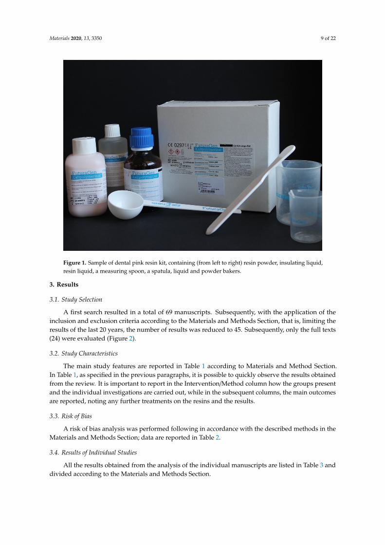
A common resin used in dentistry (FuturaGen® Schutz Dental GmbH, Rosbach, Germany), pinkin color, is presented in Figure 1, where it is possible to observe how this resin presents itself to theclinician. The resin surface and fractured surface were observed with a stereomicroscope (Leica® M125C). Once the resin was mixed according to the manufacturer’s instructions, two sample were created(4 × 2 × 1 cm); these were subsequently fractured into two equal parts, and the surface was observed.Images were modified and optimize by applying mac Os photo®.
Materials 2020, 13, 3350 8 of 22
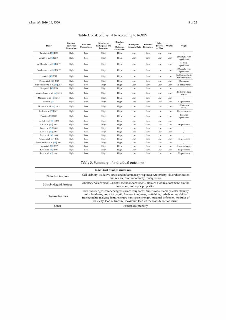
Table 2. Risk of bias table according to ROBIS.
StudyRandomSequence
Generation
AllocationConcealment
Blinding ofParticipants and
Personnel
Blindingof
OutcomeAssessment
IncompleteOutcome Data
SelectiveReporting
OtherSourcesof Bias
Overall Weight
Bacali et al. [38] 2019 High Low High High Low Low Low Low /
Alfaifi et al. [39] 2019 High Low High High Low Low Low Low 240 acrylic resinspecimens
Al-Thobity et al. [40] 2019 High Low High High Low Low Low Low 81 resinspecimens
Somkuwar et al. [41] 2017 High Low High High Low Low Low Low 180 acrylic resinspecimens
Lee et al. [42] 2017 High Low High High Low Low Low Low Six thermoplasticresin materials
Wagner et al. [43] 2015 High Low High High Low Low Low Low 20 dentures
De Sousa Porta et al. [44] 2014 High Low High High Low Low Low Low 15 participants
Wang et al. [45] 2014 High Low High High Low Low Low Low /
Akalin-Evren et al. [46] 2014 High Low High High Low Low Low Low 48 denture baseresins
Mansour et al. [47] 2013 High Low High High Low Low Low Low /
So et al. [48] High Low High High Low Low Low Low 50 specimens
Monteiro et al. [49] 2011 High Low High High Low Low Low Low 199 denturebases
Ladha et al. [50] 2011 High Low High High Low Low Low Low Denture resins
Fan et al. [51] 2011 High Low High High Low Low Low Low 160 resinspecimens
Zortuk et al. [52] 2008 High Low High High Low Low Low Low /
Puri et al. [53] 2008 High Low High High Low Low Low Low 48 specimens
Faot et al. [54] 2008 High Low High High Low Low Low Low /
Kim et al. [55] 2007 High Low High High Low Low Low Low /
Tacir et al. [56] 2006 High Low High High Low Low Low Low /
Kimoto et al. [57] 2005 High Low High High Low Low Low Low 80 specimens
Pesci-Bardon et al. [58] 2004 High Low High High Low Low Low Low /
Uzun et al. [59] 2003 High Low High High Low Low Low Low 216 specimens
Keyf et al. [60] 2003 High Low High High Low Low Low Low 16 specimens
John et al. [61] 2001 High Low High High Low Low Low Low 36 specimens
Table 3. Summary of individual outcomes.
Individual Studies Outcomes
Biological features Cell viability; oxidative stress and inflammatory response; cytotoxicity; silver distributionand release; biocompatibility, mutagenesis.
Microbiological features Antibacterial activity; C. albicans metabolic activity; C. albicans biofilm attachment; biofilmformation; antiseptic properties.
Physical features
Flexural strength; color changes; surface roughness; dimensional stability; color stability,microhardness; impact strength, fracture toughness, wettability, resin bonding ability;
fractographic analysis; denture strain; transverse strength, maximal deflection, modulus ofelasticity; load of fracture; maximum load on the load-deflection curve.
Other Patient acceptability.
Materials 2020, 13, 3350 9 of 22
Materials 2020, 13, x FOR PEER REVIEW 5 of 25
2.10. Synthesis of Results
The data obtained from the individual results are summarized in the “Results” section. The synthesis of the results was conducted manually by the individual authors, independently. Once the titles and abstracts were screened, the individual authors extrapolated the results from the individual articles and they were compared at the end of the review process.
2.11. Additional Analysis
To give the reader readiness of what has been analyzed in this review, an examination was chosen to closely observe the microscopic surface characteristics of a cold-cured resin. The resin in question is a liquid powder resin, composed of:
• Liquid: methacrylate, tetramethylene, dimethacrylate. • Powder: dibenzoyl peroxide, methyl methacrylate (does not contain cadmium).
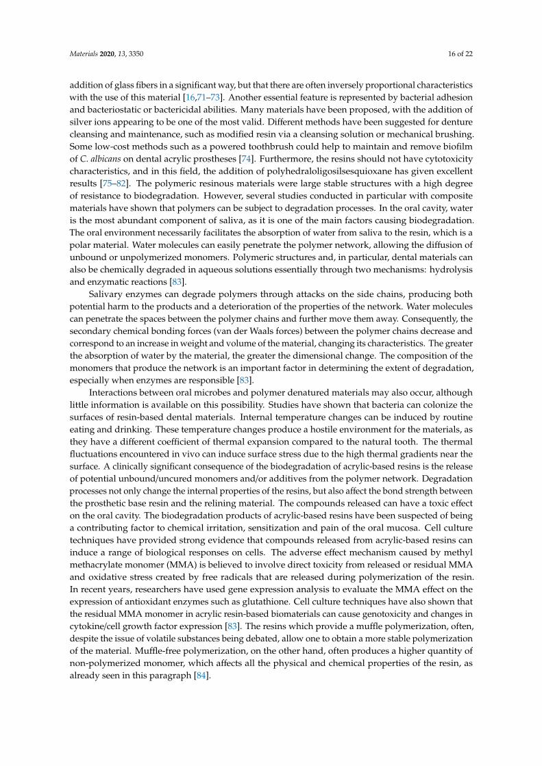
A common resin used in dentistry (FuturaGen® Schutz Dental GmbH, Rosbach, Germany), pink in color, is presented in Figure 1, where it is possible to observe how this resin presents itself to the clinician. The resin surface and fractured surface were observed with a stereomicroscope (Leica® M125 C). Once the resin was mixed according to the manufacturer’s instructions, two sample were created (4 × 2 × 1 cm); these were subsequently fractured into two equal parts, and the surface was observed. Images were modified and optimize by applying mac Os photo®.
Figure 1. Sample of dental pink resin kit, containing (from left to right) resin powder, insulating liquid, resin liquid, a measuring spoon, a spatula, liquid and powder bakers.
3. Results
3.1. Study Selection
A first search resulted in a total of 69 manuscripts. Subsequently, with the application of the inclusion and exclusion criteria according to the Materials and Methods Section, that is, limiting the
Figure 1. Sample of dental pink resin kit, containing (from left to right) resin powder, insulating liquid,resin liquid, a measuring spoon, a spatula, liquid and powder bakers.
3. Results
3.1. Study Selection
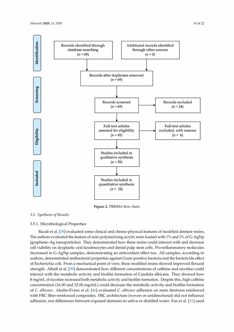
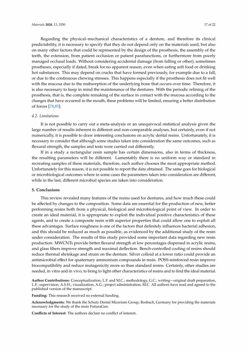
A first search resulted in a total of 69 manuscripts. Subsequently, with the application of theinclusion and exclusion criteria according to the Materials and Methods Section, that is, limiting theresults of the last 20 years, the number of results was reduced to 45. Subsequently, only the full texts(24) were evaluated (Figure 2).
3.2. Study Characteristics
The main study features are reported in Table 1 according to Materials and Method Section.In Table 1, as specified in the previous paragraphs, it is possible to quickly observe the results obtainedfrom the review. It is important to report in the Intervention/Method column how the groups presentand the individual investigations are carried out, while in the subsequent columns, the main outcomesare reported, noting any further treatments on the resins and the results.
3.3. Risk of Bias
A risk of bias analysis was performed following in accordance with the described methods in theMaterials and Methods Section; data are reported in Table 2.
3.4. Results of Individual Studies
All the results obtained from the analysis of the individual manuscripts are listed in Table 3 anddivided according to the Materials and Methods Section.
Materials 2020, 13, 3350 10 of 22
Materials 2020, 13, x FOR PEER REVIEW 6 of 25
results of the last 20 years, the number of results was reduced to 45. Subsequently, only the full texts (24) were evaluated (Figure 2).
Figure 2. PRISMA flow chart.
3.2. Study Characteristics
The main study features are reported in Table 1 according to Materials and Method Section. In Table 1, as specified in the previous paragraphs, it is possible to quickly observe the results obtained from the review. It is important to report in the Intervention/Method column how the groups present and the individual investigations are carried out, while in the subsequent columns, the main outcomes are reported, noting any further treatments on the resins and the results.
Figure 2. PRISMA flow chart.
3.5. Synthesis of Results
3.5.1. Microbiological Properties
Bacali et al. [38] evaluated some clinical and chemo-physical features of modified denture resins.The authors evaluated the feature of auto-polymerizing acrylic resin loaded with 1% and 2% of G-AgNp(graphene–Ag nanoparticles). They demonstrated how these resins could interact with and decreasecell viability on dysplastic oral keratinocytes and dental pulp stem cells. Pro-inflammatory moleculesdecreased in G-AgNp samples, demonstrating an antioxidant effect too. All samples, according toauthors, demonstrated antibacterial properties against Gram-positive bacteria and the bactericide effectof Escherichia coli. From a mechanical point of view, these modified resins showed improved flexuralstrength. Alfaifi et al. [39] demonstrated how different concentrations of caffeine and nicotine couldinteract with the metabolic activity and biofilm formation of Candida albicans. They showed how8 mg/mL of nicotine increased both metabolic activity and biofilm formation. Despite this, high caffeineconcentration (16.00 and 32.00 mg/mL) could decrease the metabolic activity and biofilm formationof C. albicans. Akalin-Evren et al. [46] evaluated C. albicans adhesion on resin dentures reinforcedwith FRC fiber-reinforced composites. FRC architecture (woven or unidirectional) did not influenceadhesion, nor differences between exposed dentures to saliva or distilled water. Fan et al. [51] used
Materials 2020, 13, 3350 11 of 22
both light-cured and chemical-cured systems to synthesize AgNPs using different concentrations of Agbenzoate (AgBz). These concentrations were verified thought an electron microscopy, and Ag benzoatedid not affect resin hardness. Antimicrobial resins released Ag+ ions in all samples, but they showedantimicrobial activity against Streptococcus mutans and showed an inhibition from 52.4% to 97.5%.Pesci-Bardon et al. [58] mixed Poly 202063, a quaternary ammonium compound polymer, with dentureresin to evaluate its antimicrobial effect. They tested some resin discs with different inoculum volumes,concluding that the specimen provided antimicrobial and antifungal effect, with better results withlow inoculum.
3.5.2. Biological Properties
Lee et al. [42] evaluated the cytotoxicity of different denture materials: polyamide, acrylic,polypropylene and a heat-polymerized acrylic resin as a control group. They obtained extracts fromspecimens of the denture materials under different condition (37 ◦C for 24 h, 70 ◦C for 24 h, and 121 ◦Cfor 1 h). The extracts were then diluted in distilled water and co-cultured for 24 h with immortalizedhuman oral keratinocytes (IHOKs) or mouse fibroblast. Greater than 70% viability was detected underall test conditions.
Denture cleansers solution can affect denture properties. de Sousa Porta et al. [44] evaluatedcolor, roughness change and biofilm formation with the use of sodium hypochlorite. The authorsevaluated patient satisfaction after 90 days of the use of this solution. They evaluated the use of a 0.5%NaOCl solution for 3 min a day on an acrylic resin denture. They showed a significant reduction inbiofilm formation with no roughness or color change, and with a better patient satisfaction after use.Monteiro et al. [49] showed silver distribution and release in antimicrobial base resin, with added silvercolloidal nanoparticles. Acrylic resin was prepared in accordance with manufacturer’s instructionsand silver nanoparticle suspension was added in different concentrations. After storing the denturesin deionized water for a time from 7 up to 120 days, they analyzed each solution. Silver was notdetected in deionized water and they showed how silver dispersion was better at a lower silverconcentration. Kim et al. [55] evaluated the biocompatibility of reinforced acrylic hybrid resin withpolyhedraloligosilsesquioxane (POSS). POSS showed improved biocompatibility (measured by ametabolic assay, an agar overlay test, and a mutagenesis assay) and lower mutagenicity.
3.5.3. Physical Properties
Al-Thobity et al. [40] evaluated the effect of cleansing solution on different denture resins(heat-polymerized, auto-polymerized, visible-light-polymerized). They evaluated the effect of distilledwater as control, as well as Corega and Renew cleansing solutions. The only color change detected wasin the visible-light-polymerized (VLP) resin treated with Corega and Renew. Surface roughness of alldenture resin increased after immersion in Corega. Immersion in Renew significantly increased surfaceroughness only in the heat-polymerized (HP) and auto-polymerized (AP) specimens. A reduction inflexural strength was detected in the HP resin after immersion in Corega. Somkuwar et al. [41] evaluatedthe effect of multiwalled carbon nanotubes (MWCNTs) on PMMA (Poly(methyl methacrylate)) dentureresin flexure strength. They demonstrated how microwave-cured denture resins have better flexurestrength than the water bath-cured type, and that a 0.025% or 0.050% MWCNT weight could improvethis physical property.
Wagner et al. [43] evaluated dimensional stability of PMMA resin dentures after microwaveirradiation. Denture bases were placed into a glass baker with 200mL of room-demineralized waterand then exposed to 420W or 700W microwave radiation for 3 min. All dentures experienced a 1- or2-mm dimensional change after each period of microwaving. Wang et al. [45] evaluated the effectof a multiwalled carbon nanotube on denture polymethyl methacrylate composite resins. In thisin vitro study, they fabricated dentures with 0.5, 1 and 2 wt% of multiwalled carbon nanotubes. Theseresins were sonically mixed for 20 min and mechanical features were measured. The results suggestedthat the interfacial bonding between MWCNTs and PMMA was weak and in need of improvement.
Materials 2020, 13, 3350 12 of 22
The addition of 0.5% and 1% MWCNTs improved the PMMA resin flexural strength and resilience,but the addition of 2% MWCNTs was not able to do so because of poor dispersion of the MWCNTs.Mansour et al. [47] evaluated the effect of mica on the flexural strength and microhardness of PMMAdenture resin. They tested two mica, W200 with an average particle sizes (d50) of 131 µm and P66 withan average particle sizes (d50) of 30 µm. Dentures were fabricated according to the resin manufacturer’sinstructions; different amounts of mica were added to each group. Mica seemed to give less flexuralstrength with high microhardness to dentures. So et al. [48] evaluated the effect of reinforcementby various concentrations of chopped E-glass fibers (0%, 1%, 2%, 3% and 5% by weight of resinpowder) and post-curing microwave irradiation (800 W for 3 min) on the flexural strength of cold-curedacrylics. According to them, at room temperature and humidity for 1 day, the group with 3% and 5%fiber reinforcements obtained significant results compared to test group. They demonstrated howthe effect that a water bath for several days could have on resin reinforcements. When the waterstorage time increased, the effect of the fiber remained, but the effect of microwave treatment vanished.Ladha et al. [50] evaluated the flexural strength of different reinforced PMMA dentures. They testedunreinforced resins, those reinforced with unidirectional stick glass fibers, woven stich net glass fibersand nylon fibers. After storing them in dry and wet conditions, they conducted a 3-point bendingflexural test. Glass fiber improved flexural strength, and nylon fiber decreased it, even more thanunreinforced acrylic resin.
Zortuk et al. [52] evaluated the influence of different concentrations of fiber glass on resin surfaceroughness. After polishing the specimens, they evaluated and calculated the surface roughnessthrough a profilometer. They observed significant differences between groups, with fiber glass groupspresenting a higher surface roughness than the non-fiber group. Puri et al. [53] evaluated the effectof ethylene glycol methacrylate phosphate (EGMP) at different concentrations on PMMA dentureresin. They did not highlight statistically significant differences regarding resin bonding ability orother mechanical features. They showed that EGMP concentrations influenced hydrophilicity in astatistically significant way. Faot et al. [54] evaluated accuracy fit, impact strength and performed afractural analysis on microwave polymerized acrylic resin, using two different polymerizing protocols.Measurements were performed immediately and after 30 days in water storage, showing a better fitfor the 30-day group. Tacir et al. [56] in their in vitro study evaluated differences in denture resinmechanical features when reinforced with fiber glass. They showed how fiber glass could improveflexure strength, but also decrease the fracture resistance of a denture in a statistically significantway. Kimoto et al. [57] evaluated dimensional accuracy of heat-cured denture resin with two differentcooling protocols, namely, a rapid cooling protocol and a bench cooling. In this last case, the flask wasleft to cool in a thermo-stabilized room for 140 min. This cooling method provided less denture straincaused by thermal shrinkage. Uzun et al. [59] evaluated the effect of different fiber reinforcement typeimmediately and after water storage on the strength properties of denture resin. They reinforced theresin with glass and aramid fiber and demonstrated how glass fiber is superior to other fibers andcould improve transverse strength.
Keyf et al. [60] evaluated the effects of hydroxyethyl-methacrylate (HEMA) and air atmosphere onglass fiber, which are used to increase the strength of denture resin. Glass fibers were surface treatedwith different air and power protocols. HEMA treatments on fiber glass resulted in the modification ofthe maximal deflection and transverse strength of the denture resin, with no differences in the modulusof elasticity between groups. A study by John et al. [61] evaluated different PMMA fiber reinforcingmethods. They highlighted differences on flexural strength of these heat-polymerized resins usingglass, aramid and nylon fibers. Glass fibers provided a better performance than the other groups. Test1 group (glass fiber) had the highest flexural strength, followed by test 2, test 3 and control. The higherthe load or force required to fracture the specimens, the higher the fracture resistance.
Materials 2020, 13, 3350 13 of 22
3.6. Additional Analysis
The data resulting from the tests carried out are visually reported in Figures 2–5 in accordancewith the Materials and Methods Section.
Materials 2020, 13, x FOR PEER REVIEW 16 of 25
polymerizing protocols. Measurements were performed immediately and after 30 days in water storage, showing a better fit for the 30-day group. Tacir et al. [56] in their in vitro study evaluated differences in denture resin mechanical features when reinforced with fiber glass. They showed how fiber glass could improve flexure strength, but also decrease the fracture resistance of a denture in a statistically significant way. Kimoto et al. [57] evaluated dimensional accuracy of heat-cured denture resin with two different cooling protocols, namely, a rapid cooling protocol and a bench cooling. In this last case, the flask was left to cool in a thermo-stabilized room for 140 min. This cooling method provided less denture strain caused by thermal shrinkage. Uzun et al. [59] evaluated the effect of different fiber reinforcement type immediately and after water storage on the strength properties of denture resin. They reinforced the resin with glass and aramid fiber and demonstrated how glass fiber is superior to other fibers and could improve transverse strength.
Keyf et al. [60] evaluated the effects of hydroxyethyl-methacrylate (HEMA) and air atmosphere on glass fiber, which are used to increase the strength of denture resin. Glass fibers were surface treated with different air and power protocols. HEMA treatments on fiber glass resulted in the modification of the maximal deflection and transverse strength of the denture resin, with no differences in the modulus of elasticity between groups. A study by John et al. [61] evaluated different PMMA fiber reinforcing methods. They highlighted differences on flexural strength of these heat-polymerized resins using glass, aramid and nylon fibers. Glass fibers provided a better performance than the other groups. Test 1 group (glass fiber) had the highest flexural strength, followed by test 2, test 3 and control. The higher the load or force required to fracture the specimens, the higher the fracture resistance.
3.6. Additional Analysis
The data resulting from the tests carried out are visually reported in Figures 2–5 in accordance with the Materials and Methods Section.
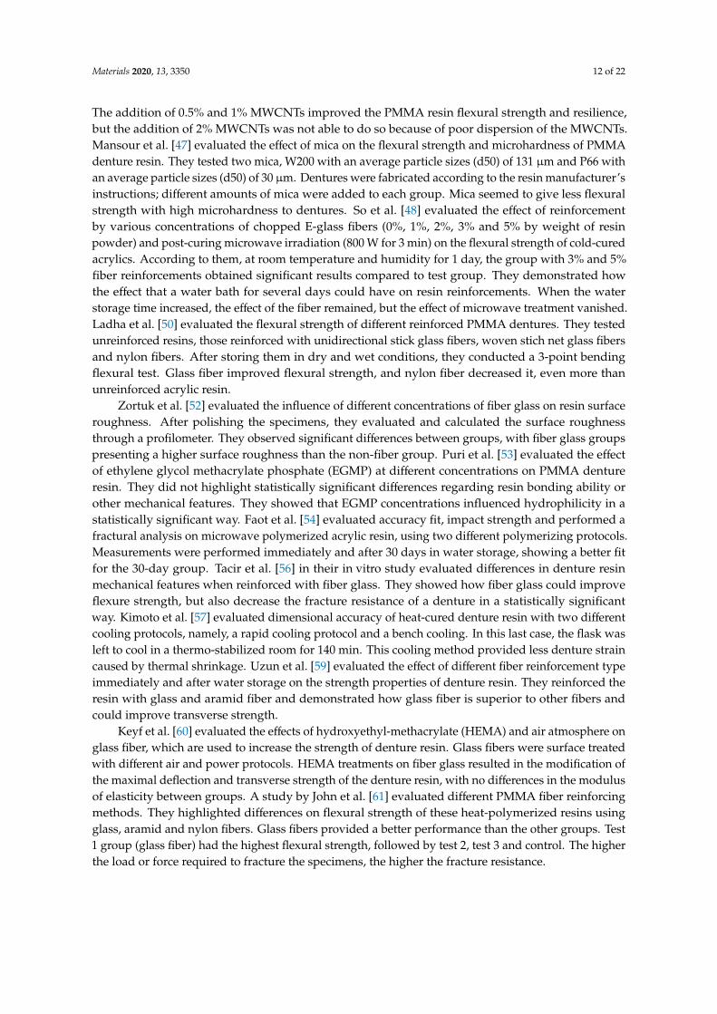
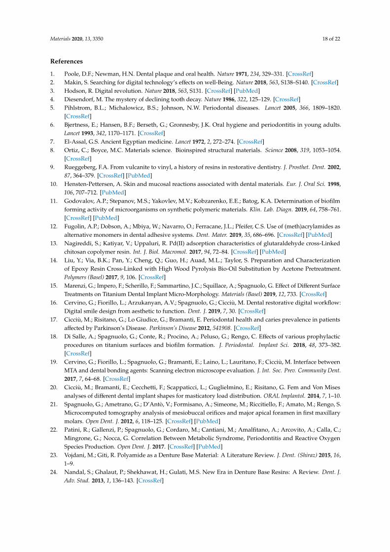
Figure 3. Polished resin sample. It shows a uniform surface with no fibers, cross linking or surface defects.
Figure 3. Polished resin sample. It shows a uniform surface with no fibers, cross linking orsurface defects.Materials 2020, 13, x FOR PEER REVIEW 17 of 25
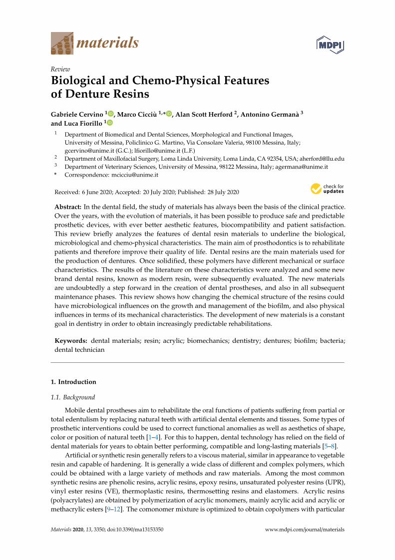
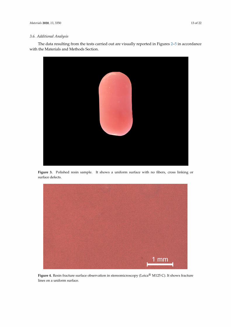
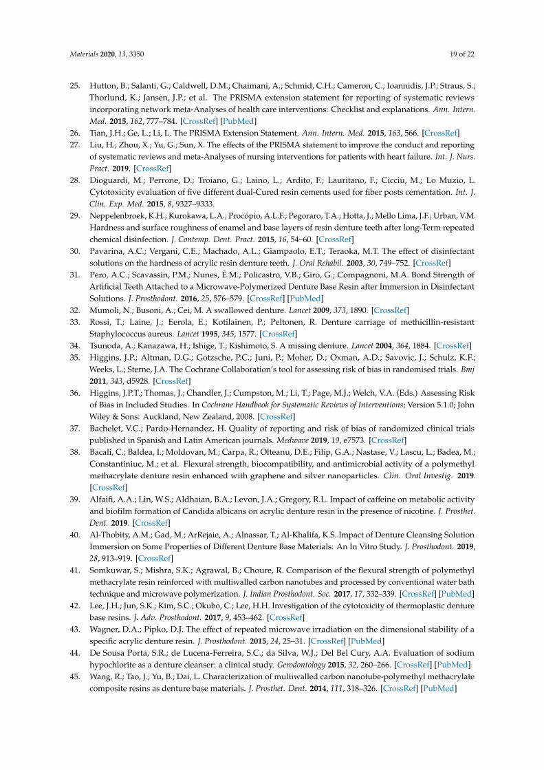
Figure 4. Resin fracture surface observation in stereomicroscopy (Leica® M125 C). It shows fracture lines on a uniform surface.
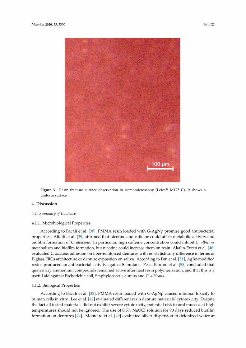
Figure 5. Resin fracture surface observation in stereomicroscopy (Leica® M125 C). It shows a uniform surface.
Figure 4. Resin fracture surface observation in stereomicroscopy (Leica® M125 C). It shows fracturelines on a uniform surface.
Materials 2020, 13, 3350 14 of 22
Materials 2020, 13, x FOR PEER REVIEW 17 of 25
Figure 4. Resin fracture surface observation in stereomicroscopy (Leica® M125 C). It shows fracture lines on a uniform surface.
Figure 5. Resin fracture surface observation in stereomicroscopy (Leica® M125 C). It shows a uniform surface. Figure 5. Resin fracture surface observation in stereomicroscopy (Leica® M125 C). It shows auniform surface.
4. Discussion
4.1. Summary of Evidence
4.1.1. Microbiological Properties
According to Bacali et al. [38], PMMA resin loaded with G-AgNp promise good antibacterialproperties. Alfaifi et al. [39] affirmed that nicotine and caffeine could affect metabolic activity andbiofilm formation of C. albicans. In particular, high caffeine concentration could inhibit C. albicansmetabolism and biofilm formation, but nicotine could increase them on resin. Akalin-Evren et al. [46]evaluated C. albicans adhesion on fiber-reinforced dentures with no statistically difference in terms ofE-glass FRCs architecture or denture exposition on saliva. According to Fan et al. [51], AgBz-modifiedresins produced an antibacterial activity against S. mutans. Pesci-Bardon et al. [58] concluded thatquaternary ammonium compounds remained active after heat resin polymerization, and that this is auseful aid against Escherichia coli, Staphylococcus aureus and C. albicans.
4.1.2. Biological Properties
According to Bacali et al. [38], PMMA resin loaded with G-AgNp caused minimal toxicity tohuman cells in vitro. Lee et al. [42] evaluated different resin denture materials’ cytotoxicity. Despitethe fact all tested materials did not exhibit severe cytotoxicity, potential risk to oral mucosa at hightemperatures should not be ignored. The use of 0.5% NaOCl solution for 90 days reduced biofilmformation on dentures [44]. Monteiro et al. [49] evaluated silver dispersion in deionized water at
Materials 2020, 13, 3350 15 of 22
37 ◦C, noting that it could affect denture resins with colloidally added antimicrobial silver. They didnot detect silver in any specimens. Fan et al. [51], instead, detected Ag+ ion release in all samples.Kim et al. [55] concluded that POSS-reinforced resin had better biocompatibility and less mutagenicitythan standard acrylic resins, but 72h of immersion results were similar [62–64].
4.1.3. Physical Properties
Flexural strength result improved in PMMA resin loaded with G-AgNp [38]. Al-Thobity et al. [40]in their study, showed how different cleansing solutions could modify denture resin properties.In particular some cleansing product could negatively affect color, surface roughness and flexuralstrength. The authors showed how heat-polymerized, auto-polymerized and visible-light-polymerizedreact differently. According to Somkuwar et al. [41], heat-polymerized denture base resins with andwithout reinforcement of MWCNTs and polymerized by the microwave technique possess higherflexural strength. MWCNTs could be used as an effective reinforcement material for the denture base.Wagner et al. [43] demonstrated how the microwaving cycle could affect the dimensional stability ofacrylic denture resin. De Sousa Porta [44] did not experienced roughness or color change in acrylic resindentures after NaOCl solution usage. Wang et al. [45] evaluated the effect of MWCNTs on mechanicalfeatures of denture resins. Despite 0.5% and 1 wt% of MWCNTs providing a better flexural strength,this was not the case for the 2 wt% group. According to the authors, it is caused by an inadequatedispersion of carbon nanotubes (that could cause agglomerates in a high percentage) and by theinterfacial bonding between MWCNTs and polymethyl methacrylate. According to Mansour et al. [47],the addition of mica addition to PMMA dentures reduced flexural strength, but it significantly increasedmicrohardness. Monteiro et al. [49] showed how antimicrobial denture base resins containing silvercolloidal nanoparticles present a better silver distribution and dispersion at a lower silver ratio.So et al. [48] concluded that glass fiber and post-curing microwaving improve the flexural strength incold-cured PMMA, but it could be influenced by the water storage time of the resin. They suggest a newmixing method called the “sprinkle method” in the fabrication of orthodontic appliances. This involvesdispensing the monomers and polymers directly onto the working model. Ladha et al. [50] evaluatedhow stick and stick net glass fiber-reinforced denture resin and improved resin flexural strengthmore so than that reinforced with nylon as well as the conventional type. Fan et al. [51] concludedthat despite further studies being necessary to evaluate the mechanical properties of AgBz-modifiedresins, the hardness of chemical-cured resins was not affected. Zortuk et al. [52] concluded that everyconcentration of fiber glass on acrylic resin affected the surface roughness negatively. Puri et al. [53]concluded that EGMP concentration did not affect the mechanical properties of PMMA dentures, but itdid improve hydrophilicity. Faot et al. [54] concluded that different microwave polymerization cyclesdid not produce different mechanical properties of resin dentures. They showed that after a 30-daystorage period in water, the dentures presented a better fit. This result is important because it couldindicate a modification process of the denture resin shape. Tacir et al. [56] concluded that glass fiberimprove flexural strength of PMMA denture resin, but also decreases fracture resistance. Accordingto the authors, these results could be useful in a clinical setting for the distal extension of partial ortotal denture bases. Kimoto et al. [57] concluded that bench-controlled cooling produced less thermalshrinkage and reduced strain on the denture. Uzun et al. [59] concluded that glass fiber was superiorto other fibers and that it improves transverse strength, deflection and elasticity. Keyf et al. [60]showed how surface treatments and chemical modification on glass fibers could improve strengthand maximal deflection of fiber-reinforced denture resin, which could subsequently reduce clinicalfailures. John et al. [61] in a study approximately 20 years ago guessed and demonstrated that glassfiber provided better results in regard to reinforcing denture resin. According to the authors, glassfiber produced better results than aramid and nylon fibers too.
The stability of the material over time is one of the characteristics that underlies excellentrehabilitation; moreover, the resin should have fracture resistance characteristics and excellent modulusof elasticity [65–70]. It has been highlighted how these two characteristics could be influenced by the
Materials 2020, 13, 3350 16 of 22
addition of glass fibers in a significant way, but that there are often inversely proportional characteristicswith the use of this material [16,71–73]. Another essential feature is represented by bacterial adhesionand bacteriostatic or bactericidal abilities. Many materials have been proposed, with the addition ofsilver ions appearing to be one of the most valid. Different methods have been suggested for denturecleansing and maintenance, such as modified resin via a cleansing solution or mechanical brushing.Some low-cost methods such as a powered toothbrush could help to maintain and remove biofilmof C. albicans on dental acrylic prostheses [74]. Furthermore, the resins should not have cytotoxicitycharacteristics, and in this field, the addition of polyhedraloligosilsesquioxane has given excellentresults [75–82]. The polymeric resinous materials were large stable structures with a high degreeof resistance to biodegradation. However, several studies conducted in particular with compositematerials have shown that polymers can be subject to degradation processes. In the oral cavity, wateris the most abundant component of saliva, as it is one of the main factors causing biodegradation.The oral environment necessarily facilitates the absorption of water from saliva to the resin, which is apolar material. Water molecules can easily penetrate the polymer network, allowing the diffusion ofunbound or unpolymerized monomers. Polymeric structures and, in particular, dental materials canalso be chemically degraded in aqueous solutions essentially through two mechanisms: hydrolysisand enzymatic reactions [83].
Salivary enzymes can degrade polymers through attacks on the side chains, producing bothpotential harm to the products and a deterioration of the properties of the network. Water moleculescan penetrate the spaces between the polymer chains and further move them away. Consequently, thesecondary chemical bonding forces (van der Waals forces) between the polymer chains decrease andcorrespond to an increase in weight and volume of the material, changing its characteristics. The greaterthe absorption of water by the material, the greater the dimensional change. The composition of themonomers that produce the network is an important factor in determining the extent of degradation,especially when enzymes are responsible [83].
Interactions between oral microbes and polymer denatured materials may also occur, althoughlittle information is available on this possibility. Studies have shown that bacteria can colonize thesurfaces of resin-based dental materials. Internal temperature changes can be induced by routineeating and drinking. These temperature changes produce a hostile environment for the materials, asthey have a different coefficient of thermal expansion compared to the natural tooth. The thermalfluctuations encountered in vivo can induce surface stress due to the high thermal gradients near thesurface. A clinically significant consequence of the biodegradation of acrylic-based resins is the releaseof potential unbound/uncured monomers and/or additives from the polymer network. Degradationprocesses not only change the internal properties of the resins, but also affect the bond strength betweenthe prosthetic base resin and the relining material. The compounds released can have a toxic effecton the oral cavity. The biodegradation products of acrylic-based resins have been suspected of beinga contributing factor to chemical irritation, sensitization and pain of the oral mucosa. Cell culturetechniques have provided strong evidence that compounds released from acrylic-based resins caninduce a range of biological responses on cells. The adverse effect mechanism caused by methylmethacrylate monomer (MMA) is believed to involve direct toxicity from released or residual MMAand oxidative stress created by free radicals that are released during polymerization of the resin.In recent years, researchers have used gene expression analysis to evaluate the MMA effect on theexpression of antioxidant enzymes such as glutathione. Cell culture techniques have also shown thatthe residual MMA monomer in acrylic resin-based biomaterials can cause genotoxicity and changes incytokine/cell growth factor expression [83]. The resins which provide a muffle polymerization, often,despite the issue of volatile substances being debated, allow one to obtain a more stable polymerizationof the material. Muffle-free polymerization, on the other hand, often produces a higher quantity ofnon-polymerized monomer, which affects all the physical and chemical properties of the resin, asalready seen in this paragraph [84].
Materials 2020, 13, 3350 17 of 22
Regarding the physical–mechanical characteristics of a denture, and therefore its clinicalpredictability, it is necessary to specify that they do not depend only on the materials used, but alsoon many other factors that could be represented by the design of the prosthesis, the assembly of theteeth, the extension, from patient occlusion or patient parafunctions, or furthermore from poorlymanaged occlusal loads. Without considering accidental damage (from falling or other), sometimesprostheses, especially if dated, break for no apparent reason, even when eating soft food or drinkinghot substances. This may depend on cracks that have formed previously, for example due to a fall,or due to the continuous chewing stresses. This happens especially if the prosthesis does not fit wellwith the mucosa due to the reabsorption of the underlying bone that occurs over time. Therefore, itis also necessary to keep in mind the maintenance of the dentures. With the periodic relining of theprosthesis, that is, the complete remaking of the surface in contact with the mucosa according to thechanges that have occurred in the mouth, these problems will be limited, ensuring a better distributionof forces [78,85].
4.2. Limitations
It is not possible to carry out a meta-analysis or an unequivocal statistical analysis given thelarge number of results inherent in different and non-comparable analyses, but certainly, even if notnumerically, it is possible to draw interesting conclusions on acrylic dental resins. Unfortunately, it isnecessary to consider that although some studies taken into consideration the same outcomes, such asflexural strength, the samples and tests were carried out differently.
If in a study a rectangular resin sample has certain dimensions, also in terms of thickness,the resulting parameters will be different. Lamentably there is no uniform way or standard inrecreating samples of these materials, therefore, each author chooses the most appropriate method.Unfortunately for this reason, it is not possible to report the data obtained. The same goes for biologicalor microbiological outcomes where in some cases the parameters taken into consideration are different,while in the last, different microbial species are taken into consideration.
5. Conclusions
This review revealed many features of the resins used for dentures, and how much these couldbe affected by changes to the composition. Some data are essential for the production of new, betterperforming resins both from a physical, biological and microbiological point of view. In order tocreate an ideal material, it is appropriate to exploit the individual positive characteristics of theseagents, and to create a composite resin with superior properties that could allow one to exploit allthese advantages. Surface roughness is one of the factors that definitely influences bacterial adhesion,and this should be reduced as much as possible, as evidenced by the additional study of the resinunder consideration. The results of this study provided some important data regarding new resinproduction: MWCNTs provide better flexural strength at low percentages dispersed in acrylic resins,and glass fibers improve strength and maximal deflection. Bench-controlled cooling of resins shouldreduce thermal shrinkage and strain on the denture. Silver colloid at a lower ratio could provide anantimicrobial effect for quaternary ammonium compounds in resin. POSS-reinforced resin improvebiocompatibility and reduce mutagenicity more so than standard resins. Certainly, other studies areneeded, in vitro and in vivo, to bring to light other characteristics of resins and to find the ideal material.
Author Contributions: Conceptualization, L.F. and M.C.; methodology, G.C.; writing—original draft preparation,L.F.; supervision, A.S.H.; visualization, A.G.; project administration, M.C. All authors have read and agreed to thepublished version of the manuscript.
Funding: This research received no external funding.
Acknowledgments: We thank the Schutz Dental Micerium Group, Rosbach, Germany for providing the materialsnecessary for the study of the resin FuturaGen.
Conflicts of Interest: The authors declare no conflict of interest.
Materials 2020, 13, 3350 18 of 22
References
1. Poole, D.F.; Newman, H.N. Dental plaque and oral health. Nature 1971, 234, 329–331. [CrossRef]2. Makin, S. Searching for digital technology’s effects on well-Being. Nature 2018, 563, S138–S140. [CrossRef]3. Hodson, R. Digital revolution. Nature 2018, 563, S131. [CrossRef] [PubMed]4. Diesendorf, M. The mystery of declining tooth decay. Nature 1986, 322, 125–129. [CrossRef]5. Pihlstrom, B.L.; Michalowicz, B.S.; Johnson, N.W. Periodontal diseases. Lancet 2005, 366, 1809–1820.
[CrossRef]6. Bjertness, E.; Hansen, B.F.; Berseth, G.; Gronnesby, J.K. Oral hygiene and periodontitis in young adults.
Lancet 1993, 342, 1170–1171. [CrossRef]7. El-Assal, G.S. Ancient Egyptian medicine. Lancet 1972, 2, 272–274. [CrossRef]8. Ortiz, C.; Boyce, M.C. Materials science. Bioinspired structural materials. Science 2008, 319, 1053–1054.
[CrossRef]9. Rueggeberg, F.A. From vulcanite to vinyl, a history of resins in restorative dentistry. J. Prosthet. Dent. 2002,
87, 364–379. [CrossRef] [PubMed]10. Hensten-Pettersen, A. Skin and mucosal reactions associated with dental materials. Eur. J. Oral Sci. 1998,
106, 707–712. [PubMed]11. Godovalov, A.P.; Stepanov, M.S.; Yakovlev, M.V.; Kobzarenko, E.E.; Batog, K.A. Determination of biofilm
forming activity of microorganisms on synthetic polymeric materials. Klin. Lab. Diagn. 2019, 64, 758–761.[CrossRef] [PubMed]
12. Fugolin, A.P.; Dobson, A.; Mbiya, W.; Navarro, O.; Ferracane, J.L.; Pfeifer, C.S. Use of (meth)acrylamides asalternative monomers in dental adhesive systems. Dent. Mater. 2019, 35, 686–696. [CrossRef] [PubMed]
13. Nagireddi, S.; Katiyar, V.; Uppaluri, R. Pd(II) adsorption characteristics of glutaraldehyde cross-Linkedchitosan copolymer resin. Int. J. Biol. Macromol. 2017, 94, 72–84. [CrossRef] [PubMed]
14. Liu, Y.; Via, B.K.; Pan, Y.; Cheng, Q.; Guo, H.; Auad, M.L.; Taylor, S. Preparation and Characterizationof Epoxy Resin Cross-Linked with High Wood Pyrolysis Bio-Oil Substitution by Acetone Pretreatment.Polymers (Basel) 2017, 9, 106. [CrossRef]
15. Marenzi, G.; Impero, F.; Scherillo, F.; Sammartino, J.C.; Squillace, A.; Spagnuolo, G. Effect of Different SurfaceTreatments on Titanium Dental Implant Micro-Morphology. Materials (Basel) 2019, 12, 733. [CrossRef]
16. Cervino, G.; Fiorillo, L.; Arzukanyan, A.V.; Spagnuolo, G.; Cicciù, M. Dental restorative digital workflow:Digital smile design from aesthetic to function. Dent. J. 2019, 7, 30. [CrossRef]
17. Cicciù, M.; Risitano, G.; Lo Giudice, G.; Bramanti, E. Periodontal health and caries prevalence in patientsaffected by Parkinson’s Disease. Parkinson’s Disease 2012, 541908. [CrossRef]
18. Di Salle, A.; Spagnuolo, G.; Conte, R.; Procino, A.; Peluso, G.; Rengo, C. Effects of various prophylacticprocedures on titanium surfaces and biofilm formation. J. Periodontal. Implant Sci. 2018, 48, 373–382.[CrossRef]
19. Cervino, G.; Fiorillo, L.; Spagnuolo, G.; Bramanti, E.; Laino, L.; Lauritano, F.; Cicciù, M. Interface betweenMTA and dental bonding agents: Scanning electron microscope evaluation. J. Int. Soc. Prev. Community Dent.2017, 7, 64–68. [CrossRef]
20. Cicciù, M.; Bramanti, E.; Cecchetti, F.; Scappaticci, L.; Guglielmino, E.; Risitano, G. Fem and Von Misesanalyses of different dental implant shapes for masticatory load distribution. ORAL Implantol. 2014, 7, 1–10.
21. Spagnuolo, G.; Ametrano, G.; D’Antò, V.; Formisano, A.; Simeone, M.; Riccitiello, F.; Amato, M.; Rengo, S.Microcomputed tomography analysis of mesiobuccal orifices and major apical foramen in first maxillarymolars. Open Dent. J. 2012, 6, 118–125. [CrossRef] [PubMed]
22. Patini, R.; Gallenzi, P.; Spagnuolo, G.; Cordaro, M.; Cantiani, M.; Amalfitano, A.; Arcovito, A.; Calla, C.;Mingrone, G.; Nocca, G. Correlation Between Metabolic Syndrome, Periodontitis and Reactive OxygenSpecies Production. Open Dent. J. 2017. [CrossRef] [PubMed]
23. Vojdani, M.; Giti, R. Polyamide as a Denture Base Material: A Literature Review. J. Dent. (Shiraz) 2015, 16,1–9.
24. Nandal, S.; Ghalaut, P.; Shekhawat, H.; Gulati, M.S. New Era in Denture Base Resins: A Review. Dent. J.Adv. Stud. 2013, 1, 136–143. [CrossRef]
Materials 2020, 13, 3350 19 of 22
25. Hutton, B.; Salanti, G.; Caldwell, D.M.; Chaimani, A.; Schmid, C.H.; Cameron, C.; Ioannidis, J.P.; Straus, S.;Thorlund, K.; Jansen, J.P.; et al. The PRISMA extension statement for reporting of systematic reviewsincorporating network meta-Analyses of health care interventions: Checklist and explanations. Ann. Intern.Med. 2015, 162, 777–784. [CrossRef] [PubMed]
26. Tian, J.H.; Ge, L.; Li, L. The PRISMA Extension Statement. Ann. Intern. Med. 2015, 163, 566. [CrossRef]27. Liu, H.; Zhou, X.; Yu, G.; Sun, X. The effects of the PRISMA statement to improve the conduct and reporting
of systematic reviews and meta-Analyses of nursing interventions for patients with heart failure. Int. J. Nurs.Pract. 2019. [CrossRef]
28. Dioguardi, M.; Perrone, D.; Troiano, G.; Laino, L.; Ardito, F.; Lauritano, F.; Cicciù, M.; Lo Muzio, L.Cytotoxicity evaluation of five different dual-Cured resin cements used for fiber posts cementation. Int. J.Clin. Exp. Med. 2015, 8, 9327–9333.
29. Neppelenbroek, K.H.; Kurokawa, L.A.; Procópio, A.L.F.; Pegoraro, T.A.; Hotta, J.; Mello Lima, J.F.; Urban, V.M.Hardness and surface roughness of enamel and base layers of resin denture teeth after long-Term repeatedchemical disinfection. J. Contemp. Dent. Pract. 2015, 16, 54–60. [CrossRef]
30. Pavarina, A.C.; Vergani, C.E.; Machado, A.L.; Giampaolo, E.T.; Teraoka, M.T. The effect of disinfectantsolutions on the hardness of acrylic resin denture teeth. J. Oral Rehabil. 2003, 30, 749–752. [CrossRef]
31. Pero, A.C.; Scavassin, P.M.; Nunes, É.M.; Policastro, V.B.; Giro, G.; Compagnoni, M.A. Bond Strength ofArtificial Teeth Attached to a Microwave-Polymerized Denture Base Resin after Immersion in DisinfectantSolutions. J. Prosthodont. 2016, 25, 576–579. [CrossRef] [PubMed]
32. Mumoli, N.; Busoni, A.; Cei, M. A swallowed denture. Lancet 2009, 373, 1890. [CrossRef]33. Rossi, T.; Laine, J.; Eerola, E.; Kotilainen, P.; Peltonen, R. Denture carriage of methicillin-resistant
Staphylococcus aureus. Lancet 1995, 345, 1577. [CrossRef]34. Tsunoda, A.; Kanazawa, H.; Ishige, T.; Kishimoto, S. A missing denture. Lancet 2004, 364, 1884. [CrossRef]35. Higgins, J.P.; Altman, D.G.; Gotzsche, P.C.; Juni, P.; Moher, D.; Oxman, A.D.; Savovic, J.; Schulz, K.F.;
Weeks, L.; Sterne, J.A. The Cochrane Collaboration’s tool for assessing risk of bias in randomised trials. Bmj2011, 343, d5928. [CrossRef]
36. Higgins, J.P.T.; Thomas, J.; Chandler, J.; Cumpston, M.; Li, T.; Page, M.J.; Welch, V.A. (Eds.) Assessing Riskof Bias in Included Studies. In Cochrane Handbook for Systematic Reviews of Interventions; Version 5.1.0; JohnWiley & Sons: Auckland, New Zealand, 2008. [CrossRef]
37. Bachelet, V.C.; Pardo-Hernandez, H. Quality of reporting and risk of bias of randomized clinical trialspublished in Spanish and Latin American journals. Medwave 2019, 19, e7573. [CrossRef]
38. Bacali, C.; Baldea, I.; Moldovan, M.; Carpa, R.; Olteanu, D.E.; Filip, G.A.; Nastase, V.; Lascu, L.; Badea, M.;Constantiniuc, M.; et al. Flexural strength, biocompatibility, and antimicrobial activity of a polymethylmethacrylate denture resin enhanced with graphene and silver nanoparticles. Clin. Oral Investig. 2019.[CrossRef]
39. Alfaifi, A.A.; Lin, W.S.; Aldhaian, B.A.; Levon, J.A.; Gregory, R.L. Impact of caffeine on metabolic activityand biofilm formation of Candida albicans on acrylic denture resin in the presence of nicotine. J. Prosthet.Dent. 2019. [CrossRef]
40. Al-Thobity, A.M.; Gad, M.; ArRejaie, A.; Alnassar, T.; Al-Khalifa, K.S. Impact of Denture Cleansing SolutionImmersion on Some Properties of Different Denture Base Materials: An In Vitro Study. J. Prosthodont. 2019,28, 913–919. [CrossRef]
41. Somkuwar, S.; Mishra, S.K.; Agrawal, B.; Choure, R. Comparison of the flexural strength of polymethylmethacrylate resin reinforced with multiwalled carbon nanotubes and processed by conventional water bathtechnique and microwave polymerization. J. Indian Prosthodont. Soc. 2017, 17, 332–339. [CrossRef] [PubMed]
42. Lee, J.H.; Jun, S.K.; Kim, S.C.; Okubo, C.; Lee, H.H. Investigation of the cytotoxicity of thermoplastic denturebase resins. J. Adv. Prosthodont. 2017, 9, 453–462. [CrossRef]
43. Wagner, D.A.; Pipko, D.J. The effect of repeated microwave irradiation on the dimensional stability of aspecific acrylic denture resin. J. Prosthodont. 2015, 24, 25–31. [CrossRef] [PubMed]
44. De Sousa Porta, S.R.; de Lucena-Ferreira, S.C.; da Silva, W.J.; Del Bel Cury, A.A. Evaluation of sodiumhypochlorite as a denture cleanser: a clinical study. Gerodontology 2015, 32, 260–266. [CrossRef] [PubMed]
45. Wang, R.; Tao, J.; Yu, B.; Dai, L. Characterization of multiwalled carbon nanotube-polymethyl methacrylatecomposite resins as denture base materials. J. Prosthet. Dent. 2014, 111, 318–326. [CrossRef] [PubMed]
Materials 2020, 13, 3350 20 of 22
46. Akalın-Evren, B.; Kulak-Özkan, Y.; Ozcan, M.; Kadir, T. Candida albicans adhesion on reinforcedpolymethylmethacrylate denture resin: effect of fibre architecture and exposure to saliva. Gerodontology 2014,31, 194–201. [CrossRef] [PubMed]
47. Mansour, M.M.; Wagner, W.C.; Chu, T.M. Effect of mica reinforcement on the flexural strength andmicrohardness of polymethyl methacrylate denture resin. J. Prosthodont. 2013, 22, 179–183. [CrossRef]
48. So, Y.C.; Tsoi, J.K.-H.; Matinlinna, J.P. A New Approach to Cure and Reinforce Cold-Cured Acrylics. Silicon2012, 4, 209–220. [CrossRef]
49. Monteiro, D.R.; Gorup, L.F.; Takamiya, A.S.; de Camargo, E.R.; Filho, A.C.; Barbosa, D.B. Silver distributionand release from an antimicrobial denture base resin containing silver colloidal nanoparticles. J. Prosthodont.2012, 21, 7–15. [CrossRef]
50. Ladha, K.; Shah, D. An in-vitro evaluation of the flexural strength of heat-Polymerized poly (methylmethacrylate) denture resin reinforced with fibers. J. Indian Prosthodont. Soc. 2011, 11, 215–220. [CrossRef]
51. Fan, C.; Chu, L.; Rawls, H.R.; Norling, B.K.; Cardenas, H.L.; Whang, K. Development of an antimicrobialresin–a pilot study. Dent. Mater. 2011, 27, 322–328. [CrossRef]
52. Zortuk, M.; Kılıc, K.; Uzun, G.; Ozturk, A.; Kesim, B. The effect of different fiber concentrations on the surfaceroughness of provisional crown and fixed partial denture resin. Eur. J. Dent. 2008, 2, 185–190. [CrossRef][PubMed]
53. Puri, G.; Berzins, D.W.; Dhuru, V.B.; Raj, P.A.; Rambhia, S.K.; Dhir, G.; Dentino, A.R. Effect of phosphategroup addition on the properties of denture base resins. J. Prosthet. Dent. 2008, 100, 302–308. [CrossRef]
54. Faot, F.; Garcia, R.C.; Del Bel Cury, A.A. Fractographic analysis, accuracy of fit and impact strength of acrylicresin. Braz Oral Res. 2008, 22, 334–339. [CrossRef] [PubMed]
55. Kim, S.K.; Heo, S.J.; Koak, J.Y.; Lee, J.H.; Lee, Y.M.; Chung, D.J.; Lee, J.I.; Hong, S.D. A biocompatibility studyof a reinforced acrylic-based hybrid denture composite resin with polyhedraloligosilsesquioxane. J. OralRehabil. 2007, 34, 389–395. [CrossRef] [PubMed]
56. Tacir, I.H.; Kama, J.D.; Zortuk, M.; Eskimez, S. Flexural properties of glass fibre reinforced acrylic resinpolymers. Aust. Dent J. 2006, 51, 52–56. [CrossRef]
57. Kimoto, S.; Kobayashi, N.; Kobayashi, K.; Kawara, M. Effect of bench cooling on the dimensional accuracy ofheat-Cured acrylic denture base material. J. Dent. 2005, 33, 57–63. [CrossRef]
58. Pesci-Bardon, C.; Fosse, T.; Madinier, I.; Serre, D. In vitro new dialysis protocol to assay the antisepticproperties of a quaternary ammonium compound polymerized with denture acrylic resin. Lett. Appl.Microbiol. 2004, 39, 226–231. [CrossRef]
59. Uzun, G.; Keyf, F. The effect of fiber reinforcement type and water storage on strength properties of aprovisional fixed partial denture resin. J. Biomater. Appl. 2003, 17, 277–286. [CrossRef]
60. Keyf, F.; Uzun, G.; Mutlu, M. The effects of HEMA-monomer and air atmosphere treatment of glass fibreon the transverse strength of a provisional fixed partial denture resin. J. Oral Rehabil. 2003, 30, 1142–1148.[CrossRef]
61. John, J.; Gangadhar, S.A.; Shah, I. Flexural strength of heat-polymerized polymethyl methacrylate dentureresin reinforced with glass, aramid, or nylon fibers. J. Prosthet. Dent. 2001, 86, 424–427. [CrossRef]
62. Riccitiello, F.; De Luise, A.; Conte, R.; D’Aniello, S.; Vittoria, V.; Di Salle, A.; Calarco, A.; Peluso, G. Effectof resveratrol release kinetic from electrospun nanofibers on osteoblast and osteoclast differentiation. Eur.Polym. J. 2018, 99, 289–297. [CrossRef]
63. Guarnieri, R.; Grande, M.; Ippoliti, S.; Iorio-Siciliano, V.; Riccitiello, F.; Farronato, D. Influence of a Laser-Loksurface on immediate functional loading of implants in single-tooth replacement: Three-year results of aprospective randomized clinical study on soft tissue response and esthetics. Int. J. Periodontics Restor. Dent.2015, 35, 864–875. [CrossRef] [PubMed]
64. De Santis, R.; Gloria, A.; Russo, T.; D’Amora, U.; Varriale, A.; Veltri, M.; Balleri, P.; Mollica, F.; Riccitiello, F.;Ambrosio, L. Reverse engineering of mandible and prosthetic framework: Effect of titanium implants inconjunction with titanium milled full arch bridge prostheses on the biomechanics of the mandible. J. Biomech.2014, 47, 3825–3829. [CrossRef]
65. D’Antò, V.; Eckhardt, A.; Hiller, K.A.; Spagnuolo, G.; Valletta, R.; Ambrosio, L.; Schmalz, G.; Schweikl, H.The influence of Ni(II) on surface antigen expression in murine macrophages. Biomaterials 2009, 30, 1492–1501.[CrossRef]
Materials 2020, 13, 3350 21 of 22
66. Krifka, S.; Petzel, C.; Bolay, C.; Hiller, K.A.; Spagnuolo, G.; Schmalz, G.; Schweikl, H. Activation ofstress-regulated transcription factors by triethylene glycol dimethacrylate monomer. Biomaterials 2011, 32,1787–1795. [CrossRef]
67. Eckhardt, A.; Müller, P.; Hiller, K.A.; Krifka, S.; Bolay, C.; Spagnuolo, G.; Schmalz, G.; Schweikl, H. Influenceof TEGDMA on the mammalian cell cycle in comparison with chemotherapeutic agents. Dental Mater. 2010,26, 232–241. [CrossRef]
68. Spagnuolo, G.; Desiderio, C.; Rivieccio, V.; Amato, M.; Rossetti, D.V.; D’Antò, V.; Schweikl, H.; Lupi, A.;Rengo, S.; Nocca, G. In vitro cellular detoxification of triethylene glycol dimethacrylate by adduct formationwith N-acetylcysteine. Dental Mater. 2013, 29, e153–e160. [CrossRef]
69. Castrogiovanni, P.; Trovato, F.M.; Szychlinska, M.A.; Nsir, H.; Imbesi, R.; Musumeci, G. The importance ofphysical activity in osteoporosis. From the molecular pathways to the clinical evidence. Histol. Histopathol.2016, 31, 1183–1194. [CrossRef]
70. Fiorillo, L.; Musumeci, G. TMJ Dysfunction and Systemic Correlation. J. Funct. Morphol. Kinesiol. 2020, 5, 20.[CrossRef]
71. Ametrano, G.; D’Antò, V.; Di Caprio, M.P.; Simeone, M.; Rengo, S.; Spagnuolo, G. Effects of sodiumhypochlorite and ethylenediaminetetraacetic acid on rotary nickel-Titanium instruments evaluated usingatomic force microscopy. Int. Endod. J. 2011, 44, 203–209. [CrossRef]
72. Spagnuolo, G.; Ametrano, G.; D’Antò, V.; Rengo, C.; Simeone, M.; Riccitiello, F.; Amato, M. Effect ofautoclaving on the surfaces of TiN-Coated and conventional nickel-titanium rotary instruments. Int. Endod.J. 2012, 45, 1148–1155. [CrossRef] [PubMed]
73. Sambataro, S.; Bocchieri, S.; Cervino, G.; La Bruna, R.; Cicciù, A.; Innorta, M.; Torrisi, B.; Cicciù, M.Correlations between Malocclusion and Postural Anomalies in Children with Mixed Dentition. J. Funct.Morphol. Kinesiol. 2019, 4, 45. [CrossRef]
74. Tan, C.-M.; Tsoi, J.K.-H.; Seneviratne, C.J.; Matinlinna, J.P. Evaluation of the Candida albicans removal andmechanical properties of denture acrylics cleaned by a low-Cost powered toothbrush. J. Prosthodont. Res.2014, 58, 243–251. [CrossRef] [PubMed]
75. Cervino, G.; Montanari, M.; Santonocito, D.; Nicita, F.; Baldari, R.; De Angelis, C.; Storni, G.; Fiorillo, L.Comparison of Two Low-Profile Prosthetic Retention System Interfaces: Preliminary Data of an In VitroStudy. Prosthesis 2019, 1, 54–60. [CrossRef]
76. Cicciù, M. Prosthesis: New Technological Opportunities and Innovative Biomedical Devices. Prosthesis 2019,1, 1. [CrossRef]
77. Fiorillo, L.; D’Amico, C.; Turkina, A.Y.; Nicita, F.; Amoroso, G.; Risitano, G. Endo and Exoskeleton: NewTechnologies on Composite Materials. Prosthesis 2020, 2, 1–9. [CrossRef]
78. Scrascia, R.; Fiorillo, L.; Gaita, V.; Secondo, L.; Nicita, F.; Cervino, G. Implant-Supported Prosthesis forEdentulous Patient Rehabilitation. From Temporary Prosthesis to Definitive with a New Protocol: A SingleCase Report. Prosthesis 2020, 2, 10–24. [CrossRef]
79. Bramanti, E.; Matacena, G.; Cecchetti, F.; Arcuri, C.; Cicciù, M. Oral health-related quality of life in partiallyedentulous patients before and after implant therapy: A 2-year longitudinal study. ORAL Implantol. 2013, 6,37–42. [CrossRef]
80. De Stefano, R. Psychological Factors in Dental Patient Care: Odontophobia. Medicina 2019, 55, 678. [CrossRef]81. Fiorillo, L.; Cervino, G.; De Stefano, R.; Iannello, G.; Cicciù, M. Socio-economic behaviours on dental
profession: an in Italy google trends investigation. In Minerva Stomatol; Minerva Medica Edition: Turin, Italy,2020. [CrossRef]
82. Militi, A.; Cicciu, M.; Sambataro, S.; Bocchieri, S.; Cervino, G.; De Stefano, R.; Fiorillo, L. Dental occlusionand sport performance. In Minerva Stomatol; Minerva Medica Edition: Turin, Italy, 2020. [CrossRef]
83. Bettencourt, A.F.; Neves, C.B.; de Almeida, M.S.; Pinheiro, L.M.; Oliveira, S.A.; Lopes, L.P.; Castro, M.F.Biodegradation of acrylic based resins: A review. Dent. Mater. 2010, 26, e171–e180. [CrossRef]
Materials 2020, 13, 3350 22 of 22
84. Consani, R.L.; Mesquita, M.F.; de Arruda Nobilo, M.A.; Henriques, G.E. Influence of simulated microwavedisinfection on complete denture base adaptation using different flask closure methods. J. Prosthet. Dent.2007, 97, 173–178. [CrossRef] [PubMed]
85. Nicholson, W.J. Titanium Alloys for Dental Implants: A Review. Prosthesis 2020, 2, 100–116. [CrossRef]
© 2020 by the authors. Licensee MDPI, Basel, Switzerland. This article is an open accessarticle distributed under the terms and conditions of the Creative Commons Attribution(CC BY) license (http://creativecommons.org/licenses/by/4.0/).