Embed Size (px)
Citation preview

Solid-State NMR 119Curr. Issues Mol. Biol. (2000) 2(4): 119-124.
© 2000 Caister Academic Press *Corresponding author. Email: [email protected]
Michèle Auger*
Département de Chimie, Centre Rech. Sci. IngénierieMacromol., Université Laval, Québec, G1K 7P4, Canada
Abstract
Nuclear magnetic resonance (NMR) spectroscopy,and particularly solid-state NMR spectroscopy, is amethod of choice to study the structure and dynamicsof both the lipid and the protein components of modeland biological membranes. Different approaches havebeen developed to study these systems in which therestricted molecular motions result in broad NMRspectra. This contribution will first present anoverview of the different techniques used to studylipid bilayers, namely 31P, 2H and 13C solid-state NMRspectroscopy. On the other hand, the study of thestructure of membrane peptides and proteins is arapidly growing field and several methods developedin the last two decades will be presented. Thesemethods allow the investigation of protein systemsfor which structural information is often difficult toobtain by techniques such as X-ray diffraction andmultidimensional solution NMR.
Introduction
During the last two decades, nuclear magnetic resonance(NMR) spectroscopy has become a method of choice forthe study of the structure and dynamics of natural andsynthetic macromolecules. In particular, the introductionof multidimensional NMR techniques has brought aversatile approach to the resolution and assignment ofresonances in complex NMR spectra, such as thoseobtained in proteins (1, 2). These methods have been usedsuccessfully to study small molecules (molecular weight< ∼30,000) in solution. However for large proteins, polymersand membrane assemblies, spectral line broadening isencountered due to reduced tumbling rates andcorrespondingly longer rotational correlation times. In suchcases, a good alternative is the solid-state NMR technique,which is particularly useful for the study of biological andmodel membranes (3, 4).
The solid-state NMR methods for determining thestructure of biological membranes have developed alongthree directions. The first involves the study of powderspectra. In particular, deuterium, phosphorus and carbonNMR methods have been widely used for the study of thestructure and dynamics of biological membranes. Thepredominant source of line broadening for deuterium isthe electric quadrupolar interaction while for phosphorusand carbon, the anisotropic chemical shift and theheteronuclear dipolar coupling with protons are both strong.The second class of techniques involves the restoration ofhigh-resolution spectra using the magic angle spinning
technique (5, 6), which replaces molecular motions as theline-narrowing mechanism. In the magic angle spinningtechnique, the sample is spun in a rotor at a spinning speedtypically between 1 and 15 kHz. This results in a NMRline-narrowing effect for large molecules or molecularassemblies similar to the effect of isotopic tumbling insolution. The orientational dependence of the magneticinteractions (chemical shift anisotropy and dipolarcoupling), which varies with (3cos2θ-1)/2, averages to zeroat the magic angle, θ=54.7°.
Finally, another class of techniques to obtain high-resolution spectra of membrane systems consists in usingeither mechanically or magnetically oriented bilayersamples in order to remove the orientation dependence ofthe different magnetic interactions discussed above.Mechanically oriented systems are usually obtained bylayering the lipid/protein mixture on thin glass plates andby hydrating these systems through the vapor phase (7,8). On the other hand, specific phospholipid mixturesnamed bicelles are known to orient in the magnetic field,with their director oriented perpendicular to the magneticfield (9, 10). These bicelles are discoidal micelles formedwhen a long chain lipid (such as dimyristoyl-phosphatidylcholine (DMPC)) is mixed either with a shortchain lipid (such as dihexanoylphosphatidylcholine(DHPC)) or a detergent (9, 10). In addition, it was recentlyobserved that the addition of small amounts ofparamagnetic ions to pure bicelles results in systems inwhich the director is oriented parallel to the magnetic field(10).
This paper will present an overview of the use of NMRspectroscopy, and particularly solid-state NMRspectroscopy, to investigate membrane structure anddynamics. The first section of the paper will be devoted tothe study of lipid bilayers by solid-state 31P, 2H and 13CNMR while the second part will present an overview ofseveral techniques to investigate the structure ofmembrane peptides and proteins.
NMR of Lipid Bilayers
Phosphorus NMR of Lipid Head GroupsSolid-state 31P NMR spectroscopy is a valuable techniqueto study the structure and dynamics of both model andbiological membranes (11-13) since phospholipids, themajor components of mammalian cell membranes, containonly one phosphate group. In addition, the naturalabundance of the 31P nucleus is 100%.
The 31P NMR lineshapes are very characteristic of thedifferent lipid phases such as the gel and liquid-crystallinelamellar phases, the inverted hexagonal phase andisotropic phases such as small vesicles and micelles (12).An example of the use of solid-state 31P NMR to investigatethe interaction between proteins and lipid bilayers is shownin Figure 1, which presents the 31P NMR spectra of anegatively charged phospholipid, dimyristoylphosphatidicacid (DMPA), in the absence and in the presence of the
Biological Membrane Structure By Solid-State NMR

120 Auger
protein cardiotoxin IIa extracted from the venom of thesnake Naja mossambica mossambica (14).
In the temperature range between 25 and 65°C, allthe pure DMPA spectra are axially symmetric andcharacteristic of lamellar phases. The spectral widthdecreases gradually with increasing temperature with amajor change between 40 and 50°C, which correspondsto the gel to liquid-crystalline phase transition temperatureof DMPA (48 to 50°C). The smaller spectral width obtainedin the liquid-crystalline phase can be explained by anadditional wobbling motion of the polar head group. Theaddition of cardiotoxin to DMPA at a lipid-to-protein molarratio of 5:1 causes the complete disappearance of thelamellar phase spectrum and only a broad isotropic peakis present (14). This suggests that at that lipid-to-proteinmolar ratio, all the phospholipids interact with cardiotoxinto form an isotropic phase in which the motions aresufficiently fast to completely average the chemical shiftanisotropy.
Another example of the use of 31P solid-state NMRspectroscopy is the study of the lateral diffusion of lipidsby two-dimensional NMR (15, 16). This technique usesthe fact that in solid-state NMR, the chemical shieldingtensor shows an orientational dependence in the staticmagnetic field. Therefore, it is possible to observecorrelation peaks in a two-dimensional spectrum maprepresenting an orientational exchange originating from thediffusion of lipids over the curved membrane surface. Figure2 shows an example of such two-dimensional 31P solid-state NMR spectra, obtained with the NOESY pulsesequence as a function of the mixing time tm, formultilamellar dispersions of dipalmitoylphosphatidylcholine(DPPC) (16). These results indicate increasing off-diagonalintensity as the mixing time increases. In addition, the
diffusion constant determined from these spectra, namelya diffusion constant of 8.8 x 10⁻⁸ cm2/s, is in agreementwith the ones determined by other techniques (16).
Deuterium NMR of Lipid BilayersDeuterium NMR spectroscopy is a valuable technique tostudy the structure and dynamics of lipid membranes inthe liquid-crystalline phase (17-19). As mentioned above,it is the orientation dependence of the electric quadrupolarinteraction that permits the study of molecular orientationalorder, structure and dynamics of lipid membranes by 2HNMR.
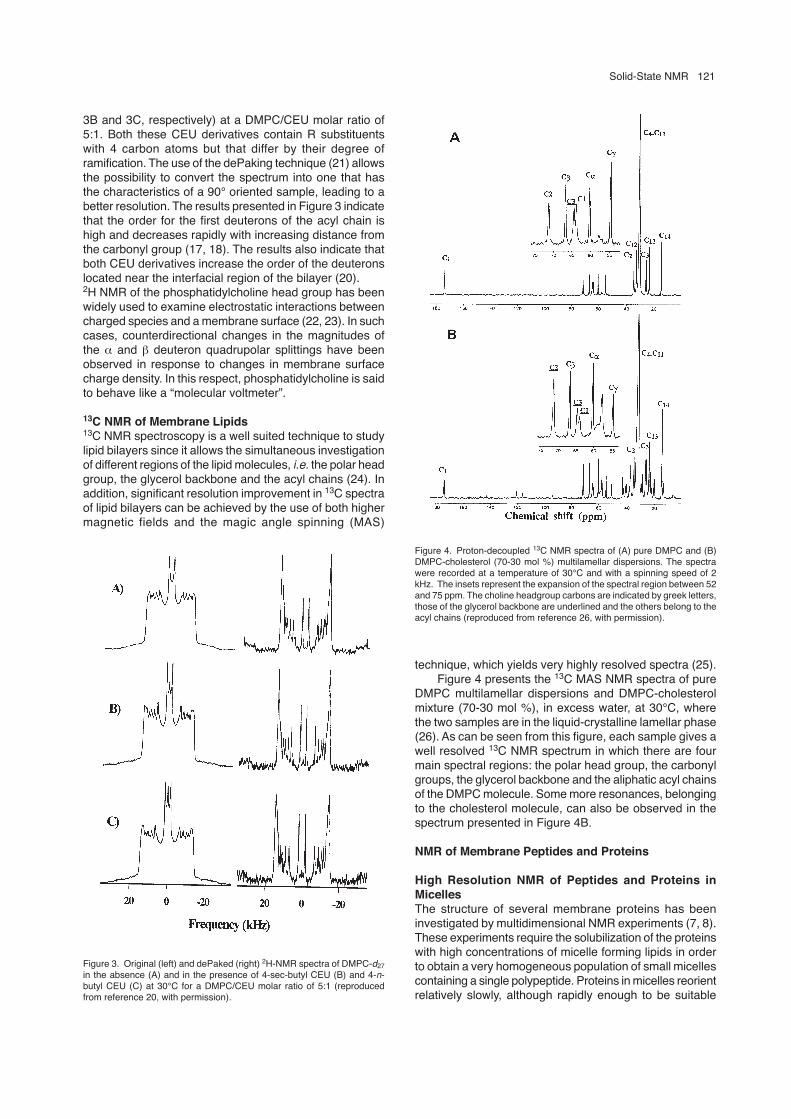
On one hand, the use of chain-perdeuterated lipids,deuterated either on one or the two acyl chains, allows thesimultaneous investigation of the structure and dynamicsof the plateau and the tail regions of the lipid chains. Figure3 shows an example of the use of deuterium NMR toinvestigate the effect of two anticancer drugs derived fromchloroethylurea (CEU) on the orientational order of lipidacyl chains (20). More specifically, the original and dePaked(deconvolved) 2H NMR spectra obtained at 30°C are shownin Figure 3 for pure DMPC deuterated on the sn-2 acylchain (DMPC-d27) (Figure 3A) and for DMPC-d27 in thepresence of 4-sec-butyl CEU and 4-n-butyl CEU (Figures
Figure 1. Temperature dependence of the 31P NMR spectra of pure DMPA(left) and the complex DMPA:cardiotoxin at a lipid-to-protein molar ratio of5:1 (right), (reproduced from reference 14, with permission).
Figure 2. 2D 31P NMR experimental spectra of pure dipalmitoyl-phosphatidylcholine (DPPC) multilamellar vesicles at 50°C for differentmixing times tm (reproduced from reference 16, with permission).
Solid-State NMR 121
3B and 3C, respectively) at a DMPC/CEU molar ratio of5:1. Both these CEU derivatives contain R substituentswith 4 carbon atoms but that differ by their degree oframification. The use of the dePaking technique (21) allowsthe possibility to convert the spectrum into one that hasthe characteristics of a 90° oriented sample, leading to abetter resolution. The results presented in Figure 3 indicatethat the order for the first deuterons of the acyl chain ishigh and decreases rapidly with increasing distance fromthe carbonyl group (17, 18). The results also indicate thatboth CEU derivatives increase the order of the deuteronslocated near the interfacial region of the bilayer (20).2H NMR of the phosphatidylcholine head group has beenwidely used to examine electrostatic interactions betweencharged species and a membrane surface (22, 23). In suchcases, counterdirectional changes in the magnitudes ofthe α and β deuteron quadrupolar splittings have beenobserved in response to changes in membrane surfacecharge density. In this respect, phosphatidylcholine is saidto behave like a “molecular voltmeter”.
13C NMR of Membrane Lipids13C NMR spectroscopy is a well suited technique to studylipid bilayers since it allows the simultaneous investigationof different regions of the lipid molecules, i.e. the polar headgroup, the glycerol backbone and the acyl chains (24). Inaddition, significant resolution improvement in 13C spectraof lipid bilayers can be achieved by the use of both highermagnetic fields and the magic angle spinning (MAS)
technique, which yields very highly resolved spectra (25).Figure 4 presents the 13C MAS NMR spectra of pure
DMPC multilamellar dispersions and DMPC-cholesterolmixture (70-30 mol %), in excess water, at 30°C, wherethe two samples are in the liquid-crystalline lamellar phase(26). As can be seen from this figure, each sample gives awell resolved 13C NMR spectrum in which there are fourmain spectral regions: the polar head group, the carbonylgroups, the glycerol backbone and the aliphatic acyl chainsof the DMPC molecule. Some more resonances, belongingto the cholesterol molecule, can also be observed in thespectrum presented in Figure 4B.
NMR of Membrane Peptides and Proteins
High Resolution NMR of Peptides and Proteins inMicellesThe structure of several membrane proteins has beeninvestigated by multidimensional NMR experiments (7, 8).These experiments require the solubilization of the proteinswith high concentrations of micelle forming lipids in orderto obtain a very homogeneous population of small micellescontaining a single polypeptide. Proteins in micelles reorientrelatively slowly, although rapidly enough to be suitable
Figure 3. Original (left) and dePaked (right) 2H-NMR spectra of DMPC-d27in the absence (A) and in the presence of 4-sec-butyl CEU (B) and 4-n-butyl CEU (C) at 30°C for a DMPC/CEU molar ratio of 5:1 (reproducedfrom reference 20, with permission).
Figure 4. Proton-decoupled 13C NMR spectra of (A) pure DMPC and (B)DMPC-cholesterol (70-30 mol %) multilamellar dispersions. The spectrawere recorded at a temperature of 30°C and with a spinning speed of 2kHz. The insets represent the expansion of the spectral region between 52and 75 ppm. The choline headgroup carbons are indicated by greek letters,those of the glycerol backbone are underlined and the others belong to theacyl chains (reproduced from reference 26, with permission).

122 Auger
for most heteronuclear multidimensional solution NMRexperiments in high-field spectrometers (8).
The steps involved in the determination of proteinstructures in micelles are the same as those used forglobular proteins in aqueous solutions and include theresolution and assignment of backbone and side-chainresonances based on through-bond and through-spaceinteractions observed in multidimensional NMR spectra.The structure of several membrane peptides and proteinshas been investigated using this approach, includingmagainin, the major coat protein of filamentousbacteriophages, peptides with sequences that correspondto the transmembrane domain of glycophorin A and peptidesequences from bacteriorhodopsin and phospholamban(for recent reviews, see references 7, 8).
High-Resolution MAS Proton Spectra of MembranePeptidesHigh-resolution 1H nuclear magnetic resonance (NMR)spectroscopy is a powerful method for protein structuredetermination in solution (1). However, this method hasnot been extensively applied to membrane protein systemssince the strong homogeneous dipole-dipole broadeningbetween neighboring hydrogen nuclei, which is averagedto zero by rapid isotropic reorientation of molecules insolution, is not completely averaged away in anisotropicnon-spinning membrane systems and leads to spectrallinewidths of 5 to 25 kHz (10 to 50 ppm depending on the
magnetic field strength) (27). However, the use of magicangle spinning rates of about 3 to 4 kHz are sufficient toobtain well resolved 1H spectra from multilamellardispersions of phospholipids in water, because of the rapidaxial diffusion of the phospholipid about its long axis.Although it is possible to obtain well-resolved lipid 1Hspectra from multilamellar dispersions, no identifiablepeptide signals can be observed at spinning speeds ofabout 3 to 4 kHz for small peptides, such as gramicidin A,incorporated in phospholipid bilayers. This is due to thepresence of additional intermediate time scale motionswhich broaden the peptide resonances. In this case, it isnecessary to spin the sample at a spinning speed fasterthan that required for phospholipid multilamellar dispersions(27).
One- and two-dimensional 1H NMR spectra ofgramicidin A incorporated in a DMPC membrane have beenobtained with high-speed MAS (27, 28). By rotating thesample at 13 kHz, it is possible to observe signals in the1H spectra between 6.0 and 9.0 ppm attributable to thearomatic protons of the tryptophan residues and the formylgroup proton of gramicidin A, as shown in Figure 5. It hasalso been shown that solvent suppression methods derivedfrom solution NMR, such as presaturation or jump-return,can be used to reduce the water resonance at relativelyhigh water content (29). In addition, regioselectiveexcitation of 1H peptide resonances promotes an efficient
Figure 5. MAS solid-state 1H spectra at 60°C of A) pure DMPC and B)DMPC/gramicidin A system initially prepared from trifluoroethanol. Thespinning speed was set to 13.000 0.002 kHz (reproduced from reference28, with permission).
Figure 6. Proton-decoupled 15N solid-state NMR spectra of PGLaincorporated into oriented POPE/POPG 3/1 phospholipid bilayers. ThePGLa concentrations in spectra B-E are 2.3 mol% and in spectrum F, 1.5mol%. Also shown are simulated 15N solid-state NMR spectra in thepresence of fast motional averaging (A) and of an amide site that is part ofan a-helix oriented parallel to the bilayer surface (G) (reproduced fromreference 31, with permission).

Solid-State NMR 123
Figure 7. 13C MAS spectra of dark-adapted bR collected at -80°C with asample spinning speed of 2.8 kHz: (a) [14-13C]retinal, [e-13C]Lys-bR, (b)unlabeled bR, and (c ) the difference spectrum obtained upon subtractionof (b) from (a). The signals at 40, 48 and 53 ppm are due to the [e-13C]Lyslabel and the signals in the 100 to 130 ppm range arise from the [14-13C]retinal marker resonances. Rotational sidebands are observed 35 ppmupfield and downfield of the centerbands (reproduced from reference 33,with permission).
suppression of lipid resonances, even in cases where theseare initially two orders of magnitude more intense (29).
NMR Spectra of Specifically Labeled MembraneProteinsSolid-state NMR is a uniquely suited technique to determinethe structure of specifically labeled membrane peptidesand proteins. Two complementary approaches are mostcommonly used, the first involving the study of orientedsamples in the magnetic field. In this case, the incompletemotional averaging allows the measurement of dipolarcouplings, chemical shift anisotropies and electricquadrupolar splittings, from which structural parameterssuch as torsional angles can be obtained. This techniquehas been applied to a variety of membrane peptides andproteins, including gramicidin A, bacteriophage proteinsand magainin (for recent reviews, see references 7, 8, 30).Figure 6 shows representative solid-state NMR spectra oforiented bilayers containing specifically 15N-labeled PGLapeptide, which is a member of the magainin family ofantibiotic peptides found in frog skin and its secretions (31).The spectra of the peptides labeled at the Ala10, Ala14,Val16 and Ala20 positions are characteristic of structuredpeptides and are indicative of an orientation of the individualN-H bond approximately perpendicular to the magnetic fielddirection.
The second approach uses the magic angle spinningtechnique to remove these tensor interactions, resulting inspectra which are only characterized by the isotropicchemical shifts and the J-couplings. This technique hasbeen widely used to investigate the structure of thechromophore and of the protein in bacteriorhodopsin andrhodopsin (for recent reviews, see references 6, 30, 32).Figure 7 shows the solid-state 13C NMR MAS spectra ofdark-adapted bacteriorhodopsin (bR) (33). The difference
spectrum shows signals due only to the [ε-13C]Lys and[14-13C]retinal labels. The signals at 110.5 ppm and 122ppm are assigned to the 14-13C of retinal of bR555 andbR568, the two components present in equilibrium in darkadapted bacteriorhodopsin. The remaining signals at 48and 55 ppm are assigned to the ε-13C label in Lys216.These spectra clearly indicate that single labeled sites canbe distinguished in magic angle spinning spectra ofmembrane proteins. The magic angle spinning technique,combined with isotopic labeling, has also been applied toother membrane proteins, including the photosyntheticreaction center and several enzymes and channel peptides(for recent reviews, see references 5, 6, 30).
Several high-resolution solid-state NMR methods havealso been developed in the last ten years to measure weakhomonuclear or heteronuclear dipolar couplings in solidsamples (for recent reviews, see references 5, 6, 30, 34-36). These methods use the magic angle spinningtechnique and include the rotational resonance and the rf-driven recoupling (RFDR) techniques for the measurementof homonuclear distances and techniques such as REDORand TEDOR for the measurement of heteronucleardistances. These techniques have been applied to a varietyof systems including the membrane proteins rhodopsin andbacteriorhodopsin, gramicidin A, glycophorin A and abacterial chemotaxis membrane receptor (5, 6, 30, 34-36).In addition, several methods have been developed recentlyfor the measurements of torsion angles from magic anglespinning spectra in solids. These methods directlyinvestigate molecular geometry by determining the relativeorientation of pairs of nuclear spin interactions (36).
Perspectives
The experiments described above for the measurementsof internuclear distances or torsional angles in membranepeptides and proteins have all been performed with spinpairs, either 13C-13C or 15N-13C. However, the synthesis ofspecifically labeled molecules is difficult and expensive incomparison with biosynthetic methods which yieldsuniformly 13C/15N labeled systems (36). Therefore, thedetermination of protein structure from completely labeledproteins would be an interesting alternative. However, themajor problem associated with a uniformly labeled samplein solid-state NMR is the multiple strong couplings whichprevent the observation of weaker couplings and whichresults in increased linewidths. Recently, a very carefulanalysis has been made on the impact of linewidths onmultidimensional solid-state NMR spectra of uniformlylabeled peptides and proteins (37). The main conclusionof this study is that the length of a peptide for which acomplete, unique assignment of 13C and 15N backboneresonances in solid-state NMR spectra is feasible isstrongly limited by spectral resolution. For peptides of 20to 30 amino acid residues, resolutions better than 0.5-1.5ppm or 0.5-1.0 ppm, respectively, are required (37). Severalavenues have recently been suggested to increase boththe spectral resolution and sensitivity (36). These includethe use of higher field spectrometers, the development ofnew labeling strategies to improve the resolution and theuse of higher dimensional experiments. These

124 Auger
developments should increase the number of membraneproteins for which the structure can be investigated by solid-state NMR spectroscopy.
References
1. Wüthrich, K. 1986. NMR of Proteins and Nucleic Acids. Wiley, NewWork.
2. Evans, J.S. 1995. Biomolecular NMR. Oxford University Press, Oxford.3. Griffin, R.G. 1981. Solid state nuclear magnetic resonance of lipid
bilayers. Methods Enzymol. 72: 108-174.4. Bloom, M. and Bayerl, T.M. 1995. Membranes studied using neutron
scattering and NMR. Can. J. Phys. 73: 687-696.5. Watts, A., Ulrich, A.S. and Middleton, D.A. 1995. Membrane protein
structure: the contribution and potential of novel solid state NMRapproaches. Mol. Membr. Biol. 12: 233-246.
6. Smith, S.O., Aschheim, K. and Groesbeek, M. 1996. Magic anglespinning NMR spectroscopy of membrane proteins. Q. Rev. Biophys.29: 395-449.
7. Opella, S.J. 1997. NMR and membrane proteins. Nature Struct. Biol.4: 845-848.
8. Opella, S.J. and Marassi, F.M. 1996. Membrane proteins. In:Encyclopedia of Nuclear Magnetic Resonance. D.M. Grant and R.K.Harris, eds. John Wiley, Toronto, Canada. p. 2994-3003.
9. Sanders, C.R. and Schwonek, J.P. 1992. Characterization ofmagnetically orientable bilayers in mixtures ofdihexanoylophosphatidylcholine and dimyristoylphosphatidyl-cholineby solid-state NMR. Biochem. 31: 8898-8909.
10. Sanders, C.R. and Prosser, R.S. 1998. Bicelles: a model membranesystem for all seasons? Structure. 6: 1227-1234.
11. Yeagle, P.L. 1996. Membranes: Phosphorus-31 NMR. In: Encyclopediaof Nuclear Magnetic Resonance. D.M. Grant and R.K. Harris, eds.John Wiley, Toronto, Canada. p. 3015-3022.
12. Smith, I.C.P. and Ekiel, I.H. 1984. Phosphorus-31 NMR ofphospholipids in membranes. In: Phosphorus-31 NMR: Principles andapplications. D. Gorenstein, ed. Academic Press Inc. London, England.p. 447-475.
13. Seelig, J. 1978. 31P nuclear magnetic resonance and the head groupstructure of phospholipids in membranes. Biochim. Biophys. Acta.515: 105-140.
14. Picard, F., Pézolet, M., Bougis, P.E. and Auger, M. 1996. Model ofinteraction between a cardiotoxin and dimyristoylphosphatidic acidbilayers determined by solid-state 31P NMR spectroscopy. Biophys.J. 70: 1737-1744.
15. Fenske, D.B. and Jarrell, H.C. 1991. Phosphorus-31 two-dimensionalsolid-state NMR: Application to model membrane and biologicalsystems. Biophys. J. 59: 55-69.
16. Picard, F., Paquet, M.-J., Dufourc, E.J. and Auger, M. 1998.Measurement of the lateral diffusion of dipalmitoylphosphatidylcholineadsorbed on silica beads in the absence and presence of melittin: A31P 2D exchange solid-state NMR study. Biophys. J. 74: 857-868.
17. Davis, J.H. 1983. The description of membrane lipid conformation,order and dynamics by 2H-NMR. Biochim. Biophys. Acta. 737: 117-171.
18. Seelig, J. 1977. Deuterium magnetic resonance: theory andapplications to lipid membranes. Q. Rev. Biophys. 10: 353-418.
19. Davis, J.H. 1996. Membranes: Deuterium NMR. In: Encyclopedia ofNuclear Magnetic Resonance. D.M. Grant and R.K. Harris, eds. JohnWiley, Toronto, Canada. p. 3008-3015.
20. Saint-Laurent, A., Boudreau, N., C.-Gaudreault, R., Poyet, P. andAuger, M. 1998. Interaction between lipid bilayers and a new class ofantineoplastic agents derived from arylchloroethylurea: A 2H solid-state NMR study. Biochem. Cell Biol. 76: 465-471.
21. Bloom, M., Davis, J.H. and MacKay, A.L. 1981. Direct determinationof the oriented sample NMR spectrum from the powder spectrum forsystems with local axial symmetry. Chem. Phys. Lett. 80: 198-202.
22. Seelig, J., Macdonald, P.M. and Scherer, P.G. 1987. Phospholipidhead groups as sensors of electric charge in membranes. Biochem.26: 7535-7541.
23. Macdonald, P.M. 1997. Deuterium NMR and the topography of surfaceelectrostatic charge. Acc. Chem. Res. 30: 196-203.
24. Separovic, F. and Cornell, B.A.. 1996. Membranes: Carbone-13 NMR.In: Encyclopedia of Nuclear Magnetic Resonance. D.M. Grant andR.K. Harris, eds. John Wiley, Toronto, Canada. p. 3003-3008.
25. Forbes, J., Bowers, J., Shan, X., Moran, L. and Oldfield, E. 1988.Some new developments in solid-state nuclear magnetic resonancespectroscopic studies of lipids and biological membranes, includingthe effects of cholesterol in model and natural systems. J. Chem.
Soc., Faraday Trans. 1. 84: 3821-3849.26. Le Guernevé, C. and Auger, M. 1995. New approach to study fast
and slow motions in lipid bilayers: Application to DMPC-cholesterolinteractions. Biophys. J. 68: 1952-1959.
27. Davis, J.H., Auger, M. and Hodges, R.S. 1995. High resolution 1HNMR of a transmembrane peptide. Biophys. J. 69: 1917-1932.
28. Bouchard, M., Davis, J.H. and Auger, M. 1995. High-speed MAS solid-state 1H NMR study of the conformation of gramicidin A in lipid bilayers.Biophys. J. 69: 1933-1938.
29. Le Guernevé, C. and Seigneuret, M. 1996. High-resolution mono-and multidimensional magic angle spinning 1H nuclear magneticresonance of membrane peptides in nondeuterated lipid membranesand H2O. Biophys. J. 71: 2633-2644.
30. Davis, J.H. and Auger, M. 1999. Static and magic angle spinning NMRof membrane peptides and proteins. Prog. NMR Spectrosc. 35: 1-84.
31. Bechinger, B., Zasloff, M. and Opella, S.J. 1998. Structure anddynamics of the antibiotic peptide PGLa in membranes by solutionand solid-state nuclear magnetic resonance spectroscopy. Biophys.J. 74: 981-987.
32. Engelhard, M. and Bechinger, B. 1996. Application of NMR-spectroscopy to retinal proteins. Israel J. Chem. 35: 273-288.
33. Farrar, M.R., Lakshmi, K.V., Smith, S.O., Brown, R.S., Raap, J.,Lugtenburg, J., Griffin, R.G. and Herzfeld, J. 1993. Solid state NMRstudy of [e-13C]Lys-bacteriorhodopsin schiff base photoisomerization.Biophys. J. 65: 310-315.
34. Auger, M. 1995. Solid state NMR study of protein structure: Methodsbased on the measurement of internuclear distances. J. Chim. Phys.92: 1751-1760.
35. Griffiths, J.M. and Griffin, R.G. 1993. Nuclear magnetic resonancemethods for measuring dipolar couplings in rotating solids. Anal. Chim.Acta. 283: 1081-1101.
36. Griffin, R.G. 1998. Dipolar recoupling in MAS spectra of biologicalsolids. Nature Struct. Biology. 5: 508-512.
37. Tycko, R. 1996. Prospects for resonance assignments inmultidimensional solid-state NMR spectra of uniformly labeledproteins. J. Biomol. NMR. 8: 239-251.

• MALDI-TOF Mass Spectrometry in Microbiology
Edited by: M Kostrzewa, S Schubert (2016) www.caister.com/malditof
• Aspergillus and Penicillium in the Post-genomic Era
Edited by: RP Vries, IB Gelber, MR Andersen (2016) www.caister.com/aspergillus2
• The Bacteriocins: Current Knowledge and Future Prospects
Edited by: RL Dorit, SM Roy, MA Riley (2016) www.caister.com/bacteriocins
• Omics in Plant Disease Resistance
Edited by: V Bhadauria (2016) www.caister.com/opdr
• Acidophiles: Life in Extremely Acidic Environments
Edited by: R Quatrini, DB Johnson (2016) www.caister.com/acidophiles
• Climate Change and Microbial Ecology: Current Research and Future Trends
Edited by: J Marxsen (2016) www.caister.com/climate
• Biofilms in Bioremediation: Current Research and Emerging Technologies
Edited by: G Lear (2016) www.caister.com/biorem
• Microalgae: Current Research and Applications
Edited by: MN Tsaloglou (2016) www.caister.com/microalgae
• Gas Plasma Sterilization in Microbiology: Theory, Applications, Pitfalls and New Perspectives
Edited by: H Shintani, A Sakudo (2016) www.caister.com/gasplasma
• Virus Evolution: Current Research and Future Directions
Edited by: SC Weaver, M Denison, M Roossinck, et al. (2016) www.caister.com/virusevol
• Arboviruses: Molecular Biology, Evolution and Control
Edited by: N Vasilakis, DJ Gubler (2016) www.caister.com/arbo
• Shigella: Molecular and Cellular Biology
Edited by: WD Picking, WL Picking (2016) www.caister.com/shigella
• Aquatic Biofilms: Ecology, Water Quality and Wastewater Treatment
Edited by: AM Romaní, H Guasch, MD Balaguer (2016) www.caister.com/aquaticbiofilms
• Alphaviruses: Current Biology
Edited by: S Mahalingam, L Herrero, B Herring (2016) www.caister.com/alpha
• Thermophilic Microorganisms
Edited by: F Li (2015) www.caister.com/thermophile
• Flow Cytometry in Microbiology: Technology and Applications
Edited by: MG Wilkinson (2015) www.caister.com/flow
• Probiotics and Prebiotics: Current Research and Future Trends
Edited by: K Venema, AP Carmo (2015) www.caister.com/probiotics
• Epigenetics: Current Research and Emerging Trends
Edited by: BP Chadwick (2015) www.caister.com/epigenetics2015
• Corynebacterium glutamicum: From Systems Biology to Biotechnological Applications
Edited by: A Burkovski (2015) www.caister.com/cory2
• Advanced Vaccine Research Methods for the Decade of Vaccines
Edited by: F Bagnoli, R Rappuoli (2015) www.caister.com/vaccines
• Antifungals: From Genomics to Resistance and the Development of Novel Agents
Edited by: AT Coste, P Vandeputte (2015) www.caister.com/antifungals
• Bacteria-Plant Interactions: Advanced Research and Future Trends
Edited by: J Murillo, BA Vinatzer, RW Jackson, et al. (2015) www.caister.com/bacteria-plant
• Aeromonas
Edited by: J Graf (2015) www.caister.com/aeromonas
• Antibiotics: Current Innovations and Future Trends
Edited by: S Sánchez, AL Demain (2015) www.caister.com/antibiotics
• Leishmania: Current Biology and Control
Edited by: S Adak, R Datta (2015) www.caister.com/leish2
• Acanthamoeba: Biology and Pathogenesis (2nd edition)
Author: NA Khan (2015) www.caister.com/acanthamoeba2
• Microarrays: Current Technology, Innovations and Applications
Edited by: Z He (2014) www.caister.com/microarrays2
• Metagenomics of the Microbial Nitrogen Cycle: Theory, Methods and Applications
Edited by: D Marco (2014) www.caister.com/n2
Caister Academic Press is a leading academic publisher of advanced texts in microbiology, molecular biology and medical research. Full details of all our publications at caister.com
Further Reading
Order from caister.com/order


![[14] NMR Experiments on Aligned Samples of Membrane Proteinsweb.mit.edu/fbml/winterschool2008/lecturenotes/pdf references Opella... · [14] nmr on aligned samples of membrane proteins](https://img.pdfslide.net/doc/110x75/5e6896d0ec57f969d73fe211/14-nmr-experiments-on-aligned-samples-of-membrane-references-opella-14.jpg)