Embed Size (px)
Citation preview
ANTIMICROBIAL AGENTS AND CHEMOTHERAPY, Mar. 1972, p. 221-234 Vol. 1, No. 3Copyright @ 1972 American Society for Microbiology Priited in U.S.A.
Biological Properties of Three3-Heterocyclic-ThiomethylCephalosporin AntibioticsWARREN E. WICK AND DAVID A. PRESTON
Thte Lilly Research Laboratories, Eli Lilly & Co., Indianapolis, lildiania 46206
Received for publication 16 November 1971
Three new cephalosporin antibiotics, prepared by substitution of heterocyclicgroups on 7-aminocephalosporanic acid, possess certain desirable chemical or bio-logical properties. All three compounds are active in vitro against a variety of gram-positive and gram-negative bacteria. Minimal inhibitory concentrations (MIC) ofthese bactericidal antibiotics were not significantly affected by changes in pH orNaCI content of nutrient broth, or by the use of different inoculum sizes. However,agar-dilution MIC values were generally two- to fourfold lower than the MICvalues in comparable broth-dilution tests. Stability to cephalosporinase by two of thecompounds extended their antibacterial spectra over cephalothin and cephaloridineto include strains of Enterobacter sp. and indole-positive Proteus sp. Binding toserum proteins of the new cephalosporins was intermediate between cephalothinand cephaloridine. Excellent concentrations of the antibiotics were attained inmouse blood, after subcutaneous administration of 20 mg per kg. In vitro biologicalcharacteristics of the antibiotics were verified by successful therapy of experimentalmouse infections. Regression lines were calculated to show the correlation of agar-dilution MIC values with zones of inhibition by the disc testing procedure. Becauseeach of the three new cephalosporins has certain advantageous properties overcephalothin and cephaloridine, additional toxicological and pharmacological datashould be obtained for all three compounds.
The clinical efficacy of parenterally adminis-tered cephalosporin antibiotics, cephalothin andcephaloridine, has been well established. How-ever, certain problems occur during therapy withthese antibiotics. For example, because of pain onintramuscular injection, cephalothin is usually ad-ministered intravenously. Cephaloridine possessesexcellent antibacterial activity and has low tox-icity, but is associated, rarely, with an adverse ef-fect on renal function. New cephalosporin anti-biotics, for which laboratory data have providedevidence for possible advantages over cephalothinor cephaloridine, may prove to be clinically usefulcompounds. Three new cephalosporin antibiotics,prepared from 7-aminocephalosporanic acid bysubstitution of heterocyclic groups (heterocycliccephalosporins), do possess certain chemical andbiological properties that make them candidatesfor clinical evaluation. One of these antibiotics,cefazolin, has been investigated extensively inJapan (10, 12-15, 19). This communication sum-marizes the data from a laboratory study withcefazolin and two chemically related cephalo-
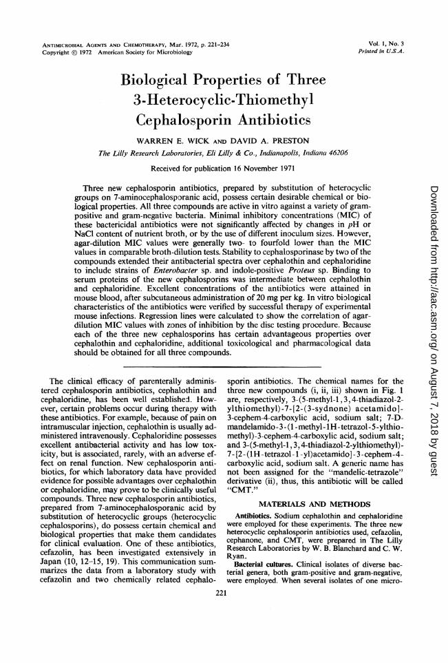
sporin antibiotics. The chemical names for thethree new compounds (i, ii, iii) shown in Fig. 1are, respectively, 3-(5-methyl-1, 3, 4-thiadiazol-2-ylthiomethyl)-7-[2-(3-sydnone) acetamido]-3-cephem-4-carboxylic acid, sodium salt; 7-D-mandelamido - 3 - (1 -methyl -1H -tetrazol - 5 -ylthio -methyl)-3-cephem-4-carboxylic acid, sodium salt;and 3-(5-methyl-1, 3, 4-thiadiazol-2-ylthiomethyl)-7- [2- (1H -tetrazol- 1 -yl)acetamido] -3 -cephem-4-carboxylic acid, sodium salt. A generic name hasnot been assigned for the "mandelic-tetrazole"derivative (ii), thus, this antibiotic will be called"CMT.")
MATERIALS AND METHODSAntibiotics. Sodium cephalothin and cephaloridine
were employed for these experiments. The three newheterocyclic cephalosporin antibiotics used, cefazolin,cephanone, and CMT, were prepared in The LillyResearch Laboratories by W. B. Blanchard and C. W.Ryan.
Bacterial cultures. Clinical isolates of diverse bac-terial genera, both gram-positive and gram-negative,were employed. When several isolates of one micro-
221
on August 7, 2018 by guest
http://aac.asm.org/
Dow
nloaded from
ANTIMICROB. AG. CHEMOTHER.
i - Cephanone (CSY)
CH-C-NHOOH tNN CH2-S--
0
COO-Na+ii - "CMT"
N=N °N-CH2-C-NH..11( N
N---/ ~~~~CH2-S{
COO-Na +
iii - Cefazolin (CEZ)
0LCH2-C-NH U cor N CH2--
COO-Na +
iv - Cephalothin (CET)'0
I CH2_C_NH Ys ~~~~~~~~+N><~~CH -N
Coo-
v - Cephaloridine (CER)
FIG. 1. Chemical structures of cephanon,cefazolin, cephalothin, and cephaloridine.
bial genus were used, each culture was siof a different strain by phage typing, serochemical reactions, or antibiotic susceptibil
Disc susceptibility testing. Strains of batested for susceptibility to the antibiotistandardized disc technique described by thoDrug Administration (5). This procedure ication of the Bauer-Kirby (or Kirby-Baueceptibility testing method (2).MIC detedmnations. Broth-dilution or as
procedures were used to determine antibioiinhibitory concentrations (MIC) for allbacteria. For aerobic gram-positive rodscocci, pseudomonads, and Enterobacter,agar-dilution method of the Internationaltive Study (ICS) as described by Ericsson;(6) was used. Mueller-Hinton agar (BB]medium employed, and the plates werewith a device similar to the Steers' replicat
Modifications of broth- or agar-diluticwere made to determine MIC values foiaerobic or anaerobic bacteria. Trypticase(BBL) was used for Corynebacteriwn sp.,cus sp., and Diplococcus pnewnoniae. Whe
streptococci or pneumococci were tested, 5% defibri-i-N nated rabbit blood was added to the broth. Final
ll inocula of 106 bacteria per ml of broth were used in`-Is CH3 these tests. For Neisseria sp. and Haemophilus sp., the
ICS agar-dilution method was modified by usingTrypticase Soy Agar with 5% rabbit blood and 1%IsoVitaleX (BBL). The agar was chocolatized forHaemophilus sp. Inoculated plates were incubated ina 10% CO2 atmosphere. For Bacteroides fragilis,
N-N Fusobacterium (Sphaerophorus) necrophorus, and the//lit clostridia, fluid thioglycolate medium was utilized,
and the tubes were inoculated with one drop fromovernight broth cultures.
CH3 MIC values for all procedures described above weredetermined after overnight incubation at 37 C.
Correlation of MIC values with disc test results.I-N Zone diameters obtained with discs containing 30 AglI C
of antibiotic were plotted against MIC values. TheSsCH3 experimentally determined points fell along a regres-
sion line, which was calculated by the method ofleast squares.
Effects of variations of pH, inoculum size, or NaCIcontent of media on antibacterial activity. The broth-dilution susceptibility test was used to assess the effecton activities of the antibiotics caused by variations in
C-CH3 media pH, size of inoculum used, or NaCl content ofthe broth. Nutrient broth (Difco) was the mediumemployed. Variations in medium pH were accom-plished by preparing the media with phosphate buffersolutions (Harleco, Na+, K+ phosphate salts) insteadof water. To determine the effects of NaCl on theMIC, various concentrations of NaCl in broth wereutilized. The inoculum size used for the pH or NaClstudies was 104 bacteria per ml of broth. Inocula sizesof 108, 104, 106, and 106, in unmodified broth, wereused to demonstrate any inoculum effects on MICvalues.
e, "CMT," Bactericidal activity. Two procedures were used todetect bactericidal activity of the antibiotics examined.In one of these methods, the surviving bacteria in all
into be clear tubes of the broth-dilution MIC series wereiowng to counted. A 1-ml sample from a 10-fold dilution ofooy, bictra each tube was placed in 20 ml of melted Trypticaseity spectra. Soy Agar and poured into a 100-mm petri dish. Afterictera were incubation for 48 hr at 37 C, the colonies were counted.cs byd thd Reduction of the original inoculum by 99.9% or more
ie oodand was considered to define bactericidal activity in theis a modifi- corresponding tube of the MIC test.:r) disc sus- A second method used to estimate bactericidal ac-
gar-dilut'ion tivity originated from the cellophane transfer tech-tic miniml nique of Chabbert (3). However, instead of cello-strains of phane, 25-mm HA (0.45 ,um) black membrane ifitersstraphylo (Millipore Corp., Bedford, Mass.) were used. Anti-
;,cstaph Yt biotic was incorporated in Mueller-Hinton agar to giveaceae, the final concentrations in a log2 dilution series. FilterCollabora- discs were placed on the surface of the agar, and eachand Sherrs filter was then seeded by pipetting one drop from anL~) was theinoculated appropriate suspension of a bacterial culture. A glassiocu at rod was used to spread the bacterial suspensions over
in methods the surfaces of the discs. The plates were then incu-r fastidious bated at 37 C. To determine the number of viable cellsSoy Broth applied to each disc, an agar-plate count was made ofStreptococ- the organisms contained in a drop delivered by then strains of pipette.
222 WICK AND PRESTON
on August 7, 2018 by guest
http://aac.asm.org/
Dow
nloaded from
3-HETEROCYCLIC-THIOMETHYL CEPHALOSPORINS
After contacts for various periods of time at 37 C,the inoculated discs were transferred to plates con-taining antibiotic-free agar. To insure that no residualantibiotic remained, discs were transferred via twointermediate antibiotic-free agar plates. All plates towhich discs were transferred were incubated at 37 Cduring the entire transfer time.An MIC was determined from the filters that re-
mained on the original antibiotic plates for 24 hr.The MIC was the lowest level of antibiotic that inhib-ited visual growth of the culture. Discs with no growthwere then transferred, and incubation was continued.After sufficient incubation for visible colony forma-tion, the colonies that grew on the surface of eachfilter were counted. Each colony represented a surviv-ing organism, and the number of survivors was com-pared with the number of bacteria used to inoculatethe membrane filter. From these data, bactericidalactivity of the antibiotics was determined; i.e., theminimal bactericidal concentration (MBC) was ob-tained from the discs that were transferred to theantibiotic-free plates at 24 hr, and was the concentra-tion of antibiotic that killed 599.9% of the inoculatedbacteria. This MBC was compared with the MIC thatwas determined from the same series of discs at 24 hr.In addition to the MBC, the rate of bactericidal activ-ity could be estimated by counting colonies on discsthat were transferred to antibiotic-free agar at incu-bation times of less than 24 hr.
Microbiological assays. Disc-plate assays with Sar-cina lutea strain PCI-1001-FDA or Bacillus subtilisstrain ATCC 6633 were used. The greatest sensitivitywas obtained by using the B. subtilis assay with Anti-biotic Medium 5 (Difco) at pH 8.0 for cephalothin,the B. subtilis assay with the agar adjusted to pH 6.0for the heterocycic compounds, and the S. lutea assaywith agar at pH 8.0 for cephaloridine.
Concentration of antibiotic in mouse blood or urine.The disc-plate assays described above were utilized todetermine antibiotic concentration in mouse blood orurine. All antibiotics were administered to mice sub-cutaneously at equal doses of 20 mg per kg of bodyweight. The blood was collected from the orbital sinusin heparinized hematocrit tubes, which were allowedto fill by capillary action (23). Paper discs (6.35 mm,Schleicher & Schuell Co., Inc.) were saturated withblood and immediately placed on inoculated assayplates. Urine specimens were diluted in saline and as-sayed as soon as they were collected.
Chemical stability studies. The stability of thecephalosporin antibiotics was studied by incubatingsolutions of the antibiotics at 4, 25, or 37 C. Sampleswere withdrawn at intervals and immediately frozenwith alcohol and dry ice. After all samples were col-lected, they were assayed by the assay procedures de-scribed above.
Metabolism of antibiotics. To assess the extent ofmetabolic degradation of the antibiotics, the paperchromatographic method of Hoehn and Pugh (9) wasemployed. Urine was collected from mice at 2 hr aftersubcutaneous administration of 20 mg of antibioticper kg. The specimens were kept frozen until proc-essed. Developed Whatman no. 1 sheets were treatedfor bioautography on B. subtilis plates.
Stability studies with bacterial enzymes. 3-Lactamaseenzymes (penicillinase and cephalosporinase) wereused to evaluate stability of the antibiotics to enzy-matic degradation. The enzymes were derived fromseveral bacterial species, including B. cereus, Staphylo-coccus aureus, Enterobacter sp., Proteus sp., Serratiasp., and Pseudomonas sp. Penicillinase (Riker) was theonly highly purified enzyme used; enzymes from otherorganisms were only partially purified preparations.Activity of the enzymes against each of the fivecephalosporins was measured by a previously de-scribed assay method (21, 24).
Effect of human sera on antibacterial activity. Thepercentage of antibacterial activity bound by humanserum was estimated by comparing serum and bufferstandard curves from assays described above.
Therapy of experimental infections. The in vivoefficacy of the antibiotics was studied by therapy ofexperimental infections in mice. Groups of eight whitemice (11 to 13 g) were treated subcutaneously atvarious times after intraperitoneal bacterial challenge.Antibiotics were usually administered at 1 and 5 hrpost-infection; however, in some experiments, otherdosage regimens were employed. Deaths and surviv-ors were recorded for a period of seven days. Theantibiotic dose effective in curing 50% of the infectedmice (EDwo) was calculated by the method of Reedand Muench (16).
RESULTS AND DISCUSSIONThe in vitro antibacterial activities of five
cephalosporin antibiotics against representativegram-positive bacteria and Neisseria sp. areshown in Tables 1 and 2. All five of the com-pounds (Fig. 1) were active against strains ofStaphylococcus sp., Streptococcus sp. (Viridansand A groups), Diplococcus pneumoniae, Clos-tridium sp., Corynebacterium sp., and Neisseriasp. (Table 1). In some instances, the three hetero-cyclic derivatives were slightly less active thancephalothin or cephaloridine.
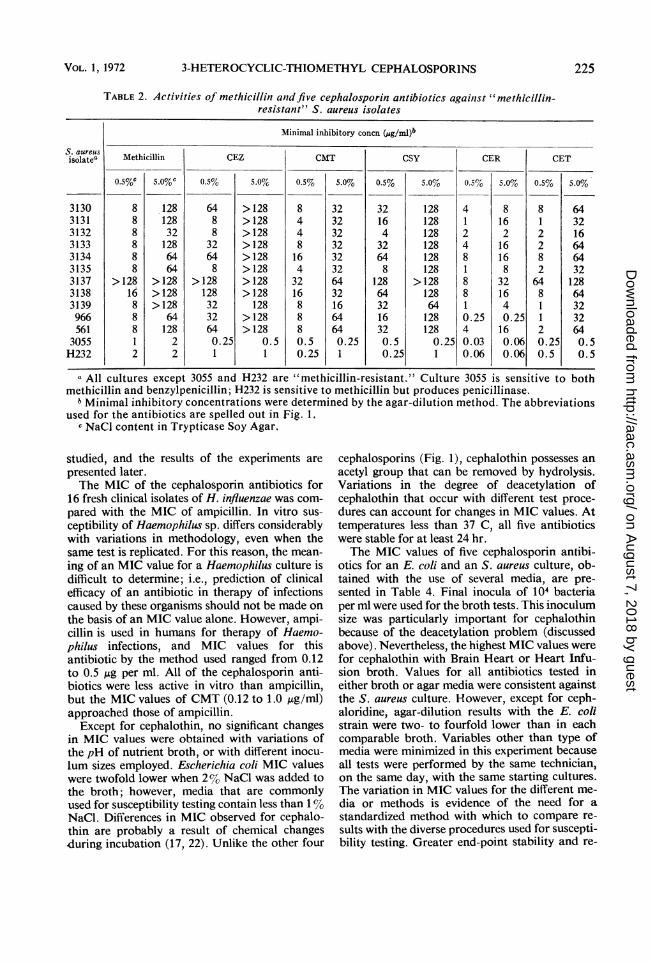
Barber (1) and Chabbert (4) have shown thatthe addition of 5% NaCl to Trypticase Soy mediaprovides conditions permitting the resistant cellswithin the heterogeneous population of methicil-lin-resistant S. aureus cultures to express theirresistance. When this procedure was used, threeof the five cephalosporin antibiotics shown weremore active than methicillin itself against thistype of S. aureus culture (Table 2). Nevertheless,a certain amount of "multiple resistance" betweenmethicillin and all of these cephalosporins wasevident.
Against gram-negative bacteria, CMT was themost active of the heterocyclic cephalosporins,followed in order by cephanone and cefazolin(Table 3). Of particular interest was the inhibi-tion of certain cultures of Enterobacter sp. andProteus sp. by CMT and cephanone. Inhibition ofthese organisms is very liely a result of increasedstability to cephalosporinase. This possibility was
223VOL. 1, 1972
on August 7, 2018 by guest
http://aac.asm.org/
Dow
nloaded from
ANTIMICROB. AG. CHEMOTHER.
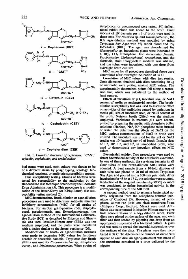
TABLE 1. Susceptibility of representative gram-positive bacteria anld Neisseria sp. to fivecephalosporin antibiotics
Minimal inhibitory concn (pg/ml)aOrganism Special identity of culture
CEZ CMT CSY CER CET
Clostridium perfringensCP23CP24CP25
C. tetani CT 1Corynebacterium
diphtheriaeCD 1CD 2
Diplococcus pneumontiaeDP 1DP 2DP14
Staphylococcus aureus(benzylpenicillin-sus-ceptible)V92
V104
SS7S. aulreuSb (penicillinase-
producing, andmethicillin-suscep-tible)H43V57H356
S. epidermidis3064
3078
3079
Streptococcus pyogenies(group A)C2031238510389
Streptococcuts sp.(Viridans group)
99439961K7
Streptococcus sp. (groupD)
99019960
Neisseria goniorrhoeae5GH
N. meningitidis OS
Harvard H-12AW
mitisgravis
Type IType IIType XIV
Phage: 47, 53, 59, 73,75
29, 52, 52A, 6, 47,54, 75, 80, 42B
77, VA4, (29, 53)
42B, 8142B, 52, 80, 8152A, 79
Benzylpenicillin-re-sistant
Benzylpenicillin-sus-ceptible
Benzylpenicillin-sus-ceptible
ATCC cultureATCC culture
0.060.120.120.06
2.02.0
0.120.120.25
0.5
0.25
0.25
0.50.50.5
0.5
0.25
0.25
0.120.120.12
0.51.00.5
32320.25
0.25
<0.030.060.5
<0.03
1.01.0
0.250.120.25
1.0
0.25
0.25
1 .00.50.5
0.5
0.25
0.25
0.030.060.06
0.50.51.0
32320.125
0.125
0.120.120.12
<0.03
1.01.0
0.120.120.25
1.0
0.25
0.25
1.01.00.5
0.5
0.25
0.25
0.030.060.06
0.50.50.25
16160.06
0.03
0.50.50.5
<0.03
0.060.06
0.060.030.06
0.06
<0.03
<0.03
0.120.120.06
0.06
<0.03
<0.03
0.0080.0080.008
0.060.030.25
16164.0
4.0
0.250.50.50.06
1.01.0
0.120.120.25
0.25
0.25
0.25
0.50.50.5
0.5
0.12
0.12
0.060.120.06
0.50.50.5
32161.0
0.5
a See Materials and Methods for procedures used to determine minimal inhibitory concentrations. Theabbreviations used for the antibiotics are spelled out in Fig. 1.
I Data for" methicillin-resistant" S. aulrelus are given in Table 2.
224 WICK AND PRESTON
on August 7, 2018 by guest
http://aac.asm.org/
Dow
nloaded from
3-HETEROCYCLIC-THIOMETHYL CEPHALOSPORINS
TABLE 2. Activities of methicillin and five cephalosporin antibiotics agailnst "methicillin-resistant" S. aureus isolates
S. aureiisisolatea
313031313132313331343135313731383139966561
3055H232
Minimal inhibitory concn (pg/il)b
Methicillin
o.s%c
888888
>1281688812
S.O%c
128128321286464
>128>128>128
6412822
CEZ
0.5%
6488
32648
>1281283232640.251
5.0%0
>128>128>128>128>128>128>128>128
128>128>128
0.51
CMT
0.5%
844816432168880.50.25
5.0%
32323232323264321664640.251
CSY
0.5%
3216432648
128643216320.50.25
5.0%
128128128128128128
>128128641281280.251
CER
0.5% 5.0%
4124818810.2540.030.06
816216168
321640.25160.060.06
CET
0.5% 5.0%
812282
6481120.250.5
643216646432128643232640.50.5
a All cultures except 3055 and H232 are "methicillin-resistant." Culture 3055 is sensitive to bothmethicillin and benzylpenicillin; H232 is sensitive to methicillin but produces penicillinase.bMinimal inhibitory concentrations were determined by the agar-dilution method. The abbreviations
used for the antibiotics are spelled out in Fig. 1.c NaCl content in Trypticase Soy Agar.
studied, and the results of the experiments arepresented later.The MIC of the cephalosporin antibiotics for
16 fresh clinical isolates of H. influenzae was com-pared with the MIC of ampicillin. In vitro sus-ceptibility of Haemophilus sp. differs considerablywith variations in methodology, even when thesame test is replicated. For this reason, the mean-ing of an MIC value for a Haemophilus culture isdifficult to determine; i.e., prediction of clinicalefficacy of an antibiotic in therapy of infectionscaused by these organisms should not be made onthe basis of an MIC value alone. However, ampi-cillin is used in humans for therapy of Haemo-philus infections, and MIC values for thisantibiotic by the method used ranged from 0.12to 0.5 ,ug per ml. All of the cephalosporin anti-biotics were less active in vitro than ampicillin,but the MIC values of CMT (0.12 to 1.0 ug/lml)approached those of ampicillin.
Except for cephalothin, no significant changesin MIC values were obtained with variations ofthe pH of nutrient broth, or with different inocu-lum sizes employed. Escherichia coli MIC valueswere twofold lower when 2% NaCl was added tothe broth; however, media that are commonlyused for susceptibility testing contain less than 1%NaCl. Differences in MIC observed for cephalo-thin are probably a result of chemical changesduring incubation (17, 22). Unlike the other four
cephalosporins (Fig. 1), cephalothin possesses anacetyl group that can be removed by hydrolysis.Variations in the degree of deacetylation ofcephalothin that occur with different test proce-dures can account for changes in MIC values. Attemperatures less than 37 C, all five antibioticswere stable for at least 24 hr.The MIC values of five cephalosporin antibi-
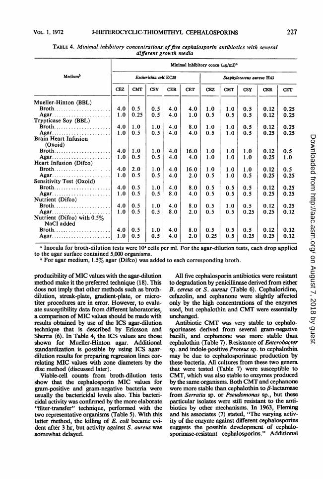
otics for an E. coli and an S. aureus culture, ob-tained with the use of several media, are pre-sented in Table 4. Final inocula of 104 bacteriaper mnl were used for the broth tests. This inoculumsize was particularly important for cephalothinbecause of the deacetylation problem (discussedabove). Nevertheless, the highest MIC values werefor cephalothin with Brain Heart or Heart Infu-sion broth. Values for all antibiotics tested ineither broth or agar media were consistent againstthe S. aureus culture. However, except for ceph-aloridine, agar-dilution results with the E. colistrain were two- to fourfold lower than in eachcomparable broth. Variables other than type ofmedia were minimized in this experiment becauseall tests were performed by the same technician,on the same day, with the same starting cultures.The variation in MIC values for the different me-dia or methods is evidence of the need for astandardized method with which to compare re-sults with the diverse procedures used for suscepti-bility testing. Greater end-point stability and re-
VOL. 1, 1972 225
on August 7, 2018 by guest
http://aac.asm.org/
Dow
nloaded from
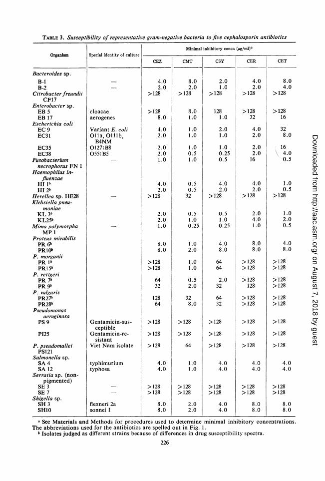
TABLE 3. Susceptibility of representative gram-negative bacteria to five cephalosporin antibiotics
Minimal inhibitory concn (ug/ml)aOrganism Special identity of culture
CEZ CMT CSY CER CET
Bacteroides sp.B-1B-2
Citrobacter freundiiCF17
Enterobacter sp.EB 5EB 17
Escherichia coliEC 9EC31
EC35EC38
Fusobacteriumnecrophorus FN 1
Haemophilus in-fluenzae
HI IbHI 2b
Herellea sp. HE28Klebsiella pneu-
moniaeKL 3bKL25b
Mima polymorphaMP 1
Proteus mirabilisPR 6bPR10O
P. morganiiPR lbPR1Sb
P. rettgeriPR 76PR 9b
P. vulgarisPR27bPR28b
Pseudomonasaeruginosa
PS 9
P125
P. pseudomalleiPS121
Salmonella sp.SA 4SA 12
Serratia sp. (non-pigmented)
SE 3SE 7
Shigella sp.SH 3SH1O
cloacaeaerogenes
Variant E. coliOlla, Olllb,B4NM
0127: B8055:B5
Gentamicin-sus-ceptible
Gentamicin-re-sistant
Viet Nam isolate
typhimuriumtyphosa
flexneri 2asonnei I
4.02.0
>128
>1288.0
4.02.0
2.02.01.0
4.02.0
>128
2.02.01.0
8.08.0
>128>128
6432
12864
>128
>128
>128
4.04.0
>128>128
8.08.0
8.02.0
>128
8.01.0
1.01.0
1.00.51.0
0.50.532
0.51.00.25
1.02.0
1.01.0
0.52.0
328.0
>128
>128
64
1.01.0
>128>128
2.02.0
2.01.0
>128
1281.0
2.01.0
1.00.250.5
4.02.0
>128
0.51.00.25
4.08.0
6464
2.032
6432
>128
>128
>128
4.04.0
>128>128
4.04.0
4.02.0
>128
>12832
4.02.0
2.02.016
4.02.0
>128
2.04.01.0
8.08.0
>128>128
>128128
>128>128
>128
>128
>128
4.04.0
>128>128
8.08.0
8.04.0
>128
>12816
328.0
169 4.0
0.5
1.00.5
>128
1.02.00.5
4.08.0
>128>128
>128>128
>128>128
>128
>128
>128
4.04.0
>128>128
8.08.0
a See Materials and Methods for procedures used to determine minimal inhibitory concentrations.The abbreviations used for the antibiotics are spelled out in Fig. 1.
b Isolates judged as different strains because of differences in drug susceptibility spectra.226
on August 7, 2018 by guest
http://aac.asm.org/
Dow
nloaded from
3-HETEROCYCLIC-THIOMETHYL CEPHALOSPORINS
TABLE 4. Minimal inhibitory concentrations offive cephalosporin antibiotics with severaldifferent growth media
Minimal inhibitory concn (g/ml)a
MediuMb Escherichia coli EC38 Staphylococcus aureus H43
CEZ CMT CSY CER CET CEZ CMT CSY CER CET
Mueller-Hinton (BBL)Broth..................... 4.0 0.5 0.5 4.0 4.0 1.0 1.0 0.5 0.12 0.25Agar..................... 1.0 0.25 0.5 4.0 1.0 0.5 0.5 0.5 0.12 0.25
Trypticase Soy (BBL)Broth..................... 4.0 1.0 1.0 4.0 8.0 1.0 1.0 0.5 0.12 0.25Agar.............. 1.0 0.5 0.5 4.0 4.0 0.5 1.0 0.5 0.25 0.25
Brain Heart Infusion(Oxoid)
Broth..................... 4.0 1.0 1.0 4.0 16.0 1.0 1.0 1.0 0.12 0.5Agar..................... 1.0 0.5 0.5 4.0 4.0 1.0 1.0 1.0 0.25 1.0
Heart Infusion (Difco)Broth.. . 4.0 2.0 1.0 4.0 16.0 1.0 1.0 1.0 0.12 0.5Agar...................... 1.0 0.5 0.5 4.0 2.0 0.5 1.0 0.5 0.25 0.25
Sensitivity Test (Oxoid)Broth...................... 4.0 0.5 1.0 4.0 8.0 0.5 0.5 0.5 0.12 0.25Agar...................... 1.0 0.5 0.5 8.0 4.0 0.5 0.5 0.5 0.25 0.25
Nutrient (Difco)Broth...................... 4.0 0.5 1.0 4.0 8.0 0.5 1.0 0.5 0.12 0.25Agar...................... 1.0 0.5 0.5 8.0 2.0 0.5 0.5 0.25 0.25 0.12
Nutrient (Difco) with 0.5%NaCl added
Broth...................... 4.0 0.5 1.0 4.0 8.0 0.5 0.5 0.5 0.12 0.12Agar...................... 1.0 0.5 0.5 4.0 2.0 0.25 0.5 0.25 0.25 0.12
a Inocula for broth-dilution tests were 104 cells per ml. For the agar-dilution tests, each drop appliedto the agar surface contained 5,000 organisms.
b For agar medium, 1.5% agar (Difco) was added to each corresponding broth.
producibility ofMIC values with the agar-dilutionmethod make it the preferred technique (18). Thisdoes not imply that other methods such as broth-dilution, streak-plate, gradient-plate, or micro-titer procedures are in error. However, to evalu-ate susceptibility data from different laboratories,a comparison of MIC values should be made withresults obtained by use of the ICS agar-dilutiontechnique that is described by Ericsson andSherris (6). In Table 4, the ICS values are thoseshown for Mueller-Hinton agar. Additionalstandardization is possible by using ICS agar-dilution results for preparing regression lines cor-relating MIC values with zone diameters by thedisc method (discussed later).
Viable-cell counts from broth-dilution testsshow that the cephalosporin MIC values forgram-positive and gram-negative bacteria wereusually the bactericidal levels also. This bacteri-cidal activity was confirmed by the more elaborate"filter-transfer" technique, performed with thetwo representative organisms (Table 5). With thislatter method, the killing of E. coli became evi-dent after 3 hr, but activity against S. aureus wassomewhat delayed.
All five cephalosporin antibiotics were resistantto degradation by penicillinase derived from eitherB. cereus or S. aureus (Table 6). Cephaloridine,cefazolin, and cephanone were slightly affectedonly by the high concentrations of the enzymesused, but cephalothin and CMT were essentiallyunchanged.
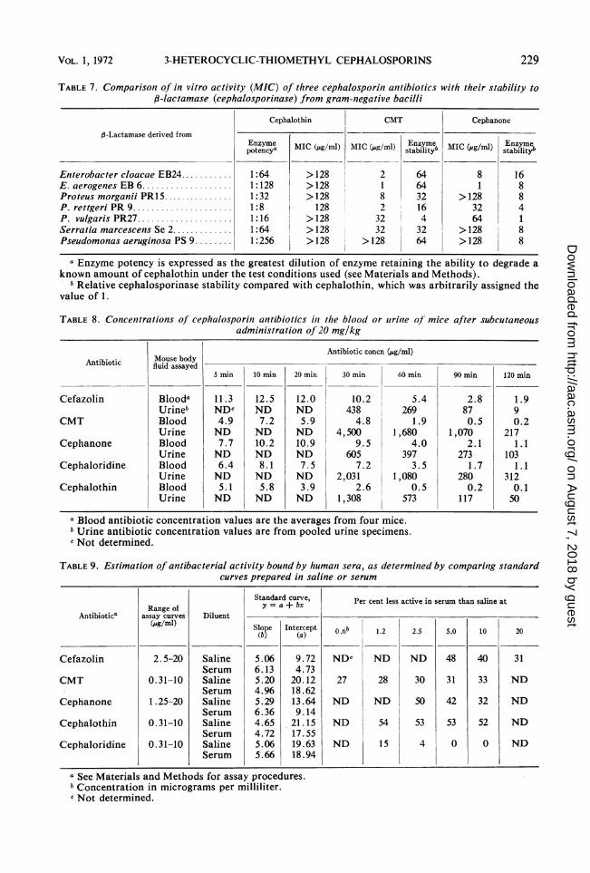
Antibiotic CMT was very stable to cephalo-sporinases derived from several gram-negativebacilli, and cephanone was more stable thancephalothin (Table 7). Resistance of Enterobactersp. and indole-positive Proteus sp. to cephalothinmay be due to cephalosporinase production bythese bacteria. All cultures from these two generathat were tested (Table 7) were susceptible toCMT, which was also stable to enzymes producedby the same organisms. BothCMT and cephanonewere more stable than cephalothin to ,B-lactamasefrom Serratia sp. or Pseudomonas sp., but theseparticular isolates were still resistant to the anti-biotics by other mechanisms. In 1963, Flemingand his associates (7) stated, "The varying activ-ity of the enzyme against different cephalosporinssuggests the possible development of cephalo-sporinase-resistant cephalosporins." Additional
227VOL. 1, 1972
on August 7, 2018 by guest
http://aac.asm.org/
Dow
nloaded from
ANTIMICROB. AG. CHEMOTHER.
TABLE 5. Summary ofdata obtained by the membraniefilter techniiquefor determilinzg bactericidal activitiesoffive cephalosporin antibiotics
Antibiotic
CefazolinCMTCephanoneCephaloridineCephalothin
CefazolinCMTCephanoneCephaloridineCephalothin
24-hr 24-hrMIC MBC
i.. /_- a f. -1 /lbpg/lmiI
1.01.00.54.08.0
0.50.50.50.120.25
Reduction of inocula by concn equal to24-hr MIBC
Wpg/m1r3 hr 6 hr 12 hr
% So %1.0 NEc 98.5 99.61.0 97.7 99.7 99.90.5 NE 98.6 >99.94.0 98.6 99.6 99.88.0 NE 99.6 99.9
0.50.50.50.120.25
NENENENENE
NENENENENE
9090.296.996.790.5
24 hr
99.9>99.9>99.9>99.9>99.9
99.9>99.999.9
>99.999.9
a MIC = lowest concentration of antibiotic to which there was no visible growth on the filter after 24hr of incubation.
b MBC = lowest concentration of antibiotic that killed >99-9%70 of the inoculated bacteria in 24 hr.c NE = the bactericidal effect could not be determined because the number of colonies on the filter
discs were too numerous to count.
TABLE 6. Stability of cephalosporint anitibiotics topenicilliniase
Amt of antibioticdegraded in 1 hr'
AntibioticPenicillinase Penicillinase
from fromB. cerez,ss S. aureus'
pg pgBenzylpenicillin .......... > 192 > 192Cefazolin 2 10Cephaloridine 101 5Cephanone. 1 5CMT 5O.S 1Cephalothin. 0 0
a All antibiotics were tested against an amountof enzyme sufficient to degrade 320 units (192,g)of benzylpenicillin.
b Penicillinase (Riker).c The staphylococcal penicillinase used was an
acetone-precipitated, water-dialyzed preparationfrom a clinical isolate of S. auireus.
studies with CMT may verify that this derivativefits this category.
Peak concentrations of cefazolin and cepha-none in mouse blood exceeded those obtainedwith similar doses of cephalothin or cephalori-dine (Table 8). The half-lives for cefazolin (36.2 43.5 min) and cephanone (29.5 i 2.2 min) weresimilar to that for cephaloridine (33.8 i 4.1min). On the other hand, CMT concentrations inblood were about equal to those of cephaloridine,but the CMT half-life (20.5 ± 2.9 min) was asrapid as for cephalothin (15.8 + 4.1 min).
Binding of antibacterial activity of the five anti-biotics, as estimated by comparing standard assaycurves prepared in either saline or human serum,followed several patterns (Table 9). The activityof cephaloridine was essentially the same in salineor serum, indicating very little binding. Bindingof CMT was not as great as with cephalothin;however, the amount of binding for both of theseantibiotics did not change with concentrations.Serum binding of cefazolin and cephanone wasconcentration-dependent, increasing as the con-centration decreased. Kind et al. (11) reportedbinding of cephalothin and cephaloridine as 65and 13%, respectively, in pooled human serum,by the ultrafiltration method. Nishida et al. (13),using essentially the same method, showed a
higher percentage of binding for cephalothin(79%) and cephaloridine (31 %), and reportedcefazolin as 74%. The ratio of 79% to 74% forcephalothin and cefazolin by these latter investi-gators compares with the 53% to 48% ratio atthe 5-,ug level as shown in Table 9. However,binding of activity against different organisms canvary (8), and may not accurately predict proteinbinding. Therefore, additional studies with pro-cedures other than those employed in this experi-ment may be necessary to predict the degrees ofbinding to serum proteins.ED50 values for subcutaneous administration of
five cephalosporins in therapy of experimental in-fections in mice are shown in Tables 10 and 11.To interpret properly the meaning of the two-doseED50 values in Table 10, the in vitro activity, con-
centrations in serum, excretion rates, and meta-
Representative bacterium
Escherichia coli(strain EC38)
Staphylococcus aureius(strain H43)
228 WICK AND PRESTON
on August 7, 2018 by guest
http://aac.asm.org/
Dow
nloaded from
3-HETEROCYCLIC-THIOMETHYL CEPHALOSPORINS
TABLE 7. Comparison of in vitro activity (MIC) of three cephalosporini anztibiotics with their stability tof3-lactamase (cephalosporinase) from gram-negative bacilli
Cephalothin CMT Cephanone
,-Lactamase derived from
pEnzyme MIC (pg/ml) MIC (pug/mi) stabilityb g/m) Etabilit
Enterobacter cloacae EB24........... 1:64 >128 2 64 8 16E. aerogenes EB 6................... 1:128 >128 1 64 1 8Proteus morganii PR15. 1:32 > 128 8 32 > 128 8P. rettgeri PR9..................... 1:8 128 2 16 32 4P. vulgaris PR27.1:16 >128 32 4 64 1Serratia marcescens Se 2............. 1:64 >128 32 I 32 >128 8Pseudomonas aeruginosa PS 9........ 1:256 >128 >128 64 >128 8
a Enzyme potency is expressed as the greatest dilution of enzyme retaining the ability to degrade aknown amount of cephalothin under the test conditions used (see Materials and Methods).
bRelative cephalosporinase stability compared with cephalothin, which was arbitrarily assigned thevalue of 1.
TABLE 8. Contcentrations of cephalosporin antibiotics in the blood or urinie of mice after subcutaneousadministration of 20 mg/kg
Antibiotic concn (pg/ml)Antibiotic Mousebodyida
5 min 10 min 20 min 30 min 60 min 90 min 120 min
Cefazolin Blooda 11.3 12.5 12.0 10.2 5.4 2.8 1.9Urineb NDc ND ND 438 269 87 9
CMT Blood 4.9 7.2 5.9 4.8 1.9 0.5 0.2Urine ND ND ND 4,500 1,680 1,070 217
Cephanone Blood 7.7 10.2 10.9 9.5 4.0 2.1 1.1Urine ND ND ND 605 397 273 103
Cephaloridine Blood 6.4 8.1 7.5 7.2 3.5 1.7 1.1Urine ND ND ND 2,031 1,080 280 312
Cephalothin Blood 5.1 5.8 3.9 2.6 0.5 0.2 0.1Urine ND ND ND 1,308 573 117 50
a Blood antibiotic concentration values are the averages from four mice.bUrine antibiotic concentration values are from pooled urine specimens.c Not determined.
TABLE 9. Estimation of antibacterial activity bound by humani sera, as determined by comparing standardcurves prepared in saline or serum
Antibiotica
Cefazolin
CMT
Cephanone
Cephalothin
Cephaloridine
Range ofassay curves
(pg/ml)
2.5-20
0.31-10
1.25-20
0. 31-10
0.31-10
Diluent
SalineSerumSalineSerumSalineSerumSalineSerumSalineSerutn
Standard curve,y = a + bx
Slope(b)
5.066.135.204.965.296.364.654.725.065.66
Intercept(a)
9.724.7320.1218.6213.649.14
21.1517.5519.6318.94
a See Materials and Methods for assay procedures.6 Concentration in micrograms per milliliter.c Not determined.
Per cent less active in serum than saline at
0 .66
NDC
27
ND
ND
ND
1.2
ND
28
ND
54
15
2.5
ND
30
50
53
4
5.0
48
31
42
53
0
10
40
33
32
52
0
20
31
ND
ND
ND
ND
229VOL. 1, 1972
on August 7, 2018 by guest
http://aac.asm.org/
Dow
nloaded from

ANTIMICROB. AG. CHEMOTHER.
TABLE 10. Activity offive cephalosporini antibiotics on experimenztal bacterial infections in mice
Subcutaneous ED6obBacterium LDloeg
CEZ CNIT CSY CER CET
Staphylococcus aureats 3055c 7,750 3.1 6.3 2.9 1.0 3.1S. aureus 3074d 5.1 3.8 13.3 3.3 2.0 26.2Streptococcus pyogenes C203 ... 1 ,580 0.6 0.6 0.3 <0.1 0.9Diplococcus pneumoniae type I. 630 1.6 15.7 1 .4 0.9 20.7Escherichia coli EC38. 10,000 12.8 15.9 9.3 20.6 95Klebsiellapneumoniae KL14 100 6.7 12.1 2.6 7.5 27.2Proteus mirabilis PR6........ 475 9.7 6.7 3.7 8.7 11.4P. morganii PR15.......... 100 >166 41.5 83 >166 <166Salmonella typhosa SA12 ..... 820 8.5 7.9 6.1 11.6 33.6Shigellaflexneri 2aSH3.. 1,280 19.3 18.6 14.4 13.5 144.5
a n U netn ls focei eurc OKl U0o n iea One LD5o = infecting dose of bacterla requi-red to kill 35()Wg of- the mice.b The EDro value is expressed as milligrams per kilogram in two treatments
The abbreviations used for the antibiotics are spelled out in Fig. 1.c Benzylpenicillin-susceptible.d Benzylpenicillin-resistant.
TABLE 11. Multiple-close therapy of experimentalD. pneumoniiae inifectionis with five cephcalosporiin
alntibiotics
Time of ED5o (mg per kg per dose)aadministration
(hr post-infection) CEZ CMT CSY CER CET
1 22.0 114 19.3 5.3 1001, 2 1.6 15.7 1.4 0.9 20.71 2 3, 4, 5 0.3 1.7 0.2 0.2 4.2
a All antibiotics were administered subcutane-ously. The abbreviations used for the antibioticsare spelled out in Fig. 1.
bolic patterns for all of the compounds must becompared.
Peak serum concentrations of cefazolin andcephanone were higher than those of cephalori-dine, and the excretion rates were similar (Table8). Therefore, the infecting bacteria were exposedin the animal to these antibiotics for similarperiods of time; ED5o values (Table 10) for thesethree antibiotics can be compared. Cephaloridine,cefazolin, and cephanone are metabolically stableand are excreted unchanged in urine. Both cefa-zolin and cephanone are less active in vitro thancephaloridine against some gram-positive cocci(Table 1). Against gram-negative bacilli, theheterocyclic compounds have activities equal toor twice those for cephaloridine (Table 3). Inaddition, cephanone exhibits a degree of stabilityto 3-lactamase derived from certain gram-nega-tive bacilli (Table 7). When all of these factsabout cefazolin and cephanone are considered,the expected ED50 values for these antibioticswould be about equal to cephaloridine against
(1 and 5 hr postinfection).
S. aureus and D. pneumoniae infections, at leasttwice that for cephaloridine against Streptococcuspyogenes infections, and one-half to about equalto those for cephaloridine in therapy of infectionscaused by E. coli, K. pneumoniae, P. mirabilis,Salmonella typhosa, and Shigella flexneri. Cefa-zolin was not expected to be effective in treatmentof infections caused by the ,-lactamase-producingP. morganii, but therapy with cephanone wouldbe successful. The data presented in Table 10 con-firmed these predictions.On the other hand, peak serum concentrations
of CMT are equal to those of cephaloridine, butthe excretion rate is rapid, like that of cephalothin(Table 8). Therefore, the infecting bacteria are incontact with cephalothin or CMT in the mousefor only short periods of time; ED50 values (Table10) for these two antibiotics can be compared.Also, unlike cephalothin, CMT is metabolicallystable. Antibiotic CMT is slightly less active thancephalothin in vitro against S. aureus or D. pneu-moniae, but about equal in activity against S. pyo-genes (Table 1). Against gram-negative bacilli,CMT is from 2 to >100 times more active thancephalothin (Table 3). In addition, CMT is stableto j3-lactamases from gram-negative bacilli (Table7). When these facts about CMT were consid-ered, the ED50 values shown in Table 10 were ex-pected; i.e., ED50 values for CMT were about one-half of those for cephalothin in therapy of all ofthe experimental infections shown. AntibioticCMT was eSfective in treating P. morganii infec-tions, most likely because of its stability tof-lactamase.
Based on the known efficacy of cephalothin andcephaloridine in therapy of bacterial infections inhumans, the in vitro and in vivo data accumulated
230 WICK AND PRESTON
on August 7, 2018 by guest
http://aac.asm.org/
Dow
nloaded from
3-HETEROCYCLIC-THIOMETHYL CEPHALOSPORINS
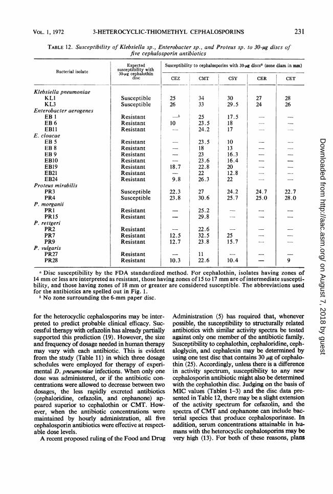
TABLE 12. Suisceptibility of Klebsiella sp., Eniterobacter sp., and Proteus sp. to 30-.sg discs offive cephalosporin antibiotics
Bacterial isolate
Klebsiella pneumoniaeKL1KL3
Eniterobacter aerogenesEB 1EB 6EB1l
E. cloacaeEB 5EB 8EB 9EBIOEB19EB21EB24
Proteus mirabilisPR3PR4
P. morganiiPRIPRI5
P. rettgeriPR2PR7PR9
P. vulgarisPR27PR28
Expectedsusceptibility with30-pg cephalothin
disc
SusceptibleSusceptible
ResistantResistantResistant
ResistantResistantResistantResistantResistantResistantResistant
SusceptibleSusceptible
ResistantResistant
ResistantResistantResistant
ResistantResistant
Susceptibility to cephalosporins with 30-pg discsa (zone diam in mm)
CEZ
2526
-b
10
18.7
9.8
22.323.8
12.512.7
10.3
CMT
3433
2523.524.2
23.5182323.622.82226.3
2730.6
25.229.8
22.632.523.8
1122.6
cSY
3029.5
17.51817
101316.316.42012.822
24.225.7
2515.7
10.4
CER
2724
24.725.0
CET
2826
22.728.0
9
a Disc susceptibility by the FDA standardized method. For cephalothin, isolates having zones of14 mm or less are interpreted as resistant, those having zones of 15 to 17 mm are of intermediate suscepti-bility, and those having zones of 18 mm or greater are considered susceptible. The abbreviations usedfor the antibiotics are spelled out in Fig. 1.bNo zone surrounding the 6-mm paper disc.
for the heterocyclic cephalosporins may be inter-preted to predict probable clinical efficacy. Suc-cessful therapy with cefazolin has already partiallysupported this prediction (19). However, the sizeand frequency of dosage needed in human therapymay vary with each antibiotic. This is evidentfrom the study (Table 11) in which three dosageschedules were employed for therapy of experi-mental D. pneumoniae infections. When only onedose was administered, or if the antibiotic con-centrations were allowed to decrease between twodosages, the less rapidly excreted antibiotics(cephaloridine, cefazolin, and cephanone) ap-peared superior to cephalothin or CMT. How-ever, when the antibiotic concentrations weremaintained by hourly administration, all fivecephalosporin antibiotics were effective at respect-able dose levels.A recent proposed ruling of the Food and Drug
Administration (5) has required that, wheneverpossible, the susceptibility to structurally relatedantibiotics with similar activity spectra be testedagainst only one member of the antibiotic family.Susceptibility to cephalothin, cephaloridine, ceph-aloglycin, and cephalexin may be determined byusing one test disc that contains 30 ,ug of cephalo-thin (25). Accordingly, unless there is a differencein activity spectrum, susceptibility to any newcephalosporin antibiotic might also be determinedwith the cephalothin disc. Judging on the basis ofMIC values (Tables 1-3) and the disc data pre-sented in Table 12, there may be a slight extensionof the activity spectrum for cefazolin, and thespectra of CMT and cephanone can include bac-terial species that produce cephalosporinase. Inaddition, serum concentrations attainable in hu-mans with the heterocyclic cephalosporins may bevery high (13). For both of these reasons, plans
231VOL. 1, 1972
on August 7, 2018 by guest
http://aac.asm.org/
Dow
nloaded from
ANTIMICROB. AG. CHEMOTHER.
CEPHANONE30 - pg DISCS
riE
Least Squares Line fory = a + bxb = - 0.3982 = Slopea =10.6847= Intercept
0 = Citrobacter sp* = Enterobacterv = Escherichia co = Klebsiella sp.
w= Prntounc en
4+\* o0 = Pseudomonas\o = Salmonella sp
* = Serratia sp.:* A = Shigella sp.
O ***e % w 0 = Staphylococc\. A = Streptococcu.
I'ISW ,
P.Sp.-oli
FS Sp.
,us aureusfs sp. (group D)
I -46 8 10 12 14 16 18 20 22 24 26 28 30 32 34 363838
Zone Diameter (mm)FDA Standardized Disc Procedure
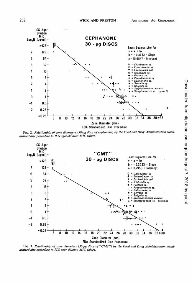
FIG. 2. Relationshlip of zone diameters (30-,ug discs of cephanionte) by the Food anid Drug Administration stanid-ardized disc procedure to ICS agar-dilulionl MIC values.
"CMT"'30 - pg DISCS Least Squares Line for
y = a + bxb = -0.3033 = Slopea = 8.7955 = Intercept
O = Citrobacter sp.Enterobacter sp.
v = Escherichia co/io = Klebsiella sp.* = Proteus sp.o = Pseudomonas sp.
O = Salmonella sp.* = Serratia sp.A = Shigella sp.
* = Staphylococcus aureusA = Streptococcus sp. (group D)
Zone Diameter (mm)FDA Standardized Disc Procedure
FIG. 3. Relationship of zone diameters (30-,ug discs of" CMT") by the Food and Drug Administration stand-ardized disc procedure to ICS agar-dilution MIC values.
ICS AgarDilutionMIC
(jg/ml)"128
128
64
32
16
8
4
2
1 _0.5
0.25
Log2N
7
6
5
4
3
2
1
0
-1
-2
ICS AgarDilutionMIC
(pg/ml)-
-128
128
moo
0
oo .
Log2N
7
6
5
4
3
2
10
-1
-2
232 WICK AND PRESTON
c--0. A
.
F
on August 7, 2018 by guest
http://aac.asm.org/
Dow
nloaded from

3-HETEROCYCLIC-THIOMETHYL CEPHALOSPORINS
to use the representative 30-Asg cephalothin discfor these new cephalosporins should be deferred.Discs for each of the heterocyclic cephalosporinsshould be used, along with cephalothin discs,should these antibiotics be used in clinical trials.
Regression lines and equations correlating MICvalues by the ICS agar-dilution method with zonediameters obtained by the Food and Drug Ad-ministration standardized disc procedure areshown in Fig. 2, 3, and 4. Data obtained by thesetwo standardized procedures should be used toselect a zone size that divides susceptible from re-sistant organisms. However, the zone diameterselected must be based on an MIC value belowwhich successful therapeutic results are expected.Therefore, zone diameters and MIC values thatdetermine susceptible bacteria must be judged onthe basis of data collected during a clinical trial.
Procedures other than the two standardizedones discussed above are often used for suscepti-bility testing. When they are, the type of mediumor the size of inoculum are two variations mostcommonly causing differences in end points. Dif-ferences in MIC values with various media usedfor broth- or agar-dilution tests have been dis-cussed previously (Table 4). When other mediawere substituted for Mueller-Hinton agar in the
Log2N
7
6
5
4
3
2
10
-1
-2
ICS AgarDilutionMIC
(jg/ml)
>128
128
64
32
16
8
4
2
1
0.5
0.25.on ')1
Food and Drug Administration disc procedure,zone diameters were about equal with TrypticaseSoy Agar (BBL), 1 mm smaller with SensitivityTest Agar (Oxoid), and 4 mm larger with NutrientAgar (Difco). A 1:100 dilution of an overnightTrypticase Soy Broth culture approximates theturbidity of the BaSO4 standard used to adjustinocula sizes for the Food and Drug Administra-tion disc test. For each 10-fold change in inoculumsize, zone diameters varied by about 2 mm.The original purpose of these comparative stud-
ies with five antibiotics was to select one of theheterocyclic cephalosporin antibiotics that mightwarrant clinical trial. However, analysis of thedata presented above makes that selection diffi-cult. In fact, each of the three newer cephalo-sporins has certain advantageous characteristics.For example, all three antibiotics have excellentin vitro activity against gram-negative bacilli,peak serum concentrations of cefazolin and ceph-anone are very high, and the better in vitro ac-tivity and stability to cephalosporinase of CMTeasily compensates for its generally lower bloodlevels. Thus, all three of the newer cephalosporinsare promising, and additional toxicological andpharmacological data should be obtained.
CEFAZOLIN
:00 30 - j9 DISCS Least Squares Line fory = a + bxb = -0.2898 = Slope
>,.# 0 a = 8.3267 = Intercept
o ** *
Citrobacter sp.* Enterobacter sp.v Escherichia colio Klebsiella sp.* Proteus sp.O = Pseudomonas sp.O = Salmonella sp.* = Serratia sp.A = Shigella sp.* = Staphylococcus aureusA = Streptococcus sp. (group 0)
U.L..6 8 10 12 14 16 18 20 22 24 26 28 30 32 34 363838
Zone Diameter (mm)FDA Standardized Disc Procedure
FIG. 4. Relationship of zone diameters (30-,ug discs of cefazolin) by the Food and Drug Administration stand-ardized disc procedure to ICS agar-dilution MIC values.
VOL. 1, 1972 233
.0 - * 9
on August 7, 2018 by guest
http://aac.asm.org/
Dow
nloaded from
WICK AND PRESTON
ACKNOWLEDGMENTS
We are grateful to W. B. Blanchard and C. W. Ryan for prepa-
ration of the cephalosporin antibiotics, and to D. Berry forchromatography.We express sincere appreciation to Lois Hawley, June Wood,
Ann Stroy, Billie Lady, and Gordon Tucker for excellent technicalassistance.
LITERATURE CITED
1. Barber, M. 1964. Naturally occurring methicillin-resistantstaphylococci. J. Gen Microbiol. 35:183-190.
2. Bauer, A. W., W. M. M. Kirby, J. C. Sherris, and M. Turck.1966. Antibiotic susceptibility testing by a standardizedsingle disc method. Amer. J. Clin. Pathol. 45:493-496.
3. Chabbert, Y. A. 1957. Une technique nouvelle d'etude del'action bactericide des associations d'antibiotiques: letransfert sur cellophane. Ann. Inst. Pasteur (Paris) 93:289-299.
4. Chabbert, Y. A. 1967. Behavior of "methicillin hetero-resistant" staphylococci to cephaloridine. Postgrad. Med.J. 43(Suppl.):40-46.
5. Department of Health, Education, and Welfare, Food andDrug Administration. 1970. Proposed rule making. FederalRegister 36:6899-6902.
6. Ericsson, H. M., and J. C. Sherris. 1971. Antibiotic sensitivitytesting. Report of an international collaborative study.Acta Pathol. Microbiol. Scand. Sect. B, Suppl. 217.
7. Fleming, P. C., M. Goldner, and D. G. Glass. 1963. Observa-tion on the nature, distrubution, and significance of cephalo-sporinase. Lancet 1:1399-1401.
8. Godzeski, C. W., G. Brier, and D. E. Pavey. 1963. Cephalo-thin, a new cephalosporin with a broad antibacterial spec-trum. I. In vitro studies employing the gradient platetechnique. Appl. Microbiol. 11:122-127.
9. Hoehn, M. M., and C. T. Pugh. 1968. Method for the detec-tion and quantitative assay of cephaloglycin and its bio-logically active metabolites. Appl. Microbiol. 16:1132-1133.
10. Kariyone, K., H. Harada, M. Kurita, and T. Takano. 1970.Cefazolin, a new semisynthetic cephalosporin antibiotic. I.
Synthesis and chemical properties of cefazolin. J. Antibiot.(Tokyo) 23:131-136.
11. Kind, A. C., D. G. Kestle, H. C. Standiford, and W. M. M.Kirby. 1969. Laboratory and clinical experience withcephalexin. Antimicrob. Ag. Chemother. 1968, p. 361-365.
12. Mine, Y., M. Nishida, S. Goto, and S. Kuwahara. 1970.Cefazolin, a new semisynthetic cephalosporin antibiotic.IV. Antigenicity of cefazolin and its cross reactivity with
ANTIMICROB. AG. CHEMOTHER.
benzylpenicillin, ampicillin, and cephaloridine. J. Anti-biot. (Tokyo) 23:195-203.
13. Nishida, M., T. Matsubara, T. Murakawa, Y. Mine, Y.Yokota, S. Kuwahara, and S. Goto. 1970. In vitro and invivo evaluation of cefazolin, a new cephalosporin C de-rivative. Antimicrob. Ag. Chemiiother. 1969, p. 236-243.
14. Nishida, M., T. Matsubara, T. Murakawa, Y. Mine, Y.Yokota, S. Goto, and S. Kuwahara. 1970. Cefazolin, a new
semisynthetic cephalosporin antibiotic. II. In vitro and invivo antimicrobial activity. J. Anitibiot. (Tokyo) 23:137-148.
15. Nishida, M., T. Matsubara, T. Murakawa, Y. Mine, Y.Yokota, S. Goto and S. Kuwahara. 1970. Cefazolin, a new
semisynthetic cephalosporin antibiotic. I11. Absorption,excretion, and tissue distribution in parenteral administra-tion. J. Antibiot. (Tokyo) 23:184-194.
16. Reed, L. J., and H. Muench. 1938. A simple method for esti-mating 50 percent endpoints. Amer. J. Hyg. 27:493-497.
17. Ronald, A. R., and M. Turck. 1967. Factors influencing invitro susceptibility to the cephalosporins and clinical trial ofan oral cephalosporin, cephaloglycin. Antimicrob. Ag.Chemother. 1966, p. 82-87.
18. Sherris, J. C., A. L. Rashad, and G. A. Liglhthart. 1967.Laboratory determination of antibiotic susceptibility toampicillin and cephalothin. Ann. N.Y. Acad. Sci. 145:248-267.
19. Shibata, K., and M. Fugii. 1971. Clinical studies of cefazolinin the surgical field. Antimicrob. Ag. Chemother. 1970,p. 467-472.
20. Steers, E., E. L. Foltz, B. S. Graves, and J. Riden. 1959. Aninocula replicating apparatus for routine testing of bacterialsusceptibility to antibiotics. Antibiot. Chemother. 9:307-311.
21. Stroy, S. A., and D. A. Preston. 1971. Specific assay ofaminoglycosidic- or polymyxin-type antibiotics present inhuman sera in combination with cephalosporins. AppI.Microbiol. 21:1002-1006.
22. Wick, W. E. 1964. Influence of antibiotic stability on theresults of in vitro testing procedures. J. Bacteriol. 87:1162-1170.
23. Wick, W. E., and W. S. Boniece. 1965. In vitro and in vivolaboratory evaluation of cephaloglycin and cephaloridine.Appl. Microbiol. 13:248-253.
24. Wick, W. E., D. H. Holmes, and W. S. Boniece. 1960. Anti-penicillinase serum: use in treatment of resistant staphy-lococcal infections in laboratory animals. Antibiot.Chemother. 10:71-77.
25. Wick, W. E., W. E. Wright, and H. V. Kuder. 1971. Cephalo-glycin and its biologically active metabolite desacetyl-cephaloglycin. Appl. Microbiol. 21:426-434.
234
on August 7, 2018 by guest
http://aac.asm.org/
Dow
nloaded from

![Synthesis of Heterocyclic [8]Circulenes and Related … review/55_synlett... · Synthesis of Heterocyclic [8]Circulenes and Related Structures ... 6 Synthesis of Other Heterocyclic](https://img.pdfslide.net/doc/110x75/5b165ee97f8b9a636d8b9414/synthesis-of-heterocyclic-8circulenes-and-related-review55synlett-synthesis.jpg)