Embed Size (px)
Citation preview
The Zika virus (ZIKV) is a mosqui-
to-borne pathogen recently linked to
certain birth defects in infants in South
and Central America and the United
States. Protein crystallography studies
performed at the ALS have now resolved
the structure of a key ZIKV protein (NS5)
to 3.0 Å. The high-resolution structure has
enabled researchers to make detailed
comparisons to similar proteins from
related viruses and gain insight into how
their differences might affect viral replica-
tion and immune response. The availability
of this structure is an important step
toward the targeted, structure-based
development (and repurposing) of drugs
capable of disrupting viral functions and
halting the spread of the disease.
ZIKV belongs in the genus Flavivirus,
which derives its name from the virus that
causes yellow fever (flavus means “yellow”
in Latin). Other members of the Flaviviridae
family include the human pathogens that
cause Japanese encephalitis (JEV) and
dengue fever (DENV). Expression of flavi-
virus genomes results in the production of
three proteins that are part of the virus
itself (structural proteins) and seven
proteins that are not part of the virus, but
could perform some viral function in the
infected cell (nonstructural proteins).
In ZIKV, nonstructural protein 5 (NS5)
includes two domains: one that facilitates
the translation of the viral genome and
helps the virus to evade the host’s immune
response (the methyltransferase domain,
MT) and another that helps start the
genetic replication process (the RNA-
dependent RNA polymerase domain,
RdRp). Because of these crucial functions,
NS5 in ZIKV is a potent drug target whose
analogues in Japanese encephalitis and
dengue fever have been used to develop
therapies that work at the molecular level.
To study the ZIKV NS5 structure in
detail, the researchers first produced and
crystallized small quantities of the full-
length NS5 protein using chemically
synthesized DNA that encoded the ZIKV
strain originally isolated from Uganda,
Africa, in 1947 (strain MR766). X-ray crys-
tallography at ALS Beamline 5.0.2, part of
Zika’s Emergence
In 1947, scientists working on a
yellow-fever research program in
Uganda, Africa, recorded a tempera-
ture spike in one of their “sentinel”
rhesus monkeys (no. 766). It had been
kept in a strip of densely forested
area along the edge of Lake Victoria
called the Zika Forest. The monkey
soon recovered, showing no other
symptoms. The scientists isolated the
agent that caused the fever and
named it the “Zika virus.” Over the
next 60 years, fewer than 20 human
Zika infections were recorded,
perhaps because infection was
associated with only mild effects.
The first large outbreak of Zika fever
occurred in 2007 in Micronesia, with
49 confirmed cases, underscoring
Zika’s potential as a newly emerging
mosquito-borne virus. In 2013,
a larger epidemic occurred in French
Polynesia, with an estimated 30,000
symptomatic infections, including
a few cases of Guillain–Barré syn-
drome, a potentially life-threatening
condition involving muscle weakness
or paralysis. In 2015, Zika reached
the Americas. In Brazil, over a million
cases were reported, along with
a dramatic increase in severe birth
defects (microcephaly) in infants.
Changes in travel, climate, viral
genetics, and population distribution
(both human and mosquito) all likely
contribute to Zika’s emergence as
a serious problem. As it follows the
path of other similar viruses to new
populations lacking natural immunity,
scientists race to develop safe and
effective treatments.
Comparison of the solved ZIKV NS5 structure to those of the Japanese encephalitis virus (JEV) and
the dengue fever virus (DENV).
Structure of a Key Protein from the Zika Virus
BIOLOGICAL SCIENCES
the Berkeley Center for Structural
Biology, was then used to solve the
atomic structure. The powerful beam and
the sensitive detector made it possible to
obtain good data despite less-than-op-
timal crystal quality. In addition, access to
the Collaborative Crystallography
program at the ALS—a fast, reliable, and
transparent mail-in crystallographic
service funded by the National Institutes
of Health—resulted in rapid turnaround
for this time-sensitive project.
The solved structure revealed
remarkable similarities to the equivalent
structures of other viruses from the Flavi-viridae family, the active sites in particular.
The results suggest that inhibitors of viral
MT activity and/or RNA synthesis in
related viruses can be developed to
inhibit ZIKV replication as well. In fact,
molecules that can inhibit RNA synthesis
in dengue virus, West Nile virus, and
yellow fever virus have recently been
shown to affect RNA synthesis by ZIKV
NS5. Solving additional structures of the
ZIKV NS5 complexed to such molecules
could aid in the development of inhibitors
with higher specificity and potency.
While the Zika virus was initially
detected in 1947 in Uganda, serious illness
associated with Zika virus infection was not
recorded until 2007. At present, the basis
for this escalation in virulence with the
more recent Zika virus outbreak remains to
be established. However, comparison of
recombinant NS5 proteins from Africa and
from Brazil revealed similar levels of RNA
synthesis. In addition, the residues of the
Brazilian ZIKV that differ from those of the
MR766 virus from Africa are mostly on the
surface of the NS5 protein and are less
likely to affect the mechanism of RNA-
dependent RNA synthesis. The changes,
however, could impact interactions with
other ZIKV proteins or with cellular
proteins. Thus, while the researchers have
gathered a great deal of information about
how to target this protein, there are still
puzzles remaining. Further studies will be
invaluable for therapeutics discovery.
357 • 07/17
Publication about this research: B. Zhao, G. Yi, F. Du, Y.-C. Chuang, R.C. Vaughan, B. Sankaran, C.C. Kao, and P. Li, “Structure and function of the Zika virus full-length NS5 protein,” Nat. Commun. 8, 14762 (2017). doi:10.1038/ncomms14762
Research conducted by: B. Zhao, F. Du, and P. Li (Texas A&M University); G. Yi, Y.-C. Chuang, R.C. Vaughan, and C.C. Kao (Indiana University); and B. Sankaran (Berkeley Lab).
Research funding: Johnson Center for Innovation and Translational Research, National Institutes of Health, and Howard Hughes Medical Institute. Operation of the ALS is supported by the U.S. Department of Energy, Office of Science, Basic Energy Sciences Program.
Published by the ADVANCED LIGHT SOURCE COMMUNICATIONS GROUP
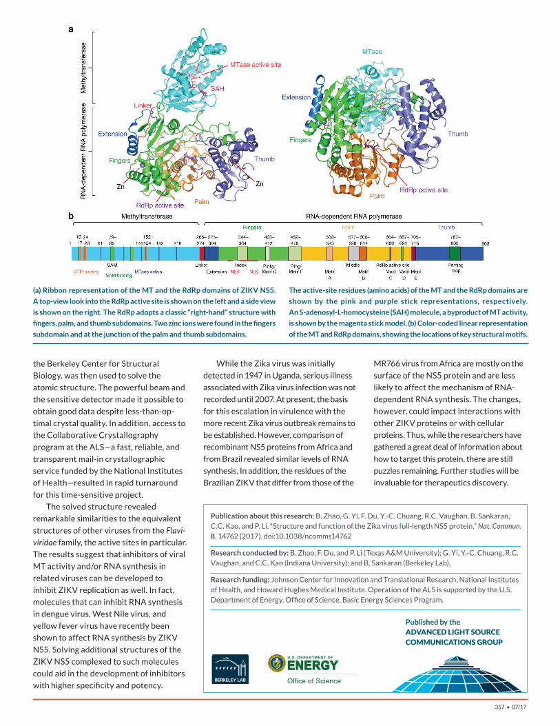
(a) Ribbon representation of the MT and the RdRp domains of ZIKV NS5.
A top-view look into the RdRp active site is shown on the left and a side view
is shown on the right. The RdRp adopts a classic “right-hand” structure with
fingers, palm, and thumb subdomains. Two zinc ions were found in the fingers
subdomain and at the junction of the palm and thumb subdomains.
The active-site residues (amino acids) of the MT and the RdRp domains are
shown by the pink and purple stick representations, respectively.
An S-adenosyl-L-homocysteine (SAH) molecule, a byproduct of MT activity,
is shown by the magenta stick model. (b) Color-coded linear representation
of the MT and RdRp domains, showing the locations of key structural motifs.

![1984 [Part 1_ Biological Sciences] __ Assembly in vitro of a Spanning Membrane Protein of the Endoplasmic Reticulum_ The](https://img.pdfslide.net/doc/110x75/613ca5b89cc893456e1e78f8/1984-part-1-biological-sciences-assembly-in-vitro-of-a-spanning-membrane-protein.jpg)