Embed Size (px)
Citation preview
Biology 1 Review:
Biology Review: Unit 2: Ch 7-11
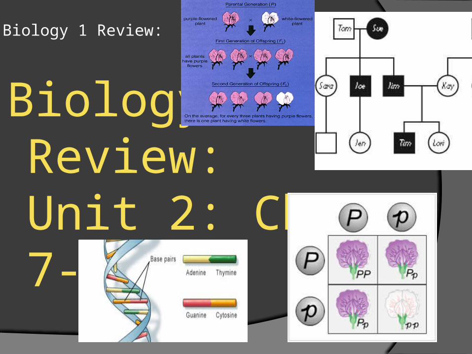
The cell cycle is a repeating sequence of cellular growth and division during the life of an organism.
A cell spends 90% of it’s time in the first three phases of the cycle, which are collectively called the interphase. A cell will enter the last two phases of the cell cycle (mitosis and cytokinesis) only if it about to divide.
The 5 phases of cell growth: First growth (G1) phase / Synthesis (S) phase / Second Growth (G2) phase / Mitosis / Cytokinesis
Stage 1: First growth (G1) During the G1 phase, a cell grows rapidly and carries out it’s routine functions. Cells that are not dividing stay in the G1 phase. For most organisms, this phase occupies the major portion of the cell’s life.
Stage 2: Synthesis (S) phase. A cell’s DNA is copied during this phase. At the end of this phase, each chromosome consists of two chromatids attached at the centromere.
Stage 3: Second Growth (G2) In the G2 phase, preparations are made for the nucleus to divide.
Stage 4: Mitosis. The process during cell division in which the nucleus of a cell is divided into two nuclei is called mitosis. Each nucleus ends up with the same number and kinds of chromosomes as the original cell.
Stage 5: Cytokinesis. The process during cell division in which the cytoplasm divides is called cytokinesis
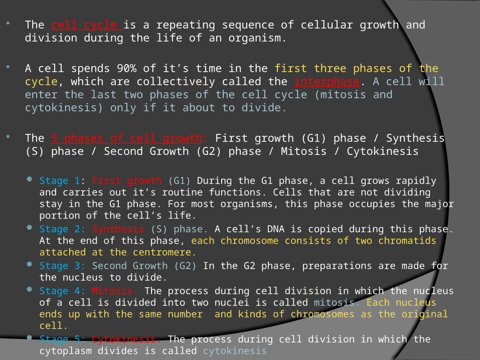
Mitosis and cytokinesis produce new cells by division that are identical to the original cells and allow organisms to grow, replace damaged tissues, and, in some organism, reproduce asexually.
Certain genes control the information that trigger the proteins that regulate cell growth and division. If one of these genes is mutated, the protein may not function and regulation of cell growth and division can be disrupted and malfunction. Cancer, the uncontrolled growth of cells, may result from this. / Cancer cells do not respond normally to the body’s control mechanisms.
_________________________________________________________ Cell division occurs in two stages; mitosis and than cytokinesis. During mitosis ,the nuclei divide to form two nuclei, each containing a
complete set of the cell’s chromosomes. During Cytokinesis, the entire cell divides into two identical cells, each
with one of the nuclei.
During mitosis, spindles play an important role in dividing the nuclei. Spindles are cell structures made up of both centrioles and individual microtubule fibers that are involved in moving chromosomes during cell division. The centrosome is an organelle that organizes the assembly of the spindle.
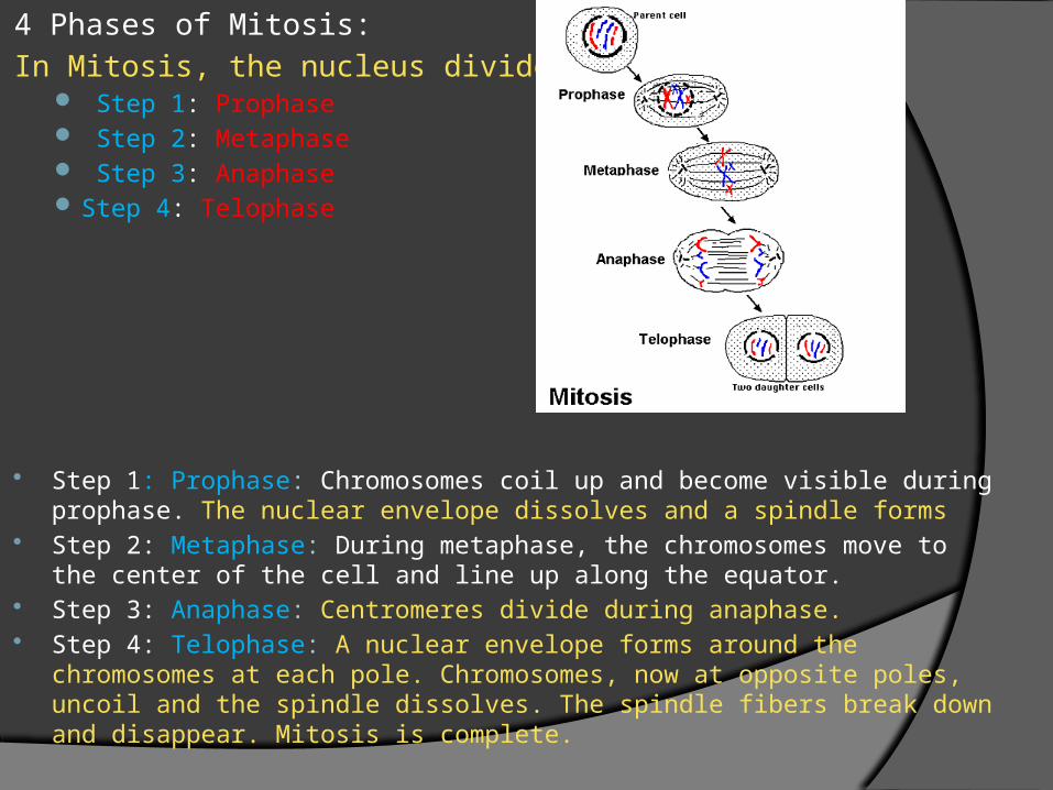
4 Phases of Mitosis: In Mitosis, the nucleus divides
Step 1: Prophase Step 2: Metaphase Step 3: Anaphase Step 4: Telophase
Step 1: Prophase: Chromosomes coil up and become visible during prophase. The nuclear envelope dissolves and a spindle forms
Step 2: Metaphase: During metaphase, the chromosomes move to the center of the cell and line up along the equator.
Step 3: Anaphase: Centromeres divide during anaphase. Step 4: Telophase: A nuclear envelope forms around the
chromosomes at each pole. Chromosomes, now at opposite poles, uncoil and the spindle dissolves. The spindle fibers break down and disappear. Mitosis is complete.
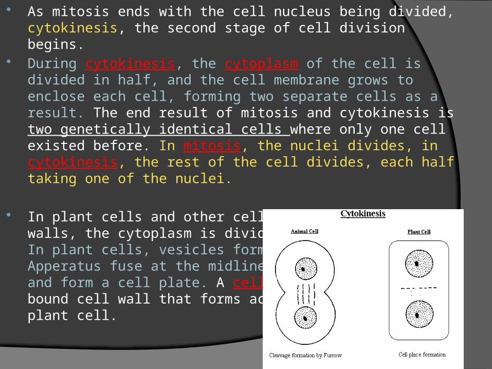
As mitosis ends with the cell nucleus being divided, cytokinesis, the second stage of cell division begins.
During cytokinesis, the cytoplasm of the cell is divided in half, and the cell membrane grows to enclose each cell, forming two separate cells as a result. The end result of mitosis and cytokinesis is two genetically identical cells where only one cell existed before. In mitosis, the nuclei divides, in cytokinesis, the rest of the cell divides, each half taking one of the nuclei.
In plant cells and other cells that have rigid cell walls, the cytoplasm is divided in a different way. In plant cells, vesicles formed by the Golgi Apperatus fuse at the midline of the dividing cell and form a cell plate. A cell plate is a membrane bound cell wall that forms across the middle of the plant cell.
Meiosis is a form of cell division that halves the number of chromosomes when forming specialized reproduction cells, such as gametes or spores.
These organisms reproduce by joining gametes to form the first cell of a new individual. The gametes are haploids; they contain only one set of chromosomes.
Before meiosis begins, DNA in the original cells is replicated. Thus meiosis starts with homologous chromosomes; chromosomes that are similar in shape, size, and genetic content.
Crossing-over occurs when portions of a chromatid on one homologous chromosome are broken and exchanged with the corresponding chromatid pieces of the other homologous chromosome. This random selection of homologous chromosomes during meiosis is called independent assortment.
In asexual reproduction, a single parent passes copies of all it’s genes to each of it’s offspring. An individual produced by asexual reproduction is a clone; genetically identical to the parent.
Prokaryotes produce by a type of asexual reproduction called binary fission.
In contrast, in sexual reproduction two parents each form reproductive cells that have one-half the chromosomes. Haploid gamates (sperm and egg cells) from both mother and father join to form a cell; a diploid zygote. Because both parents contribute half the genes, the offspring has traits from both parents.
There are several types of asexual reproduction. Here are examples of three different types of asexual reproduction.
Amoebas reproduce by fission, the separation of a parent into two or more individuals of equal size.
Some multicellular eukaryotes reproduce by a process called fragmentation where the body breaks into several pieces which each grow to a full adult.
Other organisms undergo a process called budding in which new individuals split, or bud, off from the original body and develop.
The passing of characteristics from parents to offspring is called heredity.
The study of heredity begin more than a century ago with the work of Gregor Mendel. Mendel carried out experiments in which he bred different varieties of garden peas. Mendel was the first to develop rules that accurately predicted patterns of heredity. The patterns that Mendel discovered form the basis of genetics, the branch of biology that focuses on heredity.
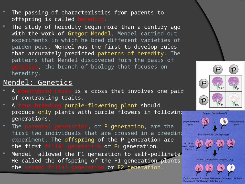
Mendel: Genetics A monohybrid cross is a cross that involves one pair of
contrasting traits. A true-breeding purple-flowering plant should produce
only plants with purple flowers in following generations. The parental generation, or P generation, are the first
two individuals that are crossed in a breeding experiment. The offspring of the P generation are the first filial generation or F1 generation.
Mendel allowed the F1 generation to self-pollinate. He called the offspring of the F1 generation plants the second filial generation or F2 generation.
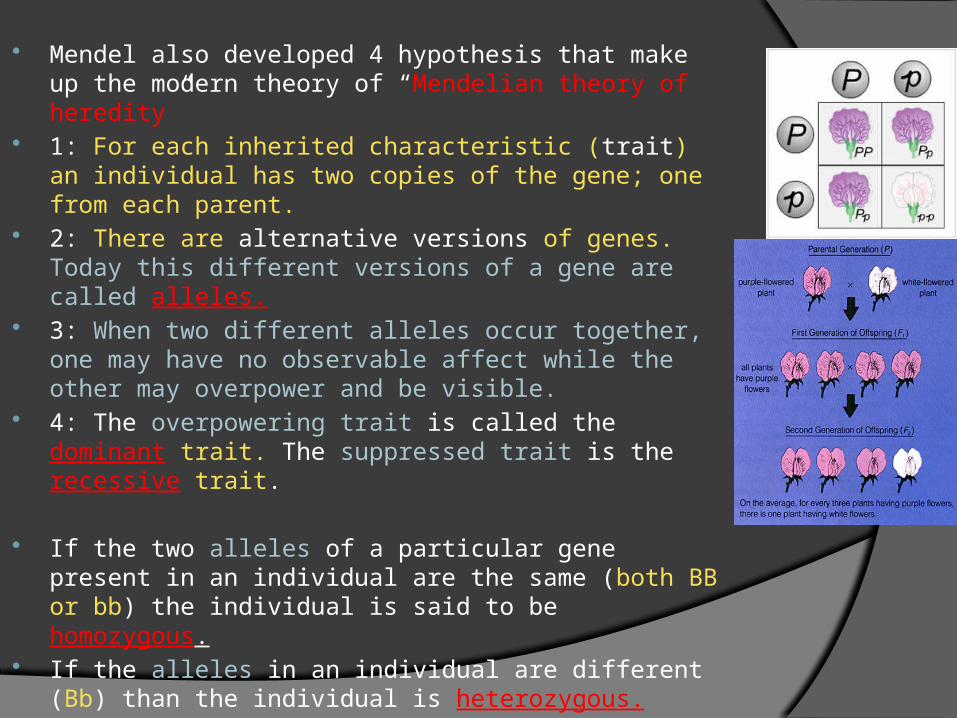
Mendel also developed 4 hypothesis that make up the modern theory of “Mendelian theory of heredity”
1: For each inherited characteristic (trait) an individual has two copies of the gene; one from each parent.
2: There are alternative versions of genes. Today this different versions of a gene are called alleles.
3: When two different alleles occur together, one may have no observable affect while the other may overpower and be visible.
4: The overpowering trait is called the dominant trait. The suppressed trait is the recessive trait.
If the two alleles of a particular gene present in an individual are the same (both BB or bb) the individual is said to be homozygous.
If the alleles in an individual are different (Bb) than the individual is heterozygous.
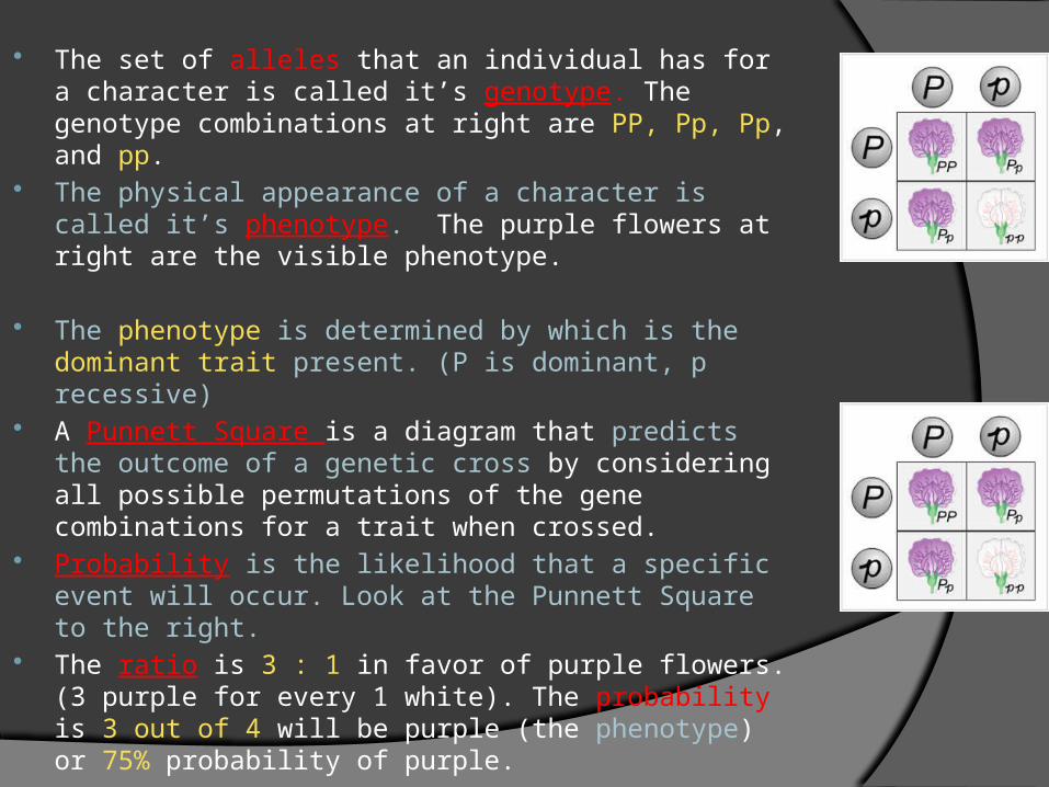
The set of alleles that an individual has for a character is called it’s genotype. The genotype combinations at right are PP, Pp, Pp, and pp.
The physical appearance of a character is called it’s phenotype. The purple flowers at right are the visible phenotype.
The phenotype is determined by which is the dominant trait present. (P is dominant, p recessive)
A Punnett Square is a diagram that predicts the outcome of a genetic cross by considering all possible permutations of the gene combinations for a trait when crossed.
Probability is the likelihood that a specific event will occur. Look at the Punnett Square to the right.
The ratio is 3 : 1 in favor of purple flowers. (3 purple for every 1 white). The probability is 3 out of 4 will be purple (the phenotype) or 75% probability of purple.
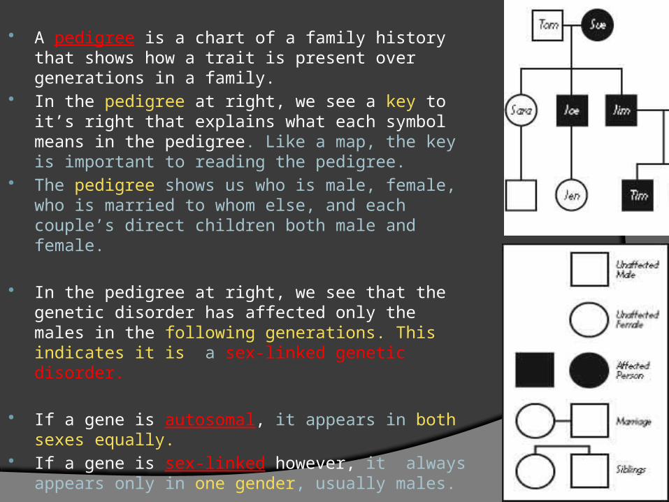
A pedigree is a chart of a family history that shows how a trait is present over generations in a family.
In the pedigree at right, we see a key to it’s right that explains what each symbol means in the pedigree. Like a map, the key is important to reading the pedigree.
The pedigree shows us who is male, female, who is married to whom else, and each couple’s direct children both male and female.
In the pedigree at right, we see that the genetic disorder has affected only the males in the following generations. This indicates it is a sex-linked genetic disorder.
If a gene is autosomal, it appears in both sexes equally.
If a gene is sex-linked however, it always appears only in one gender, usually males.
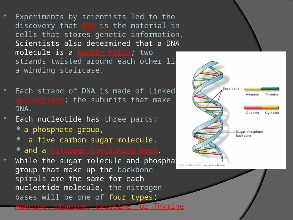
Experiments by scientists led to the discovery that DNA is the material in cells that stores genetic information. Scientists also determined that a DNA molecule is a double helix; two strands twisted around each other like a winding staircase.
Each strand of DNA is made of linked nucleotides; the subunits that make up DNA.
Each nucleotide has three parts; a phosphate group, a five carbon sugar molecule, and a nitrogen containing base.
While the sugar molecule and phosphate group that make up the backbone spirals are the same for each nucleotide molecule, the nitrogen bases will be one of four types: Adenine, Guanine, Cytosine, or Thymine
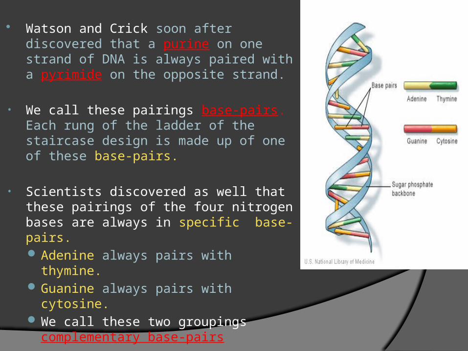
Watson and Crick soon after discovered that a purine on one strand of DNA is always paired with a pyrimide on the opposite strand.
• We call these pairings base-pairs. Each rung of the ladder of the staircase design is made up of one of these base-pairs.
• Scientists discovered as well that these pairings of the four nitrogen bases are always in specific base-pairs. Adenine always pairs with thymine. Guanine always pairs with cytosine. We call these two groupings
complementary base-pairs
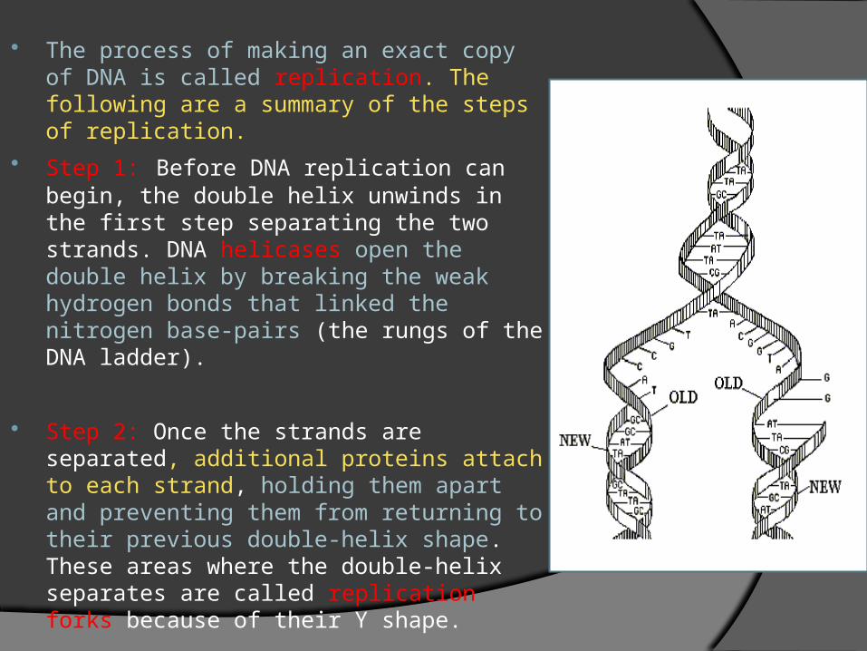
The process of making an exact copy of DNA is called replication. The following are a summary of the steps of replication.
Step 1: Before DNA replication can begin, the double helix unwinds in the first step separating the two strands. DNA helicases open the double helix by breaking the weak hydrogen bonds that linked the nitrogen base-pairs (the rungs of the DNA ladder).
Step 2: Once the strands are separated, additional proteins attach to each strand, holding them apart and preventing them from returning to their previous double-helix shape. These areas where the double-helix separates are called replication forks because of their Y shape.
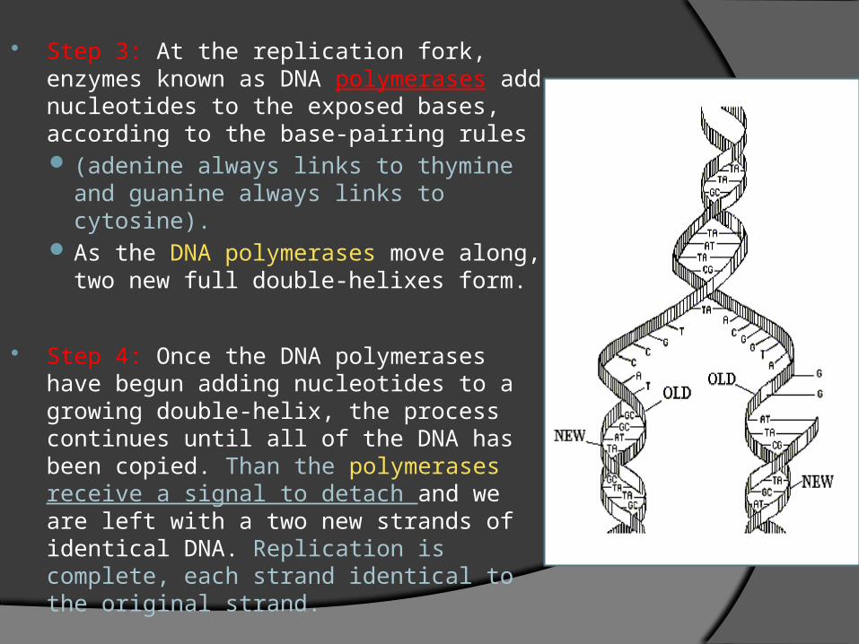
Step 3: At the replication fork, enzymes known as DNA polymerases add nucleotides to the exposed bases, according to the base-pairing rules (adenine always links to thymine and
guanine always links to cytosine). As the DNA polymerases move along,
two new full double-helixes form.
Step 4: Once the DNA polymerases have begun adding nucleotides to a growing double-helix, the process continues until all of the DNA has been copied. Than the polymerases receive a signal to detach and we are left with a two new strands of identical DNA. Replication is complete, each strand identical to the original strand.
Traits, such as eye color, are determined by proteins that are built according to instructions coded in DNA. Proteins are not built directly from DNA; ribonucleic (RNA) acid is also involved. Like DNA, ribonucleic acid is a molecule made of three nucleotides linked together. When an organism needs to build components of a new cell, a copy of the required DNA part is made. This copy is called RNA and is almost identical to DNA.
This RNA string is used by the organism as a template when it builds protein molecules, sometimes called the building blocks of the body.
A gene’s instructions for making a protein are coded in the sequence of nucleotides in the gene.
The instructions for making a protein are transferred from a gene to an RNA molecule in a process called transcription. The entire process by which proteins are made based on the information encoded in DNA is called gene expression or protein synthesis. The first step in the making of a protein, transcription, takes the information found in a gene in the DNA and transfers it to a molecule of RNA. RNA polymerase , an enzyme that adds and links complementary RNA nucleotides during transcription, is required for this process.
As transcription proceeds, the RNA polymerase eventually reaches a “stop signal” in the DNA. The stop signal is a sequence of bases that marks the end of each gene in eukaryotes, or the end of a set of genes in prokaryotes.
Transcription begins when RNA polymerase binds to the gene’s promoter; a specific sequence of DNA that acts as a “start” signal for transcription.
Transcription in prokaryote cells occurs in the cytoplasm. Transcription in eukaryote cells occurs in the nucleus, where the
DNA is located. Messenger RNA (mRNA) is a form of RNA that carries the
instructions for making a protein from a gene and delivers it to the site of translation.
The RNA instructions are written as a series of three-nucleotide sequences on the mRNA called codons. Each codon along the mRNA strand corresponds to an amino acid or signifies a start or stop signal for translation.
Ribosomal RNA molecules are RNA molecules that are part of the structure of ribosomes.
Translation is the process of synthesis of a protein by ribosomes, using mRNA as a template.
Not all genes are transcribed and translated all the time. Cells are able to regulate which genes are expressed and which are not, depending on the needs of the cell. This is called gene regulation.
During gene regulation, the piece of DNA that overlaps the promoter site and serves as the on-off switch is called an operator.
A repressor is a protein that binds to an operator and physically blocks RNA polymerase from binding to a promoter site.
In prokaryotes, gene expression is regulated by operons. Gene expression is switched OFF when repressor proteins block
RNA polymerase from transcribing a gene.
While it is tempting to think of a gene as an unbroken stretch of nucleotides that code for a protein, this simple arrangement is usually found only in prokaryotes. In eukaryotes, many genes are interrupted by introns, long series of
nucleotides that have NO coding information. (blank areas) Exons are the portions of the genes that are translated (copied or
expressed) into proteins. After a eukaryotic gene is transcribed, the introns in the resulting mRNA
are cut out by complex assemblies of RNA and protein called spliceosomes. The exons that remain are “stitched” back together by the spliceosome to form a smaller RNA molecule that is than translated.
Although changes in an organisms hereditary information are rare, they can occur. A change in the DNA of a gene is called a mutation.
Mutations that change a gene are called gene alterations. In a point mutation, a single nucleotide changes. In an insertion mutation, a sizable length of DNA is inserted into the
gene. In a deletion mutation, segments of a gene are lost, often during
meiosis.
The process of manipulating genes for practical purposes is called genetic engineering.
Genetic engineering may involve building recombinant DNA; DNA made from two or more different organisms.
Restrictive enzymes are bacterial enzymes that recognize and bind to specific short sequences of DNA, and than cut the DNA between specific nucleotides.
The DNA from a vector is also cut. A vector is an agent that is used to carry the gene of interest into another cell.
Plasmids are circular DNA molecules that can replicate independently of the main chromosomes of the bacteria.
The DNA fragments are combined with plasmid DNA fragments. We develop a hybrid plasmid made of both parts.
The host cells than take up the recombinant DNA, this plasmid hybrid.
In a process called gene cloning, many copies of the gene of interest are made each time the host cell reproduces by fission.