Embed Size (px)
Citation preview
Biology Dr. Khalida Ibrahim
The cartilage General characteristics:
1. Cartilage is a specialized type of connective tissue (supporting connective tissue).
2. Consists, like other connective tissues, of cells and extracellular matrix composed of connective tissue fibers and ground substance.
3. Does, unlike other connective tissue, not contain vessels or nerves. 4. Cartilage consists mainly of cells called chondrocytes and chondroblasts
that synthesize the extracellular matrix. 5. Is surrounded by a layer of dense connective tissue, the perichondrium. 6. Cartilage is rather rare in the adult humans, but it is very important during
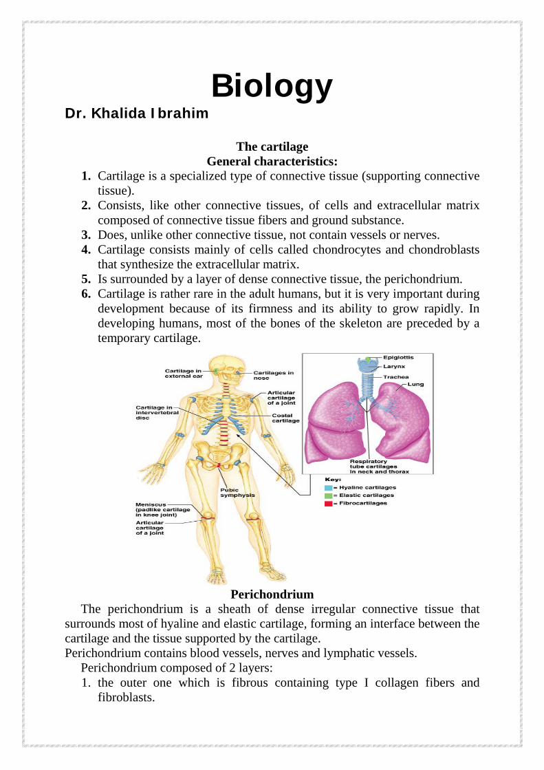
development because of its firmness and its ability to grow rapidly. In developing humans, most of the bones of the skeleton are preceded by a temporary cartilage.
Perichondrium
The perichondrium is a sheath of dense irregular connective tissue that surrounds most of hyaline and elastic cartilage, forming an interface between the cartilage and the tissue supported by the cartilage. Perichondrium contains blood vessels, nerves and lymphatic vessels. Perichondrium composed of 2 layers:
1. the outer one which is fibrous containing type I collagen fibers and fibroblasts.
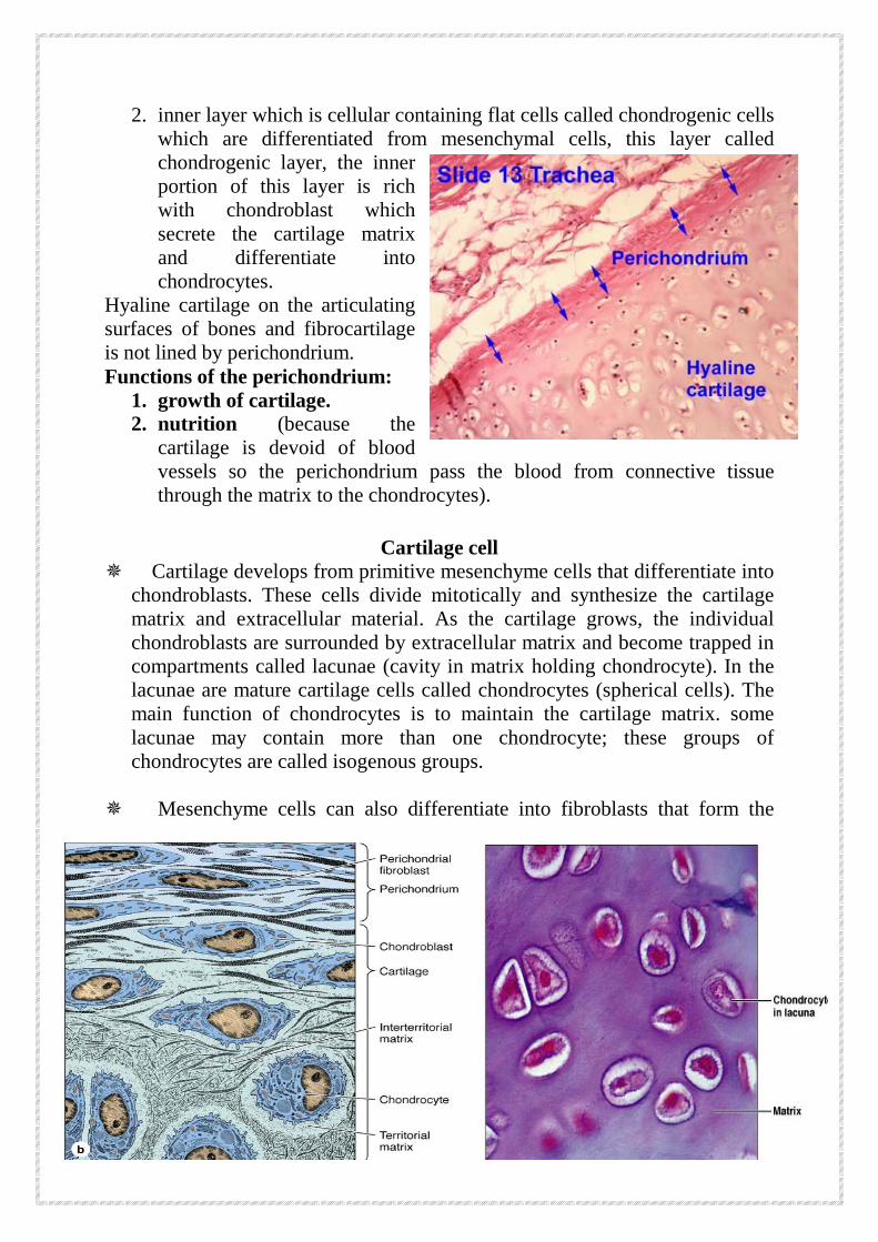
2. inner layer which is cellular containing flat cells called chondrogenic cells which are differentiated from mesenchymal cells, this layer called chondrogenic layer, the inner portion of this layer is rich with chondroblast which secrete the cartilage matrix and differentiate into chondrocytes.
Hyaline cartilage on the articulating surfaces of bones and fibrocartilage is not lined by perichondrium. Functions of the perichondrium:
1. growth of cartilage. 2. nutrition (because the
cartilage is devoid of blood vessels so the perichondrium pass the blood from connective tissue through the matrix to the chondrocytes).
Cartilage cell
Cartilage develops from primitive mesenchyme cells that differentiate into chondroblasts. These cells divide mitotically and synthesize the cartilage matrix and extracellular material. As the cartilage grows, the individual chondroblasts are surrounded by extracellular matrix and become trapped in compartments called lacunae (cavity in matrix holding chondrocyte). In the lacunae are mature cartilage cells called chondrocytes (spherical cells). The main function of chondrocytes is to maintain the cartilage matrix. some lacunae may contain more than one chondrocyte; these groups of chondrocytes are called isogenous groups.
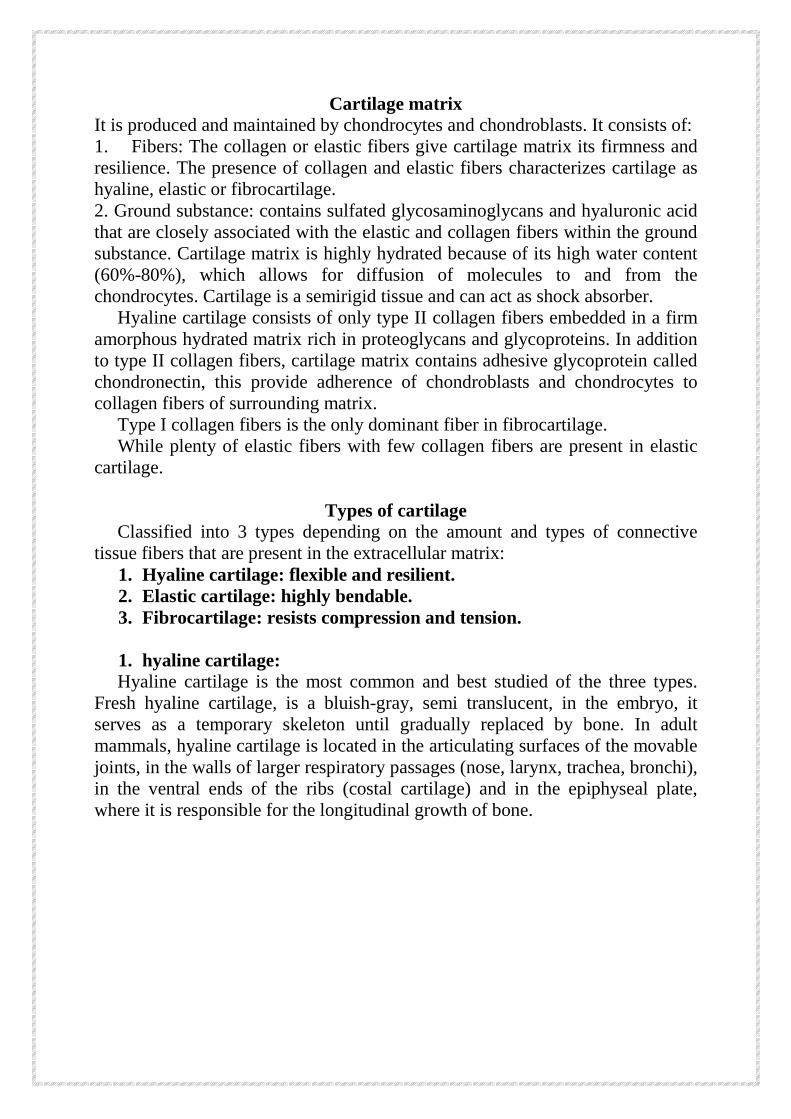
Mesenchyme cells can also differentiate into fibroblasts that form the
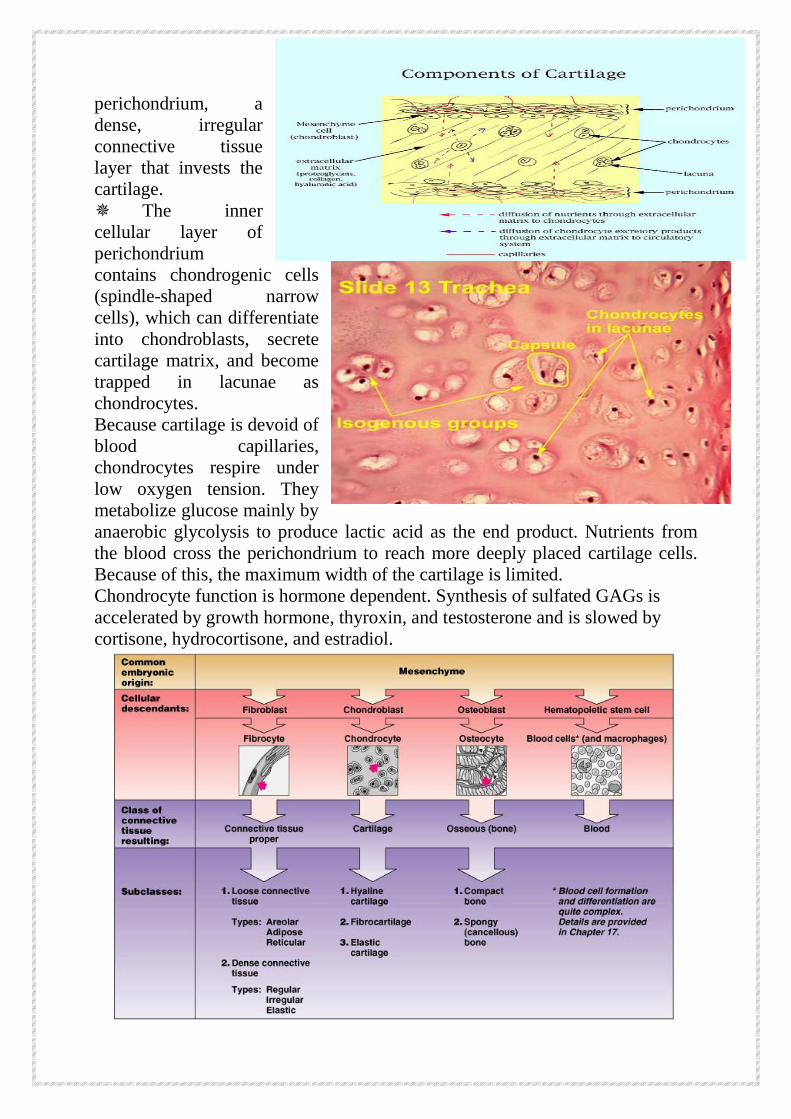
perichondrium, a dense, irregular connective tissue layer that invests the cartilage. The inner cellular layer of perichondrium contains chondrogenic cells (spindle-shaped narrow cells), which can differentiate into chondroblasts, secrete cartilage matrix, and become trapped in lacunae as chondrocytes. Because cartilage is devoid of blood capillaries, chondrocytes respire under low oxygen tension. They metabolize glucose mainly by anaerobic glycolysis to produce lactic acid as the end product. Nutrients from the blood cross the perichondrium to reach more deeply placed cartilage cells. Because of this, the maximum width of the cartilage is limited. Chondrocyte function is hormone dependent. Synthesis of sulfated GAGs is accelerated by growth hormone, thyroxin, and testosterone and is slowed by cortisone, hydrocortisone, and estradiol.
Cartilage matrix It is produced and maintained by chondrocytes and chondroblasts. It consists of: 1. Fibers: The collagen or elastic fibers give cartilage matrix its firmness and resilience. The presence of collagen and elastic fibers characterizes cartilage as hyaline, elastic or fibrocartilage. 2. Ground substance: contains sulfated glycosaminoglycans and hyaluronic acid that are closely associated with the elastic and collagen fibers within the ground substance. Cartilage matrix is highly hydrated because of its high water content (60%-80%), which allows for diffusion of molecules to and from the chondrocytes. Cartilage is a semirigid tissue and can act as shock absorber. Hyaline cartilage consists of only type II collagen fibers embedded in a firm amorphous hydrated matrix rich in proteoglycans and glycoproteins. In addition to type II collagen fibers, cartilage matrix contains adhesive glycoprotein called chondronectin, this provide adherence of chondroblasts and chondrocytes to collagen fibers of surrounding matrix. Type I collagen fibers is the only dominant fiber in fibrocartilage. While plenty of elastic fibers with few collagen fibers are present in elastic cartilage.
Types of cartilage
Classified into 3 types depending on the amount and types of connective tissue fibers that are present in the extracellular matrix:
1. Hyaline cartilage: flexible and resilient. 2. Elastic cartilage: highly bendable. 3. Fibrocartilage: resists compression and tension.
1. hyaline cartilage:
Hyaline cartilage is the most common and best studied of the three types. Fresh hyaline cartilage, is a bluish-gray, semi translucent, in the embryo, it serves as a temporary skeleton until gradually replaced by bone. In adult mammals, hyaline cartilage is located in the articulating surfaces of the movable joints, in the walls of larger respiratory passages (nose, larynx, trachea, bronchi), in the ventral ends of the ribs (costal cartilage) and in the epiphyseal plate, where it is responsible for the longitudinal growth of bone.
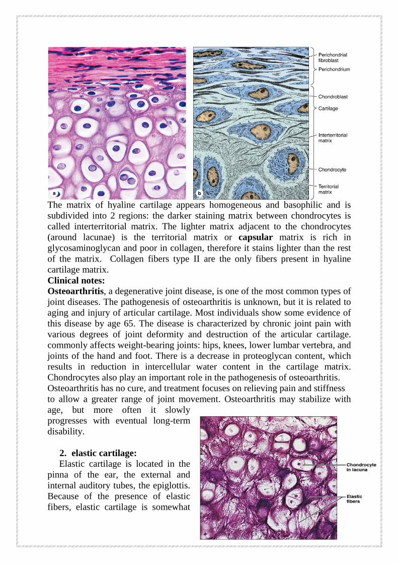
The matrix of hyaline cartilage appears homogeneous and basophilic and is subdivided into 2 regions: the darker staining matrix between chondrocytes is called interterritorial matrix. The lighter matrix adjacent to the chondrocytes (around lacunae) is the territorial matrix or capsular matrix is rich in glycosaminoglycan and poor in collagen, therefore it stains lighter than the rest of the matrix. Collagen fibers type II are the only fibers present in hyaline cartilage matrix. Clinical notes: Osteoarthritis, a degenerative joint disease, is one of the most common types of joint diseases. The pathogenesis of osteoarthritis is unknown, but it is related to aging and injury of articular cartilage. Most individuals show some evidence of this disease by age 65. The disease is characterized by chronic joint pain with various degrees of joint deformity and destruction of the articular cartilage. commonly affects weight-bearing joints: hips, knees, lower lumbar vertebra, and joints of the hand and foot. There is a decrease in proteoglycan content, which results in reduction in intercellular water content in the cartilage matrix. Chondrocytes also play an important role in the pathogenesis of osteoarthritis. Osteoarthritis has no cure, and treatment focuses on relieving pain and stiffness to allow a greater range of joint movement. Osteoarthritis may stabilize with age, but more often it slowly progresses with eventual long-term disability.
2. elastic cartilage: Elastic cartilage is located in the pinna of the ear, the external and internal auditory tubes, the epiglottis. Because of the presence of elastic fibers, elastic cartilage is somewhat
yellow and is more opaque than hyaline cartilage in the fresh state. The perichondrium is rich in elastic fibers. The matrix consist of branching elastic fibers interposed with type II collagen fiber bundles, giving it much more flexibility than hyaline cartilage.
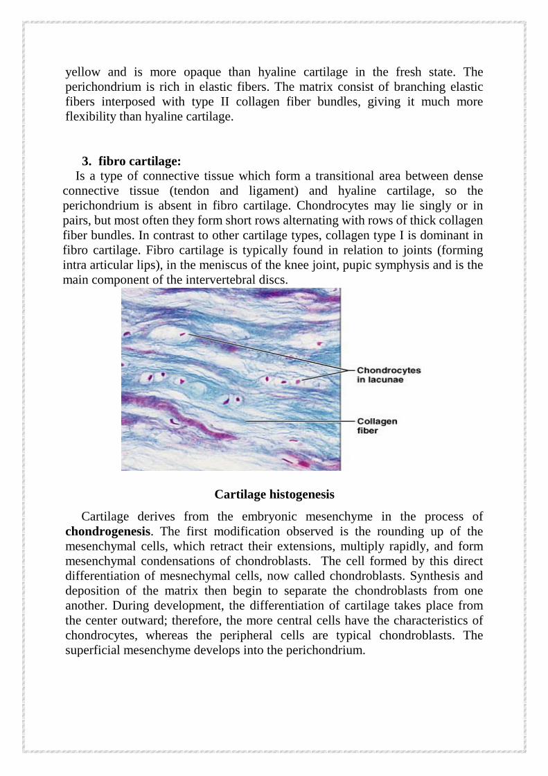
3. fibro cartilage: Is a type of connective tissue which form a transitional area between dense connective tissue (tendon and ligament) and hyaline cartilage, so the perichondrium is absent in fibro cartilage. Chondrocytes may lie singly or in pairs, but most often they form short rows alternating with rows of thick collagen fiber bundles. In contrast to other cartilage types, collagen type I is dominant in fibro cartilage. Fibro cartilage is typically found in relation to joints (forming intra articular lips), in the meniscus of the knee joint, pupic symphysis and is the main component of the intervertebral discs.
Cartilage histogenesis
Cartilage derives from the embryonic mesenchyme in the process of chondrogenesis. The first modification observed is the rounding up of the mesenchymal cells, which retract their extensions, multiply rapidly, and form mesenchymal condensations of chondroblasts. The cell formed by this direct differentiation of mesnechymal cells, now called chondroblasts. Synthesis and deposition of the matrix then begin to separate the chondroblasts from one another. During development, the differentiation of cartilage takes place from the center outward; therefore, the more central cells have the characteristics of chondrocytes, whereas the peripheral cells are typical chondroblasts. The superficial mesenchyme develops into the perichondrium.
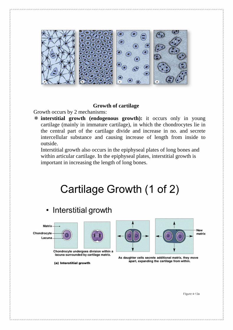
Growth of cartilage
Growth occurs by 2 mechanisms: interstitial growth (endogenous growth): it occurs only in young
cartilage (mainly in immature cartilage), in which the chondrocytes lie in the central part of the cartilage divide and increase in no. and secrete intercellular substance and causing increase of length from inside to outside. Interstitial growth also occurs in the epiphyseal plates of long bones and within articular cartilage. In the epiphyseal plates, interstitial growth is important in increasing the length of long bones.
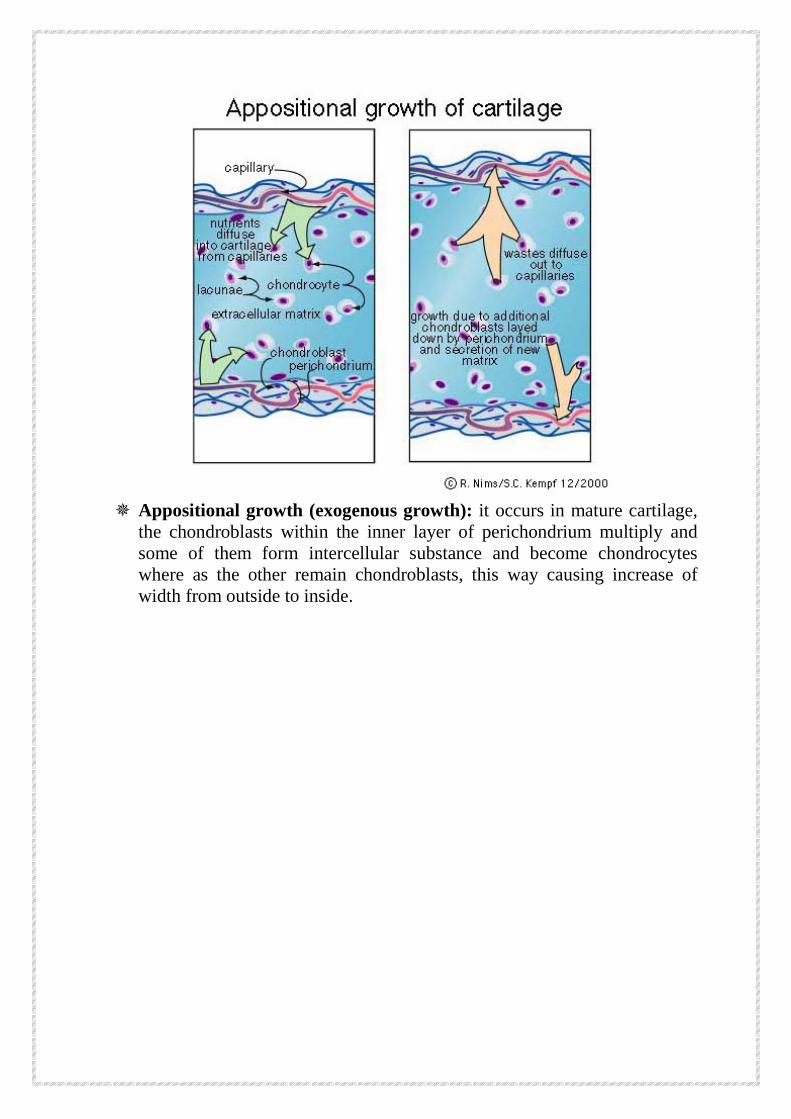
Appositional growth (exogenous growth): it occurs in mature cartilage, the chondroblasts within the inner layer of perichondrium multiply and some of them form intercellular substance and become chondrocytes where as the other remain chondroblasts, this way causing increase of width from outside to inside.
Degenerative changes in cartilage Due to the poor access of nutrients to the chondrocytes they may atrophied in deep parts of thick cartilage. Water content decreases and small cavities arise in the matrix, which often leads to the calcification of the cartilage. The chondrocytes may eventually die, and the cartilage is gradually transformed to bone. In contrast to hyaline cartilage, which can calcify with aging, the matrix of elastic cartilage does not calcify.
Regeneration of cartilage tissue
Except in young children, damaged cartilage undergoes slow and often incomplete regeneration, by activity of cells in the perichondrium which invade the injured area and generate new cartilage. In extensively damaged areas—and occasionally in small areas—the perichondrium produces a scar of dense connective tissue instead of forming new cartilage. The poor regenerative capacity of cartilage is due in part to the avascularity of this tissue.
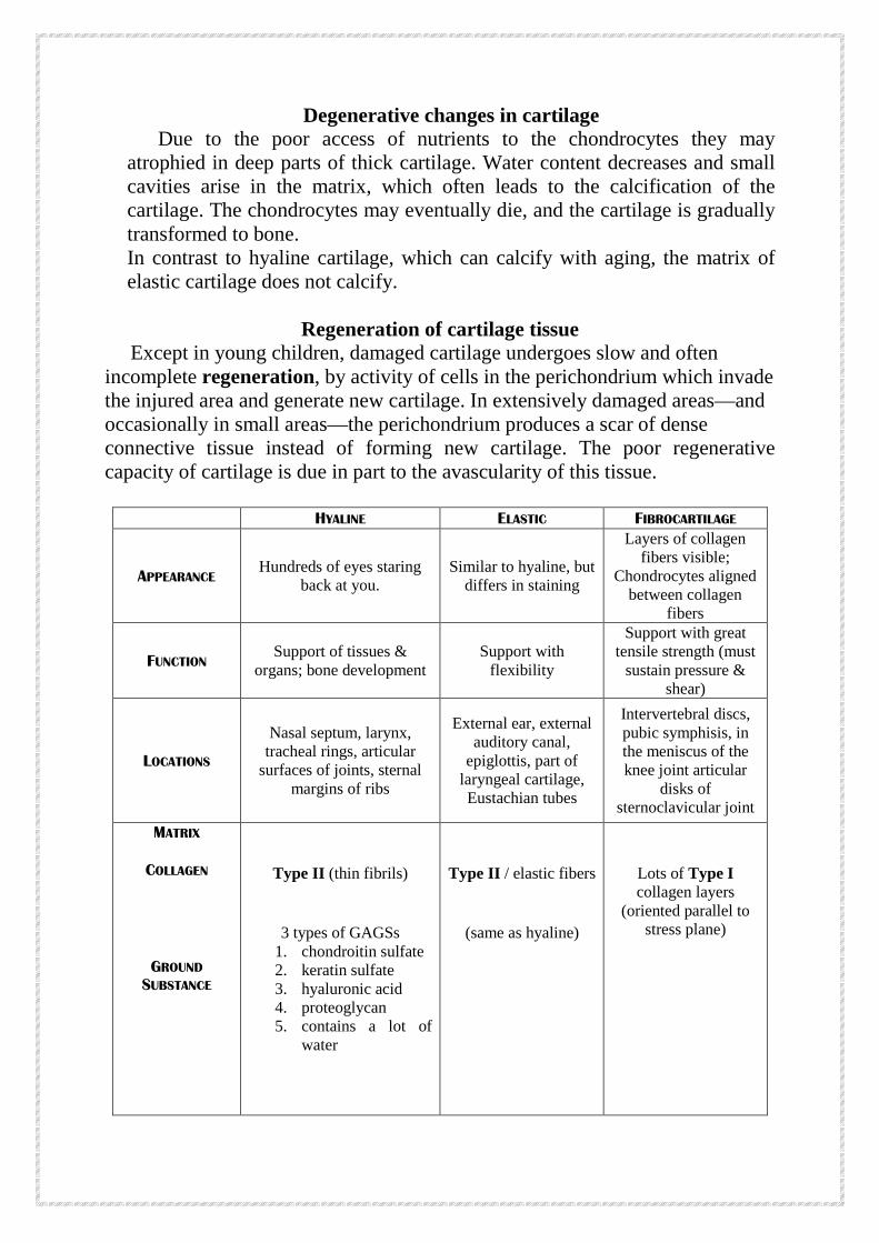
HYALINE ELASTIC FIBROCARTILAGE
APPEARANCE Hundreds of eyes staring
back at you. Similar to hyaline, but
differs in staining
Layers of collagen fibers visible;
Chondrocytes aligned between collagen
fibers
FUNCTION Support of tissues & organs; bone development
Support with flexibility
Support with great tensile strength (must
sustain pressure & shear)
LOCATIONS
Nasal septum, larynx, tracheal rings, articular
surfaces of joints, sternal margins of ribs
External ear, external auditory canal,
epiglottis, part of laryngeal cartilage, Eustachian tubes
Intervertebral discs, pubic symphisis, in the meniscus of the knee joint articular
disks of sternoclavicular joint
MATRIX
COLLAGEN
GROUND SUBSTANCE
Type II (thin fibrils)
3 types of GAGSs 1. chondroitin sulfate 2. keratin sulfate 3. hyaluronic acid 4. proteoglycan 5. contains a lot of
water
Type II / elastic fibers
(same as hyaline)
Lots of Type I collagen layers
(oriented parallel to stress plane)


![Cartilage - facultymembers.sbu.ac.irfacultymembers.sbu.ac.ir/rajabi/ppt toPDF/Cartilage [Compatibility Mode].pdfFibrocartilage • Fibrous Cartilage • is a form of connective tissue](https://img.pdfslide.net/doc/110x75/6012989a4318862a0e5813ae/cartilage-topdfcartilage-compatibility-modepdf-fibrocartilage-a-fibrous.jpg)