Embed Size (px)
Citation preview

Sylv
ia S
. Ma
der
Copyright © The McGraw Hill Companies Inc. Permission required for reproduction or display
PowerPoint® Lecture Slides are prepared by Dr. Isaac Barjis, Biology Instructor
BIOLOGY 10th Edition
1
Membrane Structure
and Function
Chapter 5: pp. 85-102
Copyright © The McGraw-Hill Companies, Inc. Permission required for reproduction or display.
Outside
Inside
plasma membrane
glycolipid
glycoprotein
integral protein
cholesterol
peripheral protein
filaments of cytoskeleton
phospholipid bilayer
extracellular matrix (ECM)
hydrophilic heads
hydrophobic tails
carbohydrate chain

2
Outline
Membrane Models
Fluid-Mosaic
Plasma Membrane Structure and Function
Phospholipids
Proteins
Plasma Membrane Permeability
Diffusion
Osmosis
Transport Via Carrier Proteins
Cell Surface Modifications

3
Structure and Function:
The Phospholipid Bilayer
The plasma membrane is common to all cells
Separates:
Internal living cytoplasmic from
External environment of cell
Phospholipid bilayer:
External surface lined with hydrophilic polar heads
Cytoplasmic surface lined with hydrophilic polar heads
Nonpolar, hydrophobic, fatty-acid tails sandwiched in
between

4
Membrane Models
Fluid-Mosaic Model
Three components: Basic membrane referred to as phospholipid
bilayer
Protein molecules Float around like icebergs on a sea Membrane proteins may be peripheral or integral
Peripheral proteins are found on the inner membrane surface
Integral proteins are partially or wholly embedded (transmembrane) in the membrane
Some have carbohydrate chains attached
Cholesterol

Animation
Please note that due to differing
operating systems, some animations
will not appear until the presentation is
viewed in Presentation Mode (Slide
Show view). You may see blank slides
in the “Normal” or “Slide Sorter” views.
All animations will appear after viewing
in Presentation Mode and playing each
animation. Most animations will require
the latest version of the Flash Player,
which is available at
http://get.adobe.com/flashplayer.

7
The Fluid Mosaic Model
Copyright © The McGraw-Hill Companies, Inc. Permission required for reproduction or display.
Outside
Inside
plasma membrane
glycolipid
glycoprotein
integral protein
cholesterol
peripheral protein
filaments of cytoskeleton
phospholipid bilayer
extracellular matrix (ECM)
hydrophilic heads
hydrophobic tails
carbohydrate chain

8
Transmembrane Proteins
Copyright © The McGraw-Hill Companies, Inc. Permission required for reproduction or display.
hydrophobic region
peripheral proteins
cholesterol
integral protein
hydrophilic regions

9
Lateral Migration of Membrane Proteins
Copyright © The McGraw-Hill Companies, Inc. Permission required for reproduction or display.
hydrophobic region
peripheral proteins
cholesterol
integral protein
hydrophilic regions

10
Functions of Membrane Proteins
Channel Proteins: Tubular Allow passage of molecules through membrane
Carrier Proteins: Combine with substance to be transported Assist passage of molecules through membrane
Cell Recognition Proteins: Provides unique chemical ID for cells Help body recognize foreign substances
Receptor Proteins: Binds with messenger molecule Causes cell to respond to message
Enzymatic Proteins: Carry out metabolic reactions directly

11
Membrane Protein Diversity
Copyright © The McGraw-Hill Companies, Inc. Permission required for reproduction or display.
Channel Protein:
Allows a particular
molecule or ion to
cross the plasma
membrane freely.
Cystic fibrosis, an
inherited disorder,
is caused by a
faulty chloride (Cl–)
channel; a thick
mucus collects in
airways and in
pancreatic and
liver ducts.
Carrier Protein:
Selectively interacts
with a specific
molecule or ion so
that it can cross the
plasma membrane.
The inability of some
persons to use
energy for sodium-
potassium (Na+–K+)
transport has been
suggested as the
cause of their obesity.
Receptor Protein:
Is shaped in such a
way that a specific
molecule can bind to
it. Pygmies are short,
not because they do
not produce enough
growth hormone, but
because their plasma
membrane growth
hormone receptors
are faulty and cannot
interact with growth
hormone.
Enzymatic Protein:
Catalyzes a specific
reaction. The membrane
protein, adenylate
cyclase, is involved in
ATP metabolism. Cholera
bacteria release a toxin
that interferes with the
proper functioning of
adenylate cyclase;
sodium (Na+) and water
leave intestinal cells, and
the individual may die
from severe diarrhea.
Junction Proteins:
Tight junctions join
cells so that a tissue
can fulfill a function, as
when a tissue pinches
off the neural tube
during development.
Without this
cooperation between
cells, an animal
embryo would have no
nervous system.
Cell Recognition
Protein:
The MHC (major
histocompatibility
complex) glycoproteins
are different for each
person, so organ
transplants are difficult
to achieve. Cells with
foreign MHC
glycoproteins are
attacked by white blood
cells responsible for
immunity.
a. b.
d. e.
c.
f.

12
Science Focus: Cell Signaling
Copyright © The McGraw-Hill Companies, Inc. Permission required for reproduction or display.
signaling molecule
receptor activation
unactivated receptor protein
nuclear envelope
b.
a. egg embryo newborn
plasma membrane
Targeted protein:
Cellular response:
enzyme
structural protein
gene regulatory
protein Nucleus Cytoplasm
Altered shape or movement
of cell
1. Receptor: Binds to a signaling molecule, becomes activated and initiates a transduction pathway
2. Transduction pathway: Series of relay proteins that ends when
a protein is activated.
3. Response:Targeted protein(s) bring about the response(s) noted.
Altered metabolism or a function of cell
Altered gene expression and the amount of a cell protein

13
Types of Transport: Active vs. Passive
Plasma membrane is differentially (selectively) permeable
Allows some material to pass
Inhibits passage of other materials
Passive Transport:
No ATP requirement
Molecules follow concentration gradient
Active Transport
Requires carrier protein
Requires energy in form of ATP

Animation
Please note that due to differing
operating systems, some animations
will not appear until the presentation is
viewed in Presentation Mode (Slide
Show view). You may see blank slides
in the “Normal” or “Slide Sorter” views.
All animations will appear after viewing
in Presentation Mode and playing each
animation. Most animations will require
the latest version of the Flash Player,
which is available at
http://get.adobe.com/flashplayer.

15
Passage of Molecules Across the
Membrane
Copyright © The McGraw-Hill Companies, Inc. Permission required for reproduction or display.

16
Types of Membrane Transport: Overview
Copyright © The McGraw-Hill Companies, Inc. Permission required for reproduction or display.
macromolecule
H2O
noncharged molecules
charged molecules and ions
protein
phospholipid molecule
17
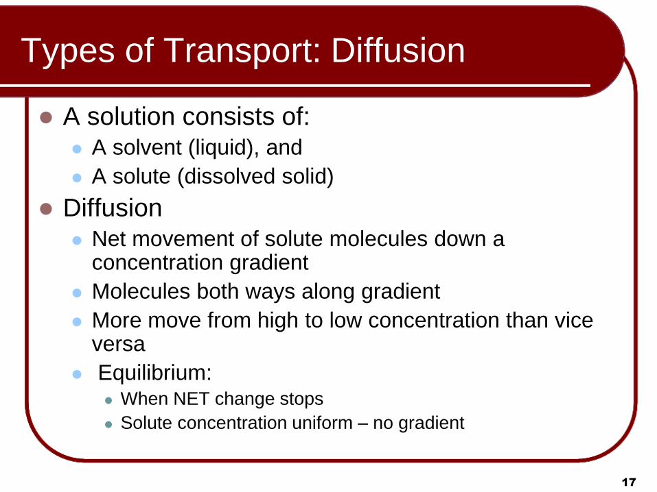
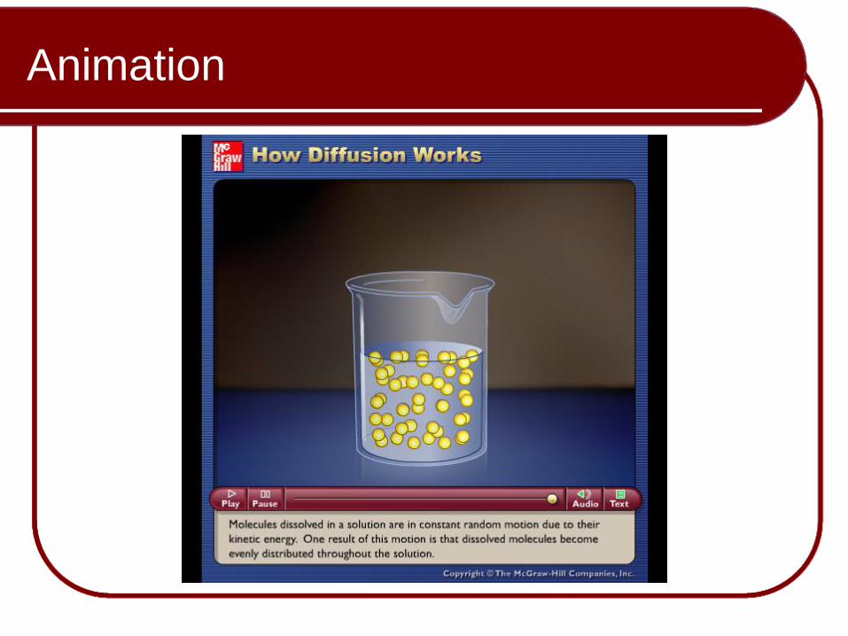
Types of Transport: Diffusion
A solution consists of: A solvent (liquid), and
A solute (dissolved solid)
Diffusion Net movement of solute molecules down a
concentration gradient
Molecules both ways along gradient
More move from high to low concentration than vice versa
Equilibrium: When NET change stops
Solute concentration uniform – no gradient
18
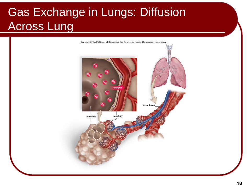
Gas Exchange in Lungs: Diffusion
Across Lung Copyright © The McGraw-Hill Companies, Inc. Permission required for reproduction or display.
capillary alveolus
bronchiole
oxygen
O2
O2
O2
O2
O2
O2 O2
O2
O2
O2
O2
O2

19
Types of Membrane Transport: Diffusion
Copyright © The McGraw-Hill Companies, Inc. Permission required for reproduction or display.
time time
a. Crystal of dye is placed in water b. Diffusion of water and dye molecules c. Equal distribution of molecules results
crystal dye

Animation
20
Please note that due to differing
operating systems, some animations
will not appear until the presentation is
viewed in Presentation Mode (Slide
Show view). You may see blank slides
in the “Normal” or “Slide Sorter” views.
All animations will appear after viewing
in Presentation Mode and playing each
animation. Most animations will require
the latest version of the Flash Player,
which is available at
http://get.adobe.com/flashplayer.
Animation
Please note that due to differing
operating systems, some animations
will not appear until the presentation is
viewed in Presentation Mode (Slide
Show view). You may see blank slides
in the “Normal” or “Slide Sorter” views.
All animations will appear after viewing
in Presentation Mode and playing each
animation. Most animations will require
the latest version of the Flash Player,
which is available at
http://get.adobe.com/flashplayer.

22
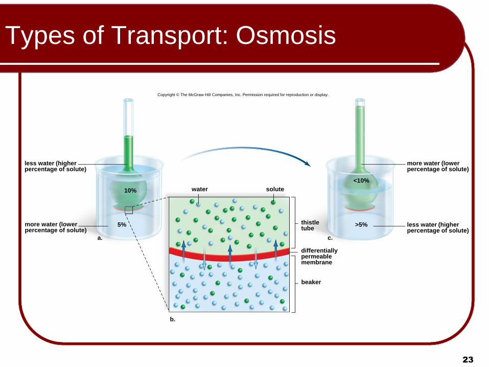
Types of Transport: Osmosis
Osmosis: Special case of diffusion Focuses on solvent (water) movement rather than
solute Diffusion of water across a differentially (selectively)
permeable membrane Solute concentration on one side high, but water
concentration low Solute concentration on other side low, but water
concentration high
Water diffuses both ways across membrane but solute can’t
Net movement of water is toward low water (high solute) concentration
Osmotic pressure is the pressure that develops due to osmosis
23
Types of Transport: Osmosis
Copyright © The McGraw-Hill Companies, Inc. Permission required for reproduction or display.
a.
less water (higher percentage of solute)
more water (lower percentage of solute)
10%
5%
<10%
>5%
solute
differentially permeable membrane
water
b.
c.
less water (higher percentage of solute)
more water (lower percentage of solute)
beaker
thistle tube

Animation
24
Please note that due to differing
operating systems, some animations
will not appear until the presentation is
viewed in Presentation Mode (Slide
Show view). You may see blank slides
in the “Normal” or “Slide Sorter” views.
All animations will appear after viewing
in Presentation Mode and playing each
animation. Most animations will require
the latest version of the Flash Player,
which is available at
http://get.adobe.com/flashplayer.

Animation
Please note that due to differing
operating systems, some animations
will not appear until the presentation is
viewed in Presentation Mode (Slide
Show view). You may see blank slides
in the “Normal” or “Slide Sorter” views.
All animations will appear after viewing
in Presentation Mode and playing each
animation. Most animations will require
the latest version of the Flash Player,
which is available at
http://get.adobe.com/flashplayer.

26
Types of Transport: Osmosis
Isotonic Solution
Solute and water concentrations equal on both sides of membrane
Hypotonic Solution
Concentration of solute lower than on other side
Cells placed in a hypotonic solution will swell
May cause cells to break – Lysis
Hypertonic Solution
Concentration of solute higher than on other side
Cells placed in a hypertonic solution will shrink –
Plasmolysis

27
Osmotic Effects on Cells
Copyright © The McGraw-Hill Companies, Inc. Permission required for reproduction or display.
Animal cells
Plant cells
plasma membrane
chloroplast
nucleus
cell wall
plasma membrane
In an isotonic solution, there is no net movement of water.
In a hypotonic solution, vacuoles fill with water, turgor pressure develops, and chloroplasts are seen next to the cell wall.
In a hypertonic solution, vacuoles lose water, the cytoplasm shrinks (plasmolysis), and chloroplasts are seen in the center of the cell.
In a hypotonic solution, water mainly enters the cell, which may burst (lysis).
In an isotonic solution, there is no net movement of water.
In a hypertonic solution, water mainly leaves the cell, which shrivels (crenation).
nucleus
central vacuole

Animation
28
Please note that due to differing
operating systems, some animations
will not appear until the presentation is
viewed in Presentation Mode (Slide
Show view). You may see blank slides
in the “Normal” or “Slide Sorter” views.
All animations will appear after viewing
in Presentation Mode and playing each
animation. Most animations will require
the latest version of the Flash Player,
which is available at
http://get.adobe.com/flashplayer.

29
Types of Transport: Carrier Proteins
Facilitated Transport
Small molecules
Can’t get through membrane lipids
Combine with carrier proteins
Follow concentration gradient
Active Transport
Small molecules
Move against concentration gradient
Combining with carrier proteins
Requires energy

Animation
30
Please note that due to differing
operating systems, some animations
will not appear until the presentation is
viewed in Presentation Mode (Slide
Show view). You may see blank slides
in the “Normal” or “Slide Sorter” views.
All animations will appear after viewing
in Presentation Mode and playing each
animation. Most animations will require
the latest version of the Flash Player,
which is available at
http://get.adobe.com/flashplayer.

31
Types of Membrane Transport:
Facilitated Transport
Copyright © The McGraw-Hill Companies, Inc. Permission required for reproduction or display.
solute
Outside
Inside
plasma membrane
carrier protein

Animation
Please note that due to differing
operating systems, some animations
will not appear until the presentation is
viewed in Presentation Mode (Slide
Show view). You may see blank slides
in the “Normal” or “Slide Sorter” views.
All animations will appear after viewing
in Presentation Mode and playing each
animation. Most animations will require
the latest version of the Flash Player,
which is available at
http://get.adobe.com/flashplayer.

33
Facilitated Transport:
The Sodium-Potassium Pump
Copyright © The McGraw-Hill Companies, Inc. Permission required for reproduction or display.
carrier protein
1. Carrier has a shape that allows it to take up 3 Na+
Outside
Inside
K+
K+
K+
K+

34
Facilitated Transport:
The Sodium-Potassium Pump
Copyright © The McGraw-Hill Companies, Inc. Permission required for reproduction or display.
carrier
protein
1. Carrier has a shape that allows it to take up 3 Na+.
2. ATP is split, and phosphate group attaches to carrier
Outside
Inside
ATP
K+
P
K+
K+
K+ K+
K+
K+
K+

35
Facilitated Transport:
The Sodium-Potassium Pump
Copyright © The McGraw-Hill Companies, Inc. Permission required for reproduction or display.
carrier protein
1. Carrier has a shape that allows it to take up 3 Na+.
3. Change in shape results and causes carrier to release 3 Na+
outside the cell.
Outside
Inside
ATP
K+
K+
K+
P
P
K+
K+
K+
Na+
K+ K+
K+
2. ATP is split, and phosphate group attaches to carrier
K+
K+
K+

36
Facilitated Transport:
The Sodium-Potassium Pump
carrier protein
1. Carrier has a shape that allows it to take up 3 Na+.
4. Carrier has a shape that allows it to take up 2K+.
3. Change in shape results and causes carrier to release 3 Na+
outside the cell.
Outside
Inside
ATP
K+
K+
K+
K+
K +
K +
K +
K +
P
P
P
K+
K+
K+
K+ K+
K+
2. ATP is split, and phosphate group attaches to carrier.
K+
K+
Copyright © The McGraw-Hill Companies, Inc. Permission required for reproduction or display.

37
Facilitated Transport:
The Sodium-Potassium Pump
carrier protein
1. Carrier has a shape that allows it to take up 3 Na+.
4. Carrier has a shape that allows it to take up 2 K+.
2. ATP is split, and phosphate group attaches to carrier.
3. Change in shape results and causes carrier to release 3 Na+
outside the cell.
5. Phosphate group is released from carrier.
Outside
Inside
ATP
K+
K+
K+
K+ P
P
P
P
K+
K+ K+
Na+
K+ K+
K+
K+ K+
K+
K+
K+
K+
K+
K+
K+
K+
Copyright © The McGraw-Hill Companies, Inc. Permission required for reproduction or display.

38
Facilitated Transport:
The Sodium-Potassium Pump
carrier protein
1. Carrier has a shape that allows it to take up 3 Na+.
4. Carrier has a shape that allows it to take up 2 K+.
2. ATP is split, and phosphate group attaches to carrier.
3. Change in shape results and causes carrier to release 3 Na+
outside the cell.
5. Phosphate group is released from carrier.
Outside
Inside
ATP
K+
K+
K+
K+ P
P
P
P
K+
K+ K+
Na+
K+ K+
K+
K+ K+
K+
K+
K+
K+
K+
K+
K+
K+
Copyright © The McGraw-Hill Companies, Inc. Permission required for reproduction or display.
6. Change in shape results and causes carrier to release 2K+
inside the cell.
K+
K
K+
Na+
K+

Animation
39
Please note that due to differing
operating systems, some animations
will not appear until the presentation is
viewed in Presentation Mode (Slide
Show view). You may see blank slides
in the “Normal” or “Slide Sorter” views.
All animations will appear after viewing
in Presentation Mode and playing each
animation. Most animations will require
the latest version of the Flash Player,
which is available at
http://get.adobe.com/flashplayer.

40
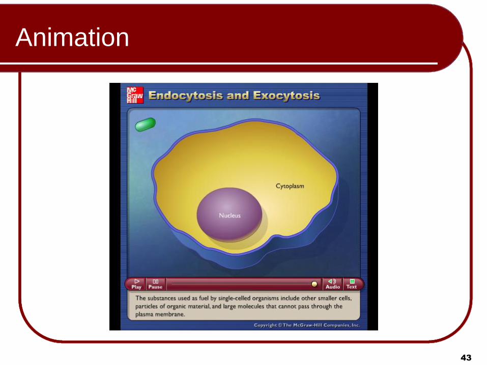
Types of Transport:
Membrane-Assisted Transport
Macromolecules transported into or out of the cell inside vesicles
Exocytosis – Vesicles fuse with plasma membrane and secrete contents
Endocytosis – Cells engulf substances into pouch which becomes a vesicle
Phagocytosis – Large, solid material into vesicle
Pinocytosis – Liquid or small, solid particles go into vesicle
Receptor-Mediated – Specific form of pinocytosis using a coated pit

41
Membrane-Assisted Transport: Exocytosis
Copyright © The McGraw-Hill Companies, Inc. Permission required for reproduction or display.
plasma membrane
Inside
Outside
secretory vesicle

42
Membrane-Assisted Transport:
Three Types of Endocytosis Copyright © The McGraw-Hill Companies, Inc. Permission required for reproduction or display.
pseudopod
paramecium
vacuole forming
vesicles forming
coated pit
coated vesicle
solute
solute
a. Phagocytosis
b. Pinocytosis
vacuole
coated vesicle
plasma membrane
receptor protein
coated pit
c. Receptor-mediated endocytosis
vesicle
0.5 m
399.9 m
Animation
43
Please note that due to differing
operating systems, some animations
will not appear until the presentation is
viewed in Presentation Mode (Slide
Show view). You may see blank slides
in the “Normal” or “Slide Sorter” views.
All animations will appear after viewing
in Presentation Mode and playing each
animation. Most animations will require
the latest version of the Flash Player,
which is available at
http://get.adobe.com/flashplayer.

44
Cell Surface Modifications: Junctions
Cell Surfaces in Animals
Junctions Between Cells
Adhesion Junctions
Intercellular filaments between cells
Tight Junctions
Form impermeable barriers
Gap Junctions
Plasma membrane channels are joined (allows
communication)

45
Cell-Surface Modifications: Junctions
Copyright © The McGraw-Hill Companies, Inc. Permission required for reproduction or display.
c. Gap junction b. Tight junction a. Adhesion junction
membrane channels
intercellular space
plasma membranes
plasma membranes
intercellular space
tight junction proteins
intercellular space
filaments of cytoskeleton
cytoplasmic plaque
intercellular filaments
plasma membranes

46
Cell Surface Modifications
Extracellular Matrix
External meshwork of polysaccharides and proteins
Found in close association with the cell that produced
them
Plant Cell Walls
Plants have freely permeable cell wall, with cellulose
as the main component
Plasmodesmata penetrate cell wall
Each contains a strand of cytoplasm
Allow passage of material between cells

47
Cell-Surface Modifications:
Extracellular Matrix
Copyright © The McGraw-Hill Companies, Inc. Permission required for reproduction or display.
collagen proteoglycan
actin filament
fibronectin
elastin
integrin
Outside (extracellular matrix)
Inside (cytoplasm)

48
Cell-Surface Modifications: Plasmodesmata
Copyright © The McGraw-Hill Companies, Inc. Permission required for reproduction or display.
cell wall
plasmodesmata
cell wall
Cell 1 Cell 2
plasma membrane
cell wall cell wall
cytoplasm
plasma membrane
cytoplasm
middle lamella
plasmodesmata
0.3mm

49
Review
Membrane Models
Fluid-Mosaic
Plasma Membrane Structure and Function
Protein Functions
Plasma Membrane Permeability
Diffusion
Osmosis
Transport Via Carrier Proteins
Cell Surface Modifications

Sylv
ia S
. Ma
der
Copyright © The McGraw Hill Companies Inc. Permission required for reproduction or display
PowerPoint® Lecture Slides are prepared by Dr. Isaac Barjis, Biology Instructor
BIOLOGY 10th Edition
50
Membrane Structure
and Function
Chapter 5: pp. 85-102
Copyright © The McGraw-Hill Companies, Inc. Permission required for reproduction or display.
Outside
Inside
plasma membrane
glycolipid
glycoprotein
integral protein
cholesterol
peripheral protein
filaments of cytoskeleton
phospholipid bilayer
extracellular matrix (ECM)
hydrophilic heads
hydrophobic tails
carbohydrate chain