Embed Size (px)
Citation preview
108 0892-6638/88/0002-0108/$01 .50. © FASEB
Biology of interleukin 1CHARLES A. DINARELLO
Department of Medicine, Tufts University and New England Medical Center Hospital, Boston, Massachusetts 02111, USA
ABSTRACT
Interleukin I (IL 1) is a polypeptide that is produced
after infection, injury, or antigenic challenge. Although
the macrophage is a primary source of IL 1, epidermal,
epithelial, lymphoid, and vascular tissues synthesize IL
1. When IL I gains access to the circulation, it acts likea hormone and induces a broad spectrum of systemic
changes in neurological, metabolic, hematologic, and
endocrinologic systems. Some of the IL 1 that is syn-
thesized remains associated with the plasma membrane
and induces changes in local tissues without producing
systemic responses. IL 1 affects mesenchymal tissueremodeling where it contributes to both destructive
and repair processes. IL I activates lymphocytes and
plays an important role in the initiation of the immuneresponse. Receptors for IL I have been identified, butreceptors are scarce and their affinities often do notmatch the potency of the biological response. The most
consistent property of IL I is up-regulation of cellular
metabolism and increased expression of several genes
coding for biologically active molecules. IL I is a
highly inflammatory molecule and stimulates the
production of arachidonic acid metabolites. IL 1 alsoacts synergistically with other cytokines, particularly
tumor necrosis factor. The multitude of biologicalresponses to IL 1 is an example of the rapid adaptive
changes that take place to increase the host’s defensive
mechanisms. - DINARELLO, C. A. Biology of inter-
leukin 1. FASEBJ 2: 108-115; 1988.
Key Words: interleukins . cytokines monocytes . lympho-
cytes . inflammation fever tumor necrosis factor . prosta-
glandin host defense
ThE POLYPEPTIDE HORMONE INTERLEUKIN 1 (IL 1) is oneof the key mediators of the body’s response to microbialinvasion, inflammation, immunological reactions, andtissue injury. IL 1 is a prominent member of a groupof polypeptide mediators now called cytokines. In thefirst hours after infection or injury, the biological effectsof IL 1 are manifested in nearly every tissue and organ.Unlike other interleukins (interleukins 2-6), IL 1 is nota newly discovered molecule. In the 1940’s, thismaterial was called endogenous pyrogen for its abilityto produce fever (1). Purified endogenous pyrogen (2,3), however, did more than cause fever. It copunifiedwith a substance called leukocytic endogenous mediator
(4), which induced hepatic acute phase protein synthesis,
decreased plasma iron and zinc levels, and produced aneutrophilia. Lymphocyte-activating factor was a pro-tein described by Gery and Waksman (5), which aug-mented T lymphocyte responses to mitogens and anti-gens, but was indistinguishable from endogenouspyrogen (reviewed in ref 6). The term IL 1 now in-cludes the originally described endogenous pyrogen,leukocytic endogenous mediator, and lymphocyte-activating factor, as well as mononuclear cell factor (7),catabolin (8), osteoclast-activating factor, and hemo-poietin 1.
MOLECULAR NATURE OF IL 1
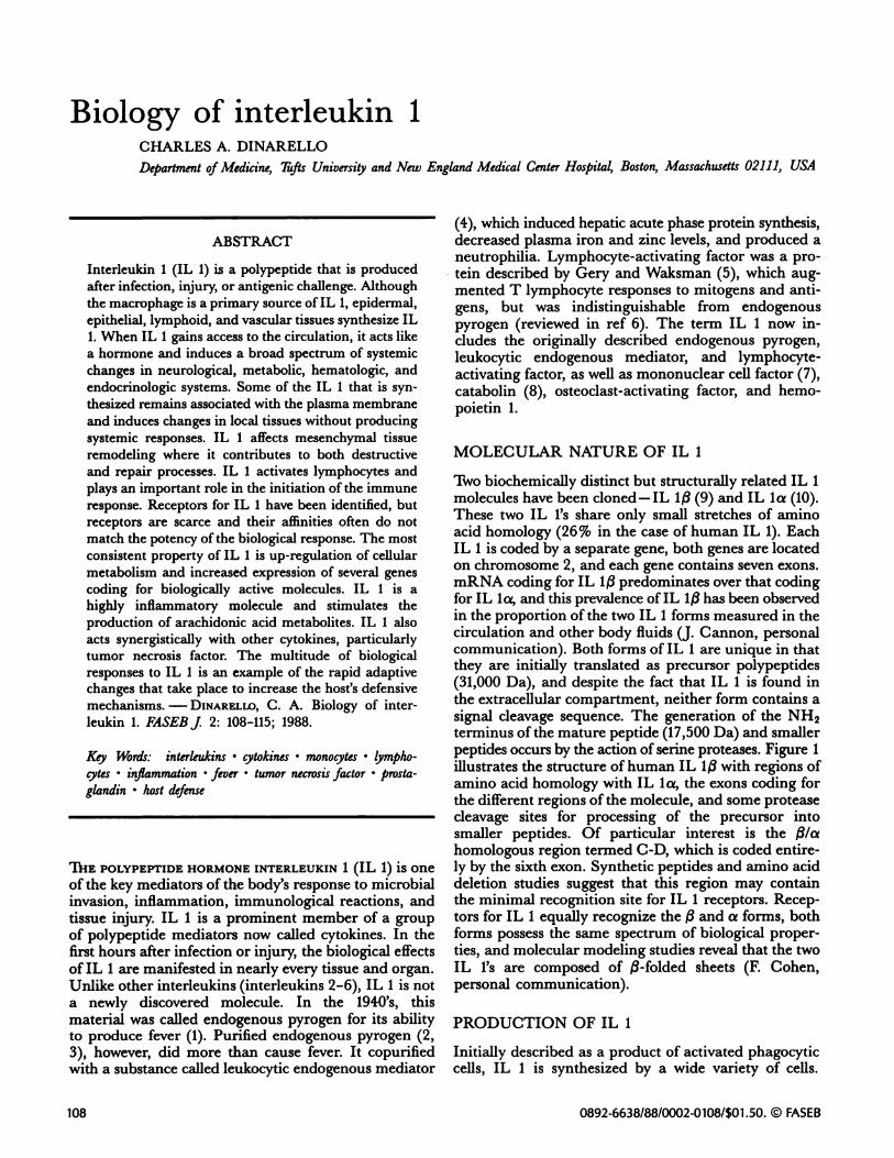
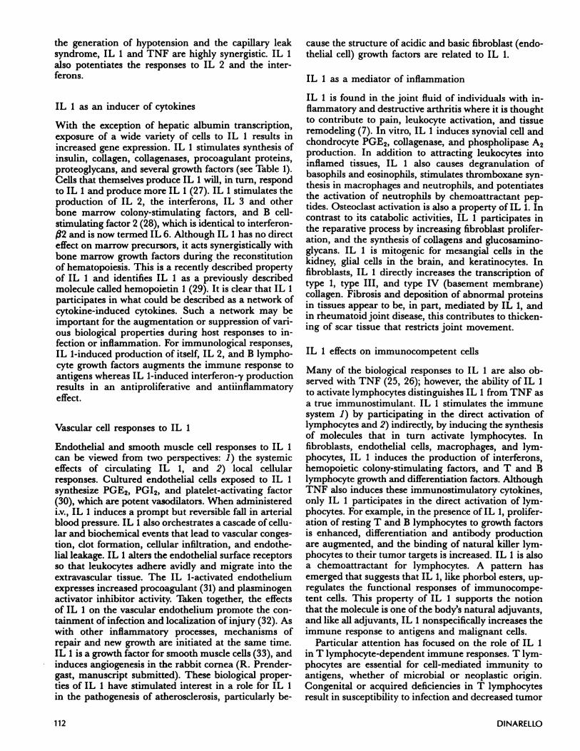
Two biochemically distinct but structurally related IL 1molecules have been cloned-IL 1/3 (9) and IL la (10).These two IL l’s share only small stretches of aminoacid homology (26% in the case of human IL 1). EachIL 1 is coded by a separate gene, both genes are locatedon chromosome 2, and each gene contains seven exons.mRNA coding for IL 1/3 predominates over that codingfor IL 1c and this prevalence of IL l3 has been observedin the proportion of the two IL 1 forms measured in thecirculation and other body fluids (J. Cannon, personalcommunication). Both forms of IL 1 are unique in thatthey are initially translated as precursor polypeptides(31,000 Da), and despite the fact that IL 1 is found inthe extracellular compartment, neither form contains asignal cleavage sequence. The generation of the NH2terminus of the mature peptide (17,500 Da) and smallerpeptides occurs by the action of senine proteases. Figure 1illustrates the structure of human IL 1/3 with regions ofamino acid homology with IL la, the exons coding forthe different regions of the molecule, and some proteasecleavage sites for processing of the precursor intosmaller peptides. Of particular interest is the /3/ahomologous region termed C-D, which is coded entire-ly by the sixth exon. Synthetic peptides and amino aciddeletion studies suggest that this region may containthe minimal recognition site for IL 1 receptors. Recep-tors for IL 1 equally recognize the /3 and a forms, bothforms possess the same spectrum of biological proper-ties, and molecular modeling studies reveal that the twoIL l’s are composed of /3-folded sheets (F. Cohen,personal communication).
PRODUCTION OF IL 1
Initially described as a product of activated phagocyticcells, IL 1 is synthesized by a wide variety of cells.
I i IIIExon I IV
h I L-13
I VII I I I -I
VII
Amino Acid 20 7682 92 150 186 208 219 236
,.
ProteinH2N-
A
.
. BN
C 0
!‘
E
-COOHII II. I__________
269
I(M.t 75)
II I(Ala117) (M.t139) (Gin197)
L6970 I11,155 -5,488
17,376 I22,310
Figure 1. Structure of human IL 1/3. The IL 1/3 precursor protein is depicted and the homologous regions shared with IL icr are shownby the stippled areas (A-E). The exons from the IL 1/3 gene coding for different segments of the protein are aligned above. Comparableexons coding for IL Ia protein exist. Some protease cleavage sites are indicated, and amino acid numbers are shown, as are the lengths
of these resulting peptide fragments. The mature IL 1(17,500 Da) NH2 terminus is indicated by the N at position 117 (alanine). Adaptedfrom refs 9, 11, and ha.
BIOLOGY OF INTERLEUKIN 1 109
These include synovial fibroblasts; keratinocytes andLangerhans cells of the skin; mesangial cells of the kid-ney; B lymphocytes; natural killer cells; astrocytes andmicroglial cells of the brain; vascular endothelial andsmooth muscle cells; corneal, gingival, and thymicepithelial cells; and some T lymphocyte cell lines. Tran-scription for IL I can be initiated in monocytes byadherence to foreign surfaces without much translation
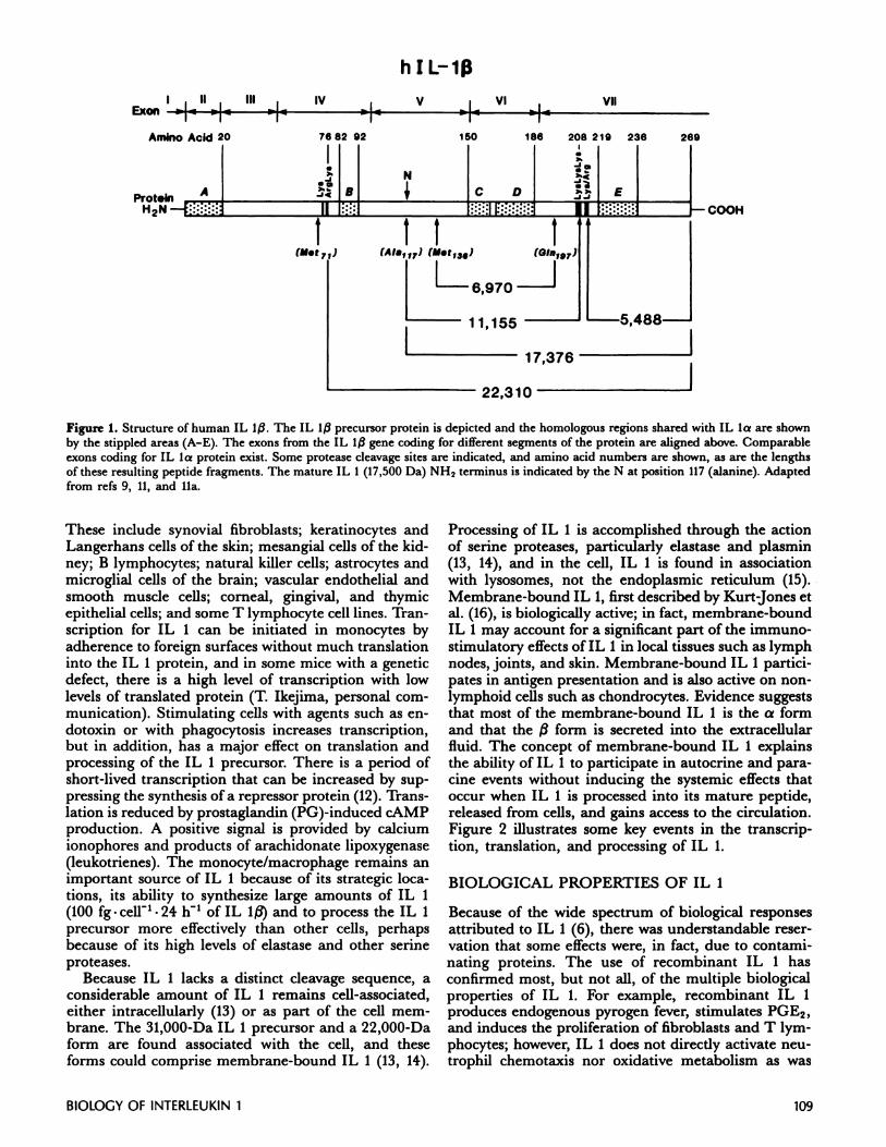
into the IL 1 protein, and in some mice with a geneticdefect, there is a high level of transcription with lowlevels of translated protein (T. Ikejima, personal com-munication). Stimulating cells with agents such as en-dotoxin or with phagocytosis increases transcription,but in addition, has a major effect on translation andprocessing of the IL 1 precursor. There is a period ofshort-lived transcription that can be increased by sup-pressing the synthesis of a repressor protein (12). Trans-lation is reduced by prostaglandin (PG)-induced cAMPproduction. A positive signal is provided by calciumionophores and products of arachidonate lipoxygenase(leukotrienes). The monocyte/macrophage remains animportant source of IL 1 because of its strategic loca-tions, its ability to synthesize large amounts of IL 1(100 fg.celr’.24 h of IL l/) and to process the IL 1precursor more effectively than other cells, perhapsbecause of its high levels of elastase and other serineproteases.
Because IL 1 lacks a distinct cleavage sequence, aconsiderable amount of IL 1 remains cell-associated,either intracellularly (13) or as part of the cell mem-brane. The 31,000-Da IL 1 precursor and a 22,000-Daform are found associated with the cell, and theseforms could comprise membrane-bound IL 1 (13, 14).
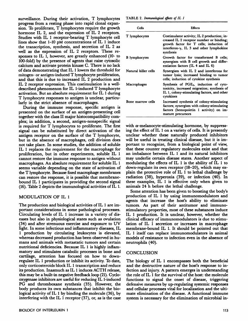
Processing of IL 1 is accomplished through the actionof serine proteases, particularly elastase and plasmin(13, 14), and in the cell, IL 1 is found in associationwith lysosomes, not the endoplasmic reticulum (15).Membrane-bound IL 1, first described by Kurt-Jones etal. (16), is biologically active; in fact, membrane-boundIL 1 may account for a significant part of the immuno-stimulatory effects of IL 1 in local tissues such as lymphnodes, joints, and skin. Membrane-bound IL 1 partici-pates in antigen presentation and is also active on non-lymphoid cells such as chondrocytes. Evidence suggeststhat most of the membrane-bound IL 1 is the a formand that the /3 form is secreted into the extracellularfluid. The concept of membrane-bound IL 1 explainsthe ability of IL 1 to participate in autocrine and para-cine events without inducing the systemic effects thatoccur when IL 1 is processed into its mature peptide,released from cells, and gains access to the circulation.Figure 2 illustrates some key events in the transcrip-tion, translation, and processing of IL I.
BIOLOGICAL PROPERTIES OF IL 1
Because of the wide spectrum of biological responsesattributed to IL 1 (6), there was understandable reser-vation that some effects were, in fact, due to contami-nating proteins. The use of recombinant IL 1 hasconfirmed most, but not all, of the multiple biologicalproperties of IL 1. For example, recombinant IL 1produces endogenous pyrogen fever, stimulates PGE2,and induces the proliferation of fibroblasts and T lym-phocytes; however, IL 1 does not directly activate neu-trophil chemota.xis nor oxidative metabolism as was
TranscriptionalRepressor
‘4
2
Activators
110 Dl NARELLO
kD
Figure 2. Steps in the transcription, translation, and processing of IL 1. Cell activators stimulate PGE, leukotrienes (LT), and intracellularcalcium levels. Transcription and translation are enhanced by calcium and LT. Transcription is short-lived because of the synthesis of atranscriptional repressor protein (or proteins), and translation is reduced by PGE-induced cAMP. The 31,000-Da IL 1 precursor (proIL 1) undergoes limited cleavage into 22,000- and 17,000-Da peptides that can be found associated with the cell as well as in the extracellu-lar compartment. Membrane-bound IL h can be the 31,000-Da precursor or the 22,000-Da product. Lysosomal enzymes and serine pro-teases are responsible for cleavage of pro IL 1 into various peptides with molecular masses of 17,000 (the prominent extracellular form)
and also of 11,000, 4000, and 2000 forms as determined by gel electrophoresis.
previously claimed. Some of the in vitro effects ofrecombinant IL 1 (either /3or a) are depicted in Table 1.The effective concentration of IL 1 that induces theseeffects ranges from I nM to 1 pM (in some cases less);
unlike interferons and bone marrow growth factors, IL Ihas no species specificity.
IL 1 specifically binds to a variety of cells, but the pre-cise nature of the IL 1 receptor or receptors and thenature of the receptor-ligand interaction remain unclear.For example, human neutrophils express specific recep-tors for IL 1 but intracellular calcium levels and oxida-tive metabolism are unaffected by IL 1. Similar dis-crepancies between specific IL 1 binding and biologicalresponses have been observed in endothelial cells.Studies with T lymphocytes and fibroblasts suggest theexistence of a single class of high-affinity receptor witha dissociation constant (KD) that varies from 5 to 50pM with 100-4000 binding sites per cell. There is alsoevidence that a low-affinity receptor exists with a KD of
300-500 pM and 15,000 sites per cell (17; N. Savage,
personal communication). The high-affinity receptorsare rapidly internalized and bind to nuclear structures,
and responsiveness to IL 1 is down-regulated (18-20).The rapid down-regulation of IL 1 receptor is specificand may account, in part, for modulating IL 1 effects
in several cells. To date, the amino acid sequence of aputative IL 1 receptor is unknown. In cells stimulatedwith IL 1, cytosolic calcium increases, sodium/potassiumion fluxes occur, and protein kinase activity increases.Many effects of IL 1 in vitro are mimicked by agentssuch as phorbol esters, which directly activate proteinkinases.
Neurological, metabolic, and endocrinologic effects
of IL 1
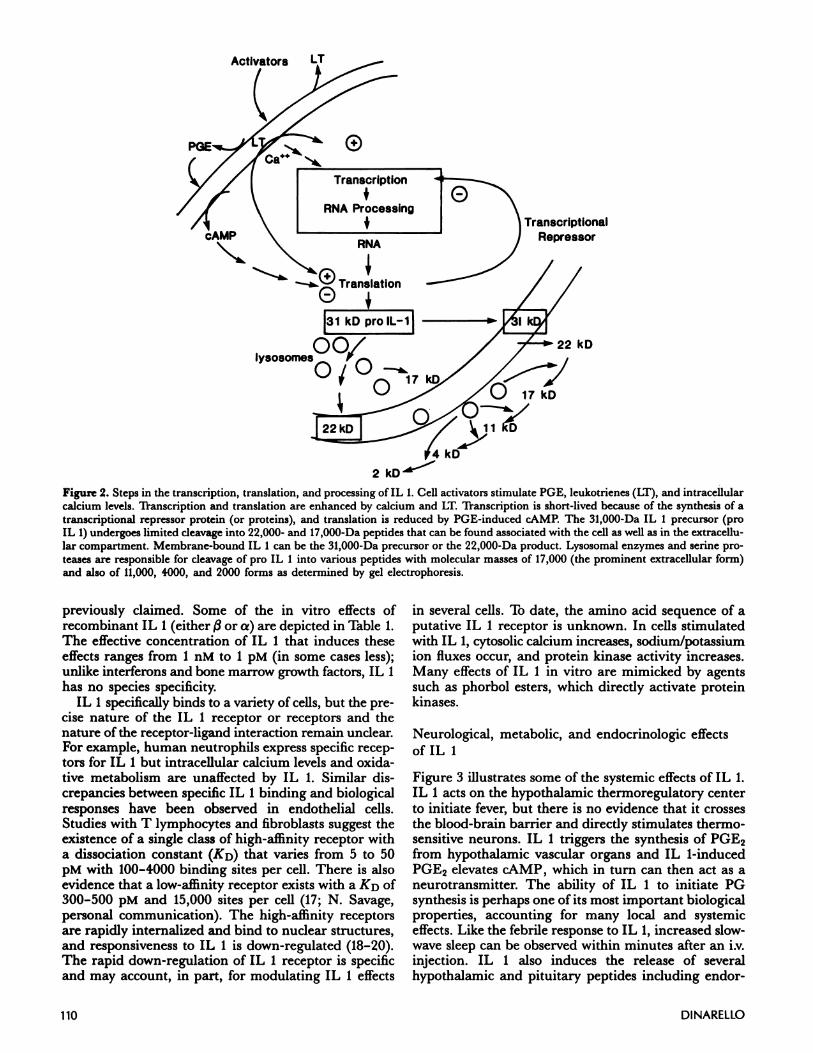
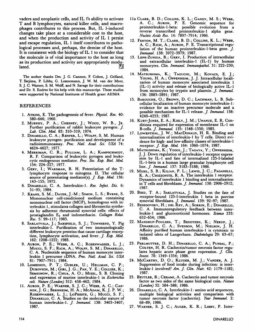
Figure 3 illustrates some of the systemic effects of IL 1.
IL 1 acts on the hypothalamic thermoregulatory centerto initiate fever, but there is no evidence that it crossesthe blood-brain barrier and directly stimulates thermo-sensitive neurons. IL I triggers the synthesis of PGE2from hypothalamic vascular organs and IL 1-inducedPGE2 elevates CAMP, which in turn can then act as aneurotransmitter. The ability of IL 1 to initiate PGsynthesis is perhaps one of its most important biologicalproperties, accounting for many local and systemic
effects. Like the febrile response to IL 1, increased slow-wave sleep can be observed within minutes after an i.v.injection. IL 1 also induces the release of severalhypothalamic and pituitary peptides including endor-
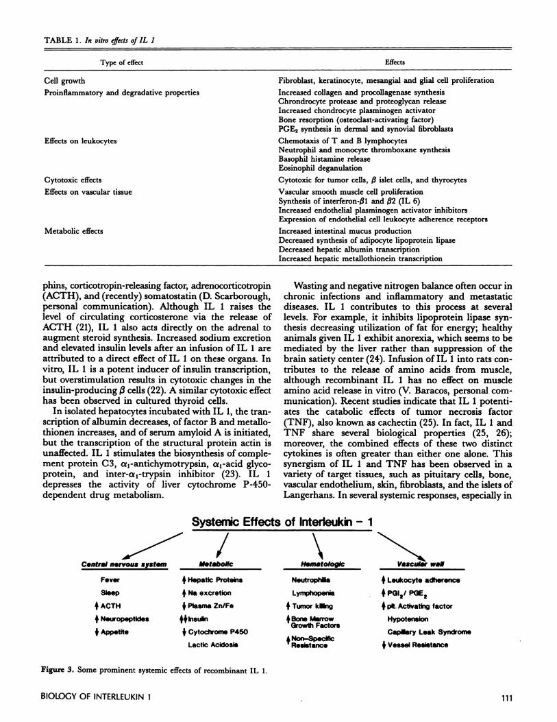
Type of effect
Cell growth
Proinflammatory and degradative properties
Effects on leukocytes
Cytotoxic effects
Effects on vascular tissue
Metabolic effects
Fever +Hepatic Proteins
Sleep +Na excretion
4ACTH 4Plasma Zn/Fe
+Neuropeptldes 4$Insulin
+Appetite $CytocIiome P450
Lactic Acidosis+Non-SpecIfic
Resistance
TABLE 1. In vitro effects oJIL 1
BIOLOGY OF INTERLEUKIN 1 111
Effects
Fibroblast, keratinocyte, mesangial and glial cell proliferation
Increased collagen and procollagenase synthesisChrondrocyte protease and proteoglycan releaseIncreased chondrocyte plasminogen activator
Bone resorption (osteoclast-activating factor)PGE2 synthesis in dermal and synovial fibroblasts
Chemotaxis of T and B lymphocytesNeutrophil and monocyte thromboxane synthesisBasophil histamine releaseEosinophil deganulation
Cytotoxic for tumor cells, /3 islet cells, and thyrocytes
Vascular smooth muscle cell proliferationSynthesis of interferon-fl and /32 (IL 6)Increased endothelial plasminogen activator inhibitorsExpression of endothelial cell leukocyte adherence receptors
Increased intestinal mucus productionDecreased synthesis of adipocyte lipoprotein lipaseDecreased hepatic albumin transcriptionIncreased hepatic metallothionein transcription
phins, corticotropin-releasing factor, adrenocorticotropin(ACTH), and (recently) somatostatin (D. Scarborough,personal communication). Although IL 1 raises thelevel of circulating corticosterone via the release ofACTH (21), IL 1 also acts directly on the adrenal toaugment steroid synthesis. Increased sodium excretionand elevated insulin levels after an infusion of IL 1 areattributed to a direct effect of IL 1 on these organs. Invitro, IL 1 is a potent inducer of insulin transcription,but overstimulation results in cytotoxic changes in theinsulin-producing /3 cells (22). A similar cytotoxic effecthas been observed in cultured thyroid cells.
In isolated hepatocytes incubated with IL 1, the tran-scription of albumin decreases, of factor B and metallo-thionen increases, and of serum amyloid A is initiated,but the transcription of the structural protein actin isunaffected. IL I stimulates the biosynthesis of comple-ment protein C3, a1-antichymotrypsin, a1-acid glyco-protein, and inter-a1-trypsin inhibitor (23). IL 1depresses the activity of liver cytochrome P-450-dependent drug metabolism.
Wasting and negative nitrogen balance often occur inchronic infections and inflammatory and metastaticdiseases. IL 1 contributes to this process at severallevels. For example, it inhibits lipoprotein lipase syn-thesis decreasing utilization of fat for energy; healthyanimals given IL 1 exhibit anorexia, which seems to bemediated by the liver rather than suppression of thebrain satiety center (24). Infusion of IL 1 into rats con-tributes to the release of amino acids from muscle,although recombinant IL 1 has no effect on muscleamino acid release in vitro (V. Baracos, personal com-munication). Recent studies indicate that IL 1 potenti-ates the catabolic effects of tumor necrosis factor(TNF), also known as cachectin (25). In fact, IL 1 andTNF share several biological properties (25, 26);moreover, the combined effects of these two distinctcytokines is often greater than either one alone. Thissynergism of IL 1 and TNF has been observed in avariety of target tissues, such as pituitary cells, bone,vascular endothelium, skin, fibroblasts, and the islets ofLangerhans. In several systemic responses, especially in
C.ntral nsrvous syst.m -
Systenic Effects of Interleukun - 1
/ _M.taboflc H.n,atotoglc
Neutrophilia
Lymphopenia
+Tumor klng
+Bone MarrowGrowth Factors
Vascular will
+Leukocyte adherence
+PGI2/
+pIt. Activating factor
Hypotenslon
Capillary Leak Syndrome
$Vessel Resistance
Figure 3. Some prominent systemic effects of recombinant IL 1.
112 DI NARELLO
the generation of hypotension and the capillary leaksyndrome, IL 1 and TNF are highly synergistic. IL 1also potentiates the responses to IL 2 and the inter-ferons.
IL I as an inducer of cytokines
With the exception of hepatic albumin transcription,exposure of a wide variety of cells to IL 1 results inincreased gene expression. IL 1 stimulates synthesis ofinsulin, collagen, collagenases, procoagulant proteins,proteoglycans, and several growth factors (see Table 1).
Cells that themselves produce IL 1 wifi, in turn, respondto IL 1 and produce more IL 1(27). IL 1 stimulates theproduction of IL 2, the interferons, IL 3 and otherbone marrow colony-stimulating factors, and B cell-stimulating factor 2 (28), which is identical to interferon-/32 and is now termed IL 6. Although IL 1 has no directeffect on marrow precursors, it acts synergistically withbone marrow growth factors during the reconstitutionof hematopoiesis. This is a recently described propertyof IL 1 and identifies IL 1 as a previously describedmolecule called hemopoietin 1 (29). It is clear that IL 1participates in what could be described as a network ofcytokine-induced cytokines. Such a network may beimportant for the augmentation or suppression of vari-ous biological properties during host responses to in-fection or inflammation. For immunological responses,IL I-induced production of itself, IL 2, and B lympho-cyte growth factors augments the immune response toantigens whereas IL 1-induced interferon--y productionresults in an antiproliferative and antiinflammatoryeffect.
Vascular cell responses to IL 1
Endothelial and smooth muscle cell responses to IL 1can be viewed from two perspectives: 1) the systemiceffects of circulating IL 1, and 2) local cellularresponses. Cultured endothelial cells exposed to IL 1synthesize PGE2, PGI2, and platelet-activating factor(30), which are potent vasodilators. When administeredi.v., IL 1 induces a prompt but reversible fall in arterialblood pressure. IL 1 also orchestrates a cascade of cellu-lar and biochemical events that lead to vascular conges-tion, clot formation, cellular infiltration, and endothe-hal leakage. IL 1 alters the endothelial surface receptorsso that leukocytes adhere avidly and migrate into theextravascular tissue. The IL 1-activated endotheliumexpresses increased procoagulant (31) and plasminogenactivator inhibitor activity. Taken together, the effectsof IL 1 on the vascular endothelium promote the con-tainment of infection and localization of injury (32). Aswith other inflammatory processes, mechanisms ofrepair and new growth are initiated at the same time.IL 1 is a growth factor for smooth muscle cells (33), andinduces angiogenesis in the rabbit cornea (R. Prender-gast, manuscript submitted). These biological proper-ties of IL 1 have stimulated interest in a role for IL 1in the pathogenesis of atherosclerosis, particularly be-
cause the structure of acidic and basic fibroblast (endo-thehial cell) growth factors are related to IL 1.
IL 1 as a mediator of inflammation
IL 1 is found in the joint fluid of individuals with in-flammatory and destructive arthritis where it is thoughtto contribute to pain, leukocyte activation, and tissueremodeling (7). In vitro, IL 1 induces synovial cell andchondrocyte PGE2, collagenase, and phospholipase A2production. In addition to attracting leukocytes intoinflamed tissues, IL 1 also causes degranulation ofbasophils and eosinophils, stimulates thromboxane syn-thesis in macrophages and neutrophils, and potentiatesthe activation of neutrophils by chemoattractant pep-tides. Osteoclast activation is also a property of IL 1. Incontrast to its catabolic activities, IL 1 participates inthe reparative process by increasing fibroblast prolifer-ation, and the synthesis of collagens and glucosamino-glycans. IL 1 is mitogenic for mesangial cells in thekidney, ghial cells in the brain, and keratinocytes. Infibroblasts, IL 1 directly increases the transcription oftype 1, type III, and type IV (basement membrane)collagen. Fibrosis and deposition of abnormal proteinsin tissues appear to be, in part, mediated by IL 1, andin rheumatoid joint disease, this contributes to thicken-ing of scar tissue that restricts joint movement.
IL I effects on immunocompetent cells
Many of the biological responses to IL 1 are also ob-served with TNF (25, 26); however, the ability of IL 1to activate lymphocytes distinguishes IL 1 from TNF asa true immunostimulant. IL 1 stimulates the immunesystem 1) by participating in the direct activation oflymphocytes and 2) indirectly, by inducing the synthesisof molecules that in turn activate lymphocytes. Infibroblasts, endothelial cells, macrophages, and lym-phocytes, IL 1 induces the production of interferons,hemopoietic colony-stimulating factors, and T and Blymphocyte growth and differentiation factors. AlthoughTNF also induces these immunostimulatory cytokines,only IL 1 participates in the direct activation of lym-phocytes. For example, in the presence of IL 1, prolifer-ation of resting T and B lymphocytes to growth factorsis enhanced, differentiation and antibody productionare augmented, and the binding of natural killer lym-phocytes to their tumor targets is increased. IL 1 is alsoa chemoattractant for lymphocytes. A pattern hasemerged that suggests that IL 1, like phorbol esters, up-regulates the functional responses of immunocompe-tent cells. This property of IL 1 supports the notionthat the molecule is one of the body’s natural adjuvants,and like all adjuvants, IL 1 nonspecifically increases theimmune response to antigens and malignant cells.
Particular attention has focused on the role of IL 1in T lymphocyte-dependent immune responses. T lym-phocytes are essential for cell-mediated immunity toantigens, whether of microbial or neoplastic origin.Congenital or acquired deficiencies in T lymphocytesresult in susceptibility to infection and decreased tumor
BIOLOGY OF INTERLEU KIN 1 113
surveillance. During their activation, T lymphocytesprogress from a resting phase into rapid clonal expan-sion. To proliferate, T lymphocytes require the growthhormone IL 2, and the expression of IL 2 receptors.Studies with IL 1 receptor-bearing T lymphocyte celllines show that 1-10 pM concentrations of IL I inducethe transcription, synthesis, and secretion of IL 2 aswell as the expression of IL 2 receptors. These re-sponses to IL 1, however, are greatly enhanced (10- to100-fold) by the presence of agents that raise cytosoliccalcium and activate protein kinase C. There is no lackof data demonstrating that IL 1 lowers the threshold formitogen- or antigen-induced T lymphocyte proliferation,and that this is due to increased IL 2 production andIL 2 receptor expression. This costimulation is a well-described phenomenon for IL 1-induced T lymphocyteactivation. But an absolute requirement for IL 1 duringT lymphocyte responses to antigen is unclear, particu-larly in the strict absence of macrophages.
During the immune response, specific antigen ispresented on the surface of an antigen-presenting celltogether with the class II major histocompatibiity com-plex; in addition, a second, antigen-nonspecific signalis required for T lymphocytes to proliferate. The firstsignal can be substituted by direct activation of theantigen receptor on the surface of the T lymphocyte,but in the absence of macrophages, cell division doesnot take place. In some studies, the addition of solubleIL 1 replaces the requirement for the macrophage forproliferation, but in other experiments, soluble IL 1cannot restore the immune response to antigen withoutmacrophages. An absolute requirement for soluble IL 1seems variable depending on the state of activation ofthe T lymphocyte. Because fixed macrophage membranescan restore the response, it is possible that membrane-bound IL 1 participates in providing the second signal(16). Table 2 depicts the immunological activities of IL 1.
MODULATION OF IL 1
The production and biological activities of IL 1 are im-portant considerations in some pathological processes.Circulating levels of IL 1 increase in a variety of dis-eases but also in physiological states such as ovulation(34) and after strenuous exercise and exposure to UVlight. In some infectious and inflammatory diseases, IL1 production by circulating leukocytes is elevated,whereas decreased production has been observed in hu-mans and animals with metastatic tumors and certainnutritional deficiencies. Because IL 1 is highly inflam-matory and stimulates catabolic processes in bone andcartilage, attention has focused on how to down-regulate IL 1 production or inhibit its activity. To date,only corticosteroids block IL 1 transcription and reduceits production. Inasmuch as IL 1 induces ACTH release,this may be a built-in negative feedback loop (21). Cyclo-oxygenase inhibitors are useful for reducing IL I-inducedPG and thromboxane synthesis (35). However, thebody produces its own substances that inhibit the bio-logical activity of IL 1 by binding the molecule (36), byinterfering with the IL 1 receptor (37), or, as is the case
TABLE 2. Immunological ejects of IL 1
Cells Effects
T lymphocytes Costimulator activity; IL 2 production; in-
creased IL 2 receptor number or binding;growth factor for T cells; induction ofinterferon-7, IL 3 and other lymphokinesynthesis
B lymphocytes Growth factor for transformed B cells;synergism with B cell growth and differ-entiation factors (IL 4 and IL 6)
Natural killer cells Synergism with IL 2 and interferons fortumor lysis; increased binding to tumorcells; induction of cytokine synthesis
Macrophages Synthesis of PGE2, induction of cyto-
toxicity, increased migration; synthesis ofIL 1, colony-stimulating factors, and othercytokines
Bone marrow cells Increased synthesis of colony-stimulatingfactors; synergism with colony-stimulatingfactors (hemopoietin 1 activity) on im-mature precursors
with a-melanocyte-stimulating hormone, by suppress-ing the effect of IL 1 on a variety of cells. It is presentlyunclear whether these naturally produced inhibitorswill be useful in treating clinical disease, but it is im-portant to recognize, from a biological point of view,that these counter regulatory molecules exist and thatan imbalance between IL I and its natural inhibitorsmay underlie certain disease states. Another aspect ofmodulating the effects of IL 1 is the ability of IL 1 todown-regulate its own receptor (18-20), which may ex-plain the protective role of IL 1 to lethal challenge byradiation (38), hyperoxia (39), or infection (40). Inthese examples, IL 1 is effective only when given toanimals 24 h before the lethal challenge.
Some attention has been given to boosting the body’sproduction of IL 1 by using immunomodulators andagents that increase the host’s ability to eliminatetumors. As part of their antitumor and immuno-stimulatory properties, most of these substances induceIL 1 production. It is unclear, however, whether theclinical efficacy of immunomodulators is due to stimu-lation of IL 1 secretion or increases in macrophagemembrane-bound IL 1. It should be pointed out thatIL 1 itself can replace immunomodulators in animalmodels of resistance to infection even in the absence ofneutrophils (40).
CONCLUSION
The biology of IL 1 encompasses both the beneficialand the destructive nature of the host’s response to in-fection and injury. A pattern emerges in understandingthe role of IL 1 for the survival of the host: the moleculefunctions to signal the onset of disease, triggeringdefensive measures by up-regulating systemic responsesand cellular processes vital for localization and the ulti-mate elimination of the disease. A functional immunesystem is necessary for the elimination of microbial in-
114 DI NARELLO
vaders and neoplastic cells, and IL l’s ability to activateT and B lymphocytes, natural killer cells, and macro-phages contributes to this process. But, IL 1-inducedchanges take place at a considerable cost to the host,and when the production and activity of IL 1 persistand escape regulation, IL 1 itself contributes to patho-logical processes and, perhaps, the demise of the host.It is consistent with the biology of IL 1 to consider thatthe molecule is of vital importance to the host as longas its production and activity are appropriately modu-lated. E!1
The author thanks Drs. J. G. Cannon, F. Cohen, J. Gelfand,
T Ikejima, P. Libby, G. Lonnemann, J. W. M. van der Meer,
S. J. C. Warner, S. M. Wolff, and N. Savage for their contributions
and Dr. S. Endres for his help with this manuscript. These studieswere supported by National Institutes of Health grant A115614.
REFERENCES
1. ATKINS, E. The pathogenesis of fever. Physiol.Rev. 40:580-646; 1960.
2. MURPHY, P. A.; CHESNEY, J.; WooD, W. B., JR.Further purification of rabbit leukocyte pyrogen. J.Lab. Gun. Med. 83: 310-319; 1974.
3. DINARELLO, C. A.; RENFER, L.; WOLFF, S. M. Humanleukocyte pyrogen: purification and development of aradioimmunoassay. Proc. Nail. Acad. Sci. USA 74:4624-4627; 1977.
4. MERRIMAN, C. R.; PULLIAM, L. A.; KAMPSCHMIDT,
R. F. Comparison of leukocytic pyrogen and leuko-cytic endogenous mediator. Proc.Soc. Exp. Biol.Med.154: 224-227; 1977.
5. GERY, I.; WAKSMAN, B. H. Potentiation of the T-lymphocyte response to mitogens. II. The cellularsource of potentiating mediator(s). J. Exp. Med. 136:143-155; 1972.
6. DINARELLO, C. A. Interleukin-1. Rev. Infect. Dis. 6:51-95; 1984.
7. KRANE, S. M.; DAYER,J.-M.; SIMON, L. S.; BYRNE, S.Mononuclear cell-conditioned medium containingmononuclear cell factor (MCF), homologous with in-terleukin 1, stimulates collagen and fibronectin synthe-sis by adherent rheumatoid synovial cells: effects ofprostaglandin E2 and indomethacin. Collagen Relat.Res. 5: 99-117; 1985.
8. SAKLATVALA, J.; SARSFIELD, S. J.; TOWNSEND, Y. Piginterleukin- 1. Purification of two immunologically
different leukocyte proteins that cause cartilage resorp-tion, lymphocyte activation, and fever. J. Exp. Med.162: 1208-1222; 1985.
9. Auto, P. E.; WEBB, A. C.; ROSENWASSER, L. J.;Muccl, S. F.; RICH, A.; WOLFF, S. M.; DINARELLO,C. A. Nucleotide sequence of human monocyte inter-leukin I precursor cDNA. Proc. Nail. Acad. Sci. USA81: 7907-7911; 1984.
10. L0MEDIc0, P. T.; GUBLER, U.; HELLMAN, C. P.;DuKovIcH, M.; Gnu,J. G.; PAN, Y. E.; COLLIER, K.;SEMI0N0w, R.; CHUA, A. 0.; MIZEL, S. B. Cloningand expression of murine interleukin- 1 in Escherichiacoli. Nature (London) 312: 458-462; 1984.
11. Auitor, P. E.; WARNER, S. J. C.; WEBB, A. C.; CAN-
NON,J. G.; BERNHEIM, H. A.; MCADAM, K.J. P. W.;RO5ENWASSER, L. J.; LOPRESTE, G.; Muccl, S. F.;DINARELLO, C. A. Studies on the molecular nature ofhuman interleukin-1. J. Immunol. 138: 3403-3407;1987.
ha CLARK, B. D.; COLLINS, K. L.; GANDY, M. S.; WEBB,
A. C.; AURON, P. E. Genomic sequence forprointerleukin-1-beta: possible evolution from areverse transcribed prointerleukin-1 alpha gene.Nucleic Acids Res. 14: 7897-7914; 1986.
12. FENTON, M. T.; CLARK, B. D.; COLLINS, K. L.; WEBB,
A. C.; RICH, A.; AURON, P. E. Transcriptional regu-lation of the human prointerleukin-1-beta gene. J.Immunol. 138: 3972-3979; 1987.
13. LEPE-ZUNIGA, B.; GERY, I. Production of intracellular
and extracellular interleukin-1 (IL-i) by human
monocytes. Clin. Immunol. Immunopathol. 31: 222-230;1984.
14. MATSUSHIMA, K.; TAGUCHI, M.; KOVACS, E. J.;YOUNG, H. A.; OPPENHEIM, J. J. Intracellular locali-zation of human monocyte associated interleukin I(IL-i) activity and release of biologically active IL-ifrom monocytes by trypsin and plasmin. J. Immunol.136: 2883-2891; 1987.
15. BAKOUCHE, 0.; BROWN, D. C.; LACHMAN, L. B. Sub-cellular localization of human monocyte interleukin- 1:evidence for an inactive precursor molecule and apossible mechanism for IL-i release.]. Immunol. 138:4249-4253; 1987.
16. KURT-JONES, E. A.; KIELY,J. M.; UNANUE, E. R. Con-ditions required for expression of membrane IL-i onB-cells. J. Immunol. 135: 1548-1550; 1985.
17. LOWENTHAL, J. W.; MACDONALD, H. R. Binding andinternalization of interleukin-1 by T cells. Direct evi-dence for high- and low-affinity classes of interleukin- 1receptor.]. Exp. Med. 164: 1060-1074; 1987.
18. MATSUSHIMA, K.; YoDoI,J.; TAGAYA, Y.; OPPENHEIM,J. J. Down regulation of interleukin- 1 receptor expres-
sion by IL-i and fate of internalized 125-I-labeledIL- 1-beta in a human large granular lymphocyte cellline.]. Immunol. 137: 3183-3188; 1986.
19. MIZEL, S. B.; KILIAN, P. L.; LEWIS,J. C.; PAGANELLI,
K. A.; CHIZZONITE, R. A. The interleukin 1 receptor.Dynamics of interleukin 1 binding and internalizationin T cells and fibroblasts. J. Immunol. 138: 2906-2912;1987.
20. BIRD, T. A.; SAKLATVALA, J. Studies on the fate ofreceptor-bound 125-I-interleukin 1 beta in porcine
synovial fibroblasts.]. Immunol. 139: 92-97; 1987.21. BESEDOVSKY, H.; DEL REY, A.; SORKIN, E.; DINARELLO,
C. A. Immunoregulatory feedback between inter-
leukin-I and glucocos-ticoid hormones. Science 233:652-654; 1986.
22. MANDRUP-POULSEN, T.; BENDTZEN, K.; NERUP, J.;DINARELLO, C. A.; SvEN50N, M.; NIELSON, J. H.Affinity purified human interleukin-1 is cytotoxic toisolated islets of Langerhans. Diabetologia 29: 63-67;1986.
23. PERLMUTTER, D. H.; DINARELLO, C. A.; PUNSAL, P.;COLTEN, H. R. Cachectin/tumor necrosis factor regu-lates hepatic acute phase gene expression. J. Clin.Invest. 78: 1349-1354; 1986.
24. MCCARTHY, D. 0.; KLUGER, M. J.; VANDER, A. J.Suppression of food intake during infection: is inter-leukin-1 involved? Am.]. Clin. NuIr. 42: 1179-1 182;1987.
25. BEUTLER, B.; CERAMI, A. Cachectin and tumor necrosisfactor as two sides of the same biological coin. Nature
(London) 32: 584-588; 1986.
26. DINARELLO, C. A. Interleukin-i: amino acid sequences,multiple biological activities and comparison withtumor necrosis factor (cachectin). Year Immunol. 2:68-89; 1986.
27. WARNER, S. J. C.; AUGER, K. R.; LIBBY, P. Inter-
BIOLOGY OF INTERLEUKIN 1 115
leukin-1 induces IL-i gene expression in smooth mus-cle cells.]. Exp. Med. 165: 1316-1331; 1987.
28. VAN DAMME, J.; BILLIAU, A. Identification of thehuman 26-kD protein, interferon beta-2, as a B cellhybridomaiplasmacytoma growth factor induced byinterleukin- 1 and tumor necrosis factor.]. Exp. Med.165: 914-9i9; 1987.
29. STANLEY, E. R.; BARTOCCI, A.; PATINKIN, D.; RosEN-
DALL, M.; BRADLEY, T. R. Regulation of very primi-tive, multipotent, hemopoietic cells by hemopoietin-1.Cell45: 667-674; 1986.
30. DEJANA, E.; BREVARIO, F.; ERRol, A.; BUSSOLINO, F.;MUSSONI, L.; BRAMSE, M.; FINTUCCI, G.; CASALI, B.;DINARELLO, C. A.; VAN DAMME, J.; MANTOVANI, A.Modulation of endothelial cell function by differentmolecular species of interleukin-i. Blood 69: 695-699;1987.
31. BEVILACQUA, M. P.; POBER, J. S.; MAJEAU, G. R.;COTRAN, R. S.; GIMBRONE, M. A., JR. Interleukin Iinduces biosynthesis and cell surface expression of pro-
coagulant activity on human vascular endothehial cells.
]. Exp. Med. 160: 618-623; 1984.32. M0vAT, H. Z.; CYBULSKY, M. I.; COLDITZ, I. G.;
CHAN, M. K. W.; DINARELLO, C. A. Acute inflamma-
tion in gram negative infection: endotoxin, inter-leukin-i, tumor necrosis factor, and neutrophils. Feder-ation Proc. 46: 97-104; 1987.
33. LIBBY, P.; WARNER, S. J. C.; FRIEDMAN, G. B.Interleukin-h: a mitogen for human vascular smooth
muscle cells that induces the release of inhibitoryprostanoids. J. C/in. Invest. In press.
34. CANNON, J. G.; DINARELLO, C. A. Increased plasmainterleukin-1 activity in women after ovulation. Science227: 1247-1248; 1985.
35. VANE, J.; BOTrING, R. Inflammation and the mecha-nism of action of anti-inflammatory drugs. FASEB].1: 89-96; 1987.
36. BROWN, K.; MUCHMORE, A. V.; ROSENSTREICH, D. L.Uromoduhin, an immunosuppressive protein derivedfrom pregnancy urine, is an inhibitor of interleukin 1.Proc. Nati. Acad. Sci. USA 83: 9119-9123; 1987.
37. BALAVOINE, J. F.; DEROCHEMONTEIX, B.; WILLIAMSON,
K.; SECKINGER, P.; CRUCHAUD, A.; DAYER, J.-M.Prostaglandin E2 and collagenase production by fibro-blasts and synovial cells is regulated by urine-derivedhuman interleukin-1 and inhibitor(s).]. Clin. Invest.78: 1120-1124; 1987.
38. WHITE, C. W.; GHEZZI, P.; DINARELLO, C. A.; CALD-
WELL, S. A.; MCMURTRY, I.J.; REPINE,J. E. Recom-binant tumor necrosis factor/cachectin and inter-leukin- I pretreatment decreases lung oxidized glu-tathione accumulation, lung injury and mortality inrats exposed to hyperoxia.]. Gun. Invest.79: 1863-1873; 1987.
39. NETA, R.; DOUCHES, S.; OPPENHEIM, J. J. Interleukin
1 is a radioprotector. ]. Immunol. 136: 2483-2488;1987.
40. VAN DER MEER, J. W. M.; BARZA, M.; WOLFF, S. M.;DINARELLO, C. A. Low dose recombinant interleukin-iprotects granulocytopenic mice from lethal gram-negative infection. Proc.Nail. Acad. Sci. USA In press.