Embed Size (px)
Citation preview
lable at ScienceDirect
Biomaterials 35 (2014) 7308e7325
Contents lists avai
Biomaterials
journal homepage: www.elsevier .com/locate/biomateria ls
Review
Microfluidic techniques for development of 3D vascularized tissue
Anwarul Hasan a,b,c,**, Arghya Paul b,c,d, Nihal E. Vrana e, Xin Zhao b,c, Adnan Memic f,Yu-Shik Hwang g, Mehmet R. Dokmeci b,c, Ali Khademhosseini b,c,d,h, i,*aBiomedical Engineering, and Department of Mechanical Engineering, American University of Beirut, Beirut 1107 2020, LebanonbBiomaterials Innovation Research Center, Division of Biomedical Engineering, Brigham and Women’s Hospital, Harvard Medical School, 65 LandsdowneStreet, Cambridge, MA 02139, USAcHarvard-MIT Division of Health Sciences and Technology, Massachusetts Institute of Technology, Cambridge, MA 02139, USAdWyss Institute for Biologically Inspired Engineering, Harvard University, Boston, MA 02115, USAe INSERM, UMR-S 1121, Biomatériaux et Bioingénierie, 11 rue Humann, F-67085 Strasbourg Cedex, FrancefCenter of Nanotechnology, King Abdulaziz University, Jeddah 21589, Saudi ArabiagDepartment of Maxillofacial Biomedical Engineering, School of Dentistry, Kyung Hee University, Seoul 130-701, Republic of KoreahWorld Premier International e Advanced Institute for Materials Research (WPI-AIMR), Tohoku University, Sendai 980-8577, JapaniDepartment of Physics, King Abdulaziz University, Jeddah 21589, Saudi Arabia
a r t i c l e i n f o
Article history:Received 4 April 2014Accepted 19 April 2014Available online 3 June 2014
Keywords:VascularizationTissue engineeringVasculogenesisAngiogenesisMicrofluidicsMicromolding
* Corresponding author. Biomaterials InnovationBiomedical Engineering, Brigham and Women’s Hosp65 Landsdowne Street, Cambridge, MA 02139, USA. T617 768 8477.** Corresponding author. Biomedical Engineering, anEngineering, American University of Beirut, Beirut 117659 7214; fax: þ961 1 744462.
E-mail addresses: [email protected] (A. [email protected] (A. Khademhosseini).
http://dx.doi.org/10.1016/j.biomaterials.2014.04.0910142-9612/� 2014 Elsevier Ltd. All rights reserved.
a b s t r a c t
Development of a vascularized tissue is one of the key challenges for the successful clinical application oftissue engineered constructs. Despite the significant efforts over the last few decades, establishing a goldstandard to develop three dimensional (3D) vascularized tissues has still remained far from reality.Recent advances in the application of microfluidic platforms to the field of tissue engineering havegreatly accelerated the progress toward the development of viable vascularized tissue constructs.Numerous techniques have emerged to induce the formation of vascular structure within tissues whichcan be broadly classified into two distinct categories, namely (1) prevascularization-based techniquesand (2) vasculogenesis and angiogenesis-based techniques. This review presents an overview of therecent advancements in the vascularization techniques using both approaches for generating 3D vascularstructure on microfluidic platforms.
� 2014 Elsevier Ltd. All rights reserved.
1. Introduction
Development of three dimensional (3D) vascularized tissue hasbeen a major challenge hindering the widespread clinical applica-tion of tissue engineering [1,2]. Due to a lack of proper vasculari-zation methods, the current techniques of tissue engineering haveencountered severe limitations when applied to the developmentof vascularized complex 3D tissues, particularly those intended forlarge vital organs such as the liver, kidney, and heart [3,4]. Adequatevascularization of tissue structures is, therefore, crucial for
Research Center, Division ofital, Harvard Medical School,el.: þ1 617 388 9271; fax: þ1
d Department of Mechanical07 2020, Lebanon. Tel.: þ961
improving survival rate and function of implanted tissue engi-neered constructs [5]. The ability to induce and control vasculari-zation of tissues will also help in advancing the clinical utility oftherapeutic angiogenesis such as in healing of tissues affected bychronic wounds associated with diabetic ulcers, myocardialischemia and peripheral arterial ischemia [6e9].
In addition, in vitromodels of blood vessel will pave the path forthe development of various vascular disease models that couldrevolutionize the new therapeutics for atherosclerosis, hyperten-sion, cardiac arrest, stroke, cancer and many other diseases [10e12]. Furthermore, the progress of patient specific smart di-agnostics and personalized medicine will greatly benefit from thedevelopment of functional healthy or diseased models of bloodvessels on microfluidic platforms. Thus, the ability to form func-tional blood vessels within engineered tissues, ranging from tens ofmicrons to several millimeters, will greatly expand the applicationof tissue engineering. However, despite the extensive research onvascularization and 3D blood vessels, the formation of a fullyfunctional tissue engineered blood vessel and/or a 3D fully func-tional vascular network have remained elusive.
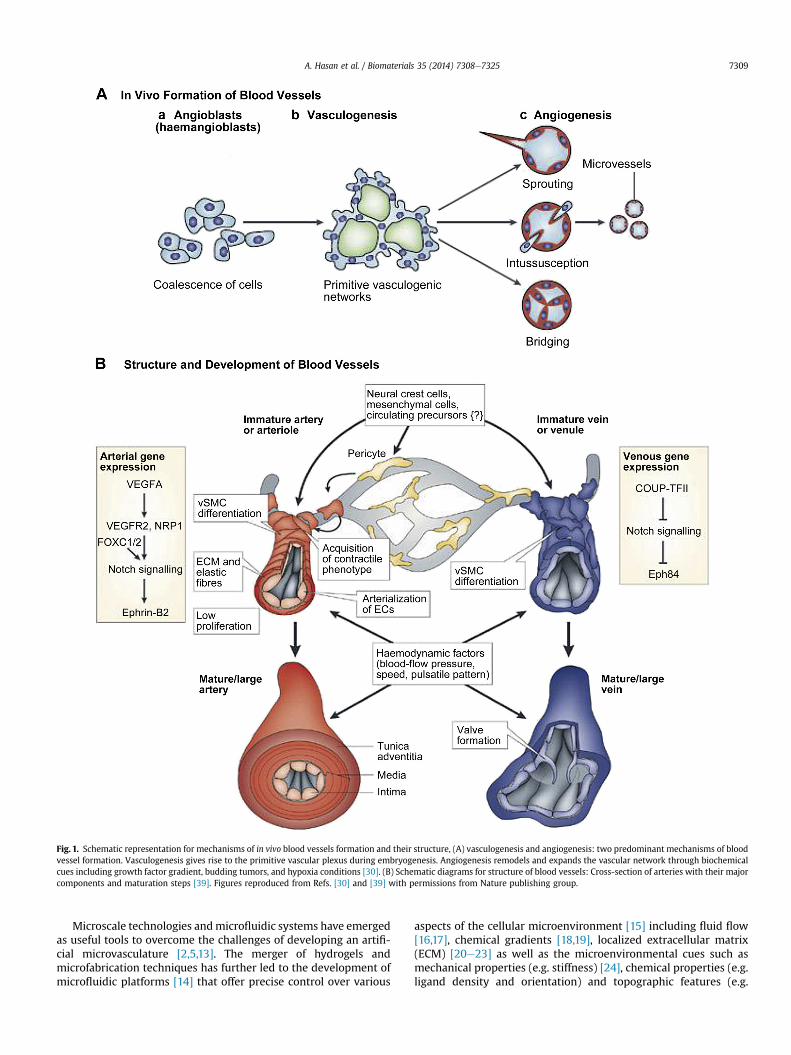
Fig. 1. Schematic representation for mechanisms of in vivo blood vessels formation and their structure, (A) vasculogenesis and angiogenesis: two predominant mechanisms of bloodvessel formation. Vasculogenesis gives rise to the primitive vascular plexus during embryogenesis. Angiogenesis remodels and expands the vascular network through biochemicalcues including growth factor gradient, budding tumors, and hypoxia conditions [30]. (B) Schematic diagrams for structure of blood vessels: Cross-section of arteries with their majorcomponents and maturation steps [39]. Figures reproduced from Refs. [30] and [39] with permissions from Nature publishing group.
A. Hasan et al. / Biomaterials 35 (2014) 7308e7325 7309
Microscale technologies andmicrofluidic systems have emergedas useful tools to overcome the challenges of developing an artifi-cial microvasculature [2,5,13]. The merger of hydrogels andmicrofabrication techniques has further led to the development ofmicrofluidic platforms [14] that offer precise control over various
aspects of the cellular microenvironment [15] including fluid flow[16,17], chemical gradients [18,19], localized extracellular matrix(ECM) [20e23] as well as the microenvironmental cues such asmechanical properties (e.g. stiffness) [24], chemical properties (e.g.ligand density and orientation) and topographic features (e.g.
A. Hasan et al. / Biomaterials 35 (2014) 7308e73257310
patterning of surfaces with substances having different cell-substrate affinity) [25,26]. All of these advantages can be used tofacilitate the formation of biomimicking in vitro blood vesselmodels and vascularized networks in 3D engineered tissue con-structs [27].
In this review we highlight the recent advancements in vascu-larization techniques for the fabrication of in vitro blood vesselmodels and vascularized networks in 3D engineered tissue con-structs. We briefly describe the different types of vascular struc-tures, the natural processes involved in their formation in vivo andthe design criteria for their in vitro fabrication. Next, we discussdifferent techniques developed so far for fabricating vascularstructures. The scope of the review is limited to the techniques forfabrication of microscale vasculatures in the size ranges of tens ofmicrons to hundreds of microns, even though some of the pre-sented techniques might also be suitable for fabrication of mediumto large size vascular structures for in vitro blood vessel models. Thetwo main types of techniques namely the prevascularization andthe vasculogenesis/angiogenesis-based ones have been discussedwith a particular focus on their use in conjunctionwithmicrofluidicplatforms.
2. Biology of native blood vessels
The successful development of biomimetic in vitro fabricatedvascularized tissue requires an in-depth understanding of thebiology of native blood vessels as it is important to replicate theanatomical, physiological and functional aspects of the native vas-culatures at different length scales [28]. In the context of tissueengineering, the development of vascularized tissues has twodistinct aims: 1) to carry the necessary oxygen and nutrients andremove the waste products from the surrounding cells in animplantable tissue engineered construct, and 2) to replace adamaged native blood vessel, or to be used as in vitro blood vesselmodels for ex-vivo studies of vascular biology, cardiovascular pa-thology, as well as pharmacological modeling, drug testing andconnecting multiple organs in body-on-a-chip applications [29].For both of these aims it is important to understand the processesof native blood vessel formation in vivo which we discuss here inbrief.
2.1. In vivo formation of native blood vessels: vasculogenesis andangiogenesis
The mechanisms of in vivo formation of native vasculature canbe classified into two types, namely, vasculogenesis and angio-genesis, Fig. 1a [30]. Vasculogenesis gives rise to the first vascularplexus and heart during embryo formation [31]. It is the processwhereby new blood vessels are formed from endothelial progenitorcells (EPCs). At first the mesodermal stem cells are differentiatedinto EPCs, also known as angioblasts. The EPCs migrate to differentregions forming discrete blood islands which eventually fusetogether to form a vascular plexus and endothelial cells (ECs). TheECs migrate and organize themselves into nascent endothelialtubes and form capillaries [32]. Vascular endothelial growth factor(VEGF) expression is required for angioblasts’ differentiation [33].The recruitment of pericytes, smooth muscle cells (SMCs) andfibroblast layers around the endothelial tubes turn them into morematured and larger blood vessels such as arterioles, arteries, ven-ioles, and veins [34]. Even though vasculogenesis is mainly adevelopmental event, it can also occur in adult mammalians in thecase of revascularization following extensive damage or duringtumor growth [14].
Angiogenesis is the expansion and remodeling of the vascularnetwork through the sprouting of ECs from existing vessels. It
results from a sequence of events influenced by cellecell and cell-ECM interactions [35]. The process starts with the sprouting ofECs and continues with the stabilization of the formed capillariesby pericytes. Angiogenesis can also happen through intussuscep-tion which is the formation of new blood vessels via splitting of anexisting one, in which case it is also commonly referred to assplitting angiogenesis [35].
2.2. Structure of native blood vessels at different length scales
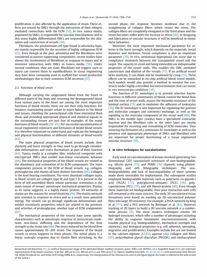
The native blood vessels have complex unique structures atdifferent length scales [10,36]. The inner diameter of blood vesselsranges from microscopic size, 5 mm for the smallest capillaries, to25 mm, for the largest artery (aorta). The blood vessels on thearterial side of the capillaries can be divided into elastic arteries,muscular arteries and arterioles while those on the venus side aredivided into veins and venioles [37]. The walls of the large vessels,namely elastic arteries, muscular arteries and veins have threedistinct layers starting from the vessel lumen: intima, tunica mediaand tunica adventitia respectively [38], Fig. 1b [39]. Intima, theinnermost layer is a thromboresistent confluent monolayer of ECswhich is attached to a basement membrane (40e120 nm) [40].Media, the middle layer, is comprised of a dense population ofconcentrically organized SMCs with bands or fibers of elastic tis-sues, and adventitia, the outermost layer is a collagenous ECMcontaining mainly fibroblasts and perivascular nerves. In arterioles(diameter w30 mm) some of these layers might be less obvious orabsent [41] while the smallest vessels, capillaries, are onlycomposed of a single monolayer of ECs, basement membrane andpericytes. An internal elastic lamina and an external elastic laminaseparate the intima from the media, and the media from theadventitia respectively [42].
The nutrition for the vascular wall itself is supplied by smallvasculatures existing throughout the adventitia. The adventitia alsocontains innervations [43]. The stress, both longitudinal and lateral,due to the pulsatile nature of the blood flow is mainly born bytunica media [44]. There is a gradient of physical properties fromcentral to peripheral vascular tree, i.e. arteries that are closer to theheart are thicker and more compliant whereas further along thevascular tree arteries are considerably thinner and stiffer. As thepressure goes down and the arteries give way to arterioles elasticproperties of tunica media and the presence of tunica adventitiabecome less prominent [45].
The ECs are adherent to the luminal surface of the vessels [46].They form a continuous monolayer, and are in direct control ofblood homeostasis, interaction with immune system cells, andregulation of themolecular exchange to and from the blood and theactivities of the surrounding SMCs. The most distinguishing prop-erty of ECs is their wide range of strong cell to cell junctions [47].They react to shear stress by increasing their surface area throughspreading. In vivo, they are elongated in the direction of the bloodflow [48]. A healthy endothelium prevents initiation of the coagu-lation cascade. Under normal conditions ECs secrete the anticoag-ulant thrombomodulin [49], but in case of an injury they start toexpress pro-platelet adhesive proteins such as selectins. Theendothelial lining also directs the behavior of SMCs and whiteblood cells by secreting various signaling molecules. One of thesesecreted bioactive molecules that have important roles in homeo-stasis of the vasculature is nitric oxide (NO). It is a free radical with awide range of functions including inhibition of vasoconstrictorsignals during vasodilatation, prevention of platelet adhesion, andexhibition of anti-inflammatory and anti-proliferative effects [50].It is important to control the EC phenotypes for a tissue engineeredblood vessel, i.e. the endothelial lining should be in a phenotypicstate in which it prevents blood coagulation. The absence of an
A. Hasan et al. / Biomaterials 35 (2014) 7308e7325 7311
intact endothelium induces conversion to a synthetic phenotypefor SMCs [51].
SMCs are in the quiescent, contractile state under normal con-ditions. Following injury, they convert to a more synthetic pheno-type resulting in cell proliferation, MMP mediated enzymaticdegradation of ECM and vessel wall remodeling via newly secreted
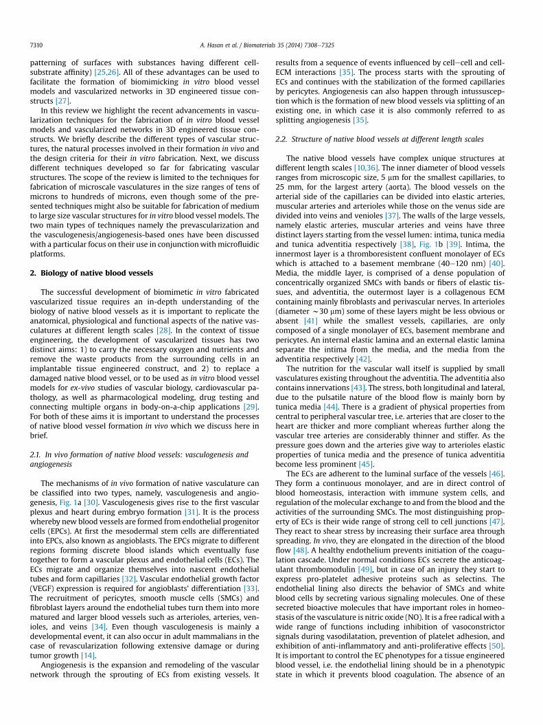
Fig. 2. Schematic diagrams of different types of in vitro vascularization techniques: (AeD)needle-based molding, (B) dissolvable-network-based sacrificial molding, (C) additive metho(D) hybrid method e Bioprinting, (EeI) various vasculogenesis and angiogenesis-based technmaterial, (H) gradient of growth factor in a microfluidic device, and (I) co-culture of multiplefrom American Chemical Society, Royal Society of Chemistry and Elsevier Science.
ECM. This is possible due to the phenotypic plasticity of SMCs,which enables them to switch between awide range of phenotypeswith distinct characteristics; defined as contractile and syntheticphenotypes for the two extreme conditions [52]. SMCs are verysensitive to strain, and dynamic culture experiments have shownthat SMCs become more oriented under controlled strain. Cell
various prevascularization techniques, (AeB) subtractive methods, (A) stainless steeld e soft lithography/PDMS stamping-based micromolding and layer-by-layer stacking,iques, (E) photolithography, (F) microcontact printing, (G) functionalization of scaffoldcells. Figures adopted and modified from Refs. [83,85,87,98,160,161] with permissions
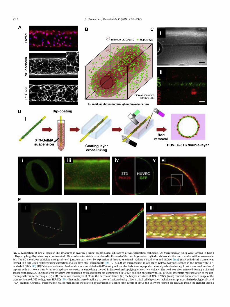
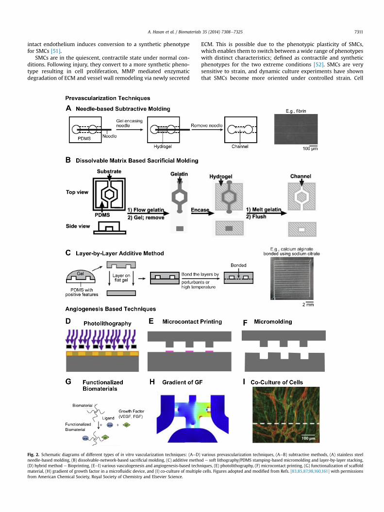
Fig. 3. Fabrication of single vascular-like structures in hydrogels using needle-based subtractive prevascularization technique. (A) Microvascular tubes were formed in type Icollagen hydrogel by extracting a pre-inserted 120-mm-diameter stainless steel needle. Removal of the needle generated cylindrical channels that were seeded with microvascularECs. The EC monolayer exhibited strong cellecell junctions as shown by expression of Prox-1, junctional markers VE-cadherin and PECAM [162]. (B) A cylindrical channel wasformed in a cell-laden hydrogel using extraction of a stainless steel microneedle [89]. (C) A 300 mm microchannel in cell-laden GelMA hydrogels seeded in the lumen with GFP-labeled-HUVECs [86]. (D) Fabrication of a vascular-like structure in cell-laden GelMA using cell transfer technique. A peptide chemically adsorbed on a gold wire was used to adsorb/capture cells that were transferred to a hydrogel construct by embedding the rod in hydrogel and applying an electrical voltage. The gold was then removed leaving a channelseeded with HUVECs. The multilayer structure was generated by an additional dip-coating step in GelMA solution enriched with 3T3 cells, (i) schematic representation of the dip-coating-cell-transfer technique, (ii) a 3D continuous monolayer of ECs in the microvasculature, (iii) the bilayer structure of 3T3-HUVECs, (iv-vi) confocal fluorescence image of thecross section, red: 3T3 cells, green: HUVECs [90]. (E) A multilayered capillary structure fabricated using a hierarchical cell deposition technique in a prevascularized polyglycolic acid(PGA) scaffold. A uniaxial microchannel was formed inside the scaffold by extraction of a silica tube. Layers of SMCs and ECs were formed sequentially inside the channel using a
A. Hasan et al. / Biomaterials 35 (2014) 7308e73257312
A. Hasan et al. / Biomaterials 35 (2014) 7308e7325 7313
proliferation is also affected by the application of strain. These ef-fects are sensed by SMCs through the interaction of their integrinmediated connections with the ECM [53]. In vivo, tunica media,populated by SMCs, is responsible for vascular tone/diameter and inthis sense highly differentiated SMCs are responsible for the dila-tation and constriction of vessels [54].
Fibroblasts, the predominant cell type found in adventitia layer,are mainly responsible for the secretion of highly collagenous ECM[55]. Even though in the past, adventitia and the fibroblasts wereconsidered as passive supporting components; recent studies haveshown the involvement of fibroblasts in response to injury and inextensive interaction with SMCs in tunica media [56]. Undernormal conditions, they are static fibroblasts but triggers such asinjury can convert them to myofibroblasts. In tissue engineeringthey have been commonly used in scaffold-free vessel productionmethodologies due to their extensive ECM secretion [57].
2.3. Functions of blood vessel
Although carrying the oxygenated blood from the heart todifferent parts of the body and returning the deoxygenated bloodfrom various parts to the heart are among the most importantfunctions of blood vessels, these are not their only functions. Forinstance, maintaining proper solute to water balance in the bloodand tissues, shielding the parenchymal cells from interstitial fluidshear, and providing appropriate physical and chemical signals tothe surrounding tissues are just few of examples of the manyfunctions of blood vessels [58e61]. For successful vascularization oftissue engineered constructs and formation of model blood vesselsit is therefore important to understand and replicate the biologicaland physical functionalities of different elements of blood vessels[62].
The main physical properties of blood vessels include theirelasticity and burst strength, as they need to go through consider-able deformations and stress fluctuations as part of their function[63]. Tunica media is a complex fiber reinforced composite withinterspersed SMCs that exhibit non-linear viscoelastic behavior[64]. The mechanical properties of the blood vessels are related tothe abundance and orientation of its ECM components. The maincomponents present in the ECM of blood vessels, e.g. collagen,proteoglycans and elastin all have distinct functions [65]. Collagenis the load bearing constituent. The most abundant collagen typesin blood vessels are collagen type III and type I. It is present in theform of self-assembled fibrils whose particular orientation is themain reason of tissues’ anisotropic mechanical properties. Elastin,as its name suggests, is a highly elastic protein. 3D networks ofelastin are the reasons for vessel elasticity [66]. The elastin networkgets oriented in response to stress and as a result store potentialenergy. The vessels can go through significant deformations andexhibit viscoelastic properties, which are related to the presenceand activities of proteoglycans, another type of ECM components[67].
The mechanical properties of the vessels have some specificcharacteristics such as anisotropic response to stress/strain condi-tions, non-linear stiffening and the dependence of the finalstrength to the strain rate [68]. The stress induced by the blood flowcauses approximately 10e20% strain. The response of the bloodvessels to stress happens in three phases. The initial phase is alinear, isotropic response due to elastin fiber stretching. In the
hierarchical cell deposition [91], (i) confocal fluorescence image of the fabricated bilayer caprepresentation of the bilayer structure. Scale bar: 200 mm in C(ii), 400 mm in E (i), and 100 mmLtd. Wiley Periodicals Inc. and Wiley-VCH Verlag GMbh & Co. respectively. (For interpretationof this article.)
second phase, the response becomes nonlinear due to thestraightening of collagen fibers which resists the stress. Thecollagen fibers are completely elongated in the third phase and thevessel becomes stiffer with the increase in stress [69]. In designingand fabrication of vascular structures it will be beneficial to mimicthese behaviors.
Moreover, the most important mechanical parameter for ar-teries is the burst strength, which depends on the materials, vesseldiameter and thickness. Vessel compliance is also an importantparameter [70]. In vivo, neointimal hyperplasia can occur due tocompliance mismatch between the transplanted vessel and thetarget. The capacity to recoil and being extensible are indispensableproperties of the vessel walls. This enables the SMC mediatedconstriction and dilation of the vessels [71]. If an artificial vessellacks elasticity, it can dilate and be weakened by creep [72]. Theseeffects can be mimicked in on-chip artificial blood vessel models.Such models would also provide a method to monitor the con-structs under highly controlled microenvironments that can mimicin vivo stress/strain conditions [10].
The function of EC monolayer is to provide selective barrierfunctions to different constituents of blood, control the blood flowand the tone of vessel walls, ensure the thrombo-resistance of theluminal surface [73] and to modulate the adhesion of leukocytes[74]. The EC monolayer is also important for regulating the gaseousand molecular (oxygen and nutrients) exchange, as well as thesignaling to the muscular component of the vessel wall [66]. TheSMCs in the middle layer (media) have a specialized contractilefunction and the fibroblast cells in the outer layer are mostlyresponsible for secreting and remodeling of ECM molecules. Thusensuring the formation of a continuous ECmonolayer as well as thepresence and appropriate phenotype of SMCs and fibroblast cellsare important for proper functioning of a tissue engineeredvascular structure [10].
3. In vitro techniques for vascularization
Early work on vascularization of tissues involved generating twodimensional (2D) vascularized constructs of non-biodegradable,e.g. silicon, pyrex [75], and PDMS [76] channels using photoli-thography and soft lithography techniques. The non-biodegradability and lack of biocompatibility of these systemsmade them unsuitable for implantation. The subsequent studiesemployed biodegradable materials such as poly(lactic-co-glycolic)acid (PLGA) [77], poly(glycerol-sebasate) (PGS) [78], poly-caprolactone (PCL) [79], and silk fibroin protein [80]. Even thoughthese materials are biodegradable, their poor interaction with cellsstill remained as the main concern. The early 3D vascularized tissueformations were based on stacking up 2D vascularized polymerfilms into large 3D structures. For example, a PLGA network by Kinget al. [77], and a PGS network by Bettinger et al. [81]. Howeverstacking of 2D layers to build a 3D vascular network is a cumber-some process [5]. The more modern techniques involve 3Dhydrogel structures, which offer a number of advantages includingthe ability to engineer biomimetic microenvironments withtunable physical (e.g. biodegradability), mechanical (strength andelasticity), and biological properties (e.g. cell adhesion, spreading,migration and proliferation). Examples include but are not limitedto the calcium alginate [82,83], agarose [84], collagen and fibrin[85], poly(ethylene glycol) diacrylate (PEGDA), and methacrylated
illary structure, green: SMCs, red: HUVECs, (ii) a magnified image of (i), (iii) schematicin E (ii). Figures modified from Refs. [87, 89, 86, 90, 91] with permissions from Elsevierof the references to color in this figure legend, the reader is referred to the web version
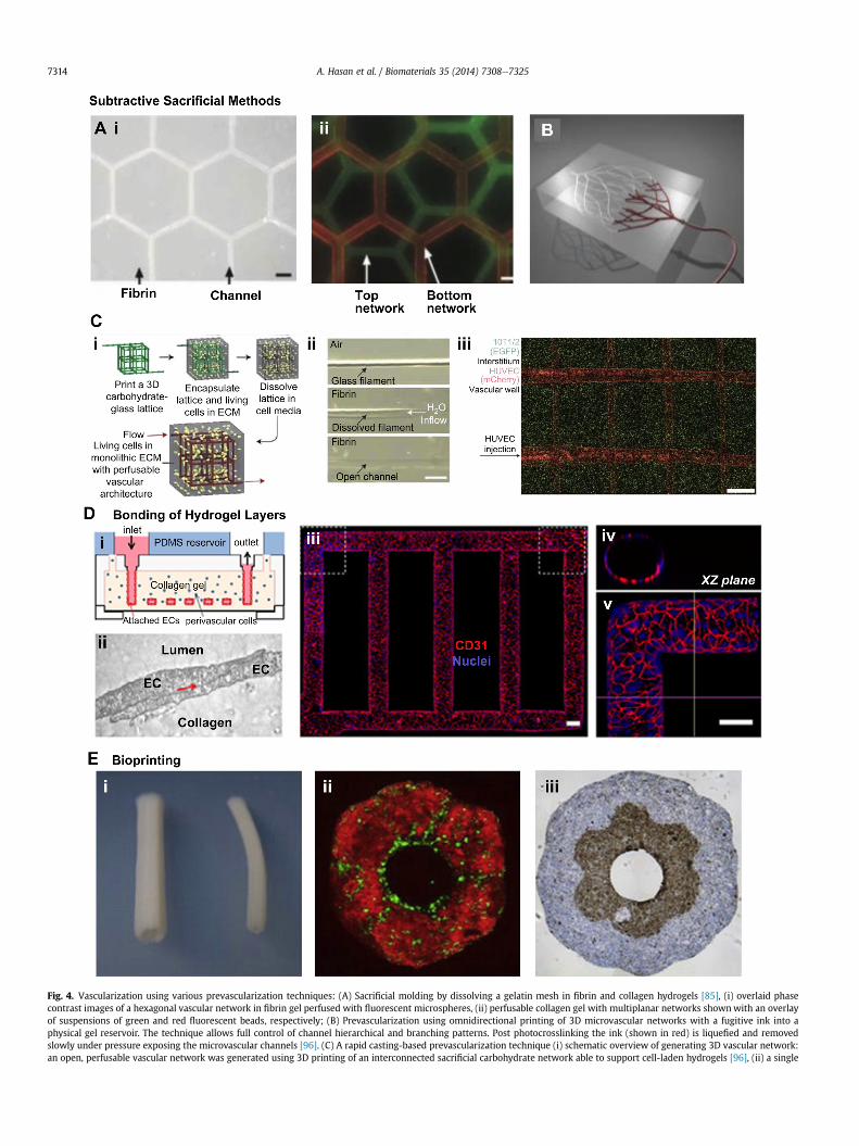
Fig. 4. Vascularization using various prevascularization techniques: (A) Sacrificial molding by dissolving a gelatin mesh in fibrin and collagen hydrogels [85], (i) overlaid phasecontrast images of a hexagonal vascular network in fibrin gel perfused with fluorescent microspheres, (ii) perfusable collagen gel with multiplanar networks shownwith an overlayof suspensions of green and red fluorescent beads, respectively; (B) Prevascularization using omnidirectional printing of 3D microvascular networks with a fugitive ink into aphysical gel reservoir. The technique allows full control of channel hierarchical and branching patterns. Post photocrosslinking the ink (shown in red) is liquefied and removedslowly under pressure exposing the microvascular channels [96]. (C) A rapid casting-based prevascularization technique (i) schematic overview of generating 3D vascular network:an open, perfusable vascular network was generated using 3D printing of an interconnected sacrificial carbohydrate network able to support cell-laden hydrogels [96], (ii) a single
A. Hasan et al. / Biomaterials 35 (2014) 7308e73257314

A. Hasan et al. / Biomaterials 35 (2014) 7308e7325 7315
gelatin (GelMA) [86] hydrogels. A schematic overview of variousrecently developed techniques for vascularization in 3D hydrogelsis presented in Fig. 2. The methods include prevascularization-based methods such as needle-based molding (Fig. 2A), sacrificialmolding (Fig. 2B), and additive bonding of prevascularized hydrogelslabs (Fig. 2C); and vasculogenesis/angiogenesis-based methodsthat use microfabrication, such as, photolithography (Fig. 2D),microcontact printing (Fig. 2E) and micromolding (Fig. 2F), as wellas vasculogenesis/angiogenesis-based methods that use function-alization of biomaterials (Fig. 2G), gradients of growth factors(Fig. 2H), and co-culture of multiple cells (Fig. 2I). These modernapproaches of vascularization can be broadly classified under twomain categories, (1) prevascularization-based techniques, and (2)vasculogenesis and angiogenesis based techniques.
3.1. Prevascularization-based techniques
Prevascularization of engineered tissue constructs is a relativelyrecent and fast growing approach that has drawn tremendousattention lately. A major advantage of these approaches lie in thatthey allow immediate perfusion of the constructs helping theproliferation and growth of the cells from the very beginning.Moreover, the delivery of oxygen and nutrients, and the removal ofmetabolic wastes can be performed continuously in a biomimeticmanner. Various prevascularization methods that have been usedso far can be grouped under (i) subtractive methods, (ii) additivemethods, and (iii) hybrid methods.
3.1.1. Subtractive methodsIn subtractive methods, a hollow structure, i.e. a single channel
or a network of channels is formed by removing a sacrificial ma-terial from a hydrogel. Examples include removal of a sacrificialtemplate either by extraction of a cylindrical object or by dissolvinga pre-formed network of a sacrificial material. Hence the subtrac-tive approach used by various researchers can be grouped into (i)needle-based molding method, and (ii) dissolvable network-basedsacrificial molding.
3.1.1.1. Needle-based molding method. When the purpose of makingblood vessels is to build experimental models for in vitro studies ofvascular functions, a single channel might be sufficient instead of abranched vascular network, and a simple method can be used bypre-inserting a needle, wire or other cylindrical structures in a pre-polymer solution followed by removal of the cylindrical object aftercross linking of the gel. Joe Tien and colleagues [87,88] developedthis “engineered” approach using stainless steel needle-removal-based subtractive method. In this approach of Chrobak et al. [87]and Price et al. [88], a cylindrical channel was formed in collagentype I hydrogel by casting the hydrogel around a stainless steelneedle. After the gel was formed, the needle was removed, creatinga cylindrical channel that could be readily perfused. ECs wereseeded into these channels forming a confluent EC monolayer thatexhibited tight EC junctions, Fig. 3A, strong barrier functions,resistance to the adhesion of leukocytes, and appropriate reactionsto the tested inflammatory cytokines.
carbohydrate-glass fiber approximately 200 mm is encapsulated in a fibrin gel showing thconstitutively expressing enhanced green fluorescent protein (eGFP) imaged with confocalization of channel walls and across the intervessel junctions. (D) A microfluidic vessel netwbonding of two hydrogel slabs [99]: (i) schematic cross-sectional view of the microfluidic col(iii) Z-stack projection of horizontal confocal sections of endothelialized microfluidic vesse100 mm) [99]. (E) Vascular tubes fabricated using bioprinting technique [110], (i) the tubularusing spheroids of ECs (green) and SMCs (red), (iii) a bilayer construct fabricated using buadventitia and media of a blood vessel. Figures reproduced and modified from Refs. [85,92,96& Co. Nature Publishing, PNAS and IOP Publishing respectively. (For interpretation of the rearticle.)
Khademhosseini and colleagues also used the needle-basedapproach to show that perfusable microchannels with EC-seededlumen can be formed in cell-laden hydrogels, Fig. 3B [89], C [86].Sadr et al. [90] combined a self-assembled-monolayer (SAM)-basedcell transfer technique with the needle-based prevascularizationmethod to obtain a free standing multilayered blood vessel with ahollow channel and controlled geometrical design. They used600 mm diameter gold sputtered rods modified with SAM-oligopeptides coating. Layers of human umbilical vein endothelialcells (HUVECs) and 3T3-fibroblast cells were formed on these rodsby dipping them in suspensions of 3T3 cells and HUVECs in GelMApre-polymer, followed by UV-cross-linking. The rod was thenremoved using an electrical stimulation after transferring the celllayers in a GelMA matrix, Fig. 3D.
Yoshida et al. [91] used a hierarchical cell manipulation tech-nique in combination with a needle-based prevascularizationmethod to form a bilayered blood capillary mimetic comprising aSMC layer and an EC layer. The authors fabricated uniaxial micro-channels in g-PGA-SS (poly (g-glutamic acid) with di-sulfide link-age) hydrogels by extracting 620 mmdiameter silica capillary tubes.Layers of umbilical artery smooth muscle cells (UASMCs),fibronectin-gelatin (FN-G) thin membrane and HUVECs wereformed inside the luminal surface by a sequential deposition toobtain a bilayer vascular structure of UASMC and HUVECs, Fig. 3E.The vascular construct exhibited strong barrier functions similar tonative blood capillaries.
Despite its numerous merits, which include its ease of fabrica-tion, the needle-based method, however, is not suitable for formingcomplex networks of interconnected channels as is required forvascularization of large tissues.
3.1.1.2. Dissolvable network-based sacrificial molding. In thedissolvable-network-based sacrificial molding, at first a 2D or 3Dnetwork of a negativemold is formed using an easily dissolvable gelor solid material. The pre-formed mesh or network is then encap-sulated in a 3D hydrogel. Finally, the sacrificial mesh or network isdissolved or melted and flushed out from the gel matrix, leavingbehind interconnected channels in the hydrogel structure. Andrewet al. [85] used this technique employing micromolded meshes ofgelatin as the sacrificial material in collagen and fibrin hydrogels,Fig. 4A(ieii). Others employed 3D printing technology for formingthe dissolvable-network of sacrificial material [92], Fig. 4B.
Recently, more complex structural motifs have been generatedfor vascularization of tissue constructs with methods such as thedirect ink writing (DIW), allowing for better replication of 3Dmicrovascular structures while retaining relatively simple fabrica-tion under benign conditions [93e95]. In the DIW method a fugi-tive organic ink is used to create uniform microchannelsinterconnected into a 3D network. However, architectural motivesthat could be fabricated with DIW were limited. A solution toovercome this challenge has been developed that creates morerepresentative biomimetic 3D interpenetrating microvascular net-works by using vertical printing combined with dual fugitive inks[93]. However, a more advanced method for fully unconstrainedvascular network printing is based on omnidirectional printing
e dissolution of the carbohydrates within hydrogel post-crosslinking, (iii) viable cellsmicroscopy z-stack to visualize two intersecting channels demonstrating endothelial-orks (mVNs) fabricated using micromolding method followed by additive stacking andlagen construct after fabrication, (ii) cellecell junction of EC seeded inside the channels,ls, (iv) view of xz plane and (v) view of a corner. Red: CD31, blue: nuclei. (Scale bar:constructs after fusing the cylindrical bioink, (ii) a bilayer tubular construct fabricatedilding blocks of fibroblast cells (outer layer) and SMCs (inner layer) representing the,99,110] with permissions from the Royal Society of Chemistry, Wiley-VCH Verlag GMbhferences to color in this figure legend, the reader is referred to the web version of this
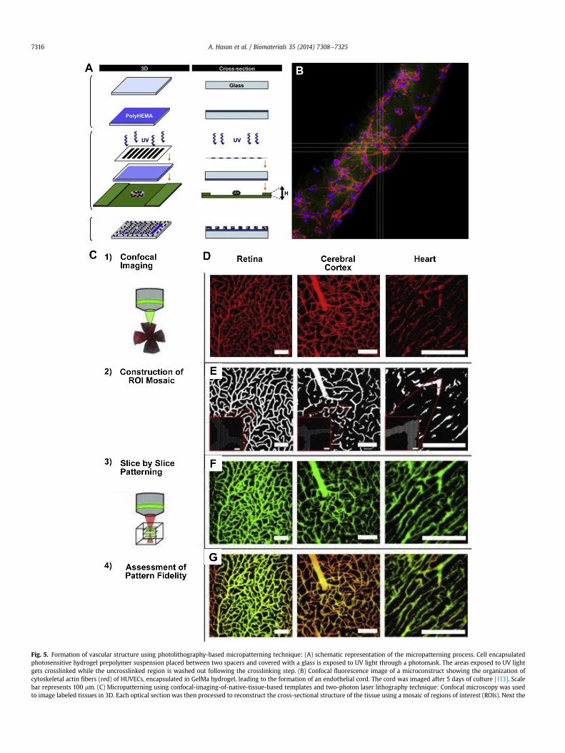
Fig. 5. Formation of vascular structure using photolithography-based micropatterning technique: (A) schematic representation of the micropatterning process. Cell encapsulatedphotosensitive hydrogel prepolymer suspension placed between two spacers and covered with a glass is exposed to UV light through a photomask. The areas exposed to UV lightgets crosslinked while the uncrosslinked region is washed out following the crosslinking step. (B) Confocal fluorescence image of a microconstruct showing the organization ofcytoskeletal actin fibers (red) of HUVECs, encapsulated in GelMa hydrogel, leading to the formation of an endothelial cord. The cord was imaged after 5 days of culture [113]. Scalebar represents 100 mm. (C) Micropatterning using confocal-imaging-of-native-tissue-based templates and two-photon laser lithography technique: Confocal microscopy was usedto image labeled tissues in 3D. Each optical section was then processed to reconstruct the cross-sectional structure of the tissue using a mosaic of regions of interest (ROIs). Next the
A. Hasan et al. / Biomaterials 35 (2014) 7308e73257316
A. Hasan et al. / Biomaterials 35 (2014) 7308e7325 7317
(ODP) method, where fugitive ink networks are printed within areservoir of a photocrosslinkable gel that provides physical supportto the designed patterns and allows for unconstrained printing ofmotifs [96].
Using a carbohydrate sacrificial material in combinationwith 3Dprinting and cell infusions Chen and colleagues employed a rapidprototyping technique for developing vascular networks. Theycreated a 3D perfusable vascular-like network architecture sur-rounded by ECMmimetic hydrogels. Prior to adding hydrogels withencapsulated cells, a 3D carbohydrate-based backbone was printedusing 3D printing technology, acting as a support during hydrogelcrosslinking. Once the gels were solidified the carbohydrate glassnetwork could easily be dissolved in water leaving behind a per-fusable, hollow, cylindrical channel network mimicking vascular-like structures and architecture of more complex tissues, Fig. 4C.The group then infused and seeded the hollow network withHUVECs. This led to the formation of an endothelialized channelnetwork that behaved similar to native microvessels. Spontaneousformation of HUVEC sprouts was observed. The authors demon-strated the functionality of the fabricated network and showed thatculturing under dynamic conditions could sustain cell viabilitydeep inside the scaffold, facilitating nutrient delivery and wasteremoval within the construct.
Additional advantages of this approach lie in the fabricationmethod. In general, the technique is highly versatile allowing se-lection of an array of cell types that can be encapsulated within arange of ECM mimic hydrogel that act as support. Specifically, uti-lizing carbohydrate glass as a sacrificial material adds and improvesto cytocompatibility. Next, the approach allows the control ofchannel diameters that compose the network. Printed filaments ofdifferent diameters can be seamlessly connected and controlled byvarying the translational velocity of the extrusion nozzle. Moreimportantly, the printed 3D channel structures can support theirown weight, paving the way for designing highly complex inter-connected structures available through 3D printer technology.Finally, carbohydrate glass-based materials offer optimal opticalproperties that do not interfere with photocrosslinking and enablecellular fluorescence and light imaging.
Miller et al. showed that emerging technologies such as 3Dprinting can provide highly versatile approaches in recreating thevasculature needed in the successful engineering of thick tissues.They clearly demonstrated a functionally perfusable hollowtubular network lined with HUVECs. The developed structuresustained the viability and proliferation of encapsulated cells anddemonstrated the formation of spontaneous intrinsic vascularsprouts.
3.1.2. Additive method-bonding of pre-formed hydrogel slabsIn the additive methods, a 3D network of interconnected
channels is formed by layer-by-layer stacking and bonding of pre-formed planer hydrogel slabs. The slabs contain network of chan-nels featured in 2D planes, pre-formed using micropatterningtechniques such as photomask lithography or micromolding insuch a way that when stacked together they result in a 3D networkof channels. The adjacent layers are bonded together irreversiblyusing partial melting or fusion of hydrogel at the interfaces therebyresulting in a 3D tightly sealed interconnected perfusable network.One such method uses the micromolding technique, in which a
ROIs were used to control precise scanning of a laser scanning microscope. To pattern a 3Dpattern each corresponding plane of the hydrogel [123]. (D) Projections of imaged vasculvasculature of various tissues at individual cross-sectional planes. (F) 3D projections of hyvarious tissues. (G) A merge of the imaged vasculature with the imaged hydrogels, with yellofor insets). Figures reproduced from Refs. [113] and [123] with permissions from Elsevier Ltd.color in this figure legend, the reader is referred to the web version of this article.)
master mold with desired microstructural features is created on asilicon wafer using microfabrication processes. A transfer mold isprepared by casting and curing an elastomeric polymer solution onthe master mold. The transfer mold with the desired microstruc-tural features is either bonded to a flat substrate resulting inmicrofluidic channels or networks, or is used as a stamp forimprinting the micropatterns to a flat hydrogel. Bonding of twoadjacent flat slabs of hydrogels to ensure a leak-proof perfusablesystem have been successfully demonstrated [97] through partiallydissolving the gel interface by chelating calcium (in case of calciumalginate hydrogel) [83], depolymerizing the gel interface usingchaotropes (for natural gels such as collagen and fibrin) [98], andtransient melting of the interface (e.g. silk fibronin) [80] andagarose gels [84].
Zheng et al. [99] recently developed a microfluidic tubularnetwork using the additive bonding of a micropatterned hydrogelslab over a flat hydrogel layer, Fig. 4D. A silicone mold was used tocast PDMS stamps that imprinted micropatterns of a vascularnetwork to a collagen casting gel. The biomimetic gel slab con-taining microscale features was then bonded to a flat collagensubstrate forming a microvascular network. The authors demon-strated the formation of a smooth and functional endothelium layerwithout any leakage that also promoted angiogenesis. The devel-oped microvascular network demonstrated various complicatedangiogenic and thrombogenic processes such as vessel sprouting,interaction of ECs with mural cells, bioactivation of ECs in responseto various biochemical agents, anti-thrombotic behavior of the ECmonolayer and its expression of a pro-thrombotic behavior inpresence of inflammatory signals. This model can be of immensevalue in designing a more complicated network, for understandingthe blood-vessel function, interaction of blood vessels with tumors,and neoangiogenesis from circulating stem cells.
3.1.3. Hybrid method-bioprintingBioprinting is the process of printing living cells in a 3D space for
the construction of a biological structure using computer-aideddesign and layer-by-layer deposition of cell-laden-matrices. It isrelatively a new method and is a growing area of research.Currently, several groups are focused on generating 3D vascularstructures and blood vessel mimics with the aid of bioprintingtechnique. As the method employs both the principles of additivelayer-by-layer deposition and the subtractive sacrificial removal offiller material, we classify it as a hybrid method.
Two predominant approaches have arisen from the bioprintingtechnology, specifically those based on inkjet printing [100e104]and mechanical extruders [105,106]. Inkjet printing is a versatile,robust and a cost effective method, as it relies on direct printing ofindividual or pockets of cells. However, substantial challengesremain with this approach, most notably the limitation in celldensity that the inkjet method can achieve, especially consideringcell densities meaningful for structures meant for fabrication ofbiomimetic organs. This barrier is further compounded by theobservation that the high speed of cell deposition can lead todamage and lower cell survival. These challenges in addition toattaining appropriate structural and functional organization withscalability, resolution, and repeatability required for construction of3D vascular networks still remain to be solved.
structure, the mosaic of ROIs for each axial cross section was utilized to sequentiallyature from the retina, cerebral cortex, and heart. (E) ROI mosaics reconstructing thedrogels with fluorescently labeled PEG-RGD patterned to mimic the vasculature fromw indicating excellent overlap between vessels and patterns. Scale bars ¼ 100 mm (5 mmandWiley-VCH Verlag GMbh & Co. respectively. (For interpretation of the references to
A. Hasan et al. / Biomaterials 35 (2014) 7308e73257318
Bioprinting can be utilized for generating small and interme-diate diameter blood vessels using mechanical extruder-basedtechniques [105,106]. In these approaches the exact purpose ofthe mechanical extruders is to place the ‘bio-ink’ or multicellularaggregated particles (that could be made of multiple cell types)with a defined composition into a supporting structure or ‘bio-paper’. High consistency is achieved by computer-generated tem-plates in order to mimic the desired biological topology [107e109].This way, layer-by-layer complex structures are fabricated.
In general, the formation of organ-like structures is made fromfusion and sorting of bio-ink particles once the printing process iscomplete [110], Fig. 4E. Several advantages are obvious in gener-ating 3D tissue constructs by employing thesemethods. Specificallyit is beneficial that the cells find themselves in a physiologicallyrelevant microenvironment both structurally for support and bio-logically for promoting adhesive cellular contacts allowing molec-ular signaling. These methods do require high initial cost for thenecessary bioprinting instrumentation [109]. However, currentresearch efforts are geared towards integrating intrinsic self-assembly principles from biology in the design of next generationof constructs, that perhaps could make these approaches morefeasible and cost effective. Furthermore, tubular and vascular-like-structure can be built with various compositions and distribution ofECs and SMCs in which fundamental principles of blood vesselformation could be studied. It is noteworthy that both the inkjetand extruder bioprinting can be made compatible with rapidprototyping.
3.2. Vasculogenesis and angiogenesis-based techniques
Vasculogenesis, as explained earlier, is the process inwhich newblood vessels are formed from EPCs through the formation ofvascular plexus, while angiogenesis is the process whereby groupsof ECs sprout, migrate and organize to form new tubular structureseventually forming blood vessels. The mechanisms of vasculo-genesis and angiogenesis can be utilized to promote the formationof vascular networks in 3D hydrogels in a controlled and regulatedmanner. In these approaches, the neovascularization in engineeredtissue constructs is promoted by providing biomimetic microen-vironments to the cells, i.e. through integrated use of biophysicaland biochemical cues. Several techniques have been developed andutilized under this category, including (i) micropatterning forvascular morphogenesis, (ii) use of functionalized biomaterials forpromoting vasculogenesis and angiogenesis, (iii) gradient ofgrowth factors for vasculogenesis and angiogenesis and (iv) con-trolling cellecell interactions for vasculogenesis and angiogenesisusing co-culture of multiple cell types.
3.2.1. Micropatterning to promote tubulogenesis and vascularmorphogenesis
There has been a rapid development in the area of micro-patterning technologies for applications in promoting the forma-tion of vascular networks in engineered tissue constructs [14,25].Micropatterning has broadened the scope to develop cell micro-patterned vascularized tissue constructs [111]. The techniquesmainly include photolithography, micromolding, microcontactprinting, laser photolithography, nanoprinting and UV light-basedchemistry.
3.2.1.1. Photolithography. In photolithography technique, usually aphotosensitive pre-polymer solution of hydrogel is exposed to thelight of certainwavelength through a photomask. The areas that areexposed to the light wave are crosslinked while the rest of thematerial remains uncrosslinked and is washed out afterward,leaving a hydrogel structure with desired micropattern on the
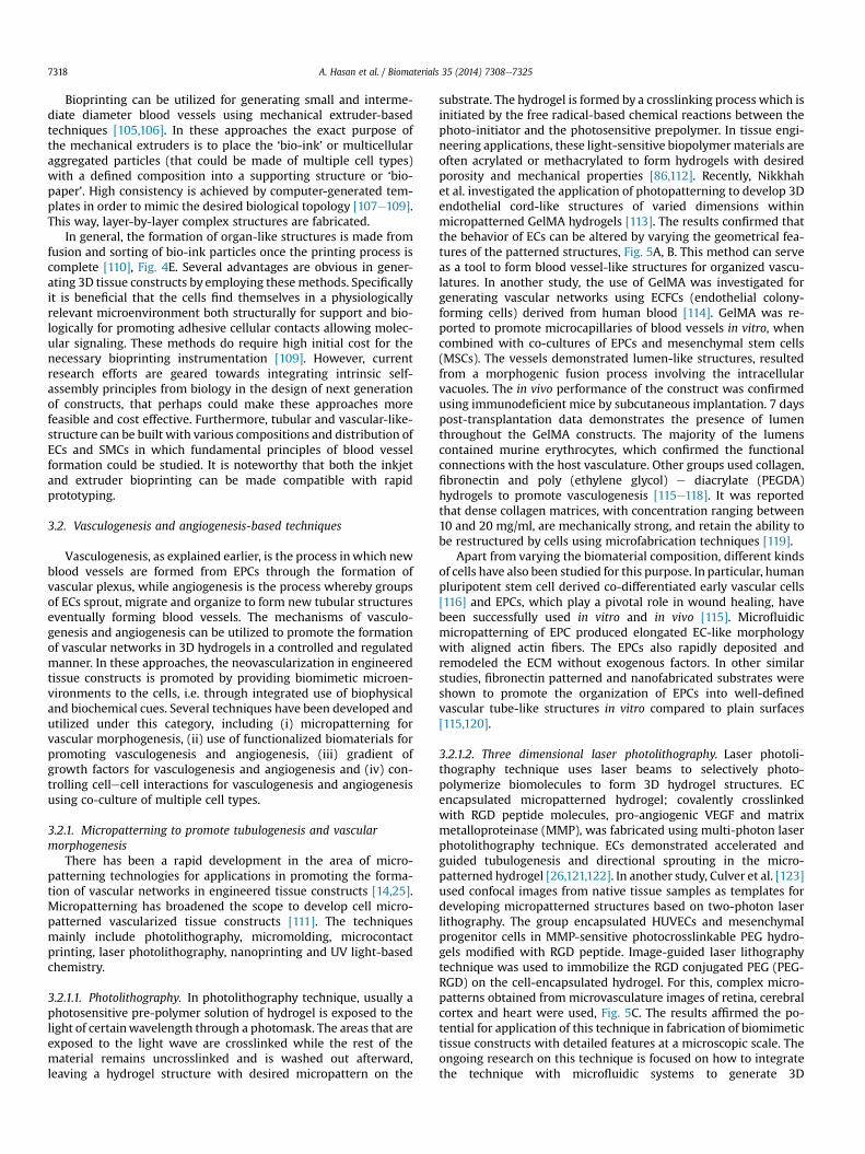
substrate. The hydrogel is formed by a crosslinking process which isinitiated by the free radical-based chemical reactions between thephoto-initiator and the photosensitive prepolymer. In tissue engi-neering applications, these light-sensitive biopolymermaterials areoften acrylated or methacrylated to form hydrogels with desiredporosity and mechanical properties [86,112]. Recently, Nikkhahet al. investigated the application of photopatterning to develop 3Dendothelial cord-like structures of varied dimensions withinmicropatterned GelMA hydrogels [113]. The results confirmed thatthe behavior of ECs can be altered by varying the geometrical fea-tures of the patterned structures, Fig. 5A, B. This method can serveas a tool to form blood vessel-like structures for organized vascu-latures. In another study, the use of GelMA was investigated forgenerating vascular networks using ECFCs (endothelial colony-forming cells) derived from human blood [114]. GelMA was re-ported to promote microcapillaries of blood vessels in vitro, whencombined with co-cultures of EPCs and mesenchymal stem cells(MSCs). The vessels demonstrated lumen-like structures, resultedfrom a morphogenic fusion process involving the intracellularvacuoles. The in vivo performance of the construct was confirmedusing immunodeficient mice by subcutaneous implantation. 7 dayspost-transplantation data demonstrates the presence of lumenthroughout the GelMA constructs. The majority of the lumenscontained murine erythrocytes, which confirmed the functionalconnections with the host vasculature. Other groups used collagen,fibronectin and poly (ethylene glycol) e diacrylate (PEGDA)hydrogels to promote vasculogenesis [115e118]. It was reportedthat dense collagen matrices, with concentration ranging between10 and 20 mg/ml, are mechanically strong, and retain the ability tobe restructured by cells using microfabrication techniques [119].
Apart from varying the biomaterial composition, different kindsof cells have also been studied for this purpose. In particular, humanpluripotent stem cell derived co-differentiated early vascular cells[116] and EPCs, which play a pivotal role in wound healing, havebeen successfully used in vitro and in vivo [115]. Microfluidicmicropatterning of EPC produced elongated EC-like morphologywith aligned actin fibers. The EPCs also rapidly deposited andremodeled the ECM without exogenous factors. In other similarstudies, fibronectin patterned and nanofabricated substrates wereshown to promote the organization of EPCs into well-definedvascular tube-like structures in vitro compared to plain surfaces[115,120].
3.2.1.2. Three dimensional laser photolithography. Laser photoli-thography technique uses laser beams to selectively photo-polymerize biomolecules to form 3D hydrogel structures. ECencapsulated micropatterned hydrogel; covalently crosslinkedwith RGD peptide molecules, pro-angiogenic VEGF and matrixmetalloproteinase (MMP), was fabricated using multi-photon laserphotolithography technique. ECs demonstrated accelerated andguided tubulogenesis and directional sprouting in the micro-patterned hydrogel [26,121,122]. In another study, Culver et al. [123]used confocal images from native tissue samples as templates fordeveloping micropatterned structures based on two-photon laserlithography. The group encapsulated HUVECs and mesenchymalprogenitor cells in MMP-sensitive photocrosslinkable PEG hydro-gels modified with RGD peptide. Image-guided laser lithographytechnique was used to immobilize the RGD conjugated PEG (PEG-RGD) on the cell-encapsulated hydrogel. For this, complex micro-patterns obtained from microvasculature images of retina, cerebralcortex and heart were used, Fig. 5C. The results affirmed the po-tential for application of this technique in fabrication of biomimetictissue constructs with detailed features at a microscopic scale. Theongoing research on this technique is focused on how to integratethe technique with microfluidic systems to generate 3D
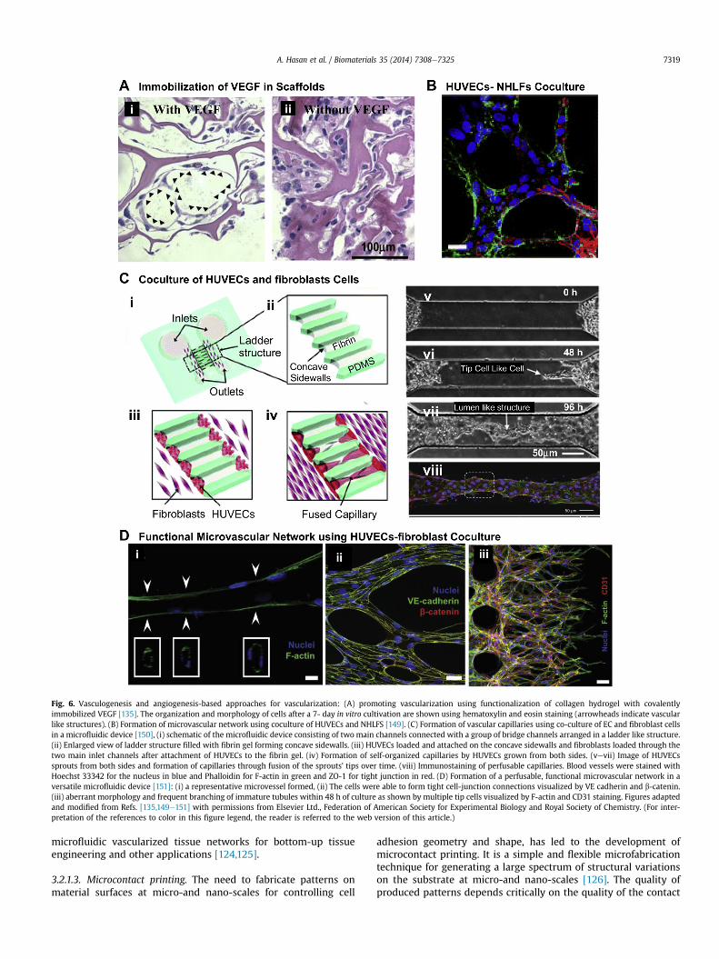
Fig. 6. Vasculogenesis and angiogenesis-based approaches for vascularization: (A) promoting vascularization using functionalization of collagen hydrogel with covalentlyimmobilized VEGF [135]. The organization and morphology of cells after a 7- day in vitro cultivation are shown using hematoxylin and eosin staining (arrowheads indicate vascularlike structures). (B) Formation of microvascular network using coculture of HUVECs and NHLFS [149]. (C) Formation of vascular capillaries using co-culture of EC and fibroblast cellsin a microfluidic device [150], (i) schematic of the microfluidic device consisting of two main channels connected with a group of bridge channels arranged in a ladder like structure.(ii) Enlarged view of ladder structure filled with fibrin gel forming concave sidewalls. (iii) HUVECs loaded and attached on the concave sidewalls and fibroblasts loaded through thetwo main inlet channels after attachment of HUVECs to the fibrin gel. (iv) Formation of self-organized capillaries by HUVECs grown from both sides. (vevii) Image of HUVECssprouts from both sides and formation of capillaries through fusion of the sprouts’ tips over time. (viii) Immunostaining of perfusable capillaries. Blood vessels were stained withHoechst 33342 for the nucleus in blue and Phalloidin for F-actin in green and ZO-1 for tight junction in red. (D) Formation of a perfusable, functional microvascular network in aversatile microfluidic device [151]: (i) a representative microvessel formed, (ii) The cells were able to form tight cell-junction connections visualized by VE cadherin and b-catenin.(iii) aberrant morphology and frequent branching of immature tubules within 48 h of culture as shown by multiple tip cells visualized by F-actin and CD31 staining. Figures adaptedand modified from Refs. [135,149e151] with permissions from Elsevier Ltd., Federation of American Society for Experimental Biology and Royal Society of Chemistry. (For inter-pretation of the references to color in this figure legend, the reader is referred to the web version of this article.)
A. Hasan et al. / Biomaterials 35 (2014) 7308e7325 7319
microfluidic vascularized tissue networks for bottom-up tissueengineering and other applications [124,125].
3.2.1.3. Microcontact printing. The need to fabricate patterns onmaterial surfaces at micro-and nano-scales for controlling cell
adhesion geometry and shape, has led to the development ofmicrocontact printing. It is a simple and flexible microfabricationtechnique for generating a large spectrum of structural variationson the substrate at micro-and nano-scales [126]. The quality ofproduced patterns depends critically on the quality of the contact
A. Hasan et al. / Biomaterials 35 (2014) 7308e73257320
between the stamp and the substrate. This technique is mainly usedto fabricate surfaces with self-assembled monolayer (SAM) regionswith different physical and chemical properties. The method hasbeen used to study the polymerization of actin and phosphoryla-tion of tyrosine in patterned vascular ECs subjected to uniaxial andcyclic strains using fibronectin microdots [127].
3.2.1.4. Micromolding. Micromolding is another widely usedmethod for micropatterning cell-laden hydrogels [128,129]. Inmicromolding, the shape of a preformed master pattern is trans-ferred onto a prepolymer solution through direct contact, duringwhich the prepolymer undergoes crosslinking. PDMS is the mostcommonly used material for master mold. This method can be usedto design constructs containing spatially patterned EC tubularstructure with varied geometrical properties [99,130,131]. Micro-molding and similar techniques have also been used to promotecapillary tubule formation by spatially patterning ECs using longtubes of collagen hydrogel [78,92,132]. The size of the lumen variedbased on both microgel size and the concentration of collagen. Thisapproach has the potential for application in developing complexblood vessel-like structures for investigating the fundamentalbehavior of ECs in initiation of tubulogenesis.
The micropatterning techniques are simple and convenient forpatterning complex structures of cell-laden hydrogels and elasto-mers on diverse substrates. They can be used to make complexpatterned network over a wide range of scale in 2D planes and canbe useful for detailed study of cell behavior under diverse micro-architectural cues. However, their applicability is mostly limited to2D, and as of now they cannot be used to develop 3D perfusableendothelial cords or vascular structures.
3.2.2. Vascularization using functionalized biomaterialsThe formation of vascular network can be promoted by func-
tionalizing the scaffold materials with various angiogenic biomol-ecular cues. It has been found that the cell migration, penetration,organization and matrix remodeling can be guided by functional-izing the scaffold materials [133], e.g. by immobilizing angiogenicgrowth factors (GFs), ECM proteins, peptides, or other bio-macromolecules within the scaffolds, thereby promoting angio-genesis and formation of vascular networks.
For example, genetically modified VEGF, where N-terminalcysteine was chemically crosslinked to a fibrin-based structure viathiol-directed cross-linker, significantly improved angiogenesis[134]. Similarly covalent immobilization of VEGF in collagenhydrogel exhibited improved vascularization compared to collagenhydrogel without any VEGF, Fig. 6A. Additionally, concomitantaddition of VEGF and angiopoietin-1 (ANG1) to collagen via EDCcrosslinking resulted in higher tube formation by ECs compared tothe collagen scaffolds with individual components [135]. Heparin,which interacts with VEGF, has also been immobilized in the scaf-fold, leading to indirect immobilization of VEGF and thus resultingin a prolonged VEGF release and improved angiogenic effect [136].
Functionalization of biomaterials can also be achieved via non-chemical immobilization instead of chemical immobilization (e.g.by promoting non-covalent-interaction mediated immobilizationor by the addition of enzyme sensitive moieties for highlycontrolled immobilization) [137]. For example, artificial ECM mol-ecules which are designed to interact with several GFs for theirnon-covalent immobilization via coiled-coil interactions have beenused to fabricate tissue engineering scaffolds and shown to pro-mote angiogenesis [138]. Advantages of this strategy includemimicking the in vivo microenvironment of angiogenesis, main-taining the angiogenic factor activities for a prolonged period oftime by protecting the compounds against fast metabolism throughroutes such as endocytosis and encapsulating and delivering
angiogenic compounds in a temporally and spatially controlledmanner [139].
3.2.3. Vascularization using gradient of growth factorsAmong the most extensively investigated methods for gener-
ating vascular network by promoting angiogenesis is the use ofconcentration-gradients of common angiogenic growth factors in acell laden or cell-seeded hydrogel, often in a microfluidic device. Inthis method, when a concentration gradient of GFs is established inthe vicinity of ECs, either in a hydrogel construct or in amicrofluidicchannel, the ECs tend to migrate from the region of low-GF-concentration toward the region of high GF concentration,thereby aligning themselves into well-organized structures andenhancing the capillary-like tubular structure formation [3]. Theevaluated GFs include VEGFs, angiopoietins, the transforminggrowth factors (TGFs), the fibroblast growth factors (FGFs) and theplatelet derived growth factors (PDGFs) [3]. These GFs can beincorporated within the scaffolds to ensure a sustained release overtime thereby creating local concentration gradients or can bedelivered through the growth media flowing through microfluidicchannels. Also a single GF can be used as well as a number of GFssimultaneously.
Upon incorporation of a single GF in a scaffold, angiogenesiswith increased capillary densities, as well as more matured capil-laries were obtained compared to scaffolds without any GF. Forexample, VEGF-loaded PLGA scaffolds resulted in double as manyblood vessel capillaries after implantation in an area of irradiatedosseous defects compared to the control scaffolds without VEGF[140]. Perets et al. [141] reported the development of hybrid scaf-fold system combining basic FGF (bFGF) encapsulated PLGA mi-crospheres within alginate gel. The in vivo results revealed thatafter 21 days of implantation, a significantly higher number ofcapillaries (70 � 7 capillaries/mm2) was observed with theinvolvement of bFGF compared to the scaffolds without bFGF(18 � 5 capillaries/mm2) [141].
Recent research has revealed that combining multiple types ofGFs enhances the formation of EC tubes and their stabilization. Forinstance, Hao et al. [142] developed an alginate-based system forthe co-delivery of VEGF and PDGF. It was found that after 4 weeks ofhydrogel injection into a rat myocardial infarction model, thescaffolds with both VEGF and PDGF resulted in a vessel density ofabout 40 capillaries/mm2 whereas scaffolds with either VEGF orPDGF had a resultant vessel density of about 30 capillaries/mm2,indicating that the sequential delivery of VEGF and PDGF may beable to facilitate the formation of capillary vessels [142]. In addition,Nillesen et al. [143] evaluated the individual and combined effectsof VEGF and FGF2 for blood vessel formation. The results showedthat the system loaded with a combination of GFs displayed a two-fold increase in the formation of mature blood vessels compared tothe system with a single GF, confirming the synergistic effect ofmultiple GFs.
GF gradient has been found to regulate the angiogenesis. Forexample, Barkefors et al. [144] studied the role of VEGF gradient inmigration of ECs. The authors designed a microfluidic device withthree inlets to generate a concentration gradient by three parallelfluid streams. The VEGF gradient was tunable by adjusting the so-lution flow rates. The results showed that the ECs migrated towardsthe high concentration of VEGF. These results also indicated thatthe steep gradients induced faster cell migration from 0 to 50 ng/ml, but no obvious migration from 50 to 100 ng/ml probably due toreceptors saturation [144].
Silvia et al. [145] further investigated the spatial and temporaleffects of GFs on angiogenesis. They studied the temporal effect ofVEGF concentration on angiogenesis using alginate hydrogelmodels and analyzed the impact of the spatial distribution of VEGF
A. Hasan et al. / Biomaterials 35 (2014) 7308e7325 7321
using murine hind limb ischemia models. A profile with a highinitial concentration of VEGF (50 ng/ml/day) and a programmedconcentration decrease was able to develop more EC sproutscompared to a constant dose of VEGF, the total amount being thesame. It was also found that a higher level of perfusion and bettervascularization performance were obtained by distributing a VEGFdose (0.1 mg/g tissue) into two streams and delivering from twodifferent locations in a murine hindlimb ischemia model. Thesefindings suggest that control and regulation of the spatial andtemporal distribution of VEGF may be effective in inducing angio-genesis in vivo [145].
Thus, it is clear that controlled GF release, right combination ofGFs, correct dosage and proper exposure time are among key fac-tors for inducing the formation and growth of functional vascula-ture and establishing appropriate vessel architecture and stability[146].
3.2.4. Vascularization using co-culture of different cellsCo-culturing different suitable cell types under proper micro-
environmental cues can induce spontaneous alignment of thevascular cells and generate vascular networks. This technique ofpromoting vascularization through angiogenesis has been widelyinvestigated lately. Some examples of these studies have reportedcapillary/microvascular tubules formation in tissue-engineeredscaffolds containing ECs or their progenitor cells. Cells co-cultured with ECs or their progenitor cells to facilitate angiogen-esis include fibroblasts, SMCs and various stem cells.
For example, Tremblay et al. [147] engineered vascularized hu-man skin by co-culturing HUVECs, dermal fibroblasts, and kerati-nocytes on 3D porous chitosan-collagen scaffolds. After 15 days ofculturing, capillary tubes were observed clearly in the co-culturesystem but not in the monoculture system. The formation of 3Dcapillary network could be attributed to the cellematrix in-teractions and the cellecell interactions of HUVECswith fibroblasts.Specifically, it was attributed to the fibroblasts which are able toproduce ECM in large amounts when co-cultured with VEGF-producing keratinocytes [147]. The angiogenic effect of cell co-culture was also investigated by Sudo et al. [148] who imple-mented a microfluidic platform made of a micropatterned PDMSwith two parallel microfluidic channels connected using an inter-vening 3D collagen gel to analyze the angiogenesis using primaryrat hepatocytes and microvascular ECs (rMVECs). The vascular andhepatic cells were cultured separately on the sidewalls of thecollagen hydrogel bridging two parallel channels in a microfluidicdevice. The results indicated that the 3D capillary-like structuresformed by rMVECs could extend across the intervening hydrogel tothe hepatic tissue in the co-culture systemwhile only 2D sheet-likestructures were observed in the rMVEC monoculture [148]. Thismay be attributed to the hepatocytes-secreted hepatocyte growthfactor which could stimulate EC motility and growth. In anotherstudy Chen et al. [149] used a similar microfluidic device withcoculture of HUVECs and normal human lungs fibroblasts (NHLFs)resulting in a well-developed microvascular network, Fig. 6B. Inaddition, Yeon et al. [150] reported the development and charac-terization of perfusable capillary networks formed by HUVECs andfibroblasts in vitro in a microfluidic device. This device containedtwo large channels, connected with eight small bridging channels(Fig. 6C i), which were completely filled with fibrin gel (Fig. 6C ii).Following gel formation, HUVECs were seeded on opposite ends ofthe fibrin gel (Fig. 6C iii) while fibroblast cells were seeded in thechannels adjacent to the gel. After 3e4 days, HUVECs migrated intothe fibrin gel from the opposite ends and connected/merged witheach other, forming a connective vessel expressing tight junctionproteins (e. g., ZO-1), which are characteristic of post-capillaryvenioles (Fig. 6.C.iv-vi). This capillary network formation was
facilitated via the recruitment of fibroblasts which were found toexpress essential matrix proteins (e.g., collagen I), supporting EClumen formation. Kim et al. [151] continued further investigationsand reported development of a more versatile, perfusable andfunctional microvascular-like network where the results demon-strated formation and morphogenesis of interconnected micro-vessels (Fig. 6D i). Furthermore, the cellecell interactions facilitatedcontinuous hollow lumen formation enclosed by ECs throughoutthe length of the vessels. The cells were able to form tight cell-junction connections visualized by VE cadherin and b-catenin(Fig. 6D-ii). The authors also demonstrated that the microfluidicplatform that they employed could also be used to model angio-genic sprout formation induced by cancer cell secreted growthfactors. They observed aberrant morphology and frequentbranching of immature tubules within 48h of culture as shown bymultiple tip cells (Fig. 6D iii) visualized by F-actin and CD31staining.
In addition, the impact of ECs on the proliferation, spreading anddifferentiation of SMCs was studied by Liu et al. [152] utilizing ahybrid hydrogel prepared from gelatin modified with meth-acrylamide, and dextran-graft-lysine modified through meth-acrylation. With the co-culture of ECs, the SMC proliferation,capillary network formation as well as elastin synthesis werepromoted within the 3D hydrogel, suggesting that co-culture of ECsand SMCs is a promising method for constructing functionalvasculature in vitro.
Among different cell types, stem cells, particularly, havedemonstrated a functional role in EC networks on in vitro 3D cul-ture, i.e., to stabilize the developed EC networks. This may be due tothe molecular machinery contained in stem cells that is also ex-pected in perivascular progenitor cells. For example, Moon et al.[153] synthesized a PEG polymer with MMP-sensitive and RGDpeptide sequences to mimic the natural provisional ECM. Whencultured in this system, HUVECs spontaneously formed capillary-like structures. These structures were stabilized by a lineage ofSMCs (differentiated from mesenchymal progenitor cells) whichdeposited laminin and collagen IV. Boyd et al. [154] have alsoshown that the co-culture of HUVECs with multi-potent MSCsderived from human embryonic stem cells increased the degree ofbranching of EC networks andmaintained the network integrity forup to 6 days compared to HUVECs alone in the 3D collagen I-fibronectin scaffolds. More recently, Chen et al. [114] found that theco-culturing of endothelial colony-forming cells (ECFCs) derivedfrom human blood with MSCs derived from bone marrow gener-ated networks of capillary-like structures when encapsulated in 3DGelMA hydrogels, whereas in the absence of MSCs, no capillary-likenetwork formation from ECFCs was observed [114]. It was also re-ported that the presence of MSCs increased the overall survival ofECFCs and enhanced the formation of capillary networks fromECFCs. Furthermore, in vivo implantation of this prevascularizedconstruct into immune-deficient mice resulted in a fast develop-ment of functional anastomoses between the native vasculature ofmouse and the engineered vascular network. Therefore, incorpo-rating appropriate cell types in a pre-designed matrix may be aneffective and safe strategy for vascularizing tissue engineeringconstructs [155].
Thus, a growing body of evidence suggests that co-culture ofmultiple cell types under proper microenvironmental cues canresult in perfusable vascular network. However, all methodshave their inherent merits and demerits. Future approaches tovascularization are, therefore, likely to be a combination ofmultiple techniques such as prevascularization or micro-fabrication using functionalized biomaterials in combinationwith multiple growth factors and co-culture of different cells[156].
A. Hasan et al. / Biomaterials 35 (2014) 7308e73257322
4. Conclusions and future directions
Vascularization is one of the biggest challenges for the wide-spread clinical application of tissue engineering. For fabrication ofimplantable thick, large and complex tissue engineered constructs,or repair and regeneration of living tissues in general, the issue ofvascularization must be resolved. In this review, we have providedan overview of the techniques available for vascularization ofengineered tissue constructs on microfluidic platforms. In general,the techniques developed to date can be classified into (i) pre-vascularization techniques and (ii) vasculogenesis andangiogenesis-based techniques. The prevascularization techniquescan be divided into (a) subtractive methods, (b) additive methods,and (c) hybrid methods, all of which are based on engineeringreadily available perfusable channels in tissue engineered con-structs. Examples of the subtractive methods include needle-basedmolding and dissolvable sacrificial network-based molding, whilethose of the additive and hybrid methods include layer-by-layerassembly of micropatterned or micromolded planer hydrogelsand bioprinting respectively. The vasculogenesis and angiogenesis-based techniques rely on modulating cellecell interactions toachieve a vascularized construct. Commonly used vasculogenesisand angiogenesis-based techniques are (a) microfabrication ofcellular networks for tubulogenesis and vascular morphogenesis,(b) functionalization of biomaterials with proangiogenic agents topromote angiogenesis, (c) utilizing either a single or multipleproangiogenic growth factors to induce angiogenesis and (d) pro-moting spontaneous vascularization by co-culture of two or moredifferent cell types including EC, SMC, fibroblasts, progenitor cellsor various stem cells. The various types of microfabrication ormicropatterning techniques that have been used so far include butare not limited to photolithography, microcontact printing, andmicromolding. The microfluidic platforms have great potential toprovide the necessary microenvironmental cues for promotingvascularization leading to formation of viable thick tissue con-structs [76e78,83,86]. Hence the practice of incorporating micro-fluidics into tissue engineering, thereby generating vascularizedconstructs has been steadily gaining ground.
Each of the vascularization methods has its own advantages andlimitations. While the prevascularization techniques providereadily available channels allowing immediate perfusion of growthmedia or blood, and are perhaps suitable for fabrication of largerblood vessels, they are not suitable for vascular microcapillary bedswith cascading bifurcations down to few micron sizes. The vascu-logenesis and angiogenesis-based approaches, on the other hand,provide very limited control over the temporal and spatial factors,require days to weeks before cells can organize and give rise toperfusable lumens, and are to date not suitable for formation ofvascular structures in the size ranges suitable for suturing andanastomosis with the host vasculatures. Since the successful im-plantation of vascularized tissue constructs lies in their ability to befunctionally integrated with the host tissue, creating upon im-plantation anastomosis contacts between the microvessels of theimplanted tissue constructs and the host’s blood vessel system, theultimate solution for proper vascularization may lie in methodscombining more than one technique.
Moreover, while many of the publications have demonstratedthe proof-of-principles and feasibility studies for different vascu-larization techniques, many remaining challenges are yet to besolved. For instance, a better understanding is required about fac-tors that determine and guide precise temporal and spatial emer-gence of biochemical cues and their effects on the formation ofstable, functional microvasculature suitable for clinical application.Amongst other factors that require further attention is the role ofmechanical stability of the constructs as more complex vascular
network structures are integrated. Furthermore, shear stressresulting from different perfusion and flow rates and the behaviorof ECs under such conditions including their attachment and pro-liferations needs to be carefully evaluated as critical design con-cepts in developing vascularized scaffolds. Other challenges includehow essentially 2D methods such as microfabrication can betranslated into a 3D vascularized tissue construct, and how tointegrate these techniques as the channel scale dimensions in-crease from microns to millimeters requiring channel integrationacross layers.
Beyond the engineering challenges, understanding the longterm biological effects is crucial for the future application of vas-cularized tissue constructs. Of particular importance are the effectsof vascularized tissue constructs on the surrounding organ, cell/tissue differentiation and vascular remodeling post implantation. Itis important to ensure that these vascularized constructs do notimpede full vascular maturation, leading to crucial physiologicalfunctions in response to biochemical or vasoactive stimuli. Addi-tionally, it is necessary to ensure proper cellular phenotypes, aswell as functional and biologically relevant cellecell communica-tions and signaling in addition to ECM proteins and cytokines se-cretions. Such challenges need to be addressedwhile incorporating,particularly for larger blood vessels, multilayered vascular wallswithin vascularized constructs with layers of fibroblasts and SMCs,in addition to the EC monolayer.
When intended for applications in brain and ocular tissues, theengineered vasculatures must exhibit the required specializedphysiological effects such as the blood-brain-barrier and blood-retina-barriers, showing that tight junction permeability is notcompromised due to the implantation of the tissue engineeredconstructs. Furthermore, effects of mechanical stimuli and shearstress have to be better understood in a biological and biochemicalcontext. Polarity of ECs must be investigated and their ability toform future instructive physical and biochemical gradients neces-sary for downstream signaling and proper function. These func-tions are essential for capillary morphogenesis dependent on bothspatial and temporal control of both biochemical and mechanicalcues. Hence, each scaffold should at least be neutral if notinstructive for these functions [157e159]. Finally, these vascular-ized tissue constructs should allow for better modeling and aid inthe study of physiology and physiopathologies of the nativevasculature and perhaps provide clues to more complex cellularprocesses.
The future direction is clear; to create implantable thick vas-cularized tissue constructs that could serve as artificial organs oraid in their repair and/or regeneration. However, in order totranslate the basic science discoveries into clinically relevanttransplants and therapies, issues down the road such as stan-dardization has to be resolved beyond the basic engineering. Withthe advancement in various microfabrication techniques, such asphotolithography and micromolding techniques, it is necessarynow to establish methods and criteria that will evaluate theperformance and clinical relevance of cell-laden vascularizedconstructs. This will aid in generating a systematic approach thatwill assess if the intended organ application requirements havebeen met. Furthermore, there is a void in the development oftools for real-time monitoring of growing tissues on microfluidicchips. With the advances in incorporation of micro- and nano-sensors it would be beneficial to integrate them into micro-fluidic tissue constructs for real-time monitoring. These sensorscould be applied in a wide variety of fashions including assessingin real-time cell viability and function. Such approaches couldlead to the development of functional materials that could bringthe necessary revolution to solve the current issues andchallenges.
A. Hasan et al. / Biomaterials 35 (2014) 7308e7325 7323
To summarize, even though significant progress has been madetoward vascularization of engineered tissue constructs during thepast decade, there is still plenty of work to be done. Even though theclinical goals are clear, the potential paths to lead toward the goalsoffer multiple alternatives. Ultimately, most likely, a combination ofmultiple approaches taking the best of what microfluidics, bio-sensing, vasculogenesis and angiogenesis-based bottom-up ap-proachesandprevascularization-based top-downapproacheshave tooffer, might lead to the achievement of the clinical goals of fullyfunctional and adequately vascularized implantable complex tissues.
Acknowledgments
Anwarul Hasan acknowledges the start up grant from AmericanUniversity of Beirut, Lebanon. Arghya Paul acknowledges post-doctoral award from FRQS (Fonds de recherché du Quebec-Sante)Quebec, Canada). Adnan Memic thanks the Strategic TechnologiesProgram of King Abdulaziz City for Science and Technology(KACST), grant number 12-MED3096-03 for their support andfunding. Nihal Engin Vrana acknowledges funding from Euro-TransBio “BiMoT” project (ETB-2012-32). Ali Khademhosseini ac-knowledges funding from the National Science Foundation CAREERAward (DMR 0847287), the office of Naval Research Young NationalInvestigator Award, the National Institutes of Health (HL092836,DE019024, EB012597, AR057837, DE021468, HL099073, EB008392),and the Presidential Early Career Award for Scientists and Engi-neers (PECASE).
References
[1] Bae H, Puranik AS, Gauvin R, Edalat F, Carrillo-Conde B, Peppas NA, et al.Building vascular networks. Sci Trans Med 2012;4:1e4.
[2] Du Y, Cropek D, Mofrad MRK, Weinberg EJ, Khademhosseinil A, Borenstein J.Microfluidic systems for engineering vascularized tissue constructs. In:Tian W-C, Finehout E, editors. Microfluidics for biological applications. NewYork: Springer; 2008.
[3] Kaully T, Kaufman-Francis K, Lesman A, Levenberg S. Vascularization e theconduit to viable engineered tissues. Tissue Eng Part B Rev 2009;15:159e69.
[4] Hasan A, Ragaert K, Swieszkowski W, Selimovi�c �S, Paul A, Camci-Unal G, et al.Biomechanical properties of native and tissue engineered heart valve con-structs. J Biomech; 2014. in press.
[5] Du Y, Lo E, Ali S, Khademhosseini A. Directed assembly of cell-ladenmicrogels for fabrication of 3D tissue constructs. Proc Natl Acad Sci U S A2008;105:9522e7.
[6] Isner JM, Asahara T. Angiogenesis and vasculogenesis as therapeutic strate-gies for postnatal neovascularization. J Clin Invest 1999;103:1231e6.
[7] Taylor RN, Yu J, Torres PB, Schickedanz AC, Park JK, Mueller MD, et al.Mechanistic and therapeutic implications of angiogenesis in endometriosis.Reprod Sci 2009;16:140e6.
[8] Polverini PJ. Angiogenesis and wound healing: basic discoveries, clinicalimplications and therapeutic opportunities. Endod Top 2012;24:130e45.
[9] Fischer C, Schneider M, Carmeliet P. Principles and therapeutic implicationsof angiogenesis, vasculogenesis and arteriogenesis. In: Moncada S, Higgs A,editors. Handbook of experimental pharmacology: the vascular endotheliumII. Berlin Heidelberg: Springer-Verlag; 2006. pp. 157e212.
[10] Hasan A, Memic A, Annabi N, Hossain M, Paul A, Dokmeci MR, et al. Elec-trospun scaffolds for tissue engineering of vascular grafts. Acta Biomater2013;10:11e25.
[11] Tsai M, Kita A, Leach J, Rounsevell R, Huang JN, Moake J, et al. In vitromodeling of the microvascular occlusion and thrombosis that occur in he-matologic diseases using microfluidic technology. J Clin Invest 2012;122:408e18.
[12] Gailloud P, Muster M, Piotin M, Mottu F, Murphy KJ, Fasel JHD, et al. In vitromodels of intracranial arteriovenous fistulas for the evaluation of newendovascular treatment materials. Am J Neuroradiol 1999;20:291e5.
[13] Paul A, Hasan A, Rodes L, Sangaralingam M, Prakash S. Bioengineeredbaculoviruses as new class of therapeutics using micro and nanotechnol-ogies: principles, prospects and challenges. Adv Drug Deliv Rev; 2014. inpress.
[14] Khademhosseini A, Langer R, Borenstein J, Vacanti JP. Microscale technolo-gies for tissue engineering and biology. Proc Natl Acad Sci U S A 2006;103:2480e7.
[15] Hsu Y-H, Moya ML, Hughes CCW, George SC, Lee AP. A microfluidic platformfor generating large-scale nearly identical human microphysiological vas-cularized tissue arrays. Lab Chip 2013;13:2990e8.
[16] Unger MA, Chou HP, Thorsen T, Scherer A, Quake SR. Monolithic micro-fabricated valves and pumps by multilayer soft lithography. Science2000;288:113e6.
[17] Gu W, Zhu X, Futai N, Cho BS, Takayama S. Computerized microfluidic cellculture using elastomeric channels and Braille displays. Proc Natl Acad Sci US A 2004;101:15861e6.
[18] Shamloo A, Heilshorn SC. Matrix density mediates polarization andlumen formation of endothelial sprouts in VEGF gradients. Lab Chip 2010;10:3061e8.
[19] Mosadegh B, Huango C, Park JW, Shin HS, Chung BG, Hwang SK, et al.Generation of stable complex gradients across two-dimensional surfaces andthree-dimensional gels. Langmuir 2007;23:10910e2.
[20] Vickerman V, Blundo J, Chung S, Kamm R. Design, fabrication and imple-mentation of a novel multi-parameter control microfluidic platform forthree-dimensional cell culture and real-time imaging. Lab Chip 2008;8:1468e77.
[21] Song JW, Bazou D, Munn LL. Anastomosis of endothelial sprouts forms newvessels in a tissue analogue of angiogenesis. Integr Biol 2012;4:857e62.
[22] Huang CP, Lu J, Seon H, Lee AP, Flanagan LA, Kim HY, et al. Engineeringmicroscale cellular niches for three-dimensional multicellular co-cultures.Lab Chip 2009;9:1740e8.
[23] Chaw KC, Manimaran M, Tay FEH, Swaminathan S. Matrigel coated poly-dimethylsiloxane based microfluidic devices for studying metastatic andnon-metastatic cancer cell invasion and migration. Biomed Microdevices2007;9:597e602.
[24] Jiang B, Waller TM, Larson JC, Appel AA, Brey EM. Fibrin-loaded porouspoly(Ethylene Glycol) hydrogels as scaffold materials for vascularized tissueformation. Tissue Eng Part A 2013;19:224e34.
[25] Nikkhah M, Edalat F, Manoucheri S, Khademhosseini A. Engineering micro-scale topographies to control the cell-substrate interface. Biomaterials2012;33:5230e46.
[26] Moon JJ, Hahn MS, Kim I, Nsiah BA, West JL. Micropatterning of poly(-ethylene glycol) diacrylate hydrogels with biomolecules to regulate andguide endothelial morphogenesis. Tissue Eng Part A 2009;15:579e85.
[27] Cuchiara MP, Gould DJ, McHale MK, Dickinson ME, West JL. Integration ofself-assembled microvascular networks with microfabricated PEG-basedhydrogels. Adv Funct Mater 2012;22:4511e8.
[28] Konig G, McAllister TN, Dusserre N, Garrido SA, Iyican C, Marini A, et al.Mechanical properties of completely autologous human tissue engineeredblood vessels compared to human saphenous vein and mammary artery.Biomaterials 2009;30:1542e50.
[29] Williamson A, Singh S, Fernekorn U, Schober A. The future of the patient-specific body-on-a-chip. Lab Chip 2013;13:3471e80.
[30] Hendrix MJC, Seftor EA, Hess AR, Seftor REB. Vasculogenic mimicryand tumour-cell plasticity: lessons from melanoma. Nat Rev Cancer 2003;3:411e21.
[31] Yancopoulos GD, Davis S, Gale NW, Rudge JS, Wiegand SJ, Holash J. Vascu-lar-specific growth factors and blood vessel formation. Nature 2000;407:242e8.
[32] Weinstein BM. What guides early embryonic blood vessel formation? DevDyn 1999;215:2e11.
[33] Patan S. Vasculogenesis and angiogenesis as mechanisms of vascularnetwork formation, growth and remodeling. J Neurooncol 2000;50:1e15.
[34] Bergers G, Song S. The role of pericytes in blood-vessel formation andmaintenance. Neuro-Oncology 2005;7:452e64.
[35] Carmeliet P, Jain RK. Molecular mechanisms and clinical applications ofangiogenesis. Nature 2011;473:298e307.
[36] Greenwald SE, Berry CL. Improving vascular grafts: the importance of me-chanical and haemodynamic properties. J Pathol 2000;190:292e9.
[37] Rhodin J. Architecture of the vessel walls. In: Bohr D, Somlyo A, Sparks H,editors. Handbook of physiology: the cardiovascular system. Bethesda,Maryland: American Physiological Society; 1980. pp. 1e31.
[38] Lafleur MA, Handsley MM, Edwards DR. Metalloproteinases and their in-hibitors in angiogenesis. Expert Rev Mol Med 2003;5:1e39.
[39] Adams RH, Alitalo K. Molecular regulation of angiogenesis and lym-phangiogenesis. Nat Rev Mol Cell Biol 2007;8:464e78.
[40] McGuigan AP, Sefton MV. The influence of biomaterials on endothelial cellthrombogenicity. Biomaterials 2007;28:2547e71.
[41] Ratcliffe A. Tissue engineering of vascular grafts. Matrix Biol 2000;19:353e7.
[42] Daamen WF, Veerkamp JH, van Hest JCM, van Kuppevelt TH. Elastin as abiomaterial for tissue engineering. Biomaterials 2007;28:4378e98.
[43] Monos E, Lóránt M, Fehér E. Influence of long-term experimental orthostaticbody position on innervation density in extremity vessels. Am J Physiol-Heart C 2001;281:H1606e12.
[44] Shadwick RE. Mechanical design in arteries. J Exp Biol 1999;202:3305e13.[45] Zhang WJ, Liu W, Cui L, Cao Y. Tissue engineering of blood vessel. J Cell Mol
Med 2007;11:945e57.[46] Deutsch M, Meinhart J, Zilla P, Howanietz N, Gorlitzer M, Froeschl A, et al.
Long-term experience in autologous in vitro endothelialization of infrain-guinal ePTFE grafts. J Vasc Surg 2009;49:352e62.
[47] Dejana E. Endothelial cell-cell junctions: happy together. Nat Rev Mol CellBiol 2004;5:261e70.
[48] Davies PF. Hemodynamic shear stress and the endothelium in cardiovascularpathophysiology. Nat Clin Pract Card 2008;6:16e26.
A. Hasan et al. / Biomaterials 35 (2014) 7308e73257324
[49] Stroncek JD, Xue Y, Haque N, Lawson JH, Reichert WM. In vitro functionaltesting of endothelial progenitor cells that overexpress thrombomodulin.Tissue Eng Part A 2011;17:2091e100.
[50] Schechter AN, Gladwin MT. Hemoglobin and the paracrine and endocrinefunctions of nitric oxide. N Engl J Med 2003;348:1483e5.
[51] Schwartz SM, Campbell GR, Campbell JH. Replication of smooth muscle cellsin vascular disease. Circ Res 1986;58:427e44.
[52] Owens GK, Kumar MS, Wamhoff BR. Molecular regulation of vascular smoothmuscle cell differentiation in development and disease. Physiol Rev 2004;84:767e801.
[53] Rensen SSM, Doevendans PAFM, Eys GJJM. Regulation and characteristics ofvascular smooth muscle cell phenotypic diversity. Neth Heart J 2007;15:100e8.
[54] Moosmang S, Schulla V, Welling A, Feil R, Feil S, Wegener JW, et al. Dominantrole of smooth muscle L-type calcium channel Cav1.2 for blood pressureregulation. EMBO J 2003;22:6027e34.
[55] Sartore S, Chiavegato A, Faggin E, Franch R, Puato M, Ausoni S, et al.Contribution of adventitial fibroblasts to neointima formation and vascularremodeling from innocent bystander to active participant. Circ Res 2001;89:1111e21.
[56] McGrath JC, Deighan C, Briones AM, Shafaroudi MM, McBride M, Adler J, et al.New aspects of vascular remodelling: the involvement of all vascular celltypes. Exp Physiol 2005;90:469e75.
[57] L’Heureux N, Dusserre N, Konig G, Victor B, Keire P, Wight TN, et al. Humantissue-engineered blood vessels for adult arterial revascularization. Nat Med2006;12:361e5.
[58] Oberleithner H, Riethmüller C, Schillers H, MacGregor GA, de Wardener HE,Hausberg M. Plasma sodium stiffens vascular endothelium and reduces nitricoxide release. Proc Natl Acad Sci U S A 2007;104:16281e6.
[59] Butcher JT, Nerem RM. Valvular endothelial cells regulate the phenotype ofinterstitial cells in co-culture: effects of steady shear stress. Tissue Eng2006;12:905e15.
[60] Butcher JT, Tressel S, Johnson T, Turner D, Sorescu G, Jo H, et al. Transcrip-tional profiles of valvular and vascular endothelial cells reveal phenotypicdifferences: influence of shear stress. Arterioscler Thromb Vasc Biol 2006;26:69e77.
[61] Epstein FH, Vane JR, Änggård EE, Botting RM. Regulatory functions of thevascular endothelium. N Engl J Med 1990;323:27e36.
[62] Kakisis JD, Liapis CD, Breuer C, Sumpio BE. Artificial blood vessel: the holygrail of peripheral vascular surgery. J Vasc Surg 2005;41:349e54.
[63] Fung YC, Liu SQ. Determination of the mechanical properties of the differentlayers of blood vessels in vivo. Proc Natl Acad Sci U S A 1995;92:2169e73.
[64] Holzapfel GA, Gasser TC, Stadler M. A structural model for the viscoelasticbehavior of arterial walls: continuum formulation and finite element anal-ysis. Eur J Mech A-Solid 2002;21:441e63.
[65] Heydarkhan-Hagvall S, Esguerra M, Helenius G, Söderberg R, Johansson BR,Risberg B. Production of extracellular matrix components in tissue-engineered blood vessels. Tissue Eng 2006;12:831e42.
[66] Stegemann JP, Kaszuba SN, Rowe SL. Review: advances in vascular tissueengineering using protein-based Biomaterials. Tissue Eng 2007;13:2601e13.
[67] Intengan HD, Schiffrin EL. Structure and mechanical properties of resistancearteries in hypertension: role of adhesion molecules and extracellular matrixdeterminants. Hypertension 2000;36:312e8.
[68] Ku DN. Blood flow in arteries. Ann Rev Fluid Mech 1997;29:399e434.[69] Holzapfel GA. Biomechanics of soft tissue. In: The handbook of materials
behavior models. Boston: Academic Press; 2001.[70] Mitchell SL, Niklason LE. Requirements for growing tissue-engineered
vascular grafts. Cardiovasc Pathol 2003;12:59e64.[71] Seliktar D, Black R, Vito R, Nerem R. Dynamic mechanical conditioning of
collagen-gel blood vessel constructs induces remodeling in vitro. Ann Bio-med Eng 2000;28:351e62.
[72] Lillie MA, Gosline JM. Limits to the durability of arterial elastic tissue. Bio-materials 2007;28:2021e31.
[73] Archipoff G, Beretz A, Bartha K, Brisson C, de la Salle C, Froget-Léon C, et al.Role of cyclic AMP in promoting the thromboresistance of human endo-thelial cells by enhancing thrombomodulin and decreasing tissue factor ac-tivities. Br J Pharmacol 1993;109:18e28.
[74] Seifalian AM, Tiwari A, Hamilton G, Salacinski HJ. Improving the clinicalpatency of prosthetic vascular and coronary bypass grafts: the role of seedingand tissue engineering. Artif Organs 2002;26:307e20.
[75] Kaihara S, Borenstein J, Koka R, Lalan S, Ochoa ER, Ravens M, et al. Siliconmicromachining to tissue engineer branched vascular channels for liverfabrication. Tissue Eng 2000;6:105e17.
[76] Borenstein JT, Terai H, King KR, Weinberg EJ, Kaazempur-Mofrad MR,Vacanti JP. Microfabrication technology for vascularized tissue engineering.Biomed Microdevices 2002;4:167e75.
[77] King KR, Wang CCJ, Kazempour-Mofrad MR, Vacanti JP, Borenstein JT.Biodegradable microfluidics. Adv Mater 2004;16:2007e12.
[78] Fidkowski C, Kaazempur-Mofrad MR, Borenstein J, Vacanti JP, Langer R,Wang Y. Endothelialized microvasculature based on a biodegradable elas-tomer. Tissue Eng 2005;11:302e9.
[79] Sarkar S, Lee GY, Wong JY, Desai TA. Development and characterization of aporous micro-patterned scaffold for vascular tissue engineering applications.Biomaterials 2006;27:4775e82.
[80] Bettinger CJ, Cyr KM, Matsumoto A, Langer R, Borenstein JT, Kaplan DL. Silkfibroin microfluidic devices. Adv Mater 2007;19:2847e50.
[81] Bettinger CJ, Weinberg EJ, Kulig KM, Vacanti JP, Wang Y, Borenstein JT, et al.Three-dimensional microfluidic tissue-engineering scaffolds using a flexiblebiodegradable polymer. Adv Mater 2006;18:165e9.
[82] Choi NW, Cabodi M, Held B, Gleghorn JP, Bonassar LJ, Stroock AD. Micro-fluidic scaffolds for tissue engineering. Nat Mater 2007;6:908e15.
[83] Cabodi M, Choi NW, Gleghorn JP, Lee CSD, Bonassar LJ, Stroock AD.A microfluidic biomaterial. J Am Chem Soc 2005;127:13788e9.
[84] Ling Y, Rubin J, Deng Y, Huang C, Demirci U, Karp JM, et al. A cell-ladenmicrofluidic hydrogel. Lab Chip 2007;7:756e62.
[85] Golden AP, Tien J. Fabrication of microfluidic hydrogels using molded gelatinas a sacrificial element. Lab Chip 2007;7:720e5.
[86] Nichol JW, Koshy ST, Bae H, Hwang CM, Yamanlar S, Khademhosseini A. Cell-laden microengineered gelatin methacrylate hydrogels. Biomaterials2010;31:5536e44.
[87] Chrobak KM, Potter DR, Tien J. Formation of perfused, functional microvas-cular tubes in vitro. Microvasc Res 2006;71:185e96.
[88] Price GM, Wong KHK, Truslow JG, Leung AD, Acharya C, Tien J. Effect ofmechanical factors on the function of engineered human blood microvesselsin microfluidic collagen gels. Biomaterials 2010;31:6182e9.
[89] Park JH, Chung BG, Lee WG, Kim J, Brigham MD, Shim J, et al. Microporouscell-laden hydrogels for engineered tissue constructs. Biotechnol Bioeng2009;106:138e48.
[90] Sadr N, Zhu M, Osaki T, Kakegawa T, Yang Y, Moretti M, et al. SAM-based celltransfer to photopatterned hydrogels for microengineering vascular-likestructures. Biomaterials 2011;32:7479e90.
[91] Yoshida H, Matsusaki M, Akashi M. Multilayered blood capillary analogs inbiodegradable hydrogels for in vitro drug permeability assays. Adv FunctMater 2012;23:1736e42.
[92] Miller JS, Stevens KR, Yang MT, Baker BM, Nguyen DH, Cohen DM, et al. Rapidcasting of patterned vascular networks for perfusable engineered three-dimensional tissues. Nat Mater 2012;11:768e74.
[93] Hansen NS, Ferguson TE, Panels JE, Park AH, Joo YL. Inorganic nanofibers withtailored placement of nanocatalysts for hydrogen production via alkalinehydrolysis of glucose. Nanotechnology 2011;22:1e13. 325302.
[94] Therriault D, White SR, Lewis JA. Chaotic mixing in three-dimensionalmicrovascular networks fabricated by direct-write assembly. Nat Mater2003;2:265e71.
[95] Toohey KS, Hansen CJ, Lewis JA, White SR, Sottos NR. Delivery of two-partself-healing chemistry via microvascular networks. Adv Funct Mater2009;19:1399e405.
[96] Wu W, DeConinck A, Lewis JA. Omnidirectional printing of 3D microvascularnetworks. Adv Mater 2011;23:H178e83.
[97] Wong KHK, Chan JM, Kamm RD, Tien J. Microfluidic models of vascularfunctions. Ann Rev Biomed Eng 2012;14:205e30.
[98] Price GM, Chu KK, Truslow JG, Tang-Schomer MD, Golden AP, Mertz J, et al.Bonding of macromolecular hydrogels using perturbants. J Am Chem Soc2008;130:6664e5.
[99] Zheng Y, Chen J, Craven M, Choi NW, Totorica S, Diaz-Santana A, et al. In vitromicrovessels for the study of angiogenesis and thrombosis. Proc Natl AcadSci U S A 2012;109:9342e7.
[100] Campbell PG, Weiss LE. Tissue engineering with the aid of inkjet printers.Expert Opin Biol Ther 2007;7:1123e7.
[101] Phillippi JA, Miller E, Weiss L, Huard J, Waggoner A, Campbell P. Microen-vironments engineered by inkjet bioprinting spatially direct adult stem cellstoward muscle- and bone-like subpopulations. Stem Cells 2008;26:127e34.
[102] Saunders RE, Gough JE, Derby B. Delivery of human fibroblast cellsby piezoelectric drop-on-demand inkjet printing. Biomaterials 2008;29:193e203.
[103] Xu T, Gregory CA, Molnar P, Cui X, Jalota S, Bhaduri SB, et al. Viability andelectrophysiology of neural cell structures generated by the inkjet printingmethod. Biomaterials 2006;27:3580e8.
[104] Xu T, Jin J, Gregory C, Hickman JJ, Boland T. Inkjet printing of viablemammalian cells. Biomaterials 2005;26:93e9.
[105] Smith CM, Christian JJ, Warren WL, Williams SK. Characterizing environ-mental factors that impact the viability of tissue-engineered constructsfabricated by a direct-write bioassembly tool. Tissue Eng 2007;13:373e83.
[106] Smith CM, Stone AL, Parkhill RL, Stewart RL, Simpkins MW, Kachurin AM,et al. Three-dimensional bioassembly tool for generating viable tissue-engineered constructs. Tissue Eng 2004;10:1566e76.
[107] Jakab K, Neagu A, Mironov V, Markwald RR, Forgacs G. Engineering biologicalstructures of prescribed shape using self-assembling multicellular systems.Proc Natl Acad Sci U S A 2004;101:2864e9.
[108] Jakab K, Norotte C, Damon B, Marga F, Neagu A, Besch-Williford CL, et al.Tissue engineering by self-assembly of cells printed into topologicallydefined structures. Tissue Eng Part A 2008;14:413e21.
[109] Norotte C, Marga FS, Niklason LE, Forgacs G. Scaffold-free vascular tissueengineering using bioprinting. Biomaterials 2009;30:5910e7.
[110] Jakab K, Norotte C, Marga F, Murphy K, Vunjak-Novakovic G, Forgacs G.Tissue engineering by self-assembly and bio-printing of living cells. Bio-fabrication 2010;2:022001.
[111] Anderson DE, Hinds MT. Endothelial cell micropatterning: methods, effects,and applications. Ann Biomed Eng 2011;39:2329e45.
A. Hasan et al. / Biomaterials 35 (2014) 7308e7325 7325
[112] Khademhosseini A, Langer R. Microengineered hydrogels for tissue engi-neering. Biomaterials 2007;28:5087e92.
[113] Nikkhah M, Eshak N, Zorlutuna P, Annabi N, Castello M, Kim K, et al. Directedendothelial cell morphogenesis in micropatterned gelatin methacrylatehydrogels. Biomaterials 2012;33:9009e18.
[114] Chen Y-C, Lin R-Z, Qi H, Yang Y, Bae H, Melero-Martin JM, et al. Functionalhuman vascular network generated in photocrosslinkable gelatin methac-rylate hydrogels. Adv Funct Mater 2012;22:2027e39.
[115] Dickinson LE, Moura ME, Gerecht S. Guiding endothelial progenitor cell tubeformation using patterned fibronectin surfaces. Soft Matter 2010;6:5109e19.
[116] Kusuma S, Shen YI, Hanjaya-Putra D, Mali P, Cheng L, Gerecht S. Self-orga-nized vascular networks from human pluripotent stem cells in a syntheticmatrix. Proc Natl Acad Sci U S A 2013;110:12601e6.
[117] Hahn MS, Taite LJ, Moon JJ, Rowland MC, Ruffino KA, West JL. Photolitho-graphic patterning of polyethylene glycol hydrogels. Biomaterials 2006;27:2519e24.
[118] Turturro MV, Christenson MC, Larson JC, Young DA, Brey EM, Papavasiliou G.MMP-sensitive PEG diacrylate hydrogels with spatial variations in matrixproperties stimulate directional vascular sprout formation. PLoS One 2013;8:e58897.
[119] Cross VL, Zheng Y, Won Choi N, Verbridge SS, Sutermaster BA, Bonassar LJ,et al. Dense type I collagen matrices that support cellular remodeling andmicrofabrication for studies of tumor angiogenesis and vasculogenesisin vitro. Biomaterials 2010;31:8596e607.
[120] Bettinger CJ, Zhang Z, Gerecht S, Borenstein JT, Langer R. Enhancement ofin vitro capillary tube formation by substrate nanotopography. Adv Mater2008;20:99e103.
[121] Leslie-Barbick JE, Saik JE, Gould DJ, Dickinson ME, West JL. The promotionof microvasculature formation in poly(ethylene glycol) diacrylate hydro-gels by an immobilized VEGF-mimetic peptide. Biomaterials 2011;32:5782e9.
[122] Lee SH, Moon JJ, West JL. Three-dimensional micropatterning of bioactivehydrogels via two-photon laser scanning photolithography for guided 3Dcell migration. Biomaterials 2008;29:2962e8.
[123] Culver JC, Hoffmann JC, Poche RA, Slater JH, West JL, Dickinson ME. Three-dimensional biomimetic patterning in hydrogels to guide cellular organi-zation. Adv Mater 2012;24:2344e8.
[124] He J, Mao M, Liu Y, Zhu L, Li D. Bottom-up fabrication of 3D cell-ladenmicrofluidic constructs. Mater Lett 2013;90:93e6.
[125] Tamayol A, Akbari M, Annabi N, Paul A, Khademhosseini A, Juncker D. Fiber-based tissue engineering: progress, challenges, and opportunities. BiotechnolAdv 2013;31:669e87.
[126] Ruiz SA, Chen CS. Microcontact printing: a tool to pattern. Soft Matter2007;3:168e77.
[127] Katanosaka Y, Bao JH, Komatsu T, Suemori T, Yamada A, Mohri S, et al.Analysis of cyclic-stretching responses using cell-adhesion-patterned cells.J Biotechnol 2008;133:82e9.
[128] Koroleva A, Gittard S, Schlie S, Deiwick A, Jockenhoevel S, Chichkov B.Fabrication of fibrin scaffolds with controlled microscale architecture by atwo-photon polymerization-micromolding technique. Biofabrication2012;4:015001.
[129] He J, Wang Y, Liu Y, Li D, Jin Z. Layer-by-layer micromolding of naturalbiopolymer scaffolds with intrinsic microfluidic networks. Biofabrication2013;5:025002.
[130] Raghavan S, Nelson CM, Baranski JD, Lim E, Chen CS. Geometricallycontrolled endothelial tubulogenesis in micropatterned gels. Tissue Eng PartA 2010;16:2255e63.
[131] Takei T, Sakai S, Ono T, Ijima H, Kawakami K. Fabrication of endothelializedtube in collagen gel as starting point for self-developing capillary-likenetwork to construct three-dimensional organs in vitro. Biotechnol Bioeng2006;95:1e7.
[132] Shin M, Matsuda K, Ishii O, Terai H, Kaazempur-Mofrad M, Borenstein J, et al.Endothelialized networks with a vascular geometry in microfabricated pol-y(dimethyl siloxane). Biomed Microdevices 2004;6:269e78.
[133] Lee KY, Mooney DJ. Hydrogels for tissue engineering. Chem Rev 2001;101:1869e80.
[134] Zisch AH, Schenk U, Schense JC, Sakiyama-Elbert SE, Hubbell JA. Covalentlyconjugated VEGFefibrin matrices for endothelialization. J Control Release2001;72:101e13.
[135] Chiu LLY, Radisic M. Scaffolds with covalently immobilized VEGF andAngiopoietin-1 for vascularization of engineered tissues. Biomaterials2010;31:226e41.
[136] Singh S, Wu BM, Dunn JCY. The enhancement of VEGF-mediated angiogen-esis by polycaprolactone scaffolds with surface cross-linked heparin. Bio-materials 2011;32:2059e69.
[137] Ito Y, Tada S. Bio-orthogonal and combinatorial approaches for the design ofbinding growth factors. Biomaterials 2013;34:7565e74.
[138] Assal Y, Mie M, Kobatake E. The promotion of angiogenesis by growth factorsintegrated with ECM proteins through coiled-coil structures. Biomaterials2013;34:3315e23.
[139] Naderi H, Matin MM, Bahrami AR. Review paper: critical issues in tissueengineering: biomaterials, cell sources, angiogenesis, and drug deliverysystems. J Biomater Appl 2011;26:383e417.
[140] Kaigler D, Wang Z, Horger K, Mooney DJ, Krebsbach PH. VEGF scaffoldsenhance angiogenesis and bone regeneration in irradiated osseous defects.J Bone Miner Res 2006;21:735e44.
[141] Perets A, Baruch Y, Weisbuch F, Shoshany G, Neufeld G, Cohen S. Enhancingthe vascularization of three-dimensional porous alginate scaffolds byincorporating controlled release basic fibroblast growth factor microspheres.J Biomed Mater Res A 2003;65A:489e97.
[142] Hao X, Silva EA, Månsson-Broberg A, Grinnemo KH, Siddiqui AJ, Dellgren G,et al. Angiogenic effects of sequential release of VEGF-A165 and PDGF-BBwith alginate hydrogels after myocardial infarction. Cardiovasc Res2007;75:178e85.
[143] Nillesen STM, Geutjes PJ, Wismans R, Schalkwijk J, Daamen WF, vanKuppevelt TH. Increased angiogenesis and blood vessel maturation in acel-lular collagen-heparin scaffolds containing both FGF2 and VEGF. Bio-materials 2007;28:1123e31.
[144] Barkefors I, Le Jan S, Jakobsson L, Hejll E, Carlson G, Johansson H, et al.Endothelial cell migration in stable gradients of vascular endothelial growthfactor a and fibroblast growth factor 2-Effects on chemotaxis and chemo-kinesis. J Biol Chem 2008;283:13905e12.
[145] Silva EA, Mooney DJ. Effects of VEGF temporal and spatial presentation onangiogenesis. Biomaterials 2010;31:1235e41.
[146] Hopkins SP, Bulgrin JP, Sims RL, Bowman B, Donovan DL, Schmidt SP.Controlled delivery of vascular endothelial growth factor promotes neo-vascularization and maintains limb function in a rabbit model of ischemia.J Vasc Surg 1998;27:886e95.
[147] Tremblay PL, Hudon V, Berthod F, Germain L, Auger FA. Inosculation oftissue-engineered capillaries with the host’s vasculature in a reconstructedskin transplanted on mice. Am J Transplant 2005;5:1002e10.
[148] Sudo R, Chung S, Zervantonakis IK, Vickerman V, Toshimitsu Y, Griffith LG,et al. Transport-mediated angiogenesis in 3D epithelial coculture. FASEB J2009;23:2155e64.
[149] Chen MB, Whisler JA, Jeon JS, Kamm RD. Mechanisms of tumor cell extrav-asation in an in vitro microvascular network platform. Integr Biol 2013;5:1262e71.
[150] Yeon JH, Ryu HR, Chung M, Hu QP, Jeon NL. In vitro formation and charac-terization of a perfusable three-dimensional tubular capillary network inmicrofluidic devices. Lab Chip 2012;12:2815e22.
[151] Kim S, Lee H, Chung M, Jeon NL. Engineering of functional, perfusable 3Dmicrovascular networks on a chip. Lab Chip 2013;13:1489e500.
[152] Liu Y, Rayatpisheh S, Chew SY, Chan-Park MB. Impact of endothelial cells on3D cultured smooth muscle cells in a biomimetic hydrogel. ACS Appl MaterInterfaces 2012;4:1378e87.
[153] Moon JJ, Saik JE, Poche RA, Leslie-Barbick JE, Lee SH, Smith AA, et al. Bio-mimetic hydrogels with pro-angiogenic properties. Biomaterials 2010;31:3840e7.
[154] Boyd NL, Nunes SS, Jokinen JD, Krishnan L, Chen YL, Smith KH, et al.Microvascular mural cell functionality of human embryonic stem cell-derived mesenchymal cells. Tissue Eng Part A 2011;17:1537e48.
[155] Levenberg S, Rouwkema J, Macdonald M, Garfein ES, Kohane DS, Darland DC,et al. Engineering vascularized skeletal muscle tissue. Nat Biotechnol2005;23:879e84.
[156] Jay SM, Shepherd BR, Bertram JP, Pober JS, Saltzman WM. Engineering ofmultifunctional gels integrating highly efficient growth factor delivery withendothelial cell transplantation. FASEB J 2008;22:2949e56.
[157] Sieminski AL, Hebbel RP, Gooch KJ. The relative magnitudes of endothelialforce generation and matrix stiffness modulate capillary morphogenesisin vitro. Exp Cell Res 2004;297:574e84.
[158] Helm CLE, Fleury ME, Zisch AH, Boschetti F, Swartz MA. Synergy betweeninterstitial flow and VEGF directs capillary morphogenesis in vitro through agradient amplification mechanism. Proc Natl Acad Sci U S A 2005;102:15779e84.
[159] Inoguchi H, Tanaka T, Maehara Y, Matsuda T. The effect of gradually gradedshear stress on the morphological integrity of a huvec-seeded compliantsmall-diameter vascular graft. Biomaterials 2007;28:486e95.
[160] Kothapalli CR, Van Veen E, De Valence S, Chung S, Zervantonakis IK,Gertler FB, et al. A high-throughput microfluidic assay to study neuriteresponse to growth factor gradients. Lab Chip 2011;11:497e507.
[161] Tsuda Y, Shimizu T, Yamato M, Kikuchi A, Sasagawa T, Sekiya S, et al. Cellularcontrol of tissue architectures using a three-dimensional tissue fabricationtechnique. Biomaterials 2007;28:4939e46.
[162] Price GM, Chrobak KM, Tien J. Effect of cyclic AMP on barrier function ofhuman lymphatic microvascular tubes. Microvasc Res 2008;76:46e51.


![Biomaterials - Khademhosseini Laboratory cell penetration and... · Biomaterials 32 (2011) 9719e9729. thismatrixwasbelow25mm[8].WhiletheCO2templatingtechnique eliminates the use of](https://img.pdfslide.net/doc/110x75/5f0930ae7e708231d425a788/biomaterials-khademhosseini-cell-penetration-and-biomaterials-32-2011-9719e9729.jpg)