Embed Size (px)
Citation preview
Volume 5 Issue 2 Article 2
Biomedical Applications of Biomedical Applications of Chasmanthera dependens stem extract stem extract mediated silver nanoparticles as Antimicrobial, Antioxidant, Anticoagulant, mediated silver nanoparticles as Antimicrobial, Antioxidant, Anticoagulant, thrombolytic, and Larvicidal agents thrombolytic, and Larvicidal agents
Daniel Ayandiran Aina Babcock University, [email protected]
Oluwafayoke Owolo Babcock University, Ilishan-Remo, Ogun State, [email protected]
Agbaje Lateef Ladoke Akintola University of Technology, Ogbomoso, [email protected]
Folasade O. Aina Babcock University, [email protected]
Abbas Saeed Hakeem King Fahd University of Petroleum and Minerals, [email protected]
See next page for additional authors
Follow this and additional works at: https://kijoms.uokerbala.edu.iq/home
Part of the Biology Commons, Chemistry Commons, Computer Sciences Commons, and the Physics Commons
Recommended Citation Recommended Citation Aina, Daniel Ayandiran; Owolo, Oluwafayoke; Lateef, Agbaje; Aina, Folasade O.; Hakeem, Abbas Saeed; Adeoye-Isijola, Morenike; Okon, Victor; Asafa, Tesleem B.; Elegbede, Joseph Adetunji; Olukanni, Olumide D.; and Adediji, Isaac (2019) "Biomedical Applications of Chasmanthera dependens stem extract mediated silver nanoparticles as Antimicrobial, Antioxidant, Anticoagulant, thrombolytic, and Larvicidal agents," Karbala International Journal of Modern Science: Vol. 5 : Iss. 2 , Article 2. Available at: https://doi.org/10.33640/2405-609X.1018
This Research Paper is brought to you for free and open access by Karbala International Journal of Modern Science. It has been accepted for inclusion in Karbala International Journal of Modern Science by an authorized editor of Karbala International Journal of Modern Science.
Biomedical Applications of Biomedical Applications of Chasmanthera dependens stem extract mediated stem extract mediated silver nanoparticles as Antimicrobial, Antioxidant, Anticoagulant, thrombolytic, silver nanoparticles as Antimicrobial, Antioxidant, Anticoagulant, thrombolytic, and Larvicidal agents and Larvicidal agents
Abstract Abstract The stem extract of Chasmanthera dependens was used in the biofabrication of silver nanoparticles (AgNPs) in this study. The AgNPs was characterised using UV-visible spectroscopy, Field Emission Scanning Electron Microscope (FESEM), EDX, and the Fourier Transform Infrared Spectroscopy (FTIR). Antibacterial, antioxidant, anticoagulant, thrombolytic and larvicidal activities of the biosynthesised nanoparticles were carried out. There was a peak at 418 nm with a strong silver peak observed around 2.7KeV when the EDX analysis was carried out. The FESEM showed a large number of cubically shaped nanoparticles with sizes ranging from 24.53 to 92.38 nm. CDE-AgNPs was effective against Klebsiella pneumoniae at 80 µg/ml and 100 µg/ml concentrations while little or no inhibition observed with other bacterial isolates used. CDE-AgNPs displayed potent activity with LC50 and LC90 of 7.15 µg/ml and 20.86 µg/ml respectively against the Aedes aegypti mosquito larva after 1hr of exposure. The synthesised nanoparticles displayed scavenging activity of DPPH at all concentrations. High anticoagulant efficacy was displayed, while 45.61% clot lysis was observed. This study therefore demonstrated the effectiveness of biosynthesised Chasmanthera dependens stem extract silver nanoparticles as antimicrobial, antioxidant, anticoagulant, thrombolytic and larvicidal agents.
Keywords Keywords Chasmanthera dependens, silver nanoparticles, biosynthesis, biomedical applications
Creative Commons License Creative Commons License
This work is licensed under a Creative Commons Attribution-Noncommercial-No Derivative Works 4.0 License.
Authors Authors Daniel Ayandiran Aina, Oluwafayoke Owolo, Agbaje Lateef, Folasade O. Aina, Abbas Saeed Hakeem, Morenike Adeoye-Isijola, Victor Okon, Tesleem B. Asafa, Joseph Adetunji Elegbede, Olumide D. Olukanni, and Isaac Adediji
This research paper is available in Karbala International Journal of Modern Science: https://kijoms.uokerbala.edu.iq/home/vol5/iss2/2
1. Introduction
Nanotechnology is a wide subject area dealing withvarious disciplines including chemistry, biology, engi-neering and physics. Nano is a Greek word meaning‘dwarf’ with particle size ranging from one to onehundred nanometres [1]. The word nano connotes ‘onebillionth of a meter [2,3]. Nanotechnology entailssynthesising particles which differ in shape, size andmorphology [1]. These particles, being little in size,possess a large surface area to volume ratio and hencedisplay unique optical, magnetic and electrical prop-erties relative to their bulk material [4]. This areapossess the potential to transform several segments andis being rapidly developed to allow inter-diversity be-tween engineering, materials and life sciences [5]. Innanotechnology, the biological fabrication of nano-particles is one of the most thriving areas of interest[6].
Recently, the uses of plants in synthesising nano-materials have been frequently reported due to theirnumerous usefulness in various fields and their phys-ico-chemical properties. Silver nanoparticles mostespecially have been fabricated from natural sourcesand studied extensively [7e12]. Silver has long beenused to prevent and kill chronic wound infection due totheir anti-inflammatory and antimicrobial abilities[13].
Some plant parts including the fruit, root, peel, seed,flowers or leaves have been reportedly utilized insynthesizing silver nanoparticles of various shapes.Stem as a source for nanoparticles synthesis is gaininggrounds recently. Shameli et al. reported synthesizingnanoparticles from the stem methanolic extract ofCallicarpa maingayi [14]. Also, Cissus quadrangularisstem extract has been reportedly used in synthesizingnanoparticles [15]. Functional groups like the amine,carboxyl and phenolic compounds which are present inthe stem extracts are responsible for reducing silverions. This study is, therefore, directed at utilizingChasmanthera dependens stem extracts for the syn-thesis of silver nanoparticles.
C. dependens (Hoschst), mostly alluded to asChasmanthera, are of the family Manispermaceae. C.dependens has been studied to have a lot of medicinaluses. Okiei et al. [16] evaluated the antimicrobialbioactivity of its stem extract and it was found activeagainst a host of pathogenic organisms. The analgesicand anti-inflammatory activity of the dried leaves'
methanolic extract on laboratory animals have alsobeen reported [17].
Since nanotechnology entails fabricating, manipu-lating and characterizing particles less than 100 nmand these particles reportedly possess novel and uniquecharacteristics [18], the stem extract of C. dependenswas used in synthesizing nanoparticles. Several bio-molecules such as alkaloids, tannins, phenolics, sapo-nins and vitamins which have been reportedly presentin the stems of C. dependens [16], could be involved inthe reduction and stabilization of silver ions. This hasmotivated the use of C. dependens stem extract for thebiosynthesis. In this study, the antimicrobial, antioxi-dant, anticoagulant, thrombolytic and larvicidal prop-erties of the biosynthesized silver nanoparticles wereevaluated.
2. Materials and methods
2.1. Sample collection
The stems of C. dependens were purchased fromBere Market, Ibadan, Nigeria. It was brought down tothe Microbiology Laboratory, Babcock University forfurther processing. The stem was chopped into smallerpieces and then blended into powdery form beforestorage at ambient temperature in air-tight container.
2.2. Collection of clinical isolates
For the antimicrobial assay of the synthesizedAgNPs, four clinical bacterial isolates were collectedfrom LAUTECH Teaching Hospital, Ogbomoso,Nigeria. They include 3 Gram negative organisms(Pseudomonas aeruginosa, Escherichia coli andKlebsiella pneumoniae) and 1 Gram positive organism(Staphylococcus aureus).
2.3. Collection of larva for the in vitro larvicidal study
The Aedes aegypti larvae that were used for thisstudy were gotten from a ditch located on the univer-sity campus, Babcock University, Ilishan-Remo. Alarge covered container was used to collect the watersample containing the larvae. The target mosquitolarva used for this study was identified by a medicalentomologist. It was the 4th instar larva of the A.aegypti mosquito which was separated into distinctcompartment, while the rest were discarded.
https://doi.org/10.33640/2405-609X.1018
2405-609X/© 2019 University of Kerbala. This is an open access article under the CC BY-NC-ND license (http://creativecommons.org/licenses/
by-nc-nd/4.0/). This is an open access article under the CC BY-NC-ND license (http://creativecommons.org/licenses/by-nc-nd/4.0/).
2.4. Collection of blood for the anticoagulant andthrombolytic study
Approximately 6 ml of veinous blood used for bothanticoagulant and thrombolytic in vitro study weredrawn from a healthy volunteer who had not been onany anticoagulant therapy before.
2.5. Preparation of extract
The aqueous extract of C. dependens stem used forthis study was prepared according to the methods ofLateef et al. [19]. Hence, the extract was deepened in a100 ml of distilled water, while 1 g of the extract wassuspended. Subsequently, this was followed by heatingfor 1 h at 60 �C in the water bath. The extract wasfurther filtered and the resulting solution was subjectedto centrifugation for 20 min at 4000 rpm and the su-pernatant that resulted was used for further experiment.
2.6. Biogenic synthesis
The biogenic synthesis of the AgNPs was carriedout by introducing 1 ml from the prepared extract into40 ml of 1 mM AgNPs and the observation for colourchange followed suit. The whole synthesis which tookapproximately 2 h was carried out at room temperature[19].
2.7. Characterisation
The AgNPs formation was first established bycharacterisation using the UVevisible spectropho-tometer (Cecil, USA). The absorbance spectrum wasmeasured at 190e900 nm. Surface structure of the Ag-NPs was characterised using Shimadzu FTIR spec-trometer, model 8400S (Shimadzu, Japan) and KBr asdiluent pressed pellets was used for the measurement.FTIR spectrum was measured between 4000 and400 cm�1 wavelength. In order to estimate the pres-ence and ratio of the elements in the particles, the EDXof the nanoparticles was carried out with the EDXSilicon-drift detector (X-MaxN, Oxford Instruments,UK). The presence, size alongside the structuralmorphological characteristics of the synthesizedAgNPs was analysed with the FESEM. Powder sam-ples were sonicated in ultrasonic probe sonicator inethanol medium to have maximum deagglomeration ofthe particles before imaging. Surface morphologies ofthe synthesised particle samples were observed with aFESEM (Lyra 3, Tescan, Czech Republic) at anaccelerating voltage of 20 up to 30 kV. Open-source
software, Image J, was used to independently analyseimage attributes and determine nanoparticles size [20].
2.8. Antimicrobial activity
To assess the efficacy of the biosynthesized CDE-AgNPs against some selected isolates of clinicalimportance, the agar well diffusion method wasemployed. An 18 h-old culture of each of the organ-isms prepared overnight in peptone broth was used toinoculate an already prepared plate of Mueller Hintonagar. Holes were bored on the inoculated plate using asterile 6 mm cork borer and the holes were labelled 10,20, 40, 60, 80 and 100 mg/ml respectively. Varyingconcentrations of 100 ml of the synthesized AgNPs wasthen introduced into the corresponding holes. Controlexperiments were set up with CDE, silver nitrate anddistilled water. The plates were incubated for 18 h at37 �C after which the zone of inhibition was read [21].The agar well diffusion test was performed intriplicate.
2.9. Larvicidal efficacy of the nanoparticles
To assay the efficacy of the AgNPs against the 4thinstar larva A. aegypti, the WHO recommended guide-line was used [22]. A 96-hole microtitre plate was used.Five larvae were introduced into each of the 5 holeslabelled 20, 40, 60, 80 and 100 mg/ml respectively.Then, 300 ml of each of the concentration of AgNPs wasdispensed into each of the corresponding holes aslabelled. A control experiment was set up in whichsterile distilled water was introduced to the larvae. Thetest was carried out in triplicate and readings taken after12 h of treatment. This was subsequently followed bystatistical evaluation of the percentage mortality andProbit analysis for the computation of LC50 and LC90.
2.10. Antioxidant activity
To evaluate the efficacy of the AgNPs to mop upfree radicals, 4 ml methanolic solution of 0.1 mMDPPH (Sigma Aldrich) was introduced into each of 4bijou bottles labelled 10, 20, 40, and 60 mg/mlrespectively. Thereafter, 1 ml of each graded concen-tration was introduced into the corresponding bottles asfast as possible, shaken and kept in a dark box at roomtemperature for 30 min. After 30 min of incubation inthe dark, the absorbance was read at 517 nm againstabsolute methanol (blank) [23]. The antioxidant ac-tivity of the biosynthesized AgNPs was calculated asfollows:
72 D.A. Aina et al. / Karbala International Journal of Modern Science 5 (2019) 71e80
%Inhibition ¼ A blank�A sample
A blank� 100%
2.11. Anticoagulant activity
The efficacy of the biosynthesized AgNPs as anti-coagulants was estimated as described by Lateef et al.[7]. Exactly 0.5 ml of the collected fresh healthy bloodwas dispensed into each of two Eppendorf tubes. Oneof the Eppendorf tubes was introduced with 0.1 ml ofthe synthesized AgNPs. The other Eppendorf tubeserved as negative control as nothing was introduced;however, a positive control in which 0.5 ml of bloodwas introduced into EDTA bottle was also set up. Thereaction mixtures were held for 1 h at room tempera-ture after which it was observed microscopically underthe photomicrograph.
2.12. Thrombolytic activity
To determine the thrombolytic activity of the syn-thesised AgNPs, 3 pre-weighed Eppendorf tubes weredispensed with 0.5 ml of blood and kept in the incubatorat 37 �C for 30 min. After the formation of clot, theserum was completely removed without disturbing thetest. The tubes were then reweighed to estimate theweight of the clotted blood. One of the tubes was treatedwith 0.1 ml AgNPs to serve as the experimental set up,while the control experiment was treated with 0.1 mlAgNO3 and 0.1 ml of the plant extract. The reactionmixtures were further incubated at 37 �C for another1 h. The tubes were brought out after this period andinverted to remove the lysed clot via decantation.Finally, the tube was reweighed to estimate the weightof blood lysed and calculate thrombolytic activity. Thismethod was as earlier described [7].
3. Results and discussion
3.1. Synthesis and characterization
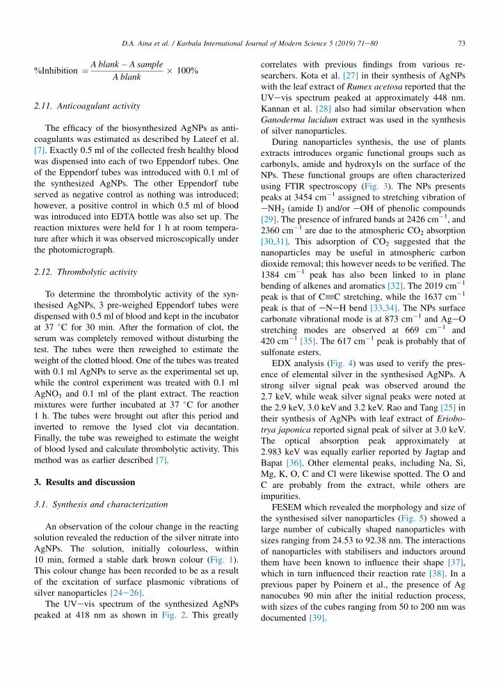
An observation of the colour change in the reactingsolution revealed the reduction of the silver nitrate intoAgNPs. The solution, initially colourless, within10 min, formed a stable dark brown colour (Fig. 1).This colour change has been recorded to be as a resultof the excitation of surface plasmonic vibrations ofsilver nanoparticles [24e26].
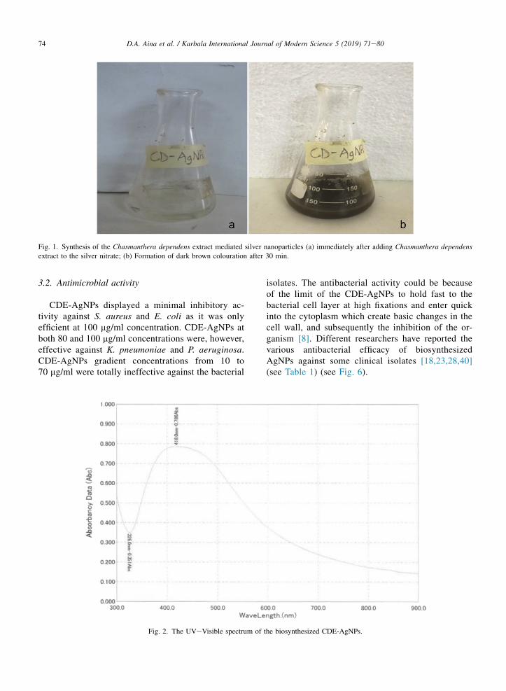
The UVevis spectrum of the synthesized AgNPspeaked at 418 nm as shown in Fig. 2. This greatly
correlates with previous findings from various re-searchers. Kota et al. [27] in their synthesis of AgNPswith the leaf extract of Rumex acetosa reported that theUVevis spectrum peaked at approximately 448 nm.Kannan et al. [28] also had similar observation whenGanoderma lucidum extract was used in the synthesisof silver nanoparticles.
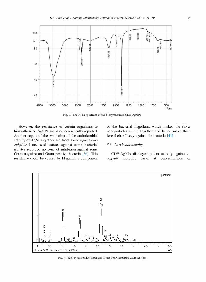
During nanoparticles synthesis, the use of plantsextracts introduces organic functional groups such ascarbonyls, amide and hydroxyls on the surface of theNPs. These functional groups are often characterizedusing FTIR spectroscopy (Fig. 3). The NPs presentspeaks at 3454 cm�1 assigned to stretching vibration ofeNH2 (amide I) and/or eOH of phenolic compounds[29]. The presence of infrared bands at 2426 cm�1, and2360 cm�1 are due to the atmospheric CO2 absorption[30,31]. This adsorption of CO2 suggested that thenanoparticles may be useful in atmospheric carbondioxide removal; this however needs to be verified. The1384 cm�1 peak has also been linked to in planebending of alkenes and aromatics [32]. The 2019 cm�1
peak is that of C^C stretching, while the 1637 cm�1
peak is that of eNeH bend [33,34]. The NPs surfacecarbonate vibrational mode is at 873 cm�1 and AgeOstretching modes are observed at 669 cm�1 and420 cm�1 [35]. The 617 cm�1 peak is probably that ofsulfonate esters.
EDX analysis (Fig. 4) was used to verify the pres-ence of elemental silver in the synthesised AgNPs. Astrong silver signal peak was observed around the2.7 keV, while weak silver signal peaks were noted atthe 2.9 keV, 3.0 keV and 3.2 keV. Rao and Tang [25] intheir synthesis of AgNPs with leaf extract of Eriobo-trya japonica reported signal peak of silver at 3.0 keV.The optical absorption peak approximately at2.983 keV was equally earlier reported by Jagtap andBapat [36]. Other elemental peaks, including Na, Si,Mg, K, O, C and Cl were likewise spotted. The O andC are probably from the extract, while others areimpurities.
FESEM which revealed the morphology and size ofthe synthesised silver nanoparticles (Fig. 5) showed alarge number of cubically shaped nanoparticles withsizes ranging from 24.53 to 92.38 nm. The interactionsof nanoparticles with stabilisers and inductors aroundthem have been known to influence their shape [37],which in turn influenced their reaction rate [38]. In aprevious paper by Poinern et al., the presence of Agnanocubes 90 min after the initial reduction process,with sizes of the cubes ranging from 50 to 200 nm wasdocumented [39].
73D.A. Aina et al. / Karbala International Journal of Modern Science 5 (2019) 71e80
3.2. Antimicrobial activity
CDE-AgNPs displayed a minimal inhibitory ac-tivity against S. aureus and E. coli as it was onlyefficient at 100 mg/ml concentration. CDE-AgNPs atboth 80 and 100 mg/ml concentrations were, however,effective against K. pneumoniae and P. aeruginosa.CDE-AgNPs gradient concentrations from 10 to70 mg/ml were totally ineffective against the bacterial
isolates. The antibacterial activity could be becauseof the limit of the CDE-AgNPs to hold fast to thebacterial cell layer at high fixations and enter quickinto the cytoplasm which create basic changes in thecell wall, and subsequently the inhibition of the or-ganism [8]. Different researchers have reported thevarious antibacterial efficacy of biosynthesizedAgNPs against some clinical isolates [18,23,28,40](see Table 1) (see Fig. 6).
Fig. 1. Synthesis of the Chasmanthera dependens extract mediated silver nanoparticles (a) immediately after adding Chasmanthera dependens
extract to the silver nitrate; (b) Formation of dark brown colouration after 30 min.
Fig. 2. The UVeVisible spectrum of the biosynthesized CDE-AgNPs.
74 D.A. Aina et al. / Karbala International Journal of Modern Science 5 (2019) 71e80
However, the resistance of certain organisms tobiosynthesised AgNPs has also been recently reported.Another report of the evaluation of the antimicrobialactivity of AgNPs synthesised from Artocarpus heter-ophyllus Lam. seed extract against some bacterialisolates recorded no zone of inhibition against someGram negative and Gram positive bacteria [36]. Thisresistance could be caused by Flagellin, a component
of the bacterial flagellum, which makes the silvernanoparticles clump together and hence make themlose their efficacy against the bacteria [41].
3.3. Larvicidal activity
CDE-AgNPs displayed potent activity against A.aegypti mosquito larva at concentrations of
Fig. 3. The FTIR spectrum of the biosynthesized CDE-AgNPs.
Fig. 4. Energy dispersive spectrum of the biosynthesized CDE-AgNPs.
75D.A. Aina et al. / Karbala International Journal of Modern Science 5 (2019) 71e80
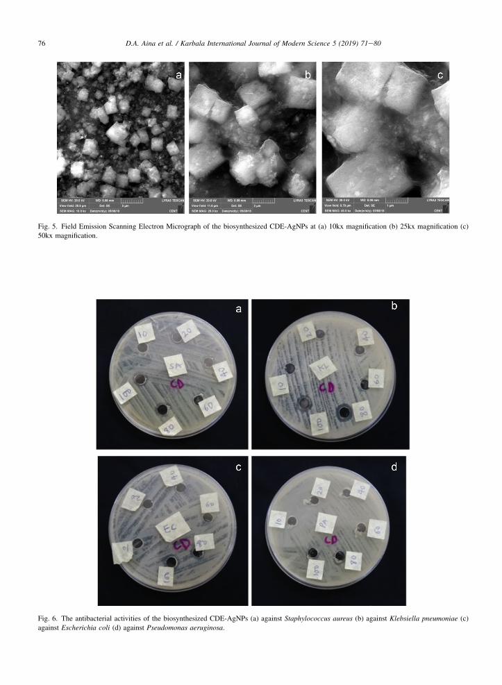
Fig. 5. Field Emission Scanning Electron Micrograph of the biosynthesized CDE-AgNPs at (a) 10kx magnification (b) 25kx magnification (c)
50kx magnification.
Fig. 6. The antibacterial activities of the biosynthesized CDE-AgNPs (a) against Staphylococcus aureus (b) against Klebsiella pneumoniae (c)
against Escherichia coli (d) against Pseudomonas aeruginosa.
76 D.A. Aina et al. / Karbala International Journal of Modern Science 5 (2019) 71e80
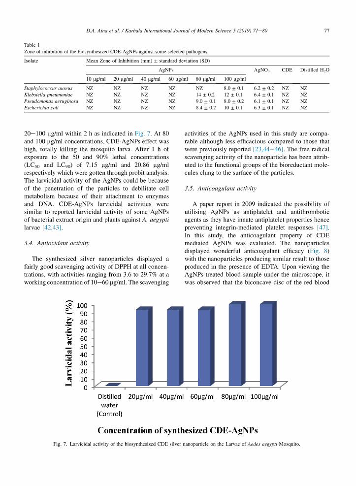
20e100 mg/ml within 2 h as indicated in Fig. 7. At 80and 100 mg/ml concentrations, CDE-AgNPs effect washigh, totally killing the mosquito larva. After 1 h ofexposure to the 50 and 90% lethal concentrations(LC50 and LC90) of 7.15 mg/ml and 20.86 mg/mlrespectively which were gotten through probit analysis.The larvicidal activity of the AgNPs could be becauseof the penetration of the particles to debilitate cellmetabolism because of their attachment to enzymesand DNA. CDE-AgNPs larvicidal activities weresimilar to reported larvicidal activity of some AgNPsof bacterial extract origin and plants against A. aegyptilarvae [42,43].
3.4. Antioxidant activity
The synthesized silver nanoparticles displayed afairly good scavenging activity of DPPH at all concen-trations, with activities ranging from 3.6 to 29.7% at aworking concentration of 10e60 mg/ml. The scavenging
activities of the AgNPs used in this study are compa-rable although less efficacious compared to those thatwere previously reported [23,44e46]. The free radicalscavenging activity of the nanoparticle has been attrib-uted to the functional groups of the bioreductant mole-cules clung to the surface of the particles.
3.5. Anticoagulant activity
A paper report in 2009 indicated the possibility ofutilising AgNPs as antiplatelet and antithromboticagents as they have innate antiplatelet properties hencepreventing integrin-mediated platelet responses [47].In this study, the anticoagulant property of CDEmediated AgNPs was evaluated. The nanoparticlesdisplayed wonderful anticoagulant efficacy (Fig. 8)with the nanoparticles producing similar result to thoseproduced in the presence of EDTA. Upon viewing theAgNPs-treated blood sample under the microscope, itwas observed that the biconcave disc of the red blood
Table 1
Zone of inhibition of the biosynthesized CDE-AgNPs against some selected pathogens.
Isolate Mean Zone of Inhibition (mm) ± standard deviation (SD)
AgNPs AgNO3 CDE Distilled H2O
10 mg/ml 20 mg/ml 40 mg/ml 60 mg/ml 80 mg/ml 100 mg/ml
Staphylococcus aureus NZ NZ NZ NZ NZ 8.0 ± 0.1 6.2 ± 0.2 NZ NZ
Klebsiella pneumoniae NZ NZ NZ NZ 14 ± 0.2 12 ± 0.1 6.4 ± 0.1 NZ NZ
Pseudomonas aeruginosa NZ NZ NZ NZ 9.0 ± 0.1 8.0 ± 0.2 6.1 ± 0.1 NZ NZ
Escherichia coli NZ NZ NZ NZ 8.4 ± 0.2 10 ± 0.1 6.3 ± 0.1 NZ NZ
Fig. 7. Larvicidal activity of the biosynthesized CDE silver nanoparticle on the Larvae of Aedes aegypti Mosquito.
77D.A. Aina et al. / Karbala International Journal of Modern Science 5 (2019) 71e80
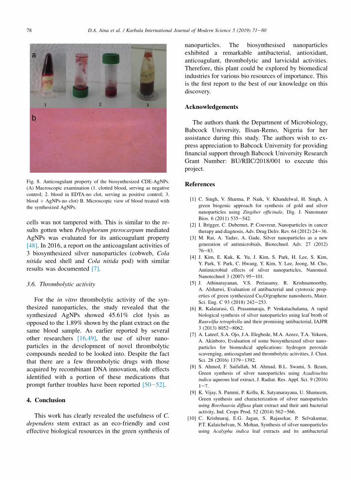
cells was not tampered with. This is similar to the re-sults gotten when Peltophorum pterocarpum mediatedAgNPs was evaluated for its anticoagulant property[48]. In 2016, a report on the anticoagulant activities of3 biosynthesized silver nanoparticles (cobweb, Colanitida seed shell and Cola nitida pod) with similarresults was documented [7].
3.6. Thrombolytic activity
For the in vitro thrombolytic activity of the syn-thesized nanoparticles, the study revealed that thesynthesized AgNPs showed 45.61% clot lysis asopposed to the 1.89% shown by the plant extract on thesame blood sample. As earlier reported by severalother researchers [16,49], the use of silver nano-particles in the development of novel thrombolyticcompounds needed to be looked into. Despite the factthat there are a few thrombolytic drugs with thoseacquired by recombinant DNA innovation, side effectsidentified with a portion of these medications thatprompt further troubles have been reported [50e52].
4. Conclusion
This work has clearly revealed the usefulness of C.dependens stem extract as an eco-friendly and costeffective biological resources in the green synthesis of
nanoparticles. The biosynthesised nanoparticlesexhibited a remarkable antibacterial, antioxidant,anticoagulant, thrombolytic and larvicidal activities.Therefore, this plant could be explored by biomedicalindustries for various bio resources of importance. Thisis the first report to the best of our knowledge on thisdiscovery.
Acknowledgements
The authors thank the Department of Microbiology,Babcock University, Ilisan-Remo, Nigeria for herassistance during this study. The authors wish to ex-press appreciation to Babcock University for providingfinancial support through Babcock University ResearchGrant Number: BU/RIIC/2018/001 to execute thisproject.
References
[1] C. Singh, V. Sharma, P. Naik, V. Khandelwal, H. Singh, A
green biogenic approach for synthesis of gold and silver
nanoparticles using Zingiber officinale, Dig. J. Nanomater
Bios. 6 (2011) 535e542.
[2] I. Brigger, C. Dubernet, P. Couvreur, Nanoparticles in cancer
therapy and diagnosis, Adv. Drug Deliv. Rev. 64 (2012) 24e36.
[3] M. Rai, A. Yadav, A. Gade, Silver nanoparticles as a new
generation of antimicrobials, Biotechnol. Adv. 27 (2012)
76e83.
[4] J. Kim, E. Kuk, K. Yu, J. Kim, S. Park, H. Lee, S. Kim,
Y. Park, Y. Park, C. Hwang, Y. Kim, Y. Lee, Jeong, M. Cho,
Antimicrobial effects of silver nanoparticles, Nanomed.
Nanotechnol 3 (2007) 95e101.
[5] J. Athinarayanan, V.S. Periasamy, R. Krishnamoorthy,
A. Alshatwi, Evaluation of antibacterial and cytotoxic prop-
erties of green synthesized Cu2O/graphene nanosheets, Mater.
Sci. Eng. C 93 (2018) 242e253.
[6] R. Kalaiarasi, G. Prasannaraja, P. Venkatachalama, A rapid
biological synthesis of silver nanoparticles using leaf broth of
Rauvolfia tetraphylla and their promising antibacterial, IAJPR
3 (2013) 8052e8062.
[7] A. Lateef, S.A. Ojo, J.A. Elegbede, M.A. Azeez, T.A. Yekeen,
A. Akinboro, Evaluation of some biosynthesized silver nano-
particles for biomedical applications: hydrogen peroxide
scavenging, anticoagulant and thrombolytic activities, J. Clust.
Sci. 28 (2016) 1379e1392.
[8] S. Ahmed, F. Saifullah, M. Ahmad, B.L. Swami, S. Ikram,
Green synthesis of silver nanoparticles using Azadirachta
indica aqueous leaf extract, J. Radiat. Res. Appl. Sci. 9 (2016)
1e7.
[9] K. Vijay, S. Pammi, P. Kollu, K. Satyanarayana, U. Shameem,
Green synthesis and characterization of silver nanoparticles
using Boerhaavia diffusa plant extract and their anti bacterial
activity, Ind. Crops Prod. 52 (2014) 562e566.
[10] C. Krishnaraj, E.G. Jagan, S. Rajasekar, P. Selvakumar,
P.T. Kalaichelvan, N. Mohan, Synthesis of silver nanoparticles
using Acalypha indica leaf extracts and its antibacterial
Fig. 8. Anticoagulant property of the biosynthesized CDE-AgNPs.
(A) Macroscopic examination (1. clotted blood, serving as negative
control; 2. blood in EDTA-no clot, serving as positive control; 3.
blood þ AgNPs-no clot) B. Microscopic view of blood treated with
the synthesized AgNPs.
78 D.A. Aina et al. / Karbala International Journal of Modern Science 5 (2019) 71e80
activity against water borne pathogens, Colloids Surf. B. 76
(2010) 50e56.
[11] S.L. Banerjee, M. Khamrai, K. Sarkar, N.K. Singha,
P.P. Kundu, Modified chitosan encapsulated core-shell Ag Nps
for superior antimicrobial and anticancer activity, Int. J. Biol.
Macromol. 85 (2016) 157e167.
[12] A.R. Allafchian, S.Z. Mirahmadi-Zare, S.A.H. Jalali,
S.S. Hashemi, M.R. Vahabi, Green synthesis of silver nano-
particles using phlomis leaf extract and investigation of their
antibacterial activity, J. Nanostructure Chem. 6 (2016)
129e135.[13] N. Krishnan, B. Velramar, B. Ramatchandirin, G.C. Abraham,
N. Duraisamy, R. Pandiyan, R.K. Velu, Effect of biogenic
silver nanocubes on matrix mettaloproteinases 2 and 9 ex-
pressions in hyperglycemic skin injury and its impact in early
wound healing in streptozotocin-induced diabetic mice, Mater.
Sci. Eng. C 91 (2018) 838e845.
[14] K. Shameli, M. Bin-Ahmad, E. Jaffar, N. Ibrahim,
P. Shabanzadeh, A. Rustaiyan, Y. Abdollahi, S. Bagheri,
S. Abdolmohammadi, M. Usman, M. Zidan, Green biosyn-
thesis of silver nanoparticles using Callicarpa maingayi stem
bark extraction, Molecules 17 (2012) 8506e8517.[15] M. Vanaja, G. Gnanajobitha, K. Paulkumar, S. Rajeshkumar,
C. Malarkodi, G. Annadurai, Phytosynthesis of silver nano-
particles by Cissus quadrangularis: influence of physico-
chemical factors, J. Nanostruct. Chem. 3 (2013) 17.
[16] W. Okiei, M. Ogunlesi, M. Ademoye, An assessment of the
antimicrobial properties of extracts of various polarities from
Chasmanthera dependens, Emilia coccinea and Cuscuta aus-
tralis, herbal medications for eye diseases, J. Appl. Sci. 9
(2009) 4076e4080.
[17] O. Morebise, E. Awe, J. Modupe, O. Olajide, Evaluation of the
anti-inflammatory and analgesic properties of Chasmanthera
dependens leaf methanol extract, Fitoterapia 72 (2001)
497e502.
[18] J. Weiss, P. Takhistov, D. McClements, Functional materials in
food nanotechnology, J. Food Sci. 71 (2006) R107eR116.
[19] A. Lateef, M.A. Azeez, T.B. Asafa, T.A. Yekeen, A. Akinboro,
I.C. Oladipo, E.B. Gueguim-Kana, L.S. Beukes, Biomedical
applications of cocoa bean extract-mediated silver nano-
particles as antimicrobial, larvicidal and anticoagulant agents,
J. Clust. Sci. 28 (2016) 149e164.
[20] A.P. Defante, W.N. Vreeland, K.D. Benkstein, D.C. Ripple,
Using image attributes to assure accurate particle size and
count using nanoparticle tracking analysis, J. Pharm. Sci.
(2018), https://doi.org/10.1016/j.xphs.2017.12.016.
[21] C. Perez, M. Paul, P. Bazerque, Antibiotic assay by agar well
diffusion method, Acta Biol. Exp. 15 (1990) 113e115.[22] WHO, Guidelines for Laboratory and Field Testing of Mos-
quito Larvicides, World Health Organization, 2005, pp. 1e41.
[23] A. Lateef, S.A. Ojo, A.S. Akinwale, L. Azeez, E.B. Gueguim-
Kana, L.S. Beukes, Biogenic synthesis of silver nanoparticles
using cell-free extract of Bacillus safensis LAU 13: antimi-
crobial, free radical scavenging and larvicidal activities, Bio-
logia 70 (2015) 1295e1306.[24] S. Ojha, A. Sett, U. Bora, Green synthesis of silver nano-
particles by Ricinus communis var. carmencita leaf extract and
its antibacterial study, Adv. Nat. Sci. Nanosci. Nanotechnol. 8
(2017), 035009.
[25] B. Rao, R. Tang, Green synthesis of silver nanoparticles with
antibacterial activities using aqueous Eriobotrya japonica leaf
extract, Adv. Nat. Sci. Nanosci. Nanotechnol. 8 (2017),
015014.
[26] A. Thirumurugan, N.A. Tomy, H.P. Kumar, P. Prakash, Bio-
logical synthesis of silver nanoparticles by Lantana camara
leaf extracts, Int. J Nanomater Biostruct. 47 (2011) 754.
[27] S. Kota, P. Dumpala, R. Anantha, M. Verma, S. Kandepu,
Evaluation of therapeutic potential of the silver/silver chloride
nanoparticles synthesized with the aqueous leaf extract of
Rumex acetosa, Sci. Rep. (2017), https://doi.org/10.1038/
s41598-017-11853-2.
[28] M. Kannan, P. Muthusamy, U. Venkatachalam,
J. Rajarajeswaran, Mycosynthesis, characterization and anti-
bacterial activity of silver nanoparticles ( Ag-NPs ) from fungus
Ganoderma lucidum, Malaya J. Biosci. 1 (2014) 134e142.[29] D. Raghunandan, B. Ravishankar, G. Sharanbasava,
D. Mahesh, V. Harsoor, M. Yalagatti, M. Bhagawanraju,
A. Venkataraman, Anti-cancer studies of noble metal nano-
particles synthesized using different plant extracts, Cancer
Nanotechnol 2 (2011) 57e65.
[30] Y. Zhao, R. Frost, W. Yang J Martens, Size and morphology
control of gallium oxide hydroxide GaO(OH), nano- to micro-
sized particles by soft-chemistry route without surfactant, J.
Phys. Chem. C 112 (2008) 3568e3579.
[31] M. Gopalakrishnan, V. Purushothaman, V. Ramakrishnan,
G. Bhalerao, K. Jeganathan, The effect of nitridation temper-
ature on the structural, optical and electrical properties of GaN
nanoparticles, Cryst. Eng. Comm. 16 (2014) 3584e3591.
[32] F. Rahmaniyan, A. Shamel, A. Shafaghatlonbar, Evaluation of
biologically synthesized silver nanoparticles by the bio-
reduction method, Synth. React. Inorg. M 45 (2014)
1495e1500.
[33] W. Lin, D. Yu, M. Yang, pH-sensitive polyelectrolyte complex
gel microspheres composed of chitosan/sodium tripolyphos-
phate/dextran sulfate: swelling kinetics and drug delivery
properties, Colloids Surf. B. 44 (2005) 143e151.
[34] L. Sun, Y. Du, L. Chen, R. Huang, X. Chen, The synthesis of
carboxymethylchitosan hydrogel and the application in drug
controlled release systems, Acta Polym. Sin. 8 (2004)
191e195.
[35] F.S. Taghavi, A. Ramazani, S. Moradi, A.P. Azimzadeh, Green
synthesis of zinc oxide nanoparticles using Arabic gum and
photocatalytic degradation of direct blue 129 dye under visible
light, J. Mater. Sci. Mater. Electron. 28 (2017) 13596e13601.
[36] U. Jagtap, V. Bapat, Green synthesis of silver nanoparticles
using Artocarpus heterophyllus Lam. seed extract and its
antibacterial activity, Ind. Crops Prod. 46 (2013) 132e137.
[37] M. Haruta, Nanoparticulate gold catalysts for low-temperature
CO oxidation, J. New Mater. Electrochem. Syst. 7 (2004)
163e172.
[38] R. Xu, D. Wang, J. Zhang, Y. Li, Shape-dependent catalytic
activity of silver nanoparticles for the oxidation of styrene,
Chem. Asian J. 1 (2006) 888e893.
[39] G. Poinern, P. Chapman, X. Le, D. Fawcett, Green biosyn-
thesis of gold nanometre scale plates using the leaf extracts
from an indigenous Australian plant Eucalyptus macrocarpa,
Gold Bull. 46 (2013) 165e173.
[40] N. Geetha, K. Harini, J. Jerlin, K.S. Priya, Biofabrication of
silver nanoparticles using leaf extract of Chromolaena odorata
(L.) king and robinson, in: International Conference on Nu-
clear Energy, Environmental and Biological Sciences, 2012,
pp. 56e59.
79D.A. Aina et al. / Karbala International Journal of Modern Science 5 (2019) 71e80
[41] A. Pan�a�cek, L. Kvítek, M. Sm�ekalov�a, R. Ve�ce�rov�a, M. Kol�a�r,
M. R€oderov�a, R. Zbo�ril, Bacterial resistance to silver nano-
particles and how to overcome it, Nat. Nanotechnol. 13 (2018)
65e71.
[42] G. Suganya, S. Karthi, M. Shivakumar, Larvicidal potential of
silver nanoparticles synthesized from Leucas aspera leaf ex-
tracts against dengue vector Aedes aegypti, Parasitol. Res. 113
(2013) 875e880.
[43] K.V. Anil, K. Ammani, R. Jobina, P. Parasuraman,
B. Siddhardha, Larvicidal activity of green synthesized silver
nanoparticles using Excoecaria agallocha L. (Euphorbiaceae)
leaf extract against Aedes aegypti, IET Nanobiotechnol. 10
(2016) 382e388.
[44] M. Abdel-Aziz, M. Shaheen, A. El-Nekeety, M. Abdel-Wah-
hab, Antioxidant and antibacterial activity of silver nano-
particles biosynthesized using Chenopodium murale leaf
extract, J. Saudi Chem. Soc. 18 (2014) 356e363.
[45] L. Azeez, A. Lateef, S.A. Adebisi, Silver nanoparticles
(AgNPs) biosynthesized using pod extract of Cola nitida en-
hances antioxidant activity and phytochemical composition of
Amaranthus caudatus Linn, Appl. Nanosci. 7 (2017) 59e66.
[46] A. Lateef, B.I. Folarin, S.M. Oladejo, P.O. Akinola, S. Lorika,
E.B. Gueguim-Kana, Anticoagulant activities of silver nano-
particles synthesized from Petiveria alliacea L. leaf extract,
Prep. Biochem. Biotechnol. 48 (2018) 646e652.
[47] S. Shrivastava, T. Bera, S. Singh, G. Singh, P. Ramachandrarao,
D. Dash, Characterization of antiplatelet properties of silver
nanoparticles, ACS Nano 3 (2009) 1357e1364.[48] S. Raja, V. Ramesh, V. Thivaharan, Antibacterial and antico-
agulant activity of silver nanoparticles synthesised from a
novel sourceepods of Peltophorum pterocarpum, J. Ind. Eng.
Chem. 29 (2015) 257e264.
[49] A. Lateef, M.A. Akande, S.A. Ojo, B.I. Folarin, E.B. Gueguim-
Kana, L.S. Beukes, Paper wasp nest-mediated biosynthesis of
silver nanoparticles for antimicrobial, catalytic, anticoagulant,
and thrombolytic applications, 3 Biotech 6 (2016) 140.
[50] D. Baruah, R. Dash, M. Chaudhari, S. Kadam, Plasminogen
activators: a comparison, Vasc. Pharmacol. 44 (2006) 1e9.
[51] A. Gallus, Thrombolytic therapy for venous thrombosis and pul-
monary embolism,Bailli�eresClin Haematol. 11 (1998) 663e673.
[52] J. Wardlaw, E. Berge, G. Del-Zoppo, T. Yamaguchi, Throm-
bolysis for acute ischemic stroke, Stroke 35 (2004) 2914e2915.
80 D.A. Aina et al. / Karbala International Journal of Modern Science 5 (2019) 71e80