Embed Size (px)
Citation preview
Biomedical Diagnostics Two
Lesson One- Basics
2
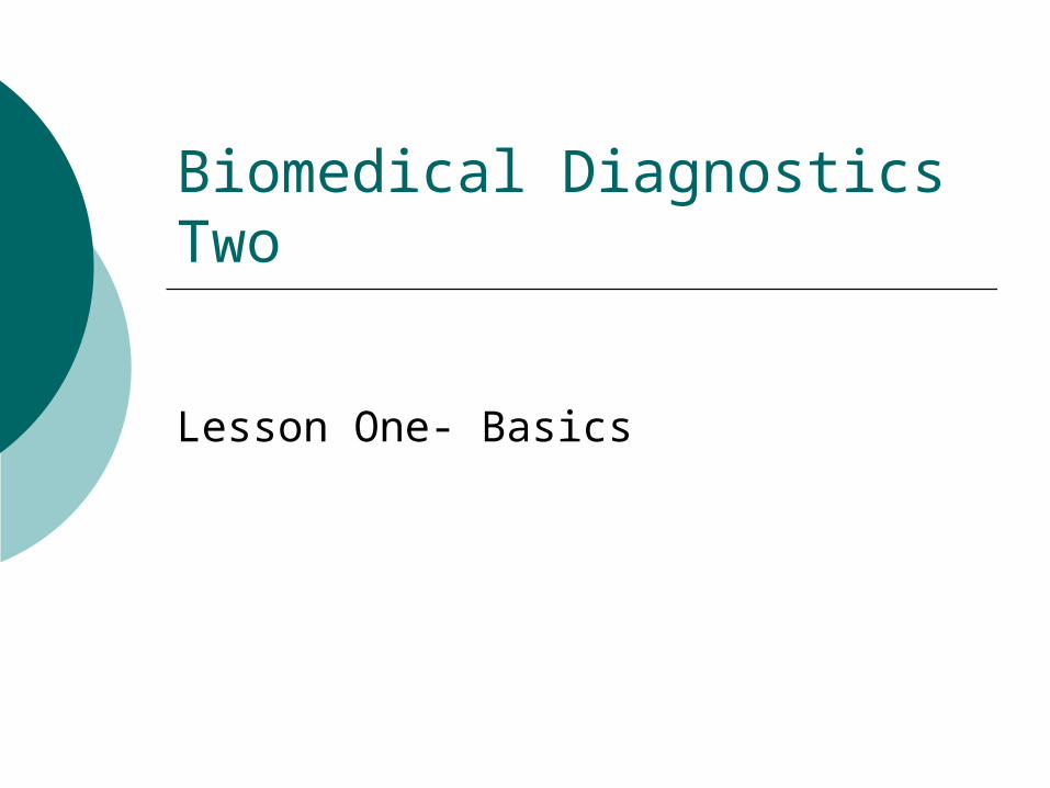
x-rayTransmission through the body
Gamma ray emission from within the body
Ultrasound echoes
Nuclear magnetic resonance induction
Basic Basic Imaging Imaging PrinciplesPrinciples
3
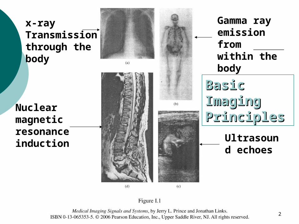
Basic Imaging Principles
What does the human body look like on the inside?What does the human body look like on the inside?Invasive Techniques:Invasive Techniques: Operation Operation EndoscopeEndoscopeNoninvasive Techniques:Noninvasive Techniques: Magnetic Resonance Imaging (MRI) Magnetic Resonance Imaging (MRI) Ultrasound ImagingUltrasound Imaging x-rayx-ray Computed Tomography (CT)Computed Tomography (CT) Nuclear MedicineNuclear Medicine Functional Magnetic Resonance Imaging (fMRI)Functional Magnetic Resonance Imaging (fMRI) Positron Emission Tomography (PET)Positron Emission Tomography (PET)
4
Basic Imaging Principles
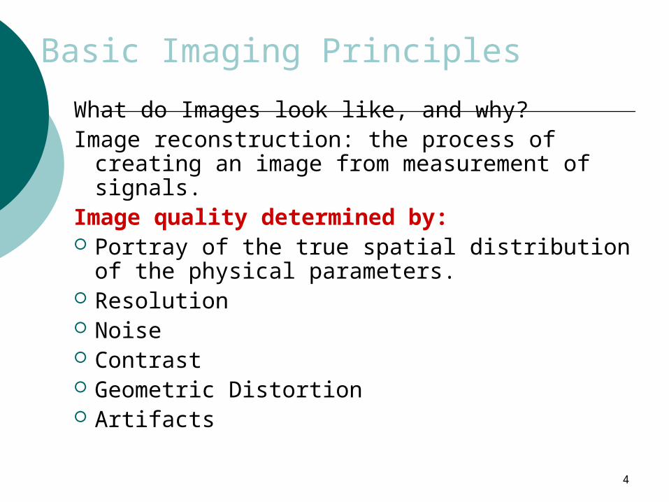
What do Images look like, and why?Image reconstruction: the process of creating an
image from measurement of signals.Image quality determined by: Portray of the true spatial distribution of the
physical parameters. Resolution Noise Contrast Geometric Distortion Artifacts
5
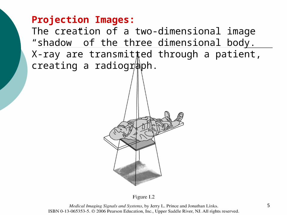
Projection Images:The creation of a two-dimensional image “shadow” of the three dimensional body. X-ray are transmitted through a patient, creating a radiograph.
6
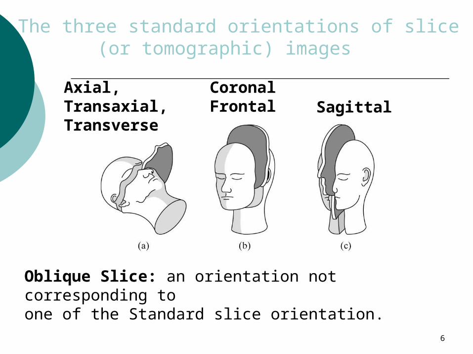
The three standard orientations of slice (or tomographic) images
Axial, Transaxial, Transverse
CoronalFrontal Sagittal
Oblique Slice: an orientation not corresponding to one of the Standard slice orientation.
7
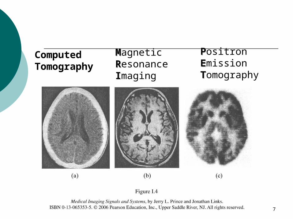
Computed Tomography
MMagnetic RResonance IImaging
PPositron EEmission TTomography
8
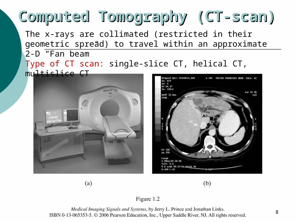
Computed Tomography (CT-Computed Tomography (CT-scan)scan)The x-rays are collimated (restricted in their geometric
spread) to travel within an approximate 2-D “Fan beam” Type of CT scan: single-slice CT, helical CT, multislice CT
9
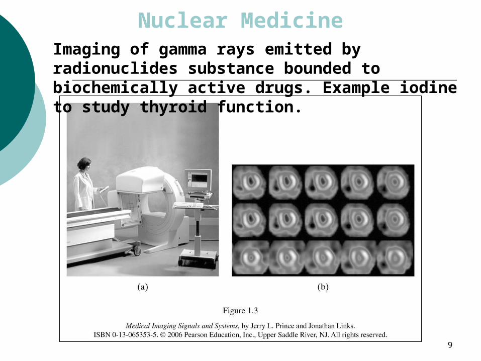
Nuclear MedicineImaging of gamma rays emitted by radionuclides substance bounded to biochemically active drugs. Example iodine to study thyroid function.
10
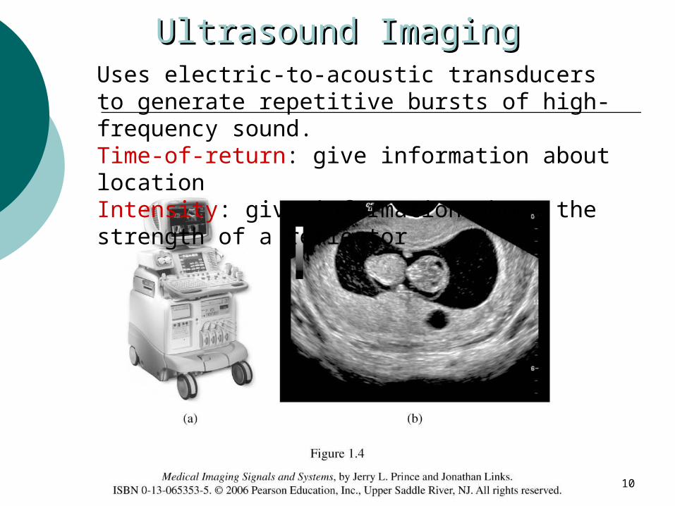
Ultrasound ImagingUltrasound ImagingUses electric-to-acoustic transducers to generate repetitive bursts of high-frequency sound.Time-of-return: give information about locationIntensity: give information about the strength of a reflector
11
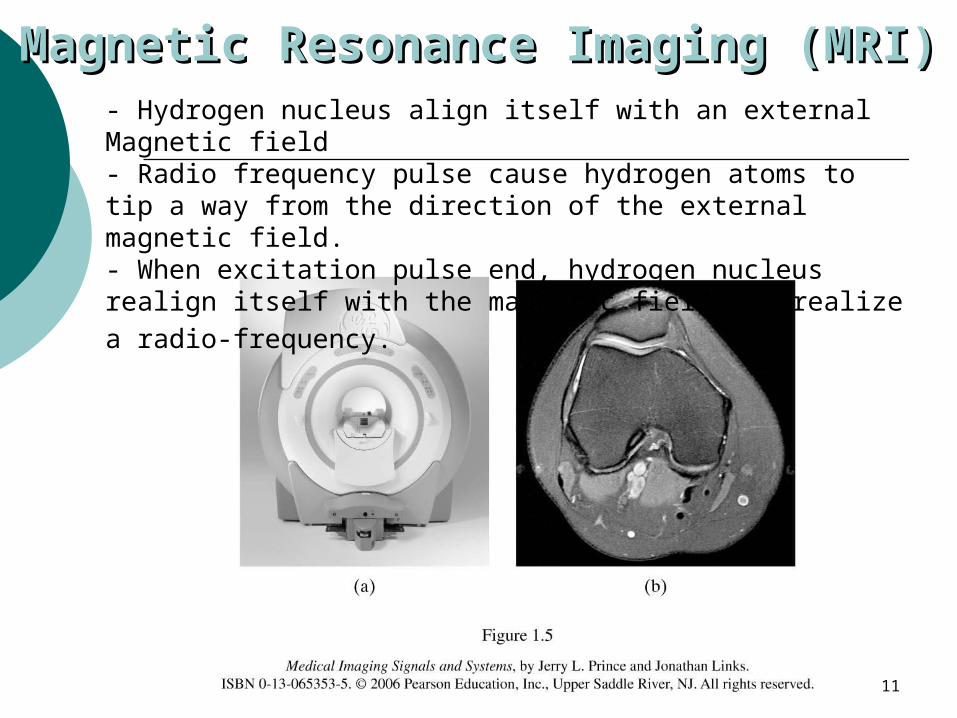
Magnetic Resonance Imaging Magnetic Resonance Imaging (MRI)(MRI)- Hydrogen nucleus align itself with an external Magnetic
field- Radio frequency pulse cause hydrogen atoms to tip a way from the direction of the external magnetic field.- When excitation pulse end, hydrogen nucleus realign itself with the magnetic field and realize a radio-frequency.
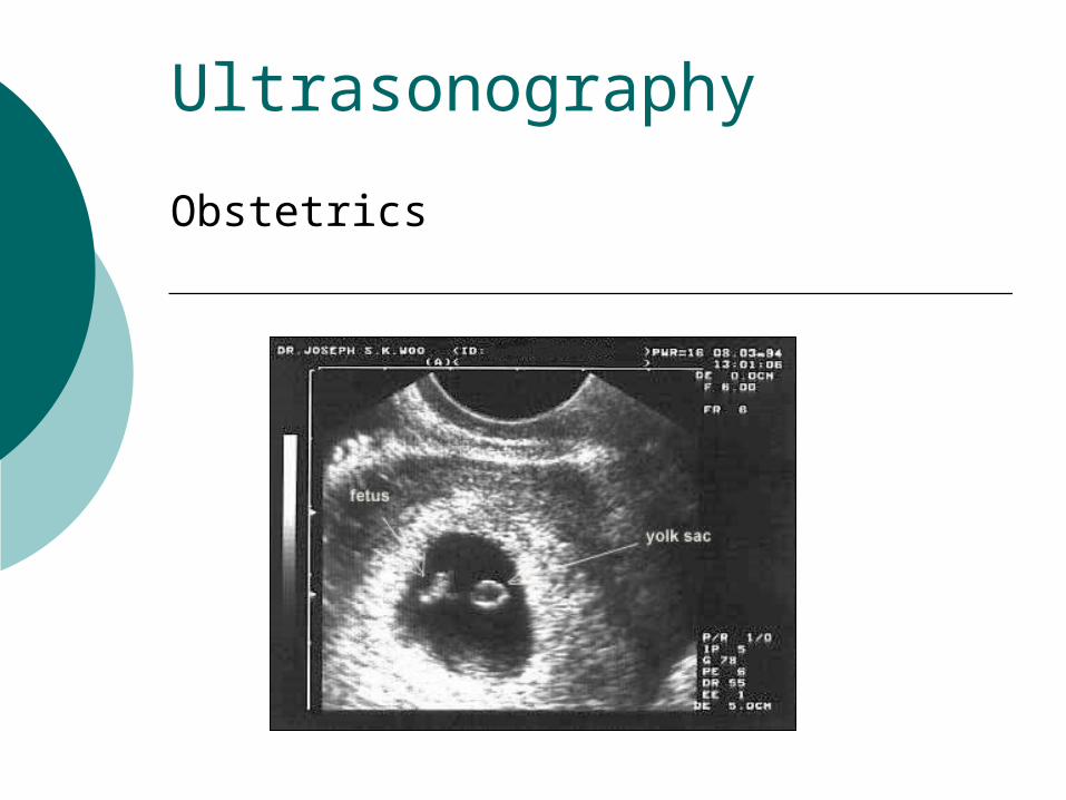
Ultrasonography
Obstetrics
13
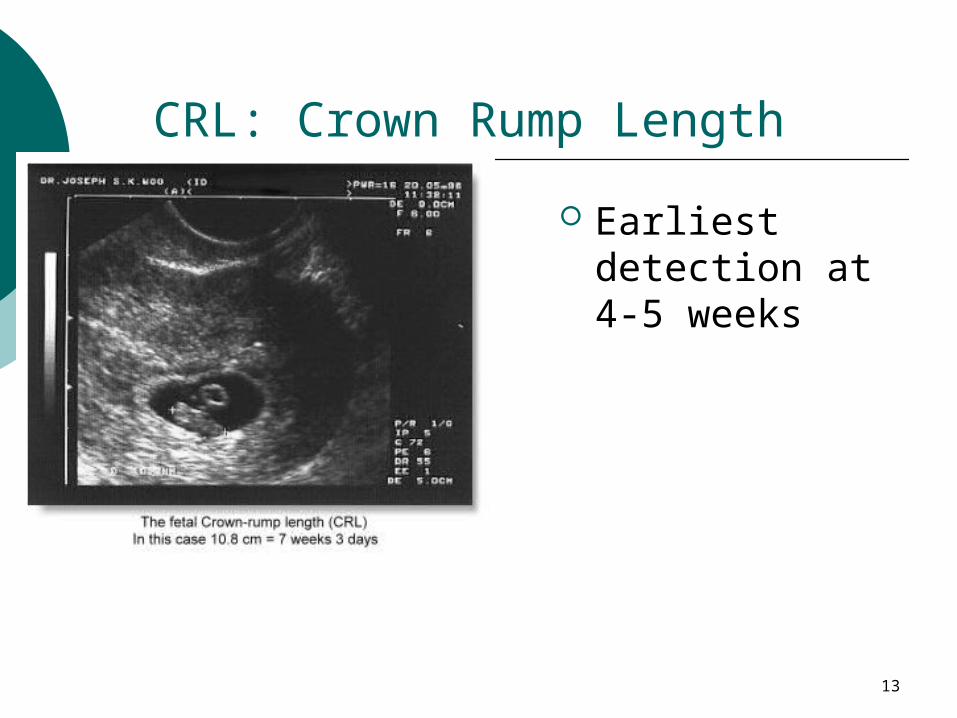
CRL: Crown Rump Length
Earliest detection at 4-5 weeks
14
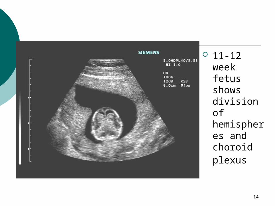
11-12 week fetus shows division of hemispheres and choroid plexus
15
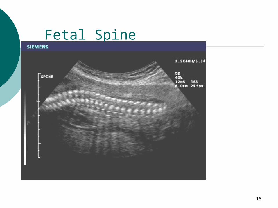
Fetal Spine
16
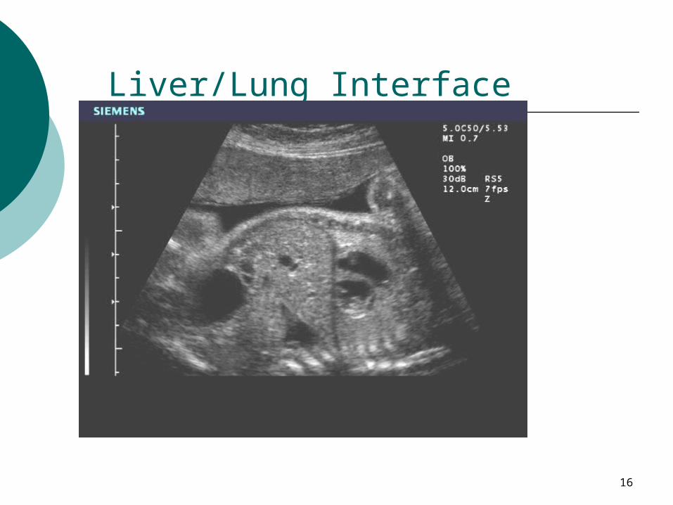
Liver/Lung Interface
17
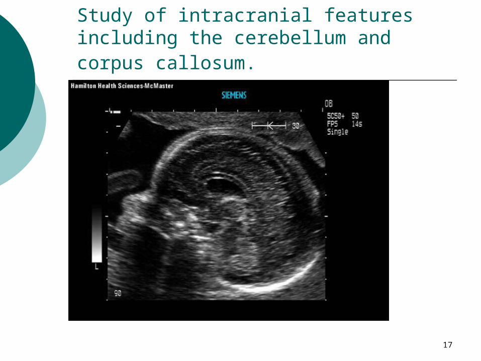
Study of intracranial features including the cerebellum and corpus callosum.
18
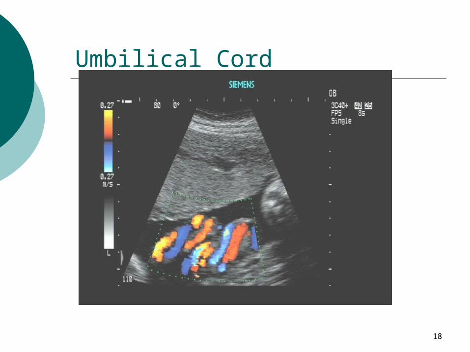
Umbilical Cord
19
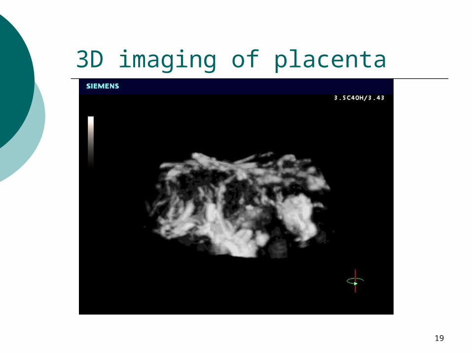
3D imaging of placenta
20
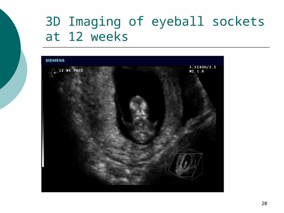
3D Imaging of eyeball sockets at 12 weeks
21
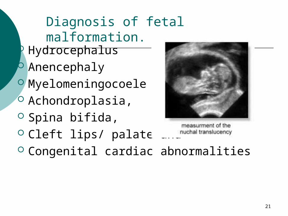
Diagnosis of fetal malformation. Hydrocephalus Anencephaly Myelomeningocoele Achondroplasia, Spina bifida, Cleft lips/ palate and Congenital cardiac abnormalities
22
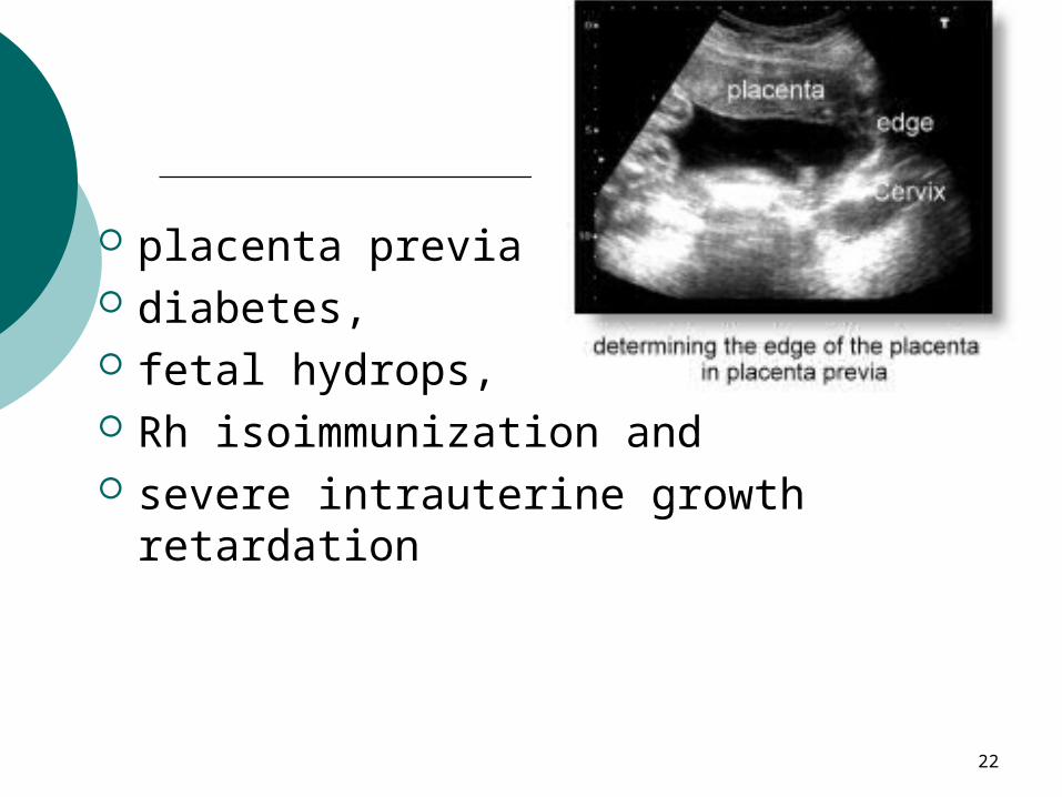
placenta previa diabetes, fetal hydrops, Rh isoimmunization and severe intrauterine growth
retardation
23
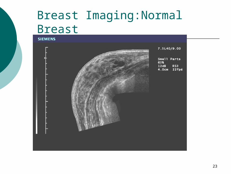
Breast Imaging:Normal Breast
24
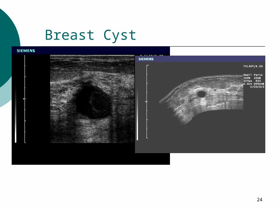
Breast Cyst
25
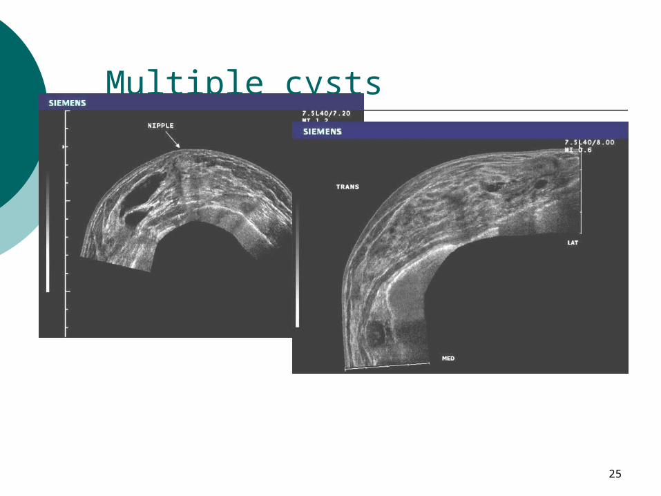
Multiple cysts
26
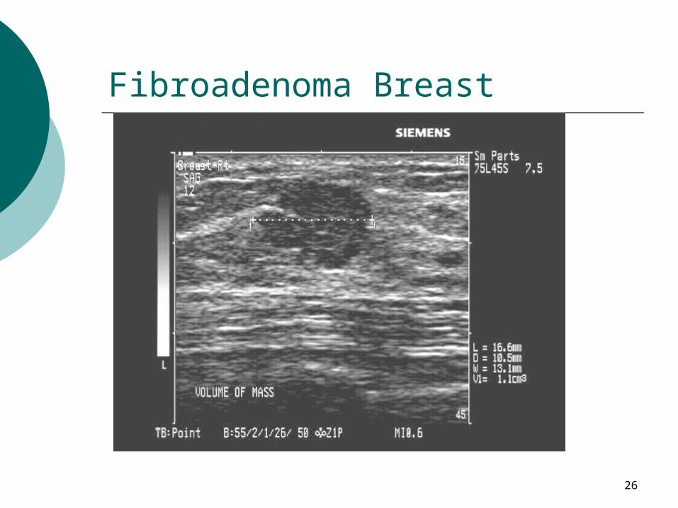
Fibroadenoma Breast
27
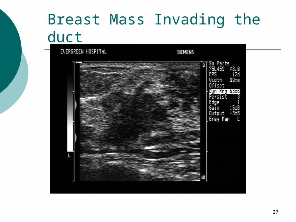
Breast Mass Invading the duct
28
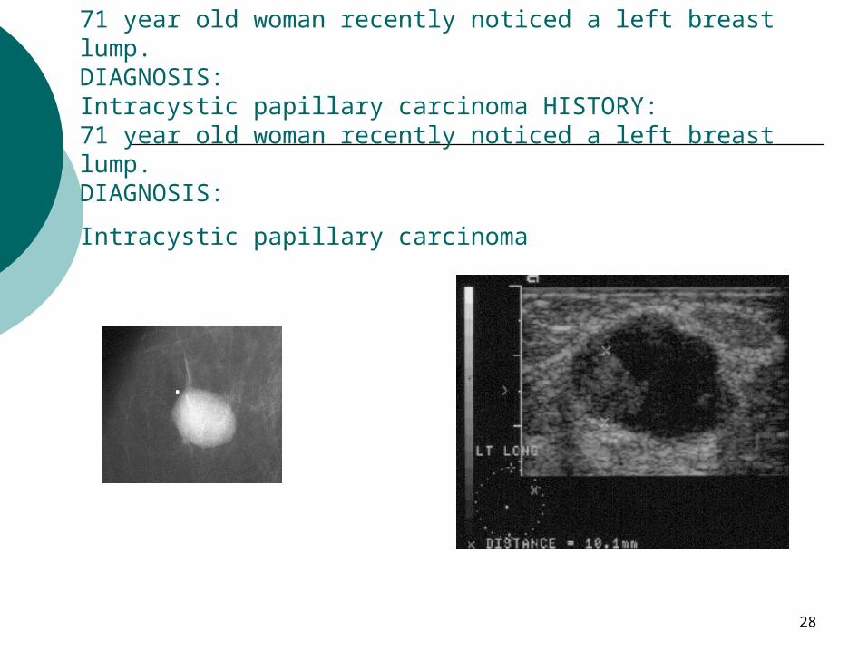
HISTORY:71 year old woman recently noticed a left breast lump. DIAGNOSIS:Intracystic papillary carcinoma HISTORY:71 year old woman recently noticed a left breast lump. DIAGNOSIS:
Intracystic papillary carcinoma
29
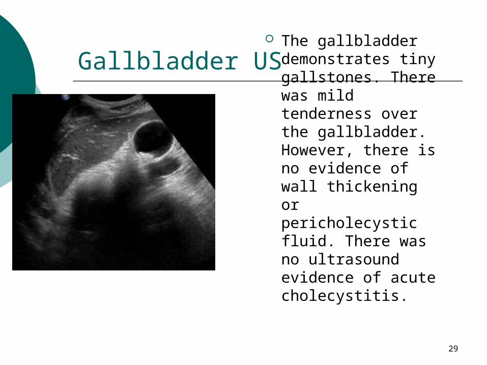
Gallbladder US The gallbladder
demonstrates tiny gallstones. There was mild tenderness over the gallbladder. However, there is no evidence of wall thickening or pericholecystic fluid. There was no ultrasound evidence of acute cholecystitis.
30
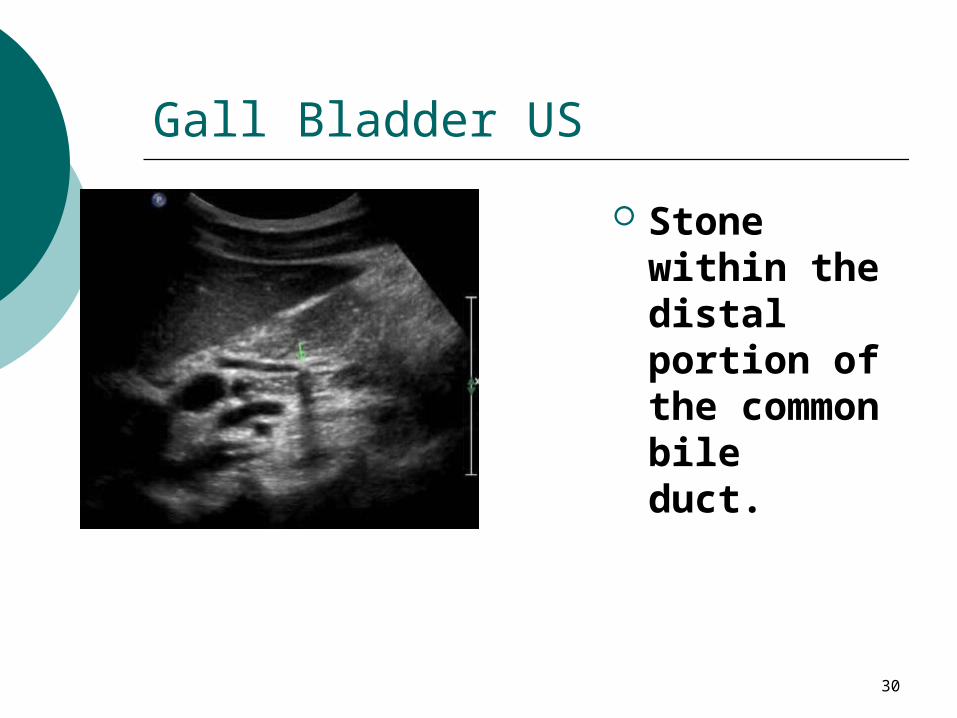
Gall Bladder US
Stone within the distal portion of the common bile duct.
31
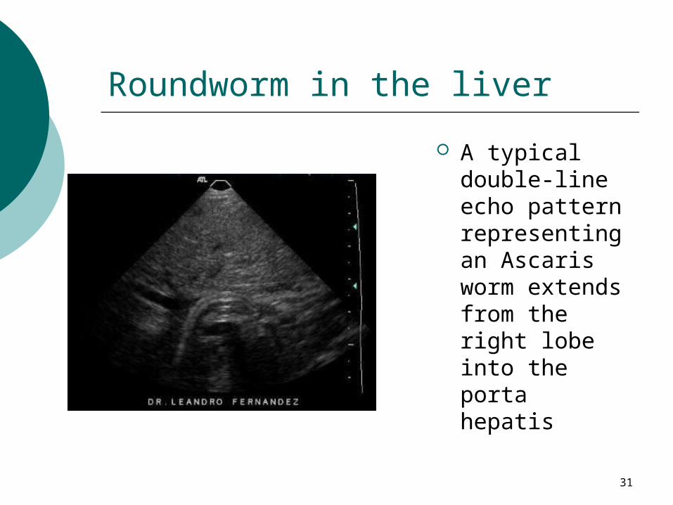
Roundworm in the liver
A typical double-line echo pattern representing an Ascaris worm extends from the right lobe into the porta hepatis